Embed Size (px)
Citation preview

Diencephalon• From interventricular foramen to posterior
commissure• Divisible in to:
ThalamusHypothalamusSubthalamusEpithalamusMetathalamus


Thalamus• Large mass of grey matter, lateral to 3rd ventricle.• Processes the affarent impulses to cerebral cortex.• Reciprocal connections with cerebral cortex & subcortical
grey masses.• Anterior & posterior ends• Surfaces: Medial- lined by ependyma; forms lateral wall
of third ventricle; interthalamic adhesion; hypothalamic sulcusSuperior- anterior tubercle; related to fornix, stria terminalis, caudate nucleus.Inferior- related to hypothalamus anteriorly, to subthalamus posteriorly; post. Surface exhibits two swellings- MGB & LGB.Lateral- in contact with internal capsule.
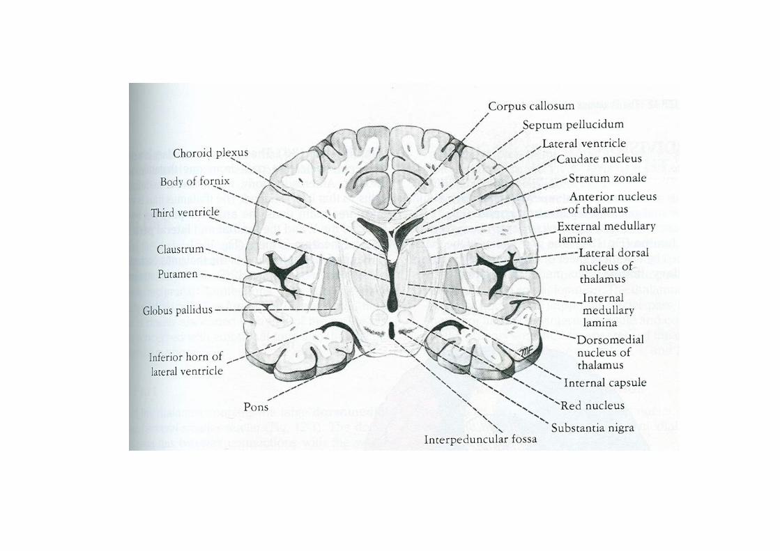

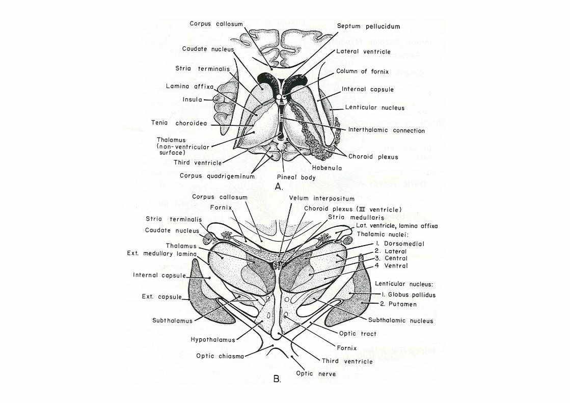
Thalamus- internal structure
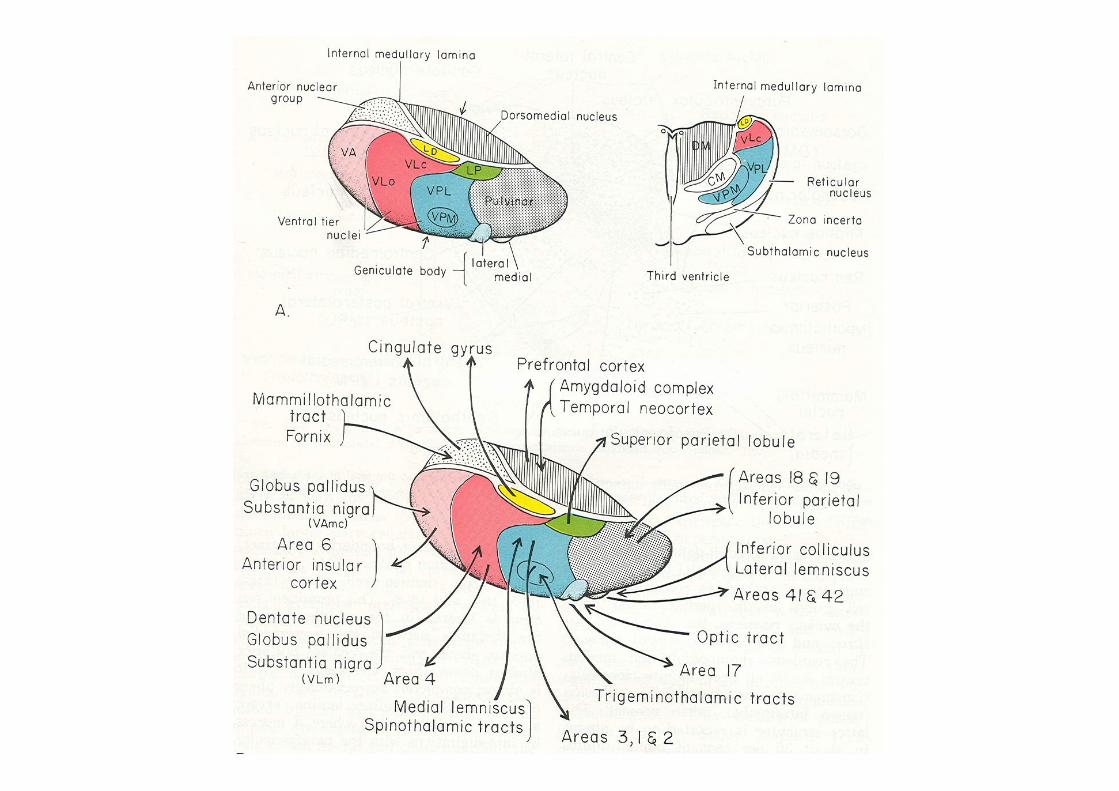
• Stratum zonale• External medullary lamina• Internal medullary lamina- divides it in to
Anterior- anteroventral, anterodorsal, anteromedial
Medial- DorsomedialLateral-Dorsal- lateral dorsal, lat. Posterior,
pulvinarVentral- ventral anterior, ventral lateral,
ventral posterior

Thalamic nuclei-contd.
• Midline nuclei- closely associated with interthalamic adhesion; concerned with visceral activity; connected to hypothalamus, dorsomedian & to intralaminar nuclei.
• Intralaminar nuclei- separate medial & lateral nuclei. Main nuclei-centromedian, parafascicular; affarents from reticular formation, fore brain, pallidal; efferent to putamen.

•Anterior nucleus:
Mamillary body AN cingulate gyrus
(mamillothalamic tract)
• Dorsomedial nucleus:Amygdaloid body DM cingulate gyrus, parietal
lobe, prefrontal cortexGlobus pallidus piriform lobe

Ventral group•Ventral anterior: Globus pallidus VAPremotor & motor c. cortex
•Ventral lateral- sub. nigra, GP, precentral C.C VM precentral C. C.•Ventral Posterior: Ventral Posteromedial (VPM) (largest somatic relay) Ventral posterolateral (VPL)Medial lemniscus VPL Sup. Thalamic radiations Sensory C. Cortex (3,1,2)Spinothalamic tract VPL ( post. Limb of Int. capsule)
Trigemino-thalamic Solitariothalamic VPM Sensory Cerebral Cortex (3,1,2)
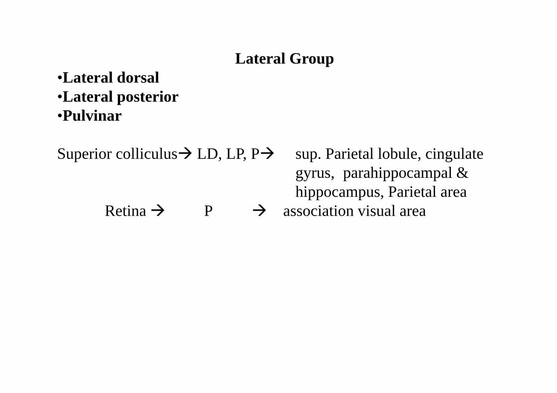
Lateral Group•Lateral dorsal•Lateral posterior•Pulvinar
Superior colliculus LD, LP, P sup. Parietal lobule, cingulate gyrus, parahippocampal & hippocampus, Parietal area
Retina P association visual area


Afferent connections1. Spinothalamic tract
Medial lemniscusTrigemino-thalamicSolitariothalamic
2. Optic tract3. Auditory pathway4. Mamillothalamic tract5. Cerebellar fibres6. Corpus striatum &
globus pallidus7. From cerebral cortex

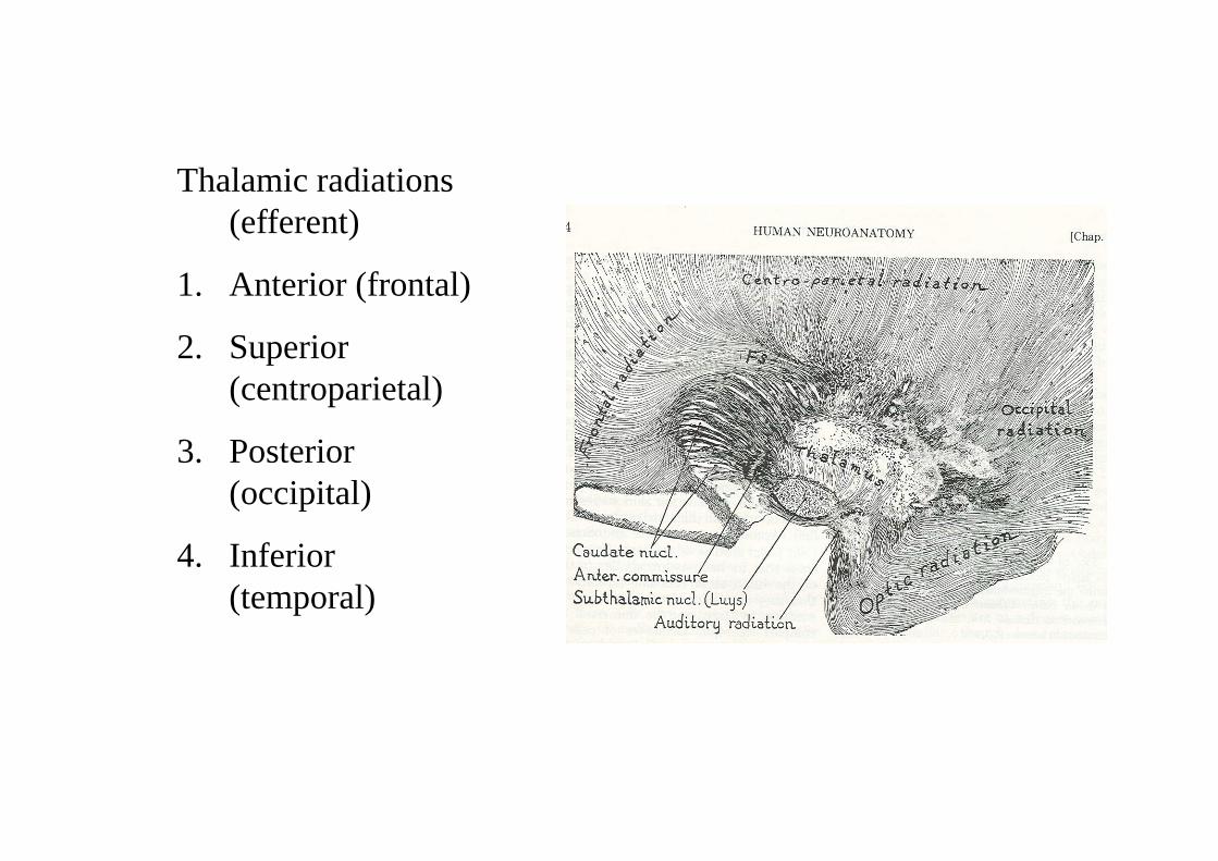
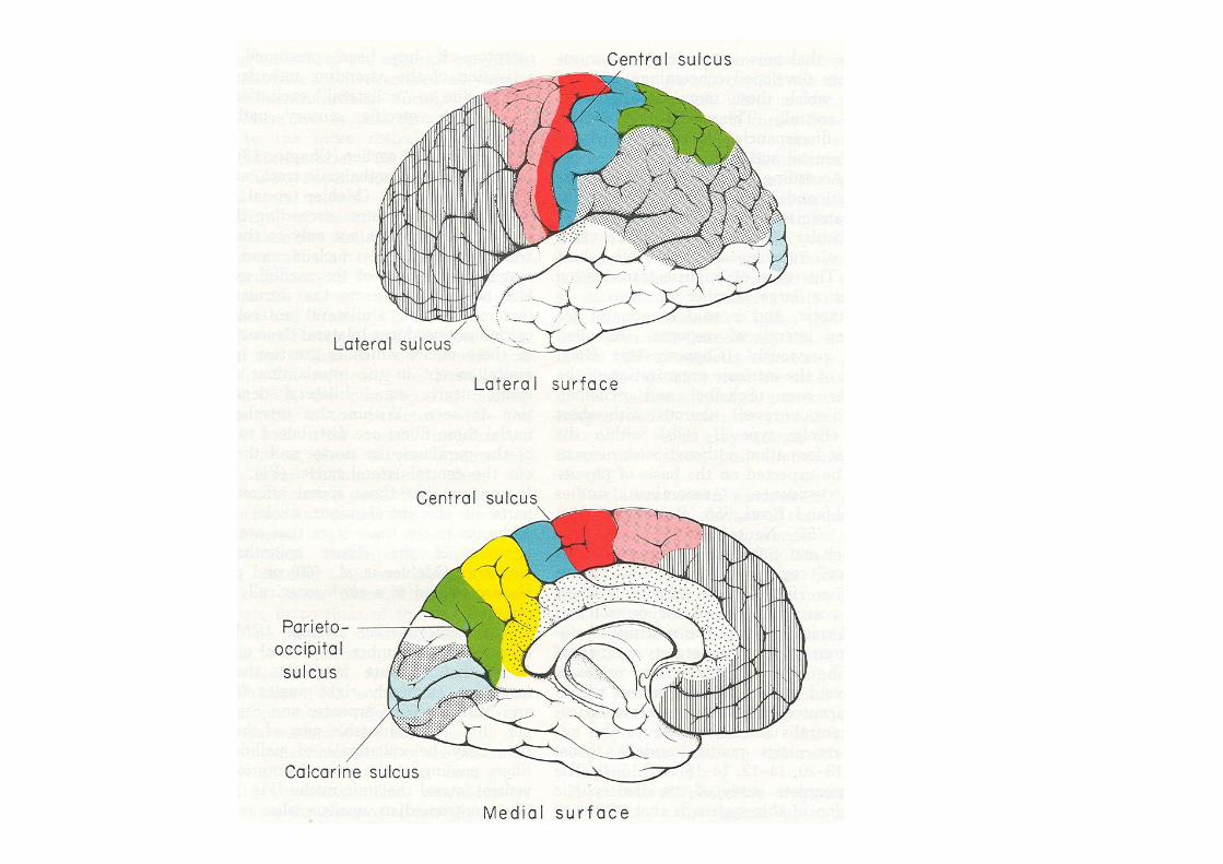
Thalamic radiations (efferent)
1. Anterior (frontal)
2. Superior (centroparietal)
3. Posterior (occipital)
4. Inferior (temporal)


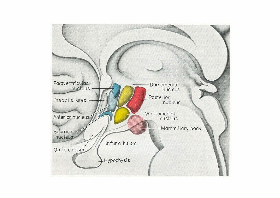
Hypothalamus
• Below the thalamus, forms lower lateral wall of 3rd ventricle.
• Laterally in contact with internal capsule & subthalamus.
• Posteriorly merges with subthalamus.• Anteriorly extends up to lamina terminalis.• Inferiorly related to structures in floor of 3rd
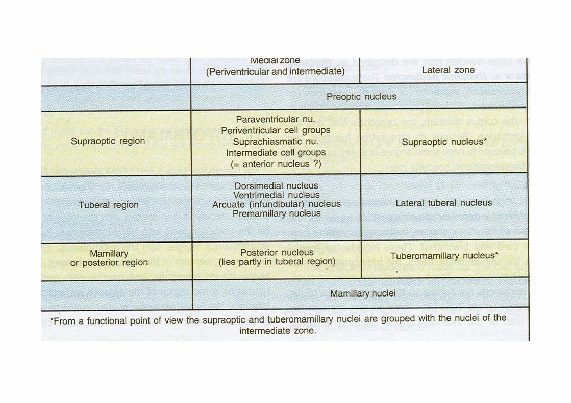
ventricle.• Medial and lateral zones


Afferent connections
1. From spinal cord & brainstem (via reticular formation)
2. Nucleus of tractus solitarius3. Olfactory pathways4. Limbic system5. Locus coeruleus6. From piriform cortex, orbital cortex7. From subthalamus & zona incerta
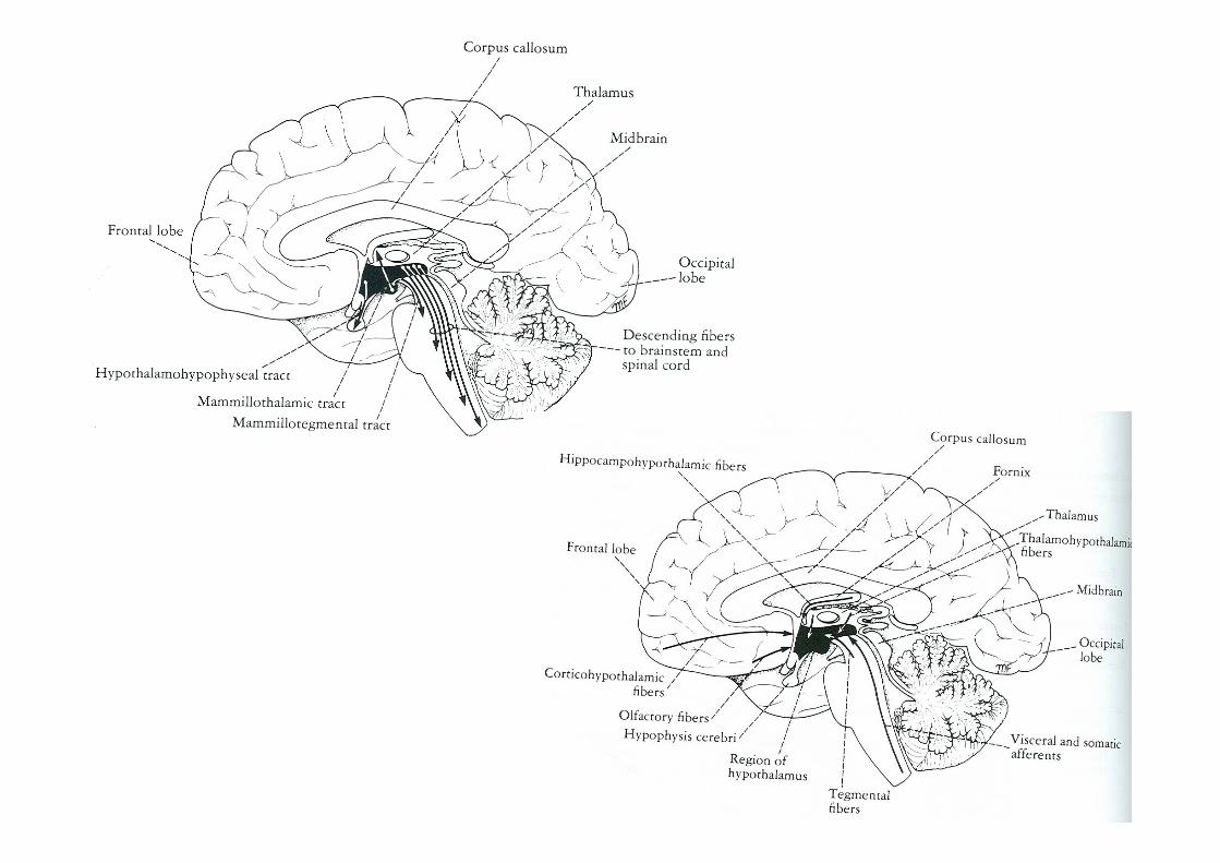

Efferent connections1. To autonomic centres in brain stem and spinal cord e.g.
tractus soiltarius, dorsal nucleus of vagus, nucleus ambiguus, intermedio-lateral grey column.
2. To hippocampal formation, septal nuclei, amygdaloid complex, tegmentum.
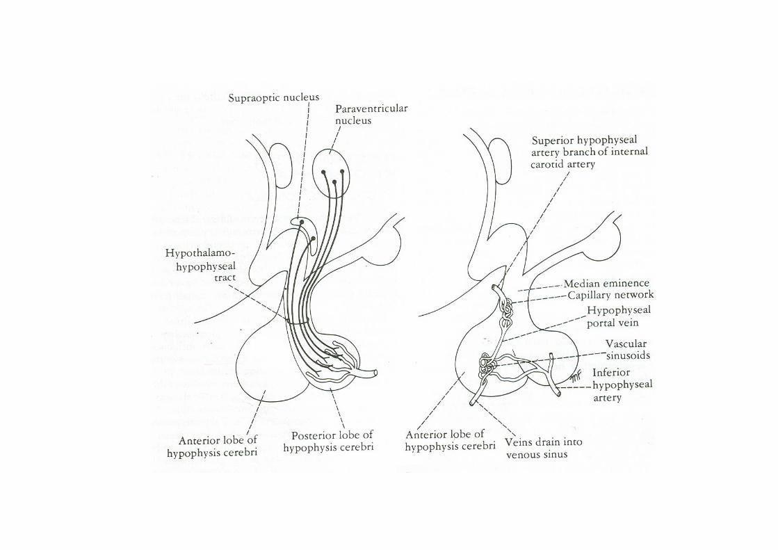
3. To anterior nucleus of thalamus (mamillothalamic tract)4. To subthalamus & tegmentum (mamillo-tegmental tract)5. To neocortex6. Control of pituitary gland
- neurosecretion- release of vasopressin (ADH); oxytocin- control of post. Neurohypophysis- production of releasing factors (tubero-hypophyseal)- hypothalamo-hypophysial portal system


Functions
1. Regulation of eating & drinking behaviour2. Regulation of sexual activity & reproduction3. Control of autonomic activity4. Control of endocrine system5. Emotional behaviour6. Response to stress7. Temperature regulation8. Biological clock

Epithalamus• Visceral efferent pathway to convey
impulses to brain stem.• Pineal body• Habenular trigone• Stria medullares

Pineal body• Cone shaped body attached to roof of 3rd ventricle• Rudimentary gland• Richly vascular connective tissue along with glia cells and
pineal cells.• Receives fibres from stria medullares, habenular nuclei &
post. Com.• Inhibits gonadal function.• After 16 yrs., calcerous bodies present which are visible in
skull x-rays.• Identification & position of pineal gland in skull films.

Metathalamus
• Medial geniculate body• Lateral geniculate body
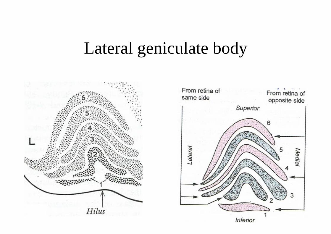
Lateral geniculate body

Subthalamus• Lies below post. part of thalamus• Inferiorly continuous with tegmentum• Laterally related to internal capsule• Reticular nucleus: separated from thalamus.somatic, visceral, auditory, reticular formation send afferents to reticular nucleus which connects to dorsal part of thalamus.
• Zona incerta: connected to reticular nucleus; function not known.
• Subthalamic nucleus: closely related to zona incerta on one side and red nucleus on the other side. Subthalamic fasciculus pass to globus pallidus.

Applied anatomy• Lesions of thalamus: sensory loss
thalamic painthalamic handabnormal involuntary movements
• Subthalamic lesions: sudden, forceful, jerky/violent involuntary movements in a contralateral extremity.
• Pineal body: pineal tumors result in alteration of reproductive function.
• Hypothalamus: Obesity/wastingSexual disordersHypo/hyperthermiaDiabetes insipidusDisturbance in sleepEmotional disorders