Embed Size (px)
Citation preview

2015 Phlebology Review Course2015 Phlebology Review Course
Diagnostic Tools and Screening
Esther Kim, MD, MPH, RPVICleveland Clinic

2015 Phlebology Review Course2015 Phlebology Review Course
Disclosures:
• Consultant ‐ Philips US

2015 Phlebology Review Course2015 Phlebology Review Course2015 Phlebology Review Course
Topics• Continuous wave Doppler• Plethysmography•Ankle‐Brachial Index• Peripheral Arterial Evaluation

2015 Phlebology Review Course2015 Phlebology Review Course
Continuous Wave Doppler• Doppler effect: change in the frequency of a detected wave when a source or the detector is moving
• Objects moving toward the detector have a higher frequency than those moving away from the detector
2015 Phlebology Review Course
www.physicsclassroom.com
20
00 Jim Doyle

2015 Phlebology Review Course2015 Phlebology Review Course
• Doppler shift occurs when reflectors move relative to the transducer
• The frequency (fr) of echo signals from moving reflectors (blood cells) is higher or lower than the frequency transmitted by the transducer (ft), depending on whether the blood cells are moving toward or away from the transducer
• The Doppler shift frequency is the difference between the received and transmitted frequencies
Continuous Wave Doppler
http://en.wikibooks.org/wiki/Basic_Physics_of_Nuclear_Medicine
fd = fr‐ft = 2ftVcosc

2015 Phlebology Review Course2015 Phlebology Review Course
• Simplest application of CW Doppler is the handheld Doppler
• Doppler shift is converted into audible sounds, processed, and emitted from speakers on the device
• Sum of all signals from moving objects; summation waves may occur (vein signal dampening an arterial signal)
Continuous Wave Doppler

2015 Phlebology Review Course2015 Phlebology Review Course
Venous applications• Determine presence of reflux• Determine sites of reflux• Assess severity of reflux• Determine origin of a varicosity• Diagnose obstruction
• (not reliable enough to diagnose or exclude DVT)
Continuous Wave Doppler

2015 Phlebology Review Course2015 Phlebology Review Course
Bedside screening for valvular incompetence using CW Doppler
veinforum.org
Normal
Normal
Reflux
Reflux
Handbook of Venous Disorders, 2nd ed.
Normal• No flow with proximal compression
or Valsalva• Antegrade flow with release of
proximal compression or Valsalva• Antegrade flow with distal
compression• No flow with release of distal
compression

2015 Phlebology Review Course2015 Phlebology Review Course
• 100 limbs in 73 patients with primary and uncomplicated vv. CWD then DUS all limbs in blinded manner
• CWD• 95% sens, 100% spec for GSV incompetence• 90% sens, 93% spec for SSV incompetence (all false positives at the SPJ)
fitsweb.uchc.eduEur J Vasc Endovasc Surg 14, 457‐461 (1997)

2015 Phlebology Review Course
• Advantages• Excellent sensitivity and acceptable specificity for venous incompetence
• Portable, in office tool
• Limitations• Operator dependent• No info re: anatomy; only that there is flow present
• Ambiguity (no depth information)–• Difficult to differentiate reflux in the deep vein vs in a superficial vein or major tributary at the SFJ or SPJ
• Reflux in deep vs perforator vein• Summation of all signals
• Vein can dampen arterial signal• Inability to standardize testing protocol –ex: tourniquet pressure
Continuous Wave Doppler

2015 Phlebology Review Course2015 Phlebology Review Course
Plethysmography• A plethysmograph is an instrument for measuring changes in volume within an organ or whole body (usually resulting from fluctuations in the amount of blood or air it contains).
• Types (not all measure volume directly):• Air (pneumato plethysmography)• Photoplethysmography• Strain gauge plethysmography• Impedance plethysmography
JVS 1987;5:148‐59
2015 Phlebology Review Course

2015 Phlebology Review Course2015 Phlebology Review Course2015 Phlebology Review Course
• Changes in blood volume in the skin is determined by measurement of backscatter of light emitted from a diode with a photosensor
• Not a true volume sensor but correlates well with intravenous and intra‐arterial pressure studies
• Arterial application TBI• Venous application incompetence or obstruction
Photoplethysmography (PPG)
http://www.thewhiteleyclinic.co.uk/

2015 Phlebology Review Course2015 Phlebology Review Course2015 Phlebology Review Course
• Can be used with maneuvers (calf muscle exercise) to determine venous refill time of the affected limb
• Use of tourniquet or cuff or manual compression of the superficial veins helps determine deep versus superficial venous disease
Photoplethysmography (PPG)
http://www.thewhiteleyclinic.co.uk/

2015 Phlebology Review Course2015 Phlebology Review Course
Venous refill time: time required for PPG tracing to return to 90% of baseline after completion of calf exercises
• VFT90 <18‐20 sec CVI• VFT90 >20 sec normal
Photoplethysmography (PPG)
Scisson R: Physiological testing techniques and interpretation, 2003.

2015 Phlebology Review Course
• Advantages• Technically simple• Equipment inexpensive and portable
• In office screening tool
• Limitations• Subjective and nonquantitative• Not able to localize site of incompetence anatomically
• Variability in sensor placement and tourniquet pressure
• Results can be influenced by body temp, vasodilation, vasoconstriciton
Photoplethysmography (PPG)

2015 Phlebology Review Course
• Assesses limb blood volume changes• Changes in limb volume measured by
displacement of air in a cuff surrounding the calf during maneuvers to empty and fill the venous system
• Cuff is inflated to a low pressure and volume changes in the limb are recorded (mL)
Air Plethysmography

2015 Phlebology Review Course
1. Assesses for obstruction to outflow2. Assess for valvular reflux3. Effectiveness of calf muscle pump4. Assess venous hypertension
Air Plethysmography
JVS 1987;5:148‐59

2015 Phlebology Review Course
Obstruction Reflux
Calf Muscle FunctionVenous Pressure
www.acimedical.com
2015 Phlebology Review Course
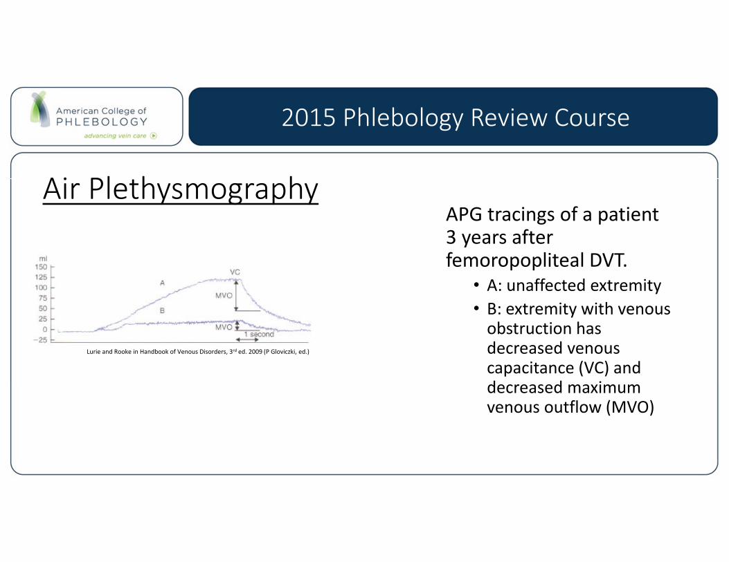
Air PlethysmographyAPG tracings of a patient 3 years after femoropopliteal DVT.
• A: unaffected extremity• B: extremity with venous obstruction has decreased venous capacitance (VC) and decreased maximum venous outflow (MVO)
Lurie and Rooke in Handbook of Venous Disorders, 3rd ed. 2009 (P Gloviczki, ed.)

2015 Phlebology Review Course
• Advantages• Assesses global lower limb hemodynamics
• Can be used to select patients who would benefit from intervention
• Can be used to evaluate effect of noninvasive therapeutic measures such as limb compression
• Limitations• Not able to identify specific incompetent valve sites
• May be difficult for patients to perform rapid maneuvers
Air Plethysmography

2015 Phlebology Review Course
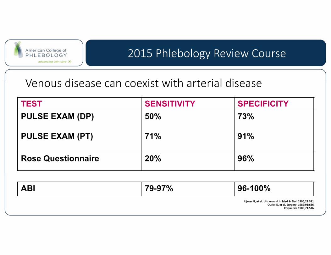
Venous disease can coexist with arterial disease
• Up to 1/3 of patients with chronic venous ulceration may have concomitant arterial insufficiency (Br J Surgery 1986;73:693‐6)
• There are case reports of compression therapy causing serious limb outcomes
• Patients with venous disease should have screening for arterial disease
2015 Phlebology Review Course
Venous disease can coexist with arterial disease
TEST SENSITIVITY SPECIFICITYPULSE EXAM (DP)
PULSE EXAM (PT)
50%
71%
73%
91%
Rose Questionnaire 20% 96%
ABI 79-97% 96-100%Lijmer G, et al. Ultrasound in Med & Biol. 1996;22:391.
Ouriel K, et al. Surgery. 1982;91:686.Criqui Circ 1985;71:516.

2015 Phlebology Review Course
ABI =
• PAD screening exam• Ankle and brachial systolic pressures taken using a Doppler sensor• Can be done bedside with a hand‐held Doppler device
Ankle‐Brachial IndexAnkle systolic pressureBrachial systolic pressure

2015 Phlebology Review Course
ABI Calculation
Normal 1.00‐1.40Borderline 0.91‐0.99PAD <0.90
Moderate PAD < 0.70Severe PAD < 0.40
Non‐compressible >1.40

2015 Phlebology Review Course
• Advantages• Simple, in office procedure• High sensitivity and specificity• Screening tool not just for PAD but also for CVD and CAD
• Limitations• Does not localize disease within the limb
• Falsely elevated ABI for patients with calcified vessels, obese
• Decreased sensitivity for mild disease
• ½ or more of patients with borderline ABI at rest will have fall in ABI with exercise
Ankle Brachial Index

2015 Phlebology Review Course
Non‐Compressible Vessels (ABI >1.4)• Ankle pressure of 255 mm Hg artifact, not severe hypertension; arteries cannot be compressed
• Partial non‐compressibility with ABI < 1.4 possible
• Occurs in 5‐8.5% of patients referred for ABI1,2,3
• Associated with vascular calcification or obesity• Diabetes mellitus• Renal failure• Hyperparathyroidism
• ABI number is NOT interpretable in this setting• Use alternate test to establish the diagnosis of PAD (Toe‐
brachial index, Pulse volume recordings, imaging (duplex, CTA, MRA))
1Allision MA, et al. JACC. 2008;51:12922Suominen V, Eur J Vasc Endovasc Surg. 2008;35:709.3Sutton‐Tyrrell, K. et al. Stroke 2008;39:863‐869

2015 Phlebology Review Course
• History:• Rose questionnaire (96% specificity)
• Physical exam:• Pulse palpation (femoral, popliteal, DP, PT)• CW Doppler • Auscultation for bruits bilateral groins
• ABI:• What if the ABI does not match the exam or the patient’s symptoms?
Peripheral Arterial EvaluationRight ABI = 0.83
Left ABI = 0.89
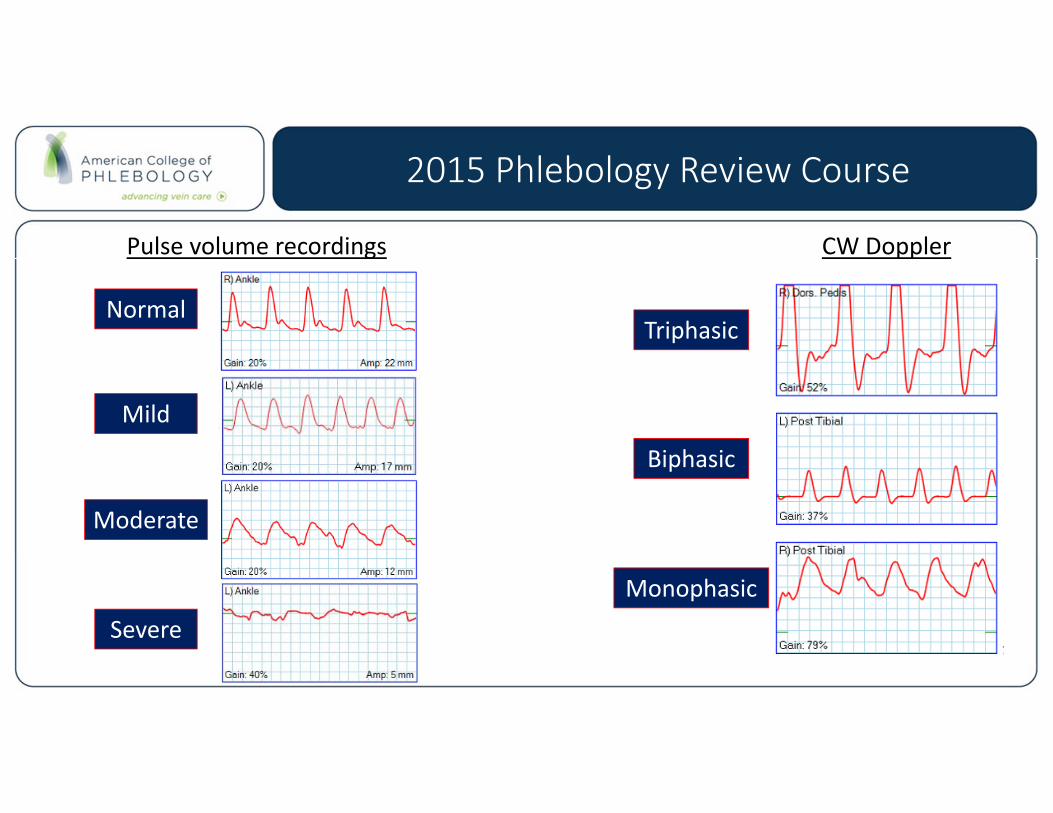
Normal
2015 Phlebology Review Course
Normal
Mild
Moderate
Severe
Triphasic
Biphasic
Monophasic
Pulse volume recordings CW Doppler

2015 Phlebology Review Course
Toe‐Brachial Index (TBI) – ABI does not match exam• Use when ABI > 1.4 or partial non‐compressible vessels as digit pressures are almost always compressible
• Measure great toe pressure using small digit cuff and a flow sensor (PPG)
• Vasoconstriction can be a confounder warm room or toe warming
• Normal TBI > 0.7
• Some labs may use a 0.8 cutpoint for TBI
Nursing. 2003;33:54.
Toe PressureBrachial Pressure
TBI =

2015 Phlebology Review Course
Non‐Compressible Vessels
• R ABI 1.10, L ABI 1.08• Waveforms do not match ABIs• R TBI 0.43, L TBI 0.69• R moderate disease • L mild disease

2015 Phlebology Review Course
Exercise ABI –ABI does not match symptoms• Lower extremity “stress test”• PAD suspected but ABI is normal or borderline at rest
• Obtain ABI and ankle PVR tracings before and immediately post exercise
• Interpretation• Normal: No significant drop in ankle pressure or ABI with exercise
• Abnormal: Fall in ankle pressure or ABI with change in PVR waveform

2015 Phlebology Review Course
57 sec ‐ onset of bilateral calf tightness2 min 31 sec ‐ tightness radiating to left thigh

2015 Phlebology Review Course
I’ve diagnosed PAD, now what?• Further imaging if revascularization is planned
• Arterial duplex ultrasound• CTA• MRA• Catheter‐based angiography
• Medical Management• Treatment of claudication symptoms (cilostazl, PAD rehab)• PAD is a CAD risk equivalent (antiplatelet, statin, antihypertensives, etc)• Referral to a cardiovascular specialist for ongoing care

2015 Phlebology Review Course
Thank You