Embed Size (px)
Citation preview

AMERICAN THORACIC SOCIETYDOCUMENTS
Diagnosis of Idiopathic Pulmonary FibrosisAn Official ATS/ERS/JRS/ALAT Clinical Practice Guideline: ExecutiveSummaryGanesh Raghu, Martine Remy-Jardin, Jeffrey L. Myers, Luca Richeldi, Christopher J. Ryerson, David J. Lederer,Juergen Behr, Vincent Cottin, Sonye K. Danoff, Ferran Morell, Kevin R. Flaherty, Athol Wells, Fernando J. Martinez,Arata Azuma, Thomas J. Bice, Demosthenes Bouros, Kevin K. Brown, Harold R. Collard, Abhijit Duggal, Liam Galvin,Yoshikazu Inoue, R. Gisli Jenkins, Takeshi Johkoh, Ella A. Kazerooni, Masanori Kitaichi, Shandra L. Knight,George Mansour, Andrew G. Nicholson, Sudhakar N. J. Pipavath, Ivette Buendıa-Roldan, Moises Selman,William D. Travis, Simon L. F. Walsh, and Kevin C. Wilson; on behalf of the American Thoracic Society, EuropeanRespiratory Society, Japanese Respiratory Society, and Latin American Thoracic Society
THIS OFFICIAL CLINICAL PRACTICE GUIDELINE OF THE AMERICAN THORACIC SOCIETY (ATS), EUROPEAN RESPIRATORY SOCIETY (ERS), JAPANESE RESPIRATORY
SOCIETY (JRS), AND LATIN AMERICAN THORACIC SOCIETY (ALAT) WAS APPROVED BY THE ATS, JRS, AND ALAT MAY 2018, AND THE ERS JUNE 2018
Background: This document provides clinical recommendationsfor the diagnosis of idiopathic pulmonary fibrosis (IPF). It representsa collaborative effort between the American Thoracic Society,European Respiratory Society, Japanese Respiratory Society, andLatin American Thoracic Society.
Methods: The evidence syntheses were discussed andrecommendations formulated by a multidisciplinary committee ofIPF experts. The evidence was appraised and recommendations wereformulated, written, and graded using the Grading ofRecommendations, Assessment, Development, and Evaluationapproach.
Results: The guideline panel updated the diagnostic criteria for IPF.Previously defined patterns of usual interstitial pneumonia (UIP)were refined to patterns of UIP, probable UIP, indeterminate forUIP, and alternate diagnosis. For patients with newly detectedinterstitial lung disease (ILD) who have a high-resolution computedtomography scan pattern of probable UIP, indeterminate for UIP, or
an alternative diagnosis, conditional recommendations were madefor performing BAL and surgical lung biopsy; due to lack of evidence,no recommendation was made for or against performingtransbronchial lung biopsy or lung cryobiopsy. In contrast, forpatients with newly detected ILD who have a high-resolutioncomputed tomography pattern of UIP, strong recommendationswere made against performing surgical lung biopsy, transbronchiallung biopsy, and lung cryobiopsy, and a conditional recommendationwas made against performing BAL. Additional recommendationsincluded a conditional recommendation for multidisciplinarydiscussion and a strong recommendation against measurement ofserum biomarkers for the sole purpose of distinguishing IPF fromother ILDs.
Conclusions: The guideline panel provided recommendationsrelated to the diagnosis of IPF.
Keywords: idiopathic pulmonary fibrosis; interstitial lung disease;pulmonary fibrosis
This Executive Summary is part of the full official ATS guideline, which readers may access online at http://www.atsjournals.org/doi/suppl/10.1164/rccm.201807-1255ST. Only the Executive Summary is appearing in the print edition of the Journal. The article of record, and the one that should be cited, is:Diagnosis of Idiopathic Pulmonary Fibrosis: An Official ATS/ERS/JRS/ALAT Clinical Practice Guideline. Am J Respir Crit Care Med 2018;198:e44–e68.Available at http://www.atsjournals.org/doi/suppl/10.1164/rccm.201807-1255ST.
ORCID IDs: 0000-0001-7506-6643 (G.R.); 0000-0001-8247-3028 (J.L.M.); 0000-0001-8594-1448 (L.R.); 0000-0001-5258-0228 (D.J.L.);0000-0002-9151-4829 (J.B.); 0000-0002-5591-0955 (V.C.); 0000-0001-9172-8977 (S.K.D.); 0000-0002-7206-4543 (F.M.); 0000-0003-2657-1314 (K.R.F.); 0000-0003-2108-6248 (A.W.); 0000-0002-2412-3182 (F.J.M.); 0000-0001-7300-3219 (T.J.B.); 0000-0002-0685-0765 (D.B.);0000-0002-8558-6711 (K.K.B.); 0000-0003-4220-2359 (A.D.); 0000-0001-5859-8744 (E.A.K.); 0000-0003-3257-102X (A.G.N.); 0000-0001-6948-2376 (S.N.J.P.); 0000-0002-8230-0749 (I.B.-R.); 0000-0002-1022-4783 (M.S.); 0000-0003-3160-6729 (W.D.T.); 0000-0003-0497-5297 (S.L.F.W.); 0000-0003-4429-2263 (K.C.W.).
Correspondence and requests for reprints should be addressed to Ganesh Raghu, M.D., Center for Interstitial Lung Diseases, University of Washington, 1959NEPacific Street, Seattle, WA 98195. E-mail: [email protected].
This article has an online supplement, which is accessible from this issue’s table of contents at www.atsjournals.org.
Am J Respir Crit Care Med Vol 198, Iss 5, pp 563–580, Sep 1, 2018
Copyright © 2018 by the American Thoracic Society
DOI: 10.1164/rccm.201807-1255ST
Internet address: www.atsjournals.org
American Thoracic Society Documents 563

ContentsSummary of RecommendationsIntroductionMethodsClinical ManifestationsDiagnosis
HRCT TechniqueHRCT PatternsSLB TechniqueHistopathology Patterns
Diagnostic Criteria for IPFDiagnostic Interventions
Question 1: Should Patientswith Newly Detected ILD ofUnknown Cause Who AreClinically Suspected of HavingIPF Undergo a Detailed,Prompted History ofMedication Use andEnvironmental Exposures atHome, Work, and Other Places
the Patient Frequently Visits toExclude Potential Causes ofthe ILD?
Question 2: Should Patientswith Newly Detected ILD ofUnknown Cause Who AreClinically Suspected of HavingIPF Undergo SerologicalTesting to Exclude CTDs asPotential Causes of the ILD?
Question 3: Should Patientswith Newly Detected ILD ofUnknown Cause Who AreClinically Suspected of HavingIPF Undergo Cellular Analysisof Their BAL Fluid?
Question 4: For Patients withNewly Detected ILD ofUnknown Cause Who AreClinically Suspected of HavingIPF, Should SLB Be Performed
to Ascertain theHistopathology Diagnosis ofUIP Pattern?
Question 5: For Patients withNewly Detected ILD ofUnknown Cause Who AreClinically Suspected of HavingIPF, Is TBBx a ReasonableAlternative to SLB to Ascertainthe Histopathology Diagnosis ofUIP Pattern?
Question 6: For Patients withNewly Detected ILD ofUnknown Cause Who AreClinically Suspected of HavingIPF, Is Lung Cryobiopsy aReasonable Alternative toSLB to Ascertain theHistopathology Diagnosis ofUIP Pattern?
Conclusions
Summary ofRecommendations
Adult patients with newly detected interstitiallung disease (ILD) of apparently unknowncause are clinically suspected of havingidiopathic pulmonary fibrosis (IPF) if theyhave unexplained symptomatic or asymptomaticpatterns of bilateralfibrosis on a chest radiographor chest computed tomography (CT), bibasilarinspiratory crackles, and an age typically olderthan 60 years. Rarely, middle-aged adults(.40 yr and ,60 yr), especially those withrisks for familial pulmonary fibrosis, mayotherwise manifest the same clinical scenarioas the typical patient older than 60 years. Therecommendations in this guideline are for thepatterns and distributions of images obtainedby high-resolution CT (HRCT) and, thus,require that patients be subjected to HRCT ofthe chest for evaluation.
For adult patients with newly detectedILD of apparently unknown cause who areclinically suspected of having IPF:
d We recommend taking a detailedhistory of both medication use andenvironmental exposures at home, work,and other places the patient frequentlyvisits to exclude potential causes of ILD(motherhood statement).
d We recommend serological testing to excludeconnective tissue disease as a potential causeof the ILD (motherhood statement).
For patients with newly detected ILDof apparently unknown cause who areclinically suspected of having IPF andhave an HRCT pattern of probable usualinterstitial pneumonia (UIP), indeterminatefor UIP, or an alternative diagnosis:
d We suggest cellular analysis of their BALfluid (conditional recommendation, verylow quality of evidence).
d We suggest surgical lung biopsy (SLB)(conditional recommendation, very lowquality of evidence).
d The panel made no recommendation for oragainst transbronchial lung biopsy (TBBx).
d The panel made no recommendation foror against lung cryobiopsy.
For patients with newly detected ILDof apparently unknown cause who areclinically suspected of having IPF and havean HRCT pattern of UIP:
d We suggest NOT performing cellularanalysis of their BAL fluid (conditionalrecommendation, very low quality of evidence).
d We recommend NOT performing SLB(strong recommendation, very low qualityof evidence).
d We recommend NOT performing TBBx(strong recommendation, very low qualityof evidence).
d We recommend NOT performing lungcryobiopsy (strong recommendation, verylow quality of evidence).
For patients with newly detected ILDof apparently unknown cause who areclinically suspected of having IPF:
d We suggest multidisciplinary discussion(MDD) for diagnostic decision-making(conditional recommendation, very lowquality of evidence).
d We recommend NOT measuring serumMMP (matrix metalloproteinase)-7, SPD(surfactant protein D), CCL (chemokineligand)-18, or KL (Krebs von den Lungen)-6 for the purpose of distinguishing IPFfrom other ILDs (strong recommendation,very low quality of evidence).
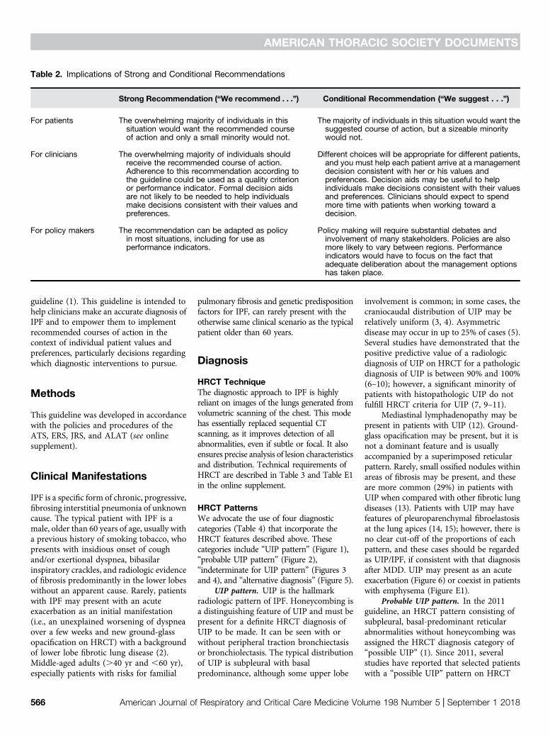
For comparison of the 2018 and 2011diagnostic recommendations, see Table 1.For an explanation of strong andconditional recommendations, see Table 2.
Introduction
The American Thoracic Society (ATS),European Respiratory Society (ERS),Japanese Respiratory Society (JRS), andLatin American Thoracic Society (ALAT)collaborated to develop clinical practiceguidelines for the diagnosis andmanagement of IPF in 2011 (1). Newevidence now enables us to improve thediagnostic criteria. The recommendationsin this 2018 guideline are revisions of thediagnostic recommendations in the 2011
AMERICAN THORACIC SOCIETY DOCUMENTS
564 American Journal of Respiratory and Critical Care Medicine Volume 198 Number 5 | September 1 2018

Tab
le1.
Com
parison
ofATS
/ERS/JRS/ALA
TRec
ommen
dations
fortheDiagn
osis
ofIPFin
the20
11an
d20
18Guidelines
2018
Guideline
HRCTPattern
ofProbab
leUIP*,
IndeterminateforUIP,an
dAlterna
tive
Diagno
sis
HRCTPattern
ofUIP*
2011
Guideline:
Did
NotDisting
uish
among
PatientswithDifferen
tHRCT
Patterns
BALce
llularan
alys
isWesu
gges
tperform
ingBAL
cellularan
alys
is(con
dition
al)
Wesu
gges
tNOTperform
ingBAL
cellularan
alys
is(con
dition
al)
“BALce
llularan
alys
issh
ould
notbe
perform
edin
thediagn
ostic
evalua
tion
ofIPFin
themajority
ofpatients,
but
may
beap
propria
tein
aminority
ofpatients.”
Surgica
llun
gbiopsy
Wesu
gges
tperform
ingsu
rgical
lung
biopsy
(con
dition
al)
Wereco
mmen
dNOTperform
ing
surgical
lung
biopsy
(stron
g)“S
urgica
llun
gbiopsy
isno
trequired
forpatientswith
anHRCTpattern
cons
istent
with
UIP.”
Tran
sbronc
hial
lung
biopsy
Noreco
mmen
dationwas
mad
eeither
foror
agains
ttran
sbronc
hial
lung
biopsy
Wereco
mmen
dNOTperform
ing
tran
sbronc
hial
lung
biopsy
(stron
g)“Trans
bronc
hial
biopsy
shou
ldno
tbe
used
intheev
alua
tionof
IPFin
the
majority
ofpatients,
but
may
be
appropria
tein
aminority
.”
Lung
cryo
biopsy
Noreco
mmen
dationwas
mad
eeither
foror
agains
tcryo
biopsy
Wereco
mmen
dNOTperform
ing
cryo
biopsy
(stron
g)Not
address
ed
Med
ical
historyof
med
icationus
ean
den
vironm
entale
xpos
ures
Wereco
mmen
dtaking
adetailedhistoryof
bothmed
icationus
ean
den
vironm
entale
xpos
ures
atho
me,
work,
andothe
rplace
sthepatient
freq
uently
visits
toex
clud
epoten
tialc
ause
sof
ILD
(mothe
rhoo
dstatem
ent)
“Diagn
osis
ofIPFrequiresex
clus
ionof
othe
rkno
wnca
uses
ofILD(e.g.,do
mestic
andoc
cupa
tiona
lenviro
nmen
tal
expo
sures,
conn
ectivetissuedisease,
anddrug
toxicity).”
Serolog
ical
testingto
exclud
eco
nnec
tivetis
suedisea
seWereco
mmen
dse
rologica
ltes
tingto
exclud
eco
nnec
tivetis
suedisea
sesas
apoten
tialc
ause
oftheILD
(mothe
rhoo
dstatem
ent)
“Diagn
osis
ofIPFrequiresex
clus
ion
ofothe
rkn
ownca
uses
ofILD
(e.g.,dom
estic
andoc
cupationa
len
vironm
entale
xpos
ures
,co
nnec
tive
tissu
edisea
se,an
ddrugtoxicity).”
Multid
isciplinarydiscu
ssion
Wesu
gges
tmultid
isciplinarydiscu
ssionfordec
ision-mak
ing(con
dition
al)
“Wereco
mmen
dthat
amultid
isciplinary
discu
ssionsh
ould
beus
edin
the
evalua
tionof
IPF.”
Serum
biomarke
rsWereco
mmen
dNOTmea
surin
gse
rum
MMP-7,SPD,CCL-18
,or
KL-6forthe
purpos
eof
distin
guishing
IPFfrom
othe
rILDs(stron
g)Not
address
ed
Definitionofabbreviatio
ns:
ALAT=Latin
Americ
anThoracic
Society;ATS=Americ
anThoracic
Society;CCL-18=chemokineligand18;ERS=EuropeanResp
iratory
Society;HRCT=
high-reso
lutio
ncomputedtomography;
ILD=interstitiallungdisease;IPF=idiopathic
pulm
onary
fibrosis;
JRS=Ja
panese
Resp
iratory
Society;KL-6
=Krebsvo
ndenLungen-6;MMP-7
=matrixmetalloproteinase
7;SPD=su
rfactantprotein
D;UIP
=usu
alinterstitialpneumonia.
Thequalityofevidenceforallrecommendatio
nsin
the2018guidelinewasvery
low.
*ThepatternsofUIP
have
beenrefinedin
these
2018guidelines,
comparedwith
the2011guidelines.
AMERICAN THORACIC SOCIETY DOCUMENTS
American Thoracic Society Documents 565
guideline (1). This guideline is intended tohelp clinicians make an accurate diagnosis ofIPF and to empower them to implementrecommended courses of action in thecontext of individual patient values andpreferences, particularly decisions regardingwhich diagnostic interventions to pursue.
Methods
This guideline was developed in accordancewith the policies and procedures of theATS, ERS, JRS, and ALAT (see onlinesupplement).
Clinical Manifestations
IPF is a specific form of chronic, progressive,fibrosing interstitial pneumonia of unknowncause. The typical patient with IPF is amale, older than 60 years of age, usually witha previous history of smoking tobacco, whopresents with insidious onset of coughand/or exertional dyspnea, bibasilarinspiratory crackles, and radiologic evidenceof fibrosis predominantly in the lower lobeswithout an apparent cause. Rarely, patientswith IPF may present with an acuteexacerbation as an initial manifestation(i.e., an unexplained worsening of dyspneaover a few weeks and new ground-glassopacification on HRCT) with a backgroundof lower lobe fibrotic lung disease (2).Middle-aged adults (.40 yr and ,60 yr),especially patients with risks for familial
pulmonary fibrosis and genetic predispositionfactors for IPF, can rarely present with theotherwise same clinical scenario as the typicalpatient older than 60 years.
Diagnosis
HRCT TechniqueThe diagnostic approach to IPF is highlyreliant on images of the lungs generated fromvolumetric scanning of the chest. This modehas essentially replaced sequential CTscanning, as it improves detection of allabnormalities, even if subtle or focal. It alsoensures precise analysis of lesion characteristicsand distribution. Technical requirements ofHRCT are described in Table 3 and Table E1in the online supplement.
HRCT PatternsWe advocate the use of four diagnosticcategories (Table 4) that incorporate theHRCT features described above. Thesecategories include “UIP pattern” (Figure 1),“probable UIP pattern” (Figure 2),“indeterminate for UIP pattern” (Figures 3and 4), and “alternative diagnosis” (Figure 5).
UIP pattern. UIP is the hallmarkradiologic pattern of IPF. Honeycombing isa distinguishing feature of UIP and must bepresent for a definite HRCT diagnosis ofUIP to be made. It can be seen with orwithout peripheral traction bronchiectasisor bronchiolectasis. The typical distributionof UIP is subpleural with basalpredominance, although some upper lobe
involvement is common; in some cases, thecraniocaudal distribution of UIP may berelatively uniform (3, 4). Asymmetricdisease may occur in up to 25% of cases (5).Several studies have demonstrated that thepositive predictive value of a radiologicdiagnosis of UIP on HRCT for a pathologicdiagnosis of UIP is between 90% and 100%(6–10); however, a significant minority ofpatients with histopathologic UIP do notfulfill HRCT criteria for UIP (7, 9–11).
Mediastinal lymphadenopathy may bepresent in patients with UIP (12). Ground-glass opacification may be present, but it isnot a dominant feature and is usuallyaccompanied by a superimposed reticularpattern. Rarely, small ossified nodules withinareas of fibrosis may be present, and theseare more common (29%) in patients withUIP when compared with other fibrotic lungdiseases (13). Patients with UIP may havefeatures of pleuroparenchymal fibroelastosisat the lung apices (14, 15); however, there isno clear cut-off of the proportions of eachpattern, and these cases should be regardedas UIP/IPF, if consistent with that diagnosisafter MDD. UIP may present as an acuteexacerbation (Figure 6) or coexist in patientswith emphysema (Figure E1).
Probable UIP pattern. In the 2011guideline, an HRCT pattern consisting ofsubpleural, basal-predominant reticularabnormalities without honeycombing wasassigned the HRCT diagnosis category of“possible UIP” (1). Since 2011, severalstudies have reported that selected patientswith a “possible UIP” pattern on HRCT
Table 2. Implications of Strong and Conditional Recommendations
Strong Recommendation (“We recommend . . .”) Conditional Recommendation (“We suggest . . .”)
For patients The overwhelming majority of individuals in thissituation would want the recommended courseof action and only a small minority would not.
The majority of individuals in this situation would want thesuggested course of action, but a sizeable minoritywould not.
For clinicians The overwhelming majority of individuals shouldreceive the recommended course of action.Adherence to this recommendation according tothe guideline could be used as a quality criterionor performance indicator. Formal decision aidsare not likely to be needed to help individualsmake decisions consistent with their values andpreferences.
Different choices will be appropriate for different patients,and you must help each patient arrive at a managementdecision consistent with her or his values andpreferences. Decision aids may be useful to helpindividuals make decisions consistent with their valuesand preferences. Clinicians should expect to spendmore time with patients when working toward adecision.
For policy makers The recommendation can be adapted as policyin most situations, including for use asperformance indicators.
Policy making will require substantial debates andinvolvement of many stakeholders. Policies are alsomore likely to vary between regions. Performanceindicators would have to focus on the fact thatadequate deliberation about the management optionshas taken place.
AMERICAN THORACIC SOCIETY DOCUMENTS
566 American Journal of Respiratory and Critical Care Medicine Volume 198 Number 5 | September 1 2018

according to the 2011 guidelines are highlylikely to have histopathologic UIP despitethe absence of radiologic honeycombing.Specifically, an HRCT pattern of possibleUIP with peripheral traction bronchiectasisor bronchiolectasis in the correct clinicalsetting likely represents histopathologicUIP on biopsy (4, 16–18). Therefore,subpleural, basal-predominant reticularabnormalities with peripheral traction
bronchiectasis or bronchiolectasis shouldbe regarded as “probable UIP.” As with aUIP pattern, ground-glass opacificationmay be present in probable UIP, but it isnot a dominant feature. Many patients withan HRCT pattern of probable UIP will bedetermined to have IPF once other factorssuch as histopathology are considered.
Indeterminate for UIP pattern. It is nowrecognized that atypical HRCT features
frequently (i.e., about 30%) accompany ahistopathologic pattern of UIP/IPF (19).Therefore, the category “indeterminate for UIPpattern” should be assigned when HRCTdemonstrates features of fibrosis but does notmeet UIP or probable UIP criteria and doesnot explicitly suggest an alternative diagnosis.This category includes a subset of patientswith very limited subpleural ground-glassopacification or reticulation without obvious
Table 3. High-Resolution Computed Tomography Scanning Parameters
Recommended Scanning Protocol Advantages of Updated Recommendations
1. Noncontrast examination —
2. Volumetric acquisition with selection of:d Sub-millimetric collimationd Shortest rotation timed Highest pitchd Tube potential and tube current appropriate to patient size:∘ Typically 120 kVp and <240 mAs∘ Lower tube potentials (e.g., 100 kVp) with adjustment of tubecurrent encouraged for thin patients
d Use of techniques available to avoid unnecessary radiationexposure (e.g., tube current modulation)
A. Acquisition covering the entire lung volume (vs. analysis of10% of lung volume with sequential scanning)d No risk of missing subtle infiltrative abnormalitiesd Possibility of multiplanar reformations, helpful for analysisof the ILD pattern and predominant distribution of lungchanges
d Possibility of post-processing to optimize detection ofsubtle hypoattenuated lesions (minimum intensityprojection) and micronodular infiltration (maximumintensity projection)
d Possibility of detection of additional lesions (e.g., incidentalidentification of lung nodule or focal consolidation in lungfibrosis that may correspond to lung carcinoma)
d Optimal to assess progression or improvement in patient’sfollow-up
B. Dramatic increase in temporal resolution and speed of dataacquisition
d Motion-free images
C. Availability of numerous dose-reduction tools
3. Reconstruction of thin-section CT images (<1.5 mm):d Contiguous or overlappingd Using a high-spatial-frequency algorithmd Iterative reconstruction algorithm if validated on the CT unit(if not, filtered back projection)
—
4. Number of acquisitions:d Supine: inspiratory (volumetric)d Supine: expiratory (can be volumetric or sequential)d Prone: only inspiratory scans (can be sequential or volumetric);optional (see text)
d Inspiratory scans obtained at full inspiration
A. Expiratory scans useful to detect air trapping
B. Prone scans allow analysis of peripheral lung changeswithout dependent lung atelectasis that may be mistakenfor abnormal lung infiltration or mimic disease (e.g.,pseudohoneycombing when combined with paraseptalemphysema)
C. Inadequate inspiration increases lung attenuation (whichshould not be interpreted as ground-glass attenuation)and is responsible for dependent lung atelectasis (whichmay mimic abnormal lung infiltration or mask subtleabnormalities)
5. Recommended radiation dose for the inspiratory volumetricacquisition:
d 1–3 mSv (i.e., “reduced” dose)d Strong recommendation to avoid “ultralow-dose CT” (,1 mSv)
A. Considerable dose reduction compared to conventionalscanning
Definition of abbreviations: CT = computed tomography; ILD = interstitial lung disease.
AMERICAN THORACIC SOCIETY DOCUMENTS
American Thoracic Society Documents 567

CT features of fibrosis for whom there is asuspicion that early UIP or probable UIPis present. In such cases, it should beconfirmed with prone inspiratory views thatthe subpleural opacities do not representdependent atelectasis (Figure E2).
Alternative diagnosis. In some casesof fibrotic lung disease, there is clinicalsuspicion of IPF, but the HRCT patternsuggests an alternative diagnosis. Examplesinclude bronchocentric fibrosis in the upperlobes or profuse mosaic attenuation thatsuggests hypersensitivity pneumonitis,posterior fibrotic retraction of the hila insarcoidosis, or extensive ground-glassopacification with subpleural sparing infibrotic nonspecific interstitial pneumonia(NSIP). Occasionally, the HRCT presentationmay be that of a UIP, probable UIP, orindeterminate for UIP pattern, but ancillaryfindings suggest an alternative diagnosis. Insuch situations, an alternative diagnosis to IPFshould be reconsidered.
CT findings in the presence of anacute exacerbation. Patients with an acuteexacerbation of IPF have bilateral ground-glass opacification with or withoutconsolidation on a background of lungfibrosis (Figure 6). In the absence of aprevious HRCT study, bilateral ground-glass opacity and/or consolidation on abackground of a UIP pattern is highlysuggestive of an acute exacerbation andcan be used to confirm an underlyingIPF diagnosis in the appropriate clinicalcontext.
SLB TechniqueVideo-assisted thoracoscopic surgery is thepreferred approach to SLB for patientswho can tolerate single-lung ventilation,rather than open thoracotomy. In patientswith severe physiologic impairment orsubstantial comorbidity, the risks of SLBmay outweigh the benefits of establishing asecure diagnosis of IPF; therefore, the final
decision regarding whether or not to pursuea biopsy must be tailored to the clinicalsituation of the individual patient. Multiplebiopsies should be obtained from two tothree lobes, because the histologic patternson SLB specimens obtained from differentsegments can be discordant (e.g., coexistingUIP pattern and fibrotic NSIP pattern fromdifferent lobes).
Histopathology PatternsWe recommend categorizing histopathologicfindings of biopsies into “UIP” (Figure 7),“probable UIP,” “indeterminate for UIP,” and“alternative diagnosis” (Table 5). Biopsiesdesignated as indeterminate for UIPdemonstrate a pattern of fibrosis that doesnot meet criteria for UIP or any otherhistopathologic pattern of fibrotic interstitialpneumonia and, in some cases, may favor analternative diagnosis while not categoricallyexcluding the possibility of sampling bias in apatient who ultimately proves to have UIP.
Table 4. High-Resolution Computed Tomography Scanning Patterns
UIP Probable UIP Indeterminate for UIP Alternative Diagnosis
Subpleural andbasal predominant;distribution is oftenheterogeneous*
Subpleural andbasal predominant;distribution is oftenheterogeneous
Subpleural and basal predominant Findings suggestive of anotherdiagnosis, including:
Honeycombing with or withoutperipheral tractionbronchiectasis orbronchiolectasis†
Reticular pattern with peripheraltraction bronchiectasis orbronchiolectasis
Subtle reticulation; may have mildGGO or distortion (“early UIPpattern”)
d CT features:
May have mild GGO
CT features and/or distribution oflung fibrosis that do not suggestany specific etiology (“trulyindeterminate for UIP”)
∘ Cysts∘ Marked mosaicattenuation
∘ Predominant GGO∘ Profuse micronodules∘ Centrilobular nodules∘ Nodules∘ Consolidation
d Predominant distribution:∘ Peribronchovascular∘ Perilymphatic∘ Upper or mid-lung
d Other:∘ Pleural plaques (considerasbestosis)
∘ Dilated esophagus(consider CTD)
∘ Distal clavicular erosions(consider RA)
∘ Extensive lymph nodeenlargement (considerother etiologies)
∘ Pleural effusions, pleuralthickening (considerCTD/drugs)
Definition of abbreviations: CT = computed tomography; CTD = connective tissue disease; GGO= ground-glass opacities; RA = rheumatoid arthritis;UIP = usual interstitial pneumonia.*Variants of distribution: occasionally diffuse, may be asymmetrical.†Superimposed CT features: mild GGO, reticular pattern, pulmonary ossification.
AMERICAN THORACIC SOCIETY DOCUMENTS
568 American Journal of Respiratory and Critical Care Medicine Volume 198 Number 5 | September 1 2018

A subset of patients with previously occultIPF may present with an acute exacerbation,which is commonly characterized by acombination of a UIP pattern complicatedby superimposed diffuse alveolar damagewith or without associated hyalinemembranes.
Diagnostic Criteria for IPF
Diagnosis of IPF requires the following:
1. Exclusion of other known causes of ILD(e.g., domestic and occupationalenvironmental exposures, connectivetissue disease [CTD], drug toxicity),and either #2 or #3
2. The presence of the HRCT pattern ofUIP (Table 4)
3. Specific combinations (Figure 8) ofHRCT patterns (Table 4) andhistopathology patterns (Table 5) inpatients subjected to lung tissuesampling
The guideline panel’s approachto diagnosis is summarized in Figures 8and 9. It is based on these 2018
guidelines and the 2011 guidelines (1)and is similar to that suggested by atask force sponsored by the FleischnerSociety (20).
Diagnostic Interventions
The questions below are specifically intendedfor patients who are “clinically suspectedof having IPF.” This classically refers topatients with unexplained symptomatic orasymptomatic bilateral pulmonary fibrosison a chest radiograph or chest CT scan,bibasilar inspiratory crackles, and an agetypically older than 60 years. It must berecognized that the questions addressed arenot restricted to patients older than 60 years,as middle-aged adults (.40 yr and ,60 yr),especially patients with risks for familialpulmonary fibrosis, can rarely present withthe otherwise same clinical scenario as thetypical patient older than 60 years. Therecommendations in this guideline are forthe patterns and distributions of imagesobtained by HRCT and, thus, require thatpatients be subjected to HRCT of the chestfor evaluation.
Question 1: Should Patients withNewly Detected ILD of UnknownCause Who Are Clinically Suspectedof Having IPF Undergo a Detailed,Prompted History of Medication Useand Environmental Exposures atHome, Work, and Other Places thePatient Frequently Visits to ExcludePotential Causes of the ILD?
Discussion. The guideline panel recognizedthere is no reasonable alternative to theproposed course of action, so a motherhoodstatement was made to take a detailed historyof medication use and environmentalexposures at home, work, and other placesthat the patient frequently visits, to identifyor exclude potential causes of ILD (e.g.,hypersensitivity pneumonitis, pneumoconiosis,drug toxicity). This is supported by anobservational study that enrolled 1,084 patientswith new-onset ILD of unknown causereporting that 47% of the patients wereidentified as having hypersensitivity pneumonitison detailed assessment, suggesting that a causecan be found in many patients who presentwith ILD (21). The panel’s clinical experienceis that identification and removal of potential
Figure 1. High-resolution computed tomography (CT) images demonstrating a usual interstitial pneumonia pattern. (A–C) Transverse CT section and(D) coronal reconstruction illustrating the presence of honeycombing with subpleural and basal predominance. Note the concurrent presence of mildground-glass opacity. (E) Magnified view of the left lower lobe showing typical characteristics of honeycombing, consisting of clustered cystic airspaceswith well-defined walls and variable diameters, seen in single or multiple layers (arrows).
AMERICAN THORACIC SOCIETY DOCUMENTS
American Thoracic Society Documents 569

causative environmental factors may result inimproved clinical outcomes.
Many panelists use publishedquestionnaires in their clinical practices toconsider environmental exposures at home,work, and frequently visited places (21–23).Such questionnaires may be tailored to culturalhabits and geographical differences. Examplesof pertinent exposures include mold, birds,down feathers, animals, metal dusts (e.g., brass,lead, steel), wood dust (e.g., pine), vegetabledust, exposure to livestock, stone polishing andcutting, medications taken, current or recentoccupations (e.g., hair dressing), and current orrecent hobbies (24–30). Although somepanelists use the presence of antibody in serumagainst specific antigen to prompt furtherevaluation for hypersensitivity pneumonitis, thetest is not standardized, and the specificityand sensitivity for the diagnosis ofhypersensitivity pneumonitis is unknown. Thepanelists who use serum antibody testingbelieve that such tests may identify an antigenthat was not suspected by clinical history and,therefore, may prompt further investigations forthe suspected etiology; also, if serum antibodytesting is negative, the results reinforce theconclusion that the patient does not havehypersensitivity pneumonitis.
ATS/ERS/JRS/ALAT recommendations.d For patients with newly detected ILD of
apparently unknown cause who areclinically suspected of having IPF,we recommend taking a detailedhistory of both medication use andenvironmental exposures at home, work,and other places the patient frequentlyvisits to exclude potential causes of theILD (motherhood statement).
Question 2: Should Patients with NewlyDetected ILD of Unknown Cause WhoAre Clinically Suspected of Having IPFUndergo Serological Testing to ExcludeCTDs as Potential Causes of the ILD?
Discussion. Diagnosis of IPF mandatesexclusion of other causes of ILD, includingCTD-related ILD (Table E2). The guidelinepanel concluded that foregoing serologicaltesting was not a reasonable alternative.Therefore, a motherhood statement was madeto perform routine serological testing in allpatients with newly identified ILD. Althoughthere was overwhelming agreement to performserological testing, there was far less agreementabout which serological tests to perform.
Themajority of panelists acknowledgedroutinely testing for CRP (C-reactive protein),erythrocyte sedimentation rate, antinuclearantibodies (by immunofluorescence),rheumatoid factor, myositis panel, andanti–cyclic citrullinated peptide. Other detailedtests are performed on a case-by-case basisaccording to associated symptoms and signs.These include muscle enzymes (creatininephosphokinase, myoglobin, and aldolase),antisynthetase antibodies (Jo-1 and othersif available), anti-MDA5 (melanomadifferentiation-associated protein 5), anti–Mi-2,anti-NXP2 (nuclear matrix protein 2), anti–TIF1-g (transcriptional intermediary factor1-g), anti-SRP (signal recognition particle),anti-HMGCR (3-hydroxy-3-methylglutaryl-CoAreductase), anti-SAE (small ubiquitin-relatedmodifier–activating enzyme), anti-U1RNP(U1 ribonucleoprotein), anti-PM/Scl75(polymyositis/scleroderma 75), anti-PM/Scl100,and anti-Ku (31). If systemic sclerosis(i.e., scleroderma) is suspected, additional testsinclude: anti–Scl-70/topoisomerase-1, anti-centromere, anti-RNA polymerase III, anti-U1RNP, anti-Th/To, anti-PMScl, U3 RNP(fibrillarin), and anti-Ku. If Sjogren syndrome issuspected, additional tests include: anti-SSA/Ro(Sjogren-specific antibody A) and anti-SSB/La.
Figure 2. Probable usual interstitial pneumonia (UIP) pattern. (A–C) Transverse computed tomography (CT) section, (D) coronal reconstruction of bothlungs, and (E) magnified sagittal view of the right lower lobe illustrating the presence of a reticular pattern with peripheral bronchiolectasis with subpleuraland basal predominance. Depending on their orientation relative to the plane of the CT section, peripheral traction bronchiolectasis appear as tubular(arrows) or cystic (arrowheads) structures. Note the concurrent presence of mild ground-glass opacities in the subpleural areas of both lungs and theabsence of honeycombing. UIP was proven at histology.
AMERICAN THORACIC SOCIETY DOCUMENTS
570 American Journal of Respiratory and Critical Care Medicine Volume 198 Number 5 | September 1 2018

If vasculitis is suspected, an additional testincludes anti-cytoplasmic antibodies. A smallminority of the panelists include all of thedetailed tests listed above as an “ILD panel” atinitial screening/baseline evaluation.
The guideline panelists do not refer allpatients with new ILD to a rheumatologist;rather, referring only those with positive clinicalmanifestations, serologies, or other characteristicsatypical for IPF (e.g., female, age,60 yr old). Inmany CTD-related ILDs, the lung disease is thefirst, dominant, or only feature of the CTDand, therefore, some patients will not fit standardrheumatologic diagnostic criteria at presentation.
ATS/ERS/JRS/ALAT recommendations.d For patients with newly detected ILD of
apparently unknown cause who areclinically suspected of having IPF, werecommend serological testing to aid inthe exclusion of CTDs as a potentialcause of the ILD (motherhood statement).
Question 3: Should Patients withNewly Detected ILD of UnknownCause Who Are Clinically Suspectedof Having IPF Undergo CellularAnalysis of Their BAL Fluid?
Evidence base. Our systematic literaturesearch yielded 2,492 titles but did not
Figure 3. Indeterminate for usual interstitial pneumonia (UIP) pattern (early UIP pattern). (A and B) Transverse computed tomography (CT) section, (C)coronal reconstruction of both lungs, and (D) magnified view of the right lung in supine position showing ground-glass opacity and subtle reticulation in thesubpleural areas (arrows) with a basal predominance. (E) Transverse CT section of the lower lung zones in prone position showing persistence of lunginfiltration in nondependent areas, thus excluding gravitational abnormalities. UIP was proven at histology.
Figure 4. Indeterminate for usual interstitial pneumonia pattern. (A–C) Transverse computedtomography sections showing extensive lung infiltration combining honeycombing, mild tomarked ground-glass opacity, asymmetrical distribution between both lungs, and no subpleuralpredominance.
AMERICAN THORACIC SOCIETY DOCUMENTS
American Thoracic Society Documents 571

identify any studies that 1) comparedclinical outcomes among patients whounderwent BAL cellular analysis to thosewho did not undergo BAL cellular analysis,or 2) reported the test characteristics ofBAL cellular analysis for distinguishing IPFfrom other ILDs. Therefore, we soughtstudies that compared BAL cell typeproportions among patients with IPF tothose among patients with other typesof ILD. The full text of 14 articles was
reviewed, and 8 were selected for analysis(32–39) (Tables E7a–E7f).
The eight studies enrolled patientswith IPF, performed BAL, and measuredcomponents of the BAL fluid, including thepercentage of neutrophils (32–37, 39),macrophages (32–36, 39), lymphocytes(32–39), and eosinophils (32, 34–37, 39), aswell as the CD4/CD8 ratio (32, 34, 36, 37).The measurements were then comparedwith similar measurement from patients
with other types of ILD, includinghypersensitivity pneumonitis (32, 33, 37),sarcoidosis (32, 36, 37), idiopathic NSIP (32,34, 37–39), cryptogenic organizingpneumonia (previously called bronchiolitisobliterans organizing pneumonia) (32–34,37), eosinophilic pneumonia (32),respiratory bronchiolitis-associated ILD(33), and lymphocytic interstitial pneumonia(33). Some BAL cell type proportions weremarkedly different in patients with IPFcompared with patients with other ILDs(Figure E3). Patients with IPF had a slightlyincreased proportion of eosinophilscompared with healthy individuals but amarkedly lower proportion of eosinophilsthan patients with eosinophilic pneumonia;thus, patients with a markedly elevatedproportion of eosinophils are more likely tohave eosinophilic pneumonia than IPF.Patients with IPF had a similar to slightlyhigher proportion of lymphocytes andCD4/CD8 ratio in their BAL than healthyindividuals but a markedly lower proportionof lymphocytes and CD4/CD8 ratio in theirBAL than patients with sarcoidosis; thus,patients with a markedly elevated proportionof lymphocytes and CD4/CD8 ratio aremore likely to have sarcoidosis than IPF.
Conclusions. When the panel weighedthe desirable consequences of BAL cellularanalysis in patients who have anHRCTpatternof probable UIP, indeterminate for UIP,or an alternative diagnosis (i.e., identifyingor excluding eosinophilic pneumonia,sarcoidosis, infection, and malignancy) versusthe undesirable consequences (i.e., risk of acomplication, burden, cost), themajority of thepanel concluded that the upsides of theprocedure outweigh the downsides in suchpatients. There was general agreement that
Figure 5. Computed tomography (CT) pattern suggestive of an alternative diagnosis for lung fibrosis. (A and B) Transverse CT sections obtained at deepinspiration showing disseminated lung infiltration, sparing some secondary pulmonary lobules in lung bases. (C) Transverse CT section obtained atexpiration confirming lobular air trapping, all findings being highly suggestive of chronic hypersensitivity pneumonitis.
Figure 6. Acute exacerbation of idiopathic pulmonary fibrosis. (A and B) Transverse computedtomography sections obtained in the upper and mid lung zones and (C and D) during acuteexacerbation showing newly developed, bilateral ground-glass opacification in both lungs on abackground of usual interstitial pneumonia pattern.
AMERICAN THORACIC SOCIETY DOCUMENTS
572 American Journal of Respiratory and Critical Care Medicine Volume 198 Number 5 | September 1 2018

BAL is appropriate when the radiologicdifferential diagnosis includes eosinophilicpneumonia, sarcoidosis, or infection. Incontrast, the panel concluded that alternativediagnoses that can be excluded by BAL cellularanalysis are sufficiently rare in patients whohave an HRCT pattern of UIP that thedownsides of the procedure typically outweighthe upsides in these patients.
ATS/ERS/JRS/ALAT recommendations.d For patients with newly detected ILD of
apparently unknown cause who are
clinically suspected of having IPF andhave an HRCT pattern of probableUIP, indeterminate for UIP, or analternative diagnosis, we suggestcellular analysis of their BAL fluid(conditional recommendation, very lowquality of evidence).
d For patients with newly detected ILD ofapparently unknown cause who areclinically suspected of having IPF andhave an HRCT pattern of UIP, wesuggest NOT performing cellular
analysis of their BAL fluid (conditionalrecommendation, very low quality ofevidence).
Question 4: For Patients with NewlyDetected ILD of Unknown Cause WhoAre Clinically Suspected of HavingIPF, Should SLB Be Performed toAscertain the Histopathology Diagnosisof UIP Pattern?
Evidence base. Our systematic literaturesearch yielded 945 titles but identified nostudies that compared clinical outcomesamong patients who underwent SLB tothose who did not. Thus, we selected studiesthat measured diagnostic yield of SLB usinga MDD as the diagnostic decision-maker.The full text of 54 articles was reviewed,and 26 were selected for analysis (40–65)(Table E8).
Pooling studies (unweighted) indicatedthat SLB obtained an adequate sample inall patients (11 studies; 918 of 918, 100%;95% confidence interval [CI], 99–100%),although the panel acknowledged that thisis not always the case in clinical practice.The proportion of SLBs that resulted in aspecific diagnosis (i.e., the diagnostic yield)was high (26 studies; 2,338 of 2,651, 88.2%;95% CI, 86.9–89.4%), with a minority beingdeemed unclassifiable (26 studies; 313of 2,651, 11.8%; 95% CI, 10.6–13.1%).Among final diagnoses, approximately one-third were IPF (24 studies; 752 of 2,360,31.9%; 95% CI, 30.0–33.8%), and manyothers were potentially treatable etiologieslike infection, sarcoidosis, hypersensitivitypneumonitis, eosinophilic pneumonia,lymphangioleiomyomatosis, cryptogenicorganizing pneumonia, and vasculitis.
Overall mortality was low (23 studies;79 of 2,268, 3.5%; 95%CI, 2.8–4.3%), but someof the deaths were probably disease related,because procedure-related mortality waslower (6 studies; 7 of 410, 1.7%; 95% CI,0.8–3.5%). Many series reported no mortality,suggesting that lower procedural mortalityis possible depending on center-specificvariables such as patient selection. Additionalcomplications included exacerbations(15 studies; 116 of 1,891, 6.1%; 95% CI,5.1–7.3%), bleeding (7 studies; 6 of 756, 0.8%;95% CI, 0.4–1.7%), severe bleeding (4 studies;1 of 461, 0.2%; 95% CI, 0.04–1.2%),prolonged air leak (13 studies; 90 of 1,527,5.9%; 95% CI, 4.8–7.2%), respiratory infection(9 studies; 32 of 496, 6.5%; 95% CI, 4.6–9.0%),neuropathic pain (1 study; 3 of 66, 4.5%; 95%
4C/FPO
Figure 7. Histopathology demonstrating usual interstitial pneumonia (UIP). (A) Low-magnificationphotomicrograph showing classical UIP/idiopathic pulmonary fibrosis (IPF) pattern characterizedby dense fibrosis with a predilection for subpleural and paraseptal parenchyma with associatedarchitectural distortion in the form of microscopic honeycomb change (arrow) juxtaposed withrelatively unaffected lung parenchyma (*). Visceral pleura is seen in the upper portion of the figure.(B) Higher-magnification photomicrograph showing subpleural scarring and honeycomb change withassociated fibroblast foci (arrow). (C) Low-magnification photomicrograph showing probable UIP/IPFpattern characterized by subpleural and paraseptal predominant patchwork fibrosis that is less welldeveloped and lacks the degree of associated architectural distortion in the form of either destructivescarring or honeycomb change illustrated in A and B. (D) Higher-magnification photomicrographshowing patchy fibrosis and fibroblast foci (*) but without the extent of scarring and honeycombchange illustrated in A and B. (E) Indeterminate for UIP/IPF pattern in which there is mild nonspecificfibrosis that lacks a well-developed patchy and predominantly subpleural/paraseptal distribution,architectural distortion, and fibroblast foci characteristic of classical UIP/IPF. There is associatedosseous metaplasia, a common but nonspecific finding in UIP. Although these findings are notdiagnostic, they do not preclude a diagnosis of UIP/IPF in a patient with supportive clinical andradiological findings.
AMERICAN THORACIC SOCIETY DOCUMENTS
American Thoracic Society Documents 573

CI, 1.6–12.5%), and delayed wound healing (4studies; 14 of 430, 3.3%; 95% CI, 2.0–5.4%).
Conclusions. When the desirableconsequences (adequate specimens in 100%,diagnosismade in 89%) were weighed againstthe undesirable consequences (surgicalcomplications including mortality,exacerbations, respiratory infection, bleeding,prolonged air leak), the guideline panelconcluded that the upsides of SLB outweighthe downsides for most patients with newlydetected ILD of uncertain etiology whoseHRCT pattern is probable UIP,indeterminate for UIP, or an alternativediagnosis. The conclusion was strengthenedby the panel’s opinion that making adiagnosis provides additional unquantifiedbenefits, such as more accurate estimatesof prognosis, cessation of additional diagnostictesting, and the initiation of more specifictreatment. The panel emphasized that thedecision to perform SLB should be made inthe context of a MDD by experiencedclinicians. The opposite was true amongpatients whose HRCT pattern is UIP, forwhom the panel was certain that thedownsides of SLB outweigh the upsides.Because the likelihood of finding an etiologyother than UIP is small in such patients, SLB isbest considered confirmatory and, therefore,was judged by the panel to not be worth therisk of complications.
ATS/ERS/JRS/ALAT recommendations.d For patients with newly detected ILD of
apparently unknown cause who areclinically suspected of having IPF and
have an HRCT pattern of probableUIP, indeterminate for UIP, or analternative diagnosis, we suggest SLB(conditional recommendation, very lowquality of evidence).
d For patients with newly detected ILD ofapparently unknown cause who areclinically suspected of having IPF andhave an HRCT pattern of UIP, werecommend NOT performing SLB (strongrecommendation, very low quality of evidence).
Question 5: For Patients with NewlyDetected ILD of Unknown Cause WhoAre Clinically Suspected of HavingIPF, Is TBBx a Reasonable Alternativeto SLB to Ascertain the HistopathologyDiagnosis of UIP Pattern?
Evidence base. Our systematic literaturesearch yielded 945 titles but identified nostudies that compared clinical outcomesamong patients who underwent TBBx tothose who did not. Thus, we selected studiesthat measured diagnostic yield of TBBx usingan MDD as the diagnostic decision-maker.The full text of 16 articles was reviewed, and 7were selected for analysis (65–71) (Table E9).
Pooling studies (unweighted) indicatedthat TBBx obtained an adequate sample inroughly three-fourths of cases (five studies;640 of 825, 77.6%; 95% CI, 74.6–80.3%).Among the adequate samples, a specificdiagnosis was obtained from roughly half(seven studies; 409 of 948, 43.1%; 95% CI,40.0–46.3%), with a slight majority deemed
unclassifiable (seven studies; 539 of 948,56.9%; 95% CI, 53.7–60.0%). Among allTBBx, only one-third yielded a specificdiagnosis (i.e., the diagnostic yield) (sevenstudies; 409 of 1,133, 36.1%; 95% CI,33.4–38.9%); however, it should be notedthat there is uncertainty whether thesespecific diagnoses were actually correct,because the small samples are susceptible tosampling error and reduced ability to detectscattered histological features such asgranulomas. There were no procedure-relateddeaths (one study; 0 of 49, 0%; 95% CI,0–7.3%), with other complications includingpneumothorax (one study; 5 of 49, 10.2%;95% CI, 4.4–21.8%) and prolonged air leak(one study; 3 of 49, 6.1%; 95% CI, 2.1–16.5%).
Conclusions. The panel believed that amajor limitation of the evidence was that thestudies did not stratify patients according toHRCT pattern. It was argued that patientswhose HRCT pattern is probable UIP,indeterminate for UIP, or an alternativediagnosis are significantly more likely to havean etiology detectable by TBBx (e.g.,sarcoidosis) than patients with an HRCTpattern of UIP. Thus, if patients had beenstratified according to their HRCT pattern,the diagnostic yield and number of SLBsavoided would probably have been higheramong those with an HRCT pattern ofprobable UIP, indeterminate for UIP, or analternative diagnosis and lower among thosewith an HRCT pattern of UIP.
No consensus was reached onwhether the desirable consequences of
Table 5. Histopathology Patterns and Features
UIP Probable UIP Indeterminate for UIP Alternative Diagnosis
d Dense fibrosis with architecturaldistortion (i.e., destructivescarring and/or honeycombing)
d Some histologic features fromcolumn 1 are present but to anextent that precludes a definitediagnosis of UIP/IPF
d Fibrosis with or withoutarchitectural distortion, withfeatures favoring either apattern other than UIP orfeatures favoring UIPsecondary to another cause*
d Features of other histologicpatterns of IIPs (e.g., absence offibroblast foci or loose fibrosis)in all biopsiesd Predominant subpleural and/or
paraseptal distribution offibrosis
And
d Some histologic features fromcolumn 1, but with otherfeatures suggesting analternative diagnosis†
d Histologic findings indicative ofother diseases (e.g.,hypersensitivity pneumonitis,Langerhans cell histiocytosis,sarcoidosis, LAM)
d Patchy involvement of lungparenchyma by fibrosis
d Absence of features to suggestan alternative diagnosis
d Fibroblast foci Ord Absence of features to suggestan alternate diagnosis
d Honeycombing only
Definition of abbreviations: IIP = idiopathic interstitial pneumonia; IPF = idiopathic pulmonary fibrosis; LAM= lymphangioleiomyomatosis; UIP = usualinterstitial pneumonia.*Granulomas, hyaline membranes (other than when associated with acute exacerbation of IPF, which may be the presenting manifestation in somepatients), prominent airway-centered changes, areas of interstitial inflammation lacking associated fibrosis, marked chronic fibrous pleuritis, organizingpneumonia. Such features may not be overt or easily seen to the untrained eye and often need to be specifically sought.†Features that should raise concerns about the likelihood of an alternative diagnosis include a cellular inflammatory infiltrate away from areas ofhoneycombing, prominent lymphoid hyperplasia including secondary germinal centers, and a distinctly bronchiolocentric distribution that could includeextensive peribronchiolar metaplasia.
AMERICAN THORACIC SOCIETY DOCUMENTS
574 American Journal of Respiratory and Critical Care Medicine Volume 198 Number 5 | September 1 2018

TBBx (adequate specimens in 78%, SLBavoided in 36%) outweigh the undesirableconsequences (nondiagnostic in 64%, riskof procedural complications) in patientswith an HRCT pattern of probable UIP,indeterminate for UIP, or an alternativediagnosis. The panel made no recommendfor or against TBBx as an alternative toSLB, meaning that until additionalevidence becomes available, TBBx shouldbe considered on a case-by-case basis. Incontrast, there was strong agreement thatpatients with an HRCT pattern of UIPshould not undergo TBBx, because thelikelihood of finding an etiology other thanUIP is small and not worth the risk ofcomplications in such patients.
Machine learning using molecularsignatures is being developed to make amolecular diagnosis of UIP in TBBxspecimens (72, 73) but is not yet available inroutine clinical practice, and further studiesto validate this are pending.
ATS/ERS/JRS/ALAT recommendations.d For patients with newly detected ILD of
apparently unknown cause who areclinically suspected of having IPF and
have an HRCT pattern of probableUIP, indeterminate for UIP, or analternative diagnosis, the panel madeno recommendation for or againstTBBx.
d For patients with newly detected ILD ofapparently unknown cause who areclinically suspected of having IPF andhave an HRCT pattern of UIP, werecommend NOT performing TBBx(strong recommendation, very low qualityof evidence).
Question 6: For Patients with NewlyDetected ILD of Unknown Cause WhoAre Clinically Suspected of HavingIPF, Is Lung Cryobiopsy a ReasonableAlternative to SLB to Ascertain theHistopathology Diagnosis of UIPPattern?
Evidence base. Our systematic literaturesearch yielded 945 titles but identified nostudies that compared clinical outcomesamong patients who underwent lungcryobiopsy to those who did not. Thus, weselected studies that measured diagnostic
yield of lung cryobiopsy using an MDD asthe diagnostic decision-maker. The full textof 25 articles was reviewed, and 13 wereselected for analysis (63, 64, 69–71, 74–81)(Table E10).
Pooling studies (unweighted) indicatedthat lung cryobiopsy obtained an adequatesample in the vastmajority of cases (10 studies;720 of 749, 96%; 95% CI, 94–97%). Amongthe adequate samples, a specific diagnosis wasobtained in more than four-fifths of cases (13studies; 692 of 833, 83%; 95% CI, 80–85%),with the remaining deemed unclassifiable (13studies; 141 of 833, 17%; 95% CI, 15–20%).Among lung cryobiopsy procedures, themajority yielded a specific diagnosis (i.e., thediagnostic yield) (13 studies; 692 of 862, 80%;95% CI, 77–83%).
Overall mortality was low (sevenstudies; 15 of 597, 2.7%; 95% CI, 1.7–4.3%),but some deaths were likely diseaserelated, because procedure-relatedmortality was even lower (three studies;1 of 427, 0.2%; 95% CI, 0.04–1.3%).Additional complications includedexacerbations (three studies; 1 of 82, 1.2%;95% CI, 0.2–6.6%), bleeding (six studies;
IPF suspected* Histopathology pattern
UIP Probable UIP
UIP IPF IPF IPF
Probable UIP IPF IPF
Indeterminatefor UIP IPF IPF (Likely)**
IPF (Likely)**
Indeterminatefor IPF***
Non-IPF dx
Indeterminate forUIP
Alternativediagnosis
HRCTpattern
Alternativediagnosis
IPF (Likely)**/non-IPF dx Non-IPF dx Non-IPF dx
Non-IPF dx
Non-IPF dx
Non-IPF dx4C/FPO
Figure 8. Idiopathic pulmonary fibrosis diagnosis based upon HRCT and biopsy patterns.*“Clinically suspected of having IPF” = unexplained symptomatic or asymptomatic patterns of bilateral pulmonary fibrosis on a chest radiograph or chestcomputed tomography, bibasilar inspiratory crackles, and age greater than 60 years. (Middle-aged adults [.40 yr and ,60 yr], especially patients withrisks for familial pulmonary fibrosis, can rarely present with the otherwise same clinical scenario as the typical patient older than 60 years.)**IPF is the likely diagnosis when any of the following features are present:
d Moderate-to-severe traction bronchiectasis/bronchiolectasis (defined as mild traction bronchiectasis/bronchiolectasis in four or more lobes includingthe lingual as a lobe, or moderate to severe traction bronchiectasis in two or more lobes) in a man over age 50 years or in a woman over age 60 years
d Extensive (.30%) reticulation on HRCT and an age .70 yearsd Increased neutrophils and/or absence of lymphocytosis in BAL fluidd Multidisciplinary discussion reaches a confident diagnosis of IPF.
***Indeterminate for IPFd Without an adequate biopsy is unlikely to be IPFd With an adequate biopsy may be reclassified to a more specific diagnosis after multidisciplinary discussion and/or additional consultation.
dx = diagnosis; HRCT = high-resolution computed tomography; IPF = idiopathic pulmonary fibrosis; UIP = usual interstitial pneumonia.
AMERICAN THORACIC SOCIETY DOCUMENTS
American Thoracic Society Documents 575

28 of 541, 5.2%; 95% CI, 3.6–7.4%), severebleeding (eight studies; 5 of 674, 0.7%; 95%CI, 0.3–1.7%), prolonged air leak (twostudies; 47 of 352, 13.4%; 95% CI,10.2–17.3%), and respiratory infection
(three studies; 3 of 409, 0.7%; 95% CI,0.2–2.1%).
Conclusions. Although the panelwas enthusiastic about the desirableconsequences of lung cryobiopsy (adequate
specimens in 96%, SLB avoided in 80%), thiswas offset by concern about the undesirableconsequences (nondiagnostic in 20%, riskof procedural complications), lack ofstandardized procedure and approach, andthe heterogeneous rates of adverse eventsnoted in previous studies (82–84). The panelidentified many questions that need to beanswered before recommending widespreaduse of cryobiopsy, including: How manyspecimens should be obtained to optimizediagnostic yield while minimizingcomplications? From which portion of thelung should they be obtained? For how longshould the probe be cooled?
The panel concluded that it isreasonable for experienced centers andexperts with a track record of performingthe procedure safely to continue performinglung cryobiopsy in patients whose HRCTpattern is probable UIP, indeterminate forUIP, or an alternative diagnosis. However,the panel believed very strongly andrecommends that such experts work towarddeveloping a standardized procedure thatoptimizes the balance between diagnosticyield and complications. Those who havenot yet begun to perform cryobiopsy shouldwait until the procedure has beenstandardized before implementing this intoclinical practice. In patients whose HRCTpattern is UIP, the panel believed that thedownsides of lung cryobiopsy outweigh theupsides. Because the likelihood of finding anetiology other than UIP is small, lungcryobiopsy is best considered a confirmatorytest and, therefore, was judged by the panelto not be worth the risk of complications.
ATS/ERS/JRS/ALAT recommendations.d For patients with newly detected ILD of
apparently unknown cause who areclinically suspected of having IPF andhave an HRCT pattern of probableUIP, indeterminate for UIP, or analternative diagnosis, the panel madeno recommendation regarding lungcryobiopsy.
d For patients with newly detected ILD ofapparently unknown cause who areclinically suspected of having IPF andhave an HRCT pattern of UIP, werecommend NOT performing lungcryobiopsy (strong recommendation,very low quality of evidence).
NOTE: Recommendations for questionsrelated to MDD and serum biomarkers areaddressed in the full-text manuscript andonline supplement (Tables E11 and E12).
No
No
Yes
Yes
Not IPFIPF per table 6
MDD
MDD
BAL
Alternativediagnosis
probable UIP,indeterminate,
alternative diagnosis
Surgical lungbiopsy*
Patient suspected to have IPF
Potential cause/associated condition
Further evaluation(including HRCT)
Specific diagnosisChest HRCT patternUIP
Figure 9. Diagnostic algorithm for idiopathic pulmonary fibrosis (IPF). Patients with suspected IPF(i.e., unexplained symptomatic or asymptomatic bilateral pulmonary infiltrates on a chest radiographor chest computed tomography [CT] scan, bibasilar inspiratory crackles, and age older than 60 yr),unexplained dyspnea on exertion, and/or cough with evidence of interstitial lung disease (ILD) shouldbe carefully evaluated for potential and/or identifiable causes of ILD, such as domestic andoccupational environmental exposures, connective tissue disease (CTD), or drug toxicity. Middle-aged adults (.40 yr and ,60 yr), especially patients with risks for familial pulmonary fibrosis, canrarely present with the otherwise same clinical scenario as the typical patient older than 60 years. If apotential cause for ILD is identified, the patient should undergo a thorough evaluation to confirm orexclude other known causes, such as hypersensitivity pneumonitis, CTD, pneumoconiosis, andiatrogenic causes (e.g., drug toxicity, irradiation). If a specific diagnosis is not made or no potentialcause for ILD is identified, further evaluation is influenced by the patterns of high-resolution CT (HRCT)images of the chest and supportive clinical findings surfaced in the course of multidisciplinarydiscussion to ascertain or exclude the diagnosis of IPF. IPF is diagnosed if the appropriatecombination of HRCT patterns and histopathological patterns are present. *Surgical lung biopsy is notindicated in patients at high risk for intra-, peri-, or postoperative complications (e.g., severehypoxemia at rest and/or severe pulmonary hypertension with a diffusion capacity less than 25% aftercorrection for hematocrit; see Reference 85). Surgical lung biopsy may be unnecessary in somefamilial cases. The panel has no recommendation for or against conventional transbronchial biopsyand/or cryobiopsy; however, if performed, histopathology may be sufficient in selected patients (seetext of Questions 5 and 6). MDD =multidisciplinary discussion; UIP = usual interstitial pneumonia.
AMERICAN THORACIC SOCIETY DOCUMENTS
576 American Journal of Respiratory and Critical Care Medicine Volume 198 Number 5 | September 1 2018

Conclusions
Evidence was discussed, diagnostic criteriafor IPF were updated, and a committee ofIPF experts formulated recommendationsfor individual diagnostic tests. A newfeature of this guideline, compared withthe prior version of the guideline (1),is that a different approach is oftenrecommended depending on whether
the patient’s HRCT pattern is UIP orsomething other than UIP (i.e., probableUIP, indeterminate for UIP, and alternativediagnosis). These recommendations shouldbe reconsidered as new evidence becomesavailable.
Although the guideline panelrecognized the need to refine and validatediagnostic approaches according to theHRCT patterns described above, the panel is
aware that other important issues exist thatneed to be addressed with future studies.These include studies of the utility of BALand lung tissue specimens (regardless ofwhether obtained by TBBx, cryobiopsy,and/or SLB) for molecular diagnostic andmachine learning tools, the impact ofdiagnosis on clinical outcomes, genetictesting, and the diagnostic utility ofcirculating biomarkers. n
This official clinical practice guideline was developed by an ad hoc subcommittee of the American Thoracic Society, EuropeanRespiratory Society, Japanese Respiratory Society, and Latin American Thoracic Society.
Members of the subcommittee are asfollows*:
GANESH RAGHU, M.D. (Chair)JEFFREY L. MYERS, M.D. (Co-Chair)MARTINE REMY-JARDIN, M.D., PH.D. (Co-Chair)LUCA RICHELDI, M.D., PH.D. (Co-Chair)ARATA AZUMA, M.D., PH.D.JUERGEN BEHR, M.D.THOMAS J. BICE, M.D., M.S.‡
DEMOSTHENES BOUROS, M.D., PH.D.KEVIN K. BROWN, M.D.IVETTE BUENDIA-ROLDAN, M.D.HAROLD R. COLLARD, M.D.VINCENT COTTIN, M.D., PH.D.SONYE K. DANOFF, M.D., PH.D.ABHIJIT DUGGAL, M.D., M.S., M.P.H.‡
KEVIN R. FLAHERTY, M.D.LIAM GALVIN
YOSHIKAZU INOUE, M.D., PH.D.R. GISLI JENKINS, B.M., PH.D.TAKESHI JOHKOH, M.D., PH.D.ELLA A. KAZEROONI, M.D., M.S.MASANORI KITAICHI, M.D.SHANDRA L. KNIGHT, M.S.DAVID J. LEDERER, M.D., M.S.GEORGE MANSOUR, M.D.‡
FERNANDO J. MARTINEZ, M.D., M.S.FERRAN MORELL, M.D.ANDREW G. NICHOLSON, D.M.SUDHAKAR N. J. PIPAVATH, M.D.CHRISTOPHER J. RYERSON, M.D.MOISES SELMAN, M.D.WILLIAM D. TRAVIS, M.D.SIMON L. F. WALSH, M.D.ATHOL WELLS, M.D., PH.D.KEVIN C. WILSON, M.D.x
*A detailed list of all the authors’ roles and
guideline panel participants can be found in the
online supplement.
‡Methodology team.
xLead methodologist and project manager.
Author Disclosures: G.R. served as aconsultant for Bellerophon, Biogen, BoehringerIngelheim, Bristol-Myers Squibb, Fibrogen,Gilead, Nitto, Patara Pharma, Promedior,Roche, Sanofi, and Veracyte. L.R. served as aconsultant for Biogen, Bristol-Myers Squibb,Celgene, ImmuneWorks, and Roche; on anadvisory committee and as a consultant forFibrogen, Nitto, Pliant Therapeutics,Promedior, and Sanofi; on an advisorycommittee and as a speaker for BoehringerIngelheim and Roche; on a steering committeefor Boehringer Ingelheim; and is editor forDynaMed. A.A. served on an advisorycommittee for Roche; on an advisorycommittee and as a speaker for BoehringerIngelheim; and received research support fromToray. J.B. served as a speaker andconsultant, and on an advisory committee forBoehringer Ingelheim and Roche; served as aconsultant for Actelion, Bayer, Biogen, andGalapagos; and served as a speaker for MerckSharp & Dohme. D.B. received researchsupport from GlaxoSmithKline and Novartis;served as a speaker for Bayer and Menarini;and served on an advisory committee andreceived travel and research support fromBoehringer Ingelheim and Roche. K.K.B.served on an advisory committee forAstraZeneca, Celgene, Galecto, Gilead,MedImmune, Prometic, and Veracyte; as aconsultant for aTyr Pharma, Bayer, Biogen,Galapagos, Genoa, Global BloodTherapeutics, Patara Pharma, and Roche; as aconsultant and on an advisory committee forAeolus, Boehringer Ingelheim, and Third Pole;and on a data and safety monitoring board forBiogen. H.R.C. served as a consultant forAdvance Medical, aTyr Pharma, Bayer,Boehringer Ingelheim, ImmuneWorks, GlobalBlood Therapeutics, Navitor, Parexel, PataraPharma, Prometic, Toray International, Unity,and Veracyte; and received research supportfrom Three Lakes Partners. V.C. served on adata and safety monitoring board for Celgeneand Promedior; as a speaker for Sanofi; as aconsultant for Galapagos; on an adjudicationcommittee for Gilead; as a consultant andspeaker, and received travel support fromBoehringer Ingelheim and Roche; and holdsstock, stock options, or other ownership inSanofi (spouse). S.K.D. served as a consultantfor Trevi Therapeutics; on a data and safetymonitoring board for Galapagos; received
research support from Bristol-Myers Squibband Roche; and served on an advisorycommittee and received research support fromBoehringer Ingelheim. K.R.F. served as aconsultant and received research support fromBoehringer Ingelheim and Roche; and servedas a consultant for Celgene, Fibrogen,ImmuneWorks, Sanofi Genzyme, andVeracyte. R.G.J. served as a consultant forHeptares, Pliant, and Pulmatrix; on an advisorycommittee for Galapagos, NuMedii, andPharmAkea; as a speaker for MedImmune andRoche; as a consultant and on an advisorycommittee for Boehringer Ingelheim andRoche; on a data and safety monitoring boardfor Roche; and received research support fromBiogen, Galecto, GlaxoSmithKline, andMedImmune. M.K. served as a speaker andreceived travel support from BoehringerIngelheim. D.J.L. served as a consultant forFibrogen, Global Blood Therapeutics, andPatara Pharma; on an advisory committee forGalapagos; on an advisory committee andreceived research support from BoehringerIngelheim, Fibrogen, and Global BloodTherapeutics; and on an advisory committeeand as a consultant for Roche, SanofiGenzyme, and Veracyte. F.J.M. receivedresearch support from AfferentPharmaceuticals, AstraZeneca, Bayer,Boehringer Ingelheim, Gilead,GlaxoSmithKline, Pearl Therapeutics,Prometic, ProTerix Bio, and Veracyte; servedon a data and safety monitoring board forBiogen, Boehringer Ingelheim, Genentech, andGlaxoSmithKline; as a consultant for Pataraand ProTerix; on an advisory committee forNovartis, Pearl Therapeutics, Veracyte, andZambon; and as a consultant and on anadvisory committee for AstraZeneca,Boehringer Ingelheim, GlaxoSmithKline, andNitto. A.G.N. served as a speaker andconsultant for Boehringer Ingelheim andRoche; as a consultant for Med QuantitativeImage Analysis and Sanofi; and as a speakerfor Pi Healthcare. S.N.J.P. served on anadvisory committee and as a consultant forBoehringer Ingelheim (past). C.J.R. servedon an advisory committee for BoehringerIngelheim, Global Blood Therapeutics,Prometic, and Roche; received researchsupport, and served on an advisory committeeand as speaker for Boehringer Ingelheim andRoche. S.L.F.W. served on an advisory
AMERICAN THORACIC SOCIETY DOCUMENTS
American Thoracic Society Documents 577

committee for Boehringer Ingelheim; and as aspeaker for Boehringer Ingelheim and Roche.A.W. served on an advisory committee and asa speaker for Bayer, Boehringer Ingelheim, andRoche; and as a consultant for Roche. J.L.M.,M.R.-J., T.J.B., I.B.-R., A.D., L.G., Y.I., T.J., E.A.K.,S.L.K., G.M., F.M., M.S., W.D.T., and K.C.W.reported no relationships with relevantcommercial interests.
Acknowledgment: The guideline panel thanksthe ATS, ERS, JRS, and ALAT for supporting thisproject. They also thank their advisors: Drs. MaryArmanios and David Schwartz (genetic factors);Dr. Virginia Steen (rheumatology); Drs. ShafKeshavjee, Walter Weder, and Michael Mulligan(thoracic surgery); and Drs. Atul Mehta andVenerino Poletti (interventional bronchoscopyand bronchoscopy procedures). The guideline
panel thanks Ms. Kimberly Lawrence,Mr. John Harmon, Ms. Judy Corn, andMs. Valerie Vaccharo from the ATS andERS staff for their project coordination andadministrative assistance. Finally, they thank themany peer reviewers and community providerswho provided input during the development ofthis guideline.
Advisor Disclosures: D.S. served on anadvisory committee for NuMedii; receivedresearch support from, holds an intellectualproperty, stock, stock options or other ownershipin, and is an employee of Eleven P15; holds anintellectual property on patent 8673565 formethods and compositions for risk prediction,diagnosis, prognosis, and treatment ofpulmonary disorders; holds an intellectualproperty on patent U.S. 2016/0060701 A1 for
methods for predicting risk of interstitialpneumonia; and has two patents pending onthe composition and methods of repeating orpreventing fibrotic diseases, and biomarkers forthe diagnosis and treatment of fibrotic diseases.V.S. received research support from BoehringerIngelheim, EMD Serono, and ReataPharmaceuticals; and served on an advisorycommittee for Reata Pharmaceuticals. S.K.received research support from UnitedTherapeutics and XVIVO Perfusion; and is afounding partner and chief scientific officer forPerfusix Canada and XOR Labs Toronto. M.M.served as a consultant for Medtronic; and on adata and safety monitoring board for Perfusix.V.P. served as a speaker for Boehringer Ingelheim,Erbe, and Roche. M.A., W.W., and A.M. reportedno relationships with relevant commercialinterests.
References
1. Raghu G, Collard HR, Egan JJ, Martinez FJ, Behr J, Brown KK, et al.;ATS/ERS/JRS/ALAT Committee on Idiopathic Pulmonary Fibrosis. Anofficial ATS/ERS/JRS/ALAT statement: idiopathic pulmonary fibrosis:evidence-based guidelines for diagnosis and management. Am JRespir Crit Care Med 2011;183:788–824.
2. Collard HR, Ryerson CJ, Corte TJ, Jenkins G, Kondoh Y, Lederer DJ,et al. Acute exacerbation of idiopathic pulmonary fibrosis: anInternational Working Group report. Am J Respir Crit Care Med 2016;194:265–275.
3. Hunninghake GW, Lynch DA, Galvin JR, Gross BH, Muller N, SchwartzDA, et al. Radiologic findings are strongly associated with apathologic diagnosis of usual interstitial pneumonia. Chest 2003;124:1215–1223.
4. Gruden JF, Panse PM, Leslie KO, Tazelaar HD, Colby TV. UIP diagnosedat surgical lung biopsy, 2000-2009: HRCT patterns and proposedclassification system. AJR Am J Roentgenol 2013;200:W458-467.
5. Tcherakian C, Cottin V, Brillet PY, Freynet O, Naggara N, Carton Z,et al. Progression of idiopathic pulmonary fibrosis: lessons fromasymmetrical disease. Thorax 2011;66:226–231.
6. Johkoh T, Muller NL, Cartier Y, Kavanagh PV, Hartman TE, Akira M, et al.Idiopathic interstitial pneumonias: diagnostic accuracy of thin-sectionCT in 129 patients. Radiology 1999;211:555–560.
7. Hunninghake GW, Zimmerman MB, Schwartz DA, King TE Jr, Lynch J,Hegele R, et al. Utility of a lung biopsy for the diagnosis of idiopathicpulmonary fibrosis. Am J Respir Crit Care Med 2001;164:193–196.
8. Nishimura K, Izumi T, Kitaichi M, Nagai S, Itoh H. The diagnosticaccuracy of high-resolution computed tomography in diffuseinfiltrative lung diseases. Chest 1993;104:1149–1155.
9. Mathieson JR, Mayo JR, Staples CA, Muller NL. Chronic diffuseinfiltrative lung disease: comparison of diagnostic accuracy of CT andchest radiography. Radiology 1989;171:111–116.
10. Swensen SJ, Aughenbaugh GL, Myers JL. Diffuse lung disease:diagnostic accuracy of CT in patients undergoing surgical biopsy ofthe lung. Radiology 1997;205:229–234.
11. Raghu G, Mageto YN, Lockhart D, Schmidt RA, Wood DE, Godwin JD.The accuracy of the clinical diagnosis of new-onset idiopathicpulmonary fibrosis and other interstitial lung disease: A prospectivestudy. Chest 1999;116:1168–1174.
12. Souza CA, Muller NL, Lee KS, Johkoh T, Mitsuhiro H, Chong S. Idiopathicinterstitial pneumonias: prevalence of mediastinal lymph nodeenlargement in 206 patients. AJR Am J Roentgenol 2006;186:995–999.
13. Egashira R, Jacob J, Kokosi MA, Brun AL, Rice A, Nicholson AG, et al.Diffuse pulmonary ossification in fibrosing interstitial lung diseases:prevalence and associations. Radiology 2017;284:255–263.
14. Reddy TL, von der Thusen J, Walsh SL. Idiopathic dendriformpulmonary ossification. J Thorac Imaging 2012;27:W108-110.
15. Reddy TL, Tominaga M, Hansell DM, von der Thusen J, Rassl D, Parfrey H,et al. Pleuroparenchymal fibroelastosis: a spectrum of histopathologicaland imaging phenotypes. Eur Respir J 2012;40:377–385.
16. Salisbury ML, Xia M, Murray S, Bartholmai BJ, Kazerooni EA, Meldrum CA,et al. Predictors of idiopathic pulmonary fibrosis in absence ofradiologic honeycombing: a cross sectional analysis in ILD patientsundergoing lung tissue sampling. Respir Med 2016;118:88–95.
17. Brownell R, Moua T, Henry TS, Elicker BM, White D, Vittinghoff E, et al.The use of pretest probability increases the value of high-resolution CT in diagnosing usual interstitial pneumonia. Thorax2017;72:424–429.
18. Raghu G, Wells AU, Nicholson AG, Richeldi L, Flaherty KR, Le Maulf F,et al. Effect of nintedanib in subgroups of idiopathic pulmonaryfibrosis by diagnostic criteria. Am J Respir Crit Care Med 2017;195:78–85.
19. Yagihashi K, Huckleberry J, Colby TV, Tazelaar HD, Zach J, SundaramB, et al.; Idiopathic Pulmonary Fibrosis Clinical Research Network(IPFnet). Radiologic-pathologic discordance in biopsy-proven usualinterstitial pneumonia. Eur Respir J 2016;47:1189–1197.
20. Lynch DA, Sverzellati N, Travis WD, Brown KK, Colby TV, Galvin JR,et al. Diagnostic criteria for idiopathic pulmonary fibrosis: aFleischner Society white paper. Lancet Respir Med [online ahead ofprint] 15 Nov 2017; DOI: 10.1016/S2213-2600(17)30433-2.
21. Singh S, Collins BF, Sharma BB, Joshi JM, Talwar D, Katiyar S, et al.Interstitial lung disease in India: results of a prospective registry. AmJ Respir Crit Care Med 2017;195:801–813.
22. Salisbury ML, Myers JL, Belloli EA, Kazerooni EA, Martinez FJ, FlahertyKR. Diagnosis and treatment of fibrotic hypersensitivity pneumonia:where we stand and where we need to go. Am J Respir Crit CareMed 2017;196:690–699.
23. Vasakova M, Morell F, Walsh S, Leslie K, Raghu G. Hypersensitivitypneumonitis: perspectives in diagnosis and management. Am JRespir Crit Care Med 2017;196:680–689.
24. Iwai K, Mori T, Yamada N, Yamaguchi M, Hosoda Y. Idiopathicpulmonary fibrosis: epidemiologic approaches to occupationalexposure. Am J Respir Crit Care Med 1994;150:670–675.
25. Hubbard R, Lewis S, Richards K, Johnston I, Britton J. Occupationalexposure to metal or wood dust and aetiology of cryptogenicfibrosing alveolitis. Lancet 1996;347:284–289.
26. Baumgartner KB, Samet JM, Stidley CA, Colby TV, Waldron JA.Cigarette smoking: a risk factor for idiopathic pulmonary fibrosis. AmJ Respir Crit Care Med 1997;155:242–248.
27. Miyake Y, Sasaki S, Yokoyama T, Chida K, Azuma A, Suda T, et al.Occupational and environmental factors and idiopathic pulmonaryfibrosis in Japan. Ann Occup Hyg 2005;49:259–265.
28. Johnston ID, Prescott RJ, Chalmers JC, Rudd RM; Fibrosing AlveolitisSubcommittee of the Research Committee of the British ThoracicSociety. British Thoracic Society study of cryptogenic fibrosingalveolitis: current presentation and initial management. Thorax 1997;52:38–44.
AMERICAN THORACIC SOCIETY DOCUMENTS
578 American Journal of Respiratory and Critical Care Medicine Volume 198 Number 5 | September 1 2018

29. Hubbard R, Cooper M, Antoniak M, Venn A, Khan S, Johnston I, et al.Risk of cryptogenic fibrosing alveolitis in metal workers. Lancet 2000;355:466–467.
30. Gustafson T, Dahlman-Hoglund A, Nilsson K, Strom K, Tornling G,Toren K. Occupational exposure and severe pulmonary fibrosis.Respir Med 2007;101:2207–2212.
31. Lega JC, Reynaud Q, Belot A, Fabien N, Durieu I, Cottin V. Idiopathicinflammatory myopathies and the lung. Eur Respir Rev 2015;24:216–238. [Published erratum appears in Eur Respir Rev 24:545.]
32. Lee W, Chung WS, Hong KS, Huh J. Clinical usefulness ofbronchoalveolar lavage cellular analysis and lymphocyte subsets indiffuse interstitial lung diseases. Ann Lab Med 2015;35:220–225.
33. Schildge J, Frank J, Klar B. The role of bronchoalveolar lavage in thediagnosis of idiopathic pulmonary fibrosis: an investigation of therelevance of the protein content [in German]. Pneumologie 2016;70:435–441.
34. Nagai S, Kitaichi M, Itoh H, Nishimura K, Izumi T, Colby TV. Idiopathicnonspecific interstitial pneumonia/fibrosis: comparison withidiopathic pulmonary fibrosis and BOOP. Eur Respir J 1998;12:1010–1019.
35. Ohshimo S, Bonella F, Cui A, Beume M, Kohno N, Guzman J, et al.Significance of bronchoalveolar lavage for the diagnosis ofidiopathic pulmonary fibrosis. Am J Respir Crit Care Med 2009;179:1043–1047.
36. Efared B, Ebang-Atsame G, Rabiou S, Diarra AS, Tahiri L, Hammas N,et al. The diagnostic value of the bronchoalveolar lavage in interstitiallung diseases. J Negat Results Biomed 2017;16:4.
37. Welker L, Jorres RA, Costabel U, Magnussen H. Predictive value ofBAL cell differentials in the diagnosis of interstitial lung diseases. EurRespir J 2004;24:1000–1006.
38. Ryu YJ, Chung MP, Han J, Kim TS, Lee KS, Chun EM, et al.Bronchoalveolar lavage in fibrotic idiopathic interstitial pneumonias.Respir Med 2007;101:655–660.
39. Veeraraghavan S, Latsi PI, Wells AU, Pantelidis P, Nicholson AG, ColbyTV, et al. BAL findings in idiopathic nonspecific interstitial pneumoniaand usual interstitial pneumonia. Eur Respir J 2003;22:239–244.
40. Ayed AK. Video-assisted thoracoscopic lung biopsy in the diagnosis ofdiffuse interstitial lung disease: a prospective study. J CardiovascSurg (Torino) 2003;44:115–118.
41. Morris D, Zamvar V. The efficacy of video-assisted thoracoscopicsurgery lung biopsies in patients with Interstitial Lung Disease: aretrospective study of 66 patients. J Cardiothorac Surg 2014;9:45–52.
42. Bagheri R, Haghi SZ, Attaran D, Hashem Asnaashari AM, Basiri R,Rajabnejad A. Efficacy of minimally invasive surgery in diagnosis ofinterstitial lung disease. Asian Cardiovasc Thorac Ann 2015;23:851–854.
43. Bando M, Ohno S, Hosono T, Yanase K, Sato Y, Sohara Y, et al. Risk ofacute exacerbation after video-assisted thoracoscopic lung biopsyfor interstitial lung disease. J Bronchology Interv Pulmonol 2009;16:229–235.
44. Blackhall V, Asif M, Renieri A, Civitelli S, Kirk A, Jilaihawi A, et al. Therole of surgical lung biopsy in the management of interstitial lungdisease: experience from a single institution in the UK. InteractCardiovasc Thorac Surg 2013;17:253–257.
45. Blanco M, Obeso GA, Duran JC, Rivo JE, Garcıa-Fontan E, Peña E,et al. Surgical lung biopsy for diffuse lung disease: our experience inthe last 15 years. Rev Port Pneumol 2013;19:59–64.
46. Blewett CJ, Bennett WF, Miller JD, Urschel JD. Open lung biopsy as anoutpatient procedure. Ann Thorac Surg 2001;71:1113–1115.
47. Fibla JJ, Brunelli A, Allen MS, Wigle D, Shen R, Nichols F, et al. Dothe number and volume of surgical lung biopsies influence thediagnostic yield in interstitial lung disease? A propensity scoreanalysis. Arch Bronconeumol 2015;51:76–79.
48. Findikcioglu A, Karadayi S. Is surgical biopsy necessary for diagnosisof interstitial lung diseases: a retrospective clinical study. J Clin AnalMed 2014;5:204–208.
49. Guerra M, Miranda JA, Leal F, Vouga L. Interstitial lung disease:diagnostic accuracy and safety of surgical lung biopsy. Rev PortPneumol 2009;15:433–442.
50. Ishie RT, Cardoso JJD, Silveira RJ, Stocco L. Video-assistedthoracoscopy for the diagnosis of diffuse parenchymal lung disease.J Bras Pneumol 2009;35:234–241.
51. Kayatta MO, Ahmed S, Hammel JA, Fernandez F, Pickens A, MillerD, et al. Surgical biopsy of suspected interstitial lung disease issuperior to radiographic diagnosis. Ann Thorac Surg 2013;96:399–401.
52. Khalil M, Cowen M, Chaudhry M, Loubani M. Single versus multiplelung biopsies for suspected interstitial lung disease. AsianCardiovasc Thorac Ann 2016;24:788–791.
53. Kreider ME, Hansen-Flaschen J, Ahmad NN, Rossman MD, Kaiser LR,Kucharczuk JC, et al. Complications of video-assisted thoracoscopiclung biopsy in patients with interstitial lung disease. Ann ThoracSurg 2007;83:1140–1144.
54. Luo Q, Han Q, Chen X, Xie J, Wu L, Chen R. The diagnosis efficacy andsafety of video-assisted thoracoscopy surgery (VATS) in undefinedinterstitial lung diseases: a retrospective study. J Thorac Dis 2013;5:283–288.
55. Miller JD, Urschel JDA, Cox G, Olak J, Young JE, Kay JM, et al. Arandomized, controlled trial comparing thoracoscopy and limitedthoracotomy for lung biopsy in interstitial lung disease. Ann ThoracSurg 2000;70:1647–1650.
56. Ooi A, Iyenger S, Ferguson J, Ritchie AJ. VATS lung biopsy insuspected, diffuse interstitial lung disease provides diagnosis, andalters management strategies. Heart Lung Circ 2005;14:90–92.
57. Pompeo E, Rogliani P, Cristino B, Schillaci O, Novelli G, Saltini C.Awake thoracoscopic biopsy of interstitial lung disease. Ann ThoracSurg 2013;95:445–452.
58. Qureshi RA, Ahmed TA, Grayson AD, Soorae AS, Drakeley MJ, PageRD. Does lung biopsy help patients with interstitial lung disease? EurJ Cardiothorac Surg 2002;21:621–626. [Discussion, p. 626.]
59. Rotolo N, Imperatori A, Dominioni L, Facchini A, Conti V, Castiglioni M,et al. Efficacy and safety of surgical lung biopsy for interstitialdisease: experience of 161 consecutive patients from a singleinstitution in Italy. Sarcoidosis Vasc Diffuse Lung Dis 2015;32:251–258.
60. Samejima J, Tajiri M, Ogura T, Baba T, Omori T, Tsuboi M, et al.Thoracoscopic lung biopsy in 285 patients with diffuse pulmonarydisease. Asian Cardiovasc Thorac Ann 2015;23:191–197.
61. Sigurdsson MI, Isaksson HJ, Gudmundsson G, Gudbjartsson T.Diagnostic surgical lung biopsies for suspected interstitial lungdiseases: a retrospective study. Ann Thorac Surg 2009;88:227–232.
62. Sonobe M, Handa T, Tanizawa K, Sato M, Sato T, Chen F, et al.Videothoracoscopy-assisted surgical lung biopsy for interstitial lungdiseases. Gen Thorac Cardiovasc Surg 2014;62:376–382.
63. Tomassetti S, Wells AU, Costabel U, Cavazza A, Colby TV, Rossi G,et al. Bronchoscopic lung cryobiopsy increases diagnosticconfidence in the multidisciplinary diagnosis of idiopathic pulmonaryfibrosis. Am J Respir Crit Care Med 2016;193:745–752.
64. Ravaglia C, Bonifazi M, Wells AU, Tomassetti S, Gurioli C, Piciucchi S,et al. Safety and diagnostic yield of transbronchial lung cryobiopsy indiffuse parenchymal lung diseases: a comparative study versusvideo-assisted thoracoscopic lung biopsy and a systematic review ofthe literature. Respiration 2016;91:215–227.
65. Morell F, Reyes L, Domenech G, De Gracia J, Majo J, Ferrer J.Diagnoses and diagnostic procedures in 500 consecutive patientswith clinical suspicion of interstitial lung disease [in Spanish]. ArchBronconeumol 2008;44:185–191.
66. Han Q, Luo Q, Chen X, Xie J, Wu L, Chen R. The evaluation of clinicalusefulness of transbrochoscopic lung biopsy in undefined interstitiallung diseases: a retrospective study. Clin Respir J 2017;11:168–175.
67. Sindhwani G, Shirazi N, Sodhi R, Raghuvanshi S, Rawat J.Transbronchial lung biopsy in patients with diffuse parenchymal lungdisease without ‘idiopathic pulmonary fibrosis pattern’ on HRCTscan: experience from a tertiary care center of North India. Lung India2015;32:453–456.
68. Sheth JS, Belperio JA, Fishbein MC, Kazerooni EA, Lagstein A, Murray S,et al. Utility of transbronchial vs surgical lung biopsy in the diagnosis ofsuspected fibrotic interstitial lung disease. Chest 2017;151:389–399.
69. Pajares V, Puzo C, Castillo D, Lerma E, Montero MA, Ramos-Barbon D,et al. Diagnostic yield of transbronchial cryobiopsy in interstitial lungdisease: a randomized trial. Respirology 2014;19:900–906.
AMERICAN THORACIC SOCIETY DOCUMENTS
American Thoracic Society Documents 579

70. Pourabdollah M, Shamaei M, Karimi S, Karimi M, Kiani A, Jabbari HR.Transbronchial lung biopsy: the pathologist’s point of view. ClinRespir J 2016;10:211–216.
71. Ramaswamy A, Homer R, Killam J, Pisani MA, Murphy TE, Araujo K,et al. Comparison of transbronchial and cryobiopsies in evaluation ofdiffuse parenchymal lung disease. J Bronchology Interv Pulmonol2016;23:14–21.
72. Kim SY, Diggans J, Pankratz D, Huang J, Pagan M, Sindy N, et al.Classification of usual interstitial pneumonia in patients withinterstitial lung disease: assessment of a machine learning approachusing high-dimensional transcriptional data. Lancet Respir Med2015;3:473–482.
73. Pankratz DG, Choi Y, Imtiaz U, Fedorowicz GM, Anderson JD, ColbyTV, et al. Usual interstitial pneumonia can be detected intransbronchial biopsies using machine learning. Ann Am Thorac Soc2017;14:1646–1654.
74. Cascante JA, Cebollero P, Herrero S, Yague A, Echegoyen A, ElizaldeJ, et al. Transbronchial cryobiopsy in interstitial lung disease: are weon the right path? J Bronchology Interv Pulmonol 2016;23:204–209.
75. Fruchter O, Fridel L, El Raouf BA, Abdel-Rahman N, Rosengarten D,Kramer MR. Histological diagnosis of interstitial lung diseases bycryo-transbronchial biopsy. Respirology 2014;19:683–688.
76. Griff S, Schonfeld N, Ammenwerth W, Blum TG, Grah C, Bauer TT, et al.Diagnostic yield of transbronchial cryobiopsy in non-neoplastic lungdisease: a retrospective case series. BMC Pulm Med 2014;14:171.
77. Hagmeyer L, Theegarten D, Treml M, Priegnitz C, Randerath W.Validation of transbronchial cryobiopsy in interstitial lung disease:interim analysis of a prospective trial and critical review of theliterature. Sarcoidosis Vasc Diffuse Lung Dis 2016;33:2–9.
78. Hernandez-Gonzalez F, Lucena CM, Ramırez J, Sanchez M, JimenezMJ, Xaubet A, et al. Cryobiopsy in the diagnosis of diffuse interstitiallung disease: yield and cost-effectiveness analysis. ArchBronconeumol 2015;51:261–267.
79. Kronborg-White S, Folkersen B, Rasmussen TR, Voldby N, Madsen LB,Rasmussen F, et al.; Kronberg- White S. Introduction of cryobiopsiesin the diagnostics of interstitial lung diseases: experiences in a referralcenter. Eur Clin Respir J 2017;4:1274099.
80. Kropski JA, Pritchett JM, Mason WR, Sivarajan L, Gleaves LA, JohnsonJE, et al. Bronchoscopic cryobiopsy for the diagnosis of diffuseparenchymal lung disease. PLoS One 2013;8:e78674
81. Ussavarungsi K, Kern RM, Roden AC, Ryu JH, Edell ES. Transbronchialcryobiopsy in diffuse parenchymal lung disease: retrospectiveanalysis of 74 cases. Chest 2017;151:400–408.
82. Sharp C, McCabe M, Adamali H, Medford AR. Use of transbronchialcryobiopsy in the diagnosis of interstitial lung disease-a systematicreview and cost analysis. QJM 2017;110:207–214.
83. Ganganah O, Guo SL, Chiniah M, Li YS. Efficacy and safety of cryobiopsyversus forceps biopsy for interstitial lung diseases and lung tumours: asystematic review and meta-analysis. Respirology 2016;21:834–841.
84. Hetzel J, Maldonado F, Ravaglia C, Wells AU, Colby TV, Tomassetti S,et al. Transbronchial cryobiopsies for the diagnosis of diffuseparenchymal lung diseases: expert statement from the cryobiopsyworking group on safety and utility and a call for standardization ofthe procedure. Respiration 2018;95:188–200.
85. Hutchinson JP, Fogarty AW, McKeever TM, Hubbard RB. In-hospitalmortality after surgical lung biopsy for interstitial lung disease in theUnited States: 2000 to 2011. Am J Respir Crit Care Med 2016;193:1161–1167.
AMERICAN THORACIC SOCIETY DOCUMENTS
580 American Journal of Respiratory and Critical Care Medicine Volume 198 Number 5 | September 1 2018