Embed Size (px)
Citation preview

Development/Plasticity/Repair
�-Adrenoceptor Blockers Increase Cardiac SympatheticInnervation by Inhibiting Autoreceptor Suppression ofAxon Growth
Gwenaelle L. Clarke, Aritra Bhattacherjee, Sarah E. Tague, Wohaib Hasan, and Peter G. SmithDepartment of Molecular and Integrative Physiology, and Kansas Intellectual and Developmental Disabilities Research Center, University of KansasMedical Center, Kansas City, Kansas 66160
�-Adrenoceptor antagonists are used widely to reduce cardiovascular sympathetic tone, but withdrawal is accompanied by sympathetichyperactivity. Receptor supersensitivity accounts for some but not all aspects of this withdrawal syndrome. Therefore, we investigatedeffects of �-blockers on sympathetic innervation. Rats received infusions of adrenergic receptor blockers or saline for 1 week. Thenonselective �-blocker propranolol and the �1-antagonist metoprolol both increased myocardial sympathetic axon density. At 2 d afterpropranolol discontinuation, �-receptor sensitivity and responsiveness to isoproterenol were similar to controls. However, tyramine-induced mobilization of norepinephrine stores produced elevated ventricular contractility consistent with enhanced sympathetic neu-roeffector properties. In addition, rats undergoing discontinuation showed exaggerated increases in mean arterial pressure in responseto air puff or noise startle. In sympathetic neuronal cell cultures, both propranolol and metoprolol increased axon outgrowth but the�2-blocker ICI 118551 did not. Norepinephrine synthesis suppression by �-methyl-p-tyrosine also increased sprouting and concurrentdobutamine administration reduced it, confirming that locally synthesized norepinephrine inhibits outgrowth via �1-adrenoceptors.Immunohistochemistry revealed �1-adrenoceptor protein on sympathetic axon terminations. In rats with coronary artery ligation,propranolol reversed heart failure-induced ventricular myocardial sympathetic axon depletion, but did not affect infarct-associatedsympathetic hyperinnervation. We conclude that sympathetic neurons possess �1-autoreceptors that negatively regulate axon out-growth. Chronic �-adrenoceptor blockade disrupts this feedback system, leading to ventricular sympathetic axon proliferation andincreased neuroeffector gain, which are likely to contribute to �-blocker withdrawal syndrome.
Introduction�-Adrenergic receptor (AR) antagonists are commonly usedclinically to treat conditions associated with excessive effects ofnorepinephrine (NE) and epinephrine, including hypertension,congestive heart failure, arrhythmias, and risk of sudden cardiacdeath (Yusuf et al., 1985; Viskin et al., 1995; Rodriguez-Ospinaand Montano-Soto, 2008; Germino, 2009). In the heart,�-blockers inhibit catecholamine interactions with �-adrenergicG-protein-coupled receptors, thus preventing Gs-protein activa-tion, formation of cAMP, and activation of protein kinase A(Lefkowitz, 2004). As a result, calcium influx and contractileforce are decreased (Wang et al., 2004), thus reducing cardiac
inotropy, chronotropy, and dromotropy, and decreasing arterialblood pressure and myocardial oxygen demand.
Although �-blockers are generally considered safe and effec-tive, cessation of treatment can be accompanied by chest pain,hypertension, arrhythmias, myocardial infarction, and increasedrisk of mortality (Miller et al., 1975; Harrison and Alderman,1976; Nattel et al., 1979; Hoeks et al., 2007). Adverse effects areattributed to exaggerated sympathoadrenal responsiveness, gen-erally ascribed to increased �-receptor density and sensitivity re-sulting from long-term receptor blockade (Aarons et al., 1980;Heilbrunn et al., 1989). Although altered �-AR responsivenesscertainly contributes to increased sympathoadrenal tone, it doesnot explain all facets of the withdrawal syndrome (Dollery andMaling, 1979). For example, after �-blocker discontinuation, se-rum NE is paradoxically elevated (Nattel et al., 1979; Vincent etal., 2009), suggesting increased sympathetic transmitter release.
We hypothesized that, similar to some central neurons(Haydon et al., 1984; Whitaker-Azmitia and Azmitia, 1986), pe-ripheral sympathetic neurons possess prejunctional autorecep-tors that inhibit axonal outgrowth. If so, then chronic ARblockade could promote axonal growth, thus leading to increasedsympathetic innervation density. Since sympathetic hyperinner-vation is believed to underlie enhanced end-organ responsive-ness (Kondo et al., 1996), this could contribute to exaggeratedsympathetic activity after �-blocker withdrawal.
Received March 30, 2010; revised June 30, 2010; accepted July 27, 2010.This work was supported by National Institutes of Health Grant HL079652 with core support from Grant
HD002528. We thank Timothy Donohue, Dora Krizsan-Agbas, Zhaohui Liao, Elza Kharatyan, Michelle Winter, andKenneth McCarson for their assistance.
The authors declare no competing financial interests.Correspondence should be addressed to Dr. Peter G. Smith, Hemenway Life Sciences Innovation Center, Mail Stop
3051, 3901 Rainbow Boulevard, University of Kansas Medical Center, Kansas City, KS 66160. E-mail address:[email protected].
W. Hasan’s present address: Department of Physiology and Pharmacology, Oregon Health and Science Univer-sity, Portland, OR 97239.
DOI:10.1523/JNEUROSCI.1667-10.2010Copyright © 2010 the authors 0270-6474/10/3012446-09$15.00/0
12446 • The Journal of Neuroscience, September 15, 2010 • 30(37):12446 –12454
Materials and MethodsAdrenoceptor blockade. All experiments conformed to the Guide for theCare and Use of Laboratory Animals (National Institutes of Health pub-lication No. 85–23, 1996) and were approved by the University of KansasMedical Center Animal Care and Use Committee. Unless otherwise in-dicated, drugs and reagents were purchased from Sigma. Female SpragueDawley rats (Harlan Laboratories) aged 60 d were anesthetized by amixture of ketamine HCl, atropine sulfate, and xylazine (Wernli et al.,2009), and minipumps (Durect Corporation) were implanted. Rats re-ceived weeklong infusions of saline, propranolol (13.2 mg/kg/d) (Conlonet al., 1991), phentolamine (1 mg/kg/d) (Steinle and Smith, 2002), ormetoprolol (36 mg/kg/d) (Ablad et al., 1975; DiBona and Sawin, 1999).Effectiveness of blockade was confirmed 7 d after minipump implan-tation in rats anesthetized with urethane (1.5 g/kg supplemented asneeded). Phentolamine prevented normal �-AR-mediated increases inpupil diameter after application of 1% NE to the eye. Propranolol andmetoprolol blocked tachycardic responses to intravenous isoproterenolby 79 and 45%, respectively, assessed electrocardiographically. The selec-tivity of metopropolol for �1-AR (Ablad et al., 1975; DiBona and Sawin,1999) was confirmed by showing that isoproterenol-induced�2-AR-mediatedincreases in gracilis muscle blood flow measured by laser Doppler flowmetry(OxyFlow, Oxford Optronix Ltd) were unimpaired.
Cardiac sympathetic neuroeffector function after �-blocker discontinua-tion. Minipumps containing propranolol or saline were removed withketamine-xylazine-atropine anesthesia, and, 48 h later, a calibrated cath-eter (SPR 838, Millar Instruments) for measuring pressure–volume rela-tionships was introduced into the left ventricle (Pacher et al., 2008) underurethane anesthesia. After recording resting cardiac hemodynamics, theautonomic ganglionic blocker chlorisondamine (2.5 mg/kg; Ciba Pharma-ceuticals) was administered intravenously. Tyramine was administered ingraded doses until maximum changes were achieved (70 �g/kg), and afterreturn to baseline values this was repeated using isoproterenol (0.35 �g/kg).Neuroeffector function and receptor sensitivity was calculated using
LabChart software (AD Instruments) andPVAN Software (Millar Instruments) (Pacheret al., 2008).
Cardiovascular reactivity to stress after dis-continuation of �-AR blockade was assessed ina second group of awake unanesthetized rats.Two days after pump removal, rats were placedin restrainers set on a warming platform andoccluding/pressure sensor cuffs positioned atthe base of the tail. After an acclimation periodof 40 min, rats received an air puff directedtoward the whiskers. After a recovery periodduring which blood pressure returned to base-line, rats were subjected to a loud noise (�110db) generated using an air horn. Systolic anddiastolic pressures were recorded during con-trol periods and after startle using a CODA ratblood pressure system (Kent Scientific), anddata are expressed as mean arterial pressures.
Ventricular sympathetic innervation den-sity. After week-long minipump infusions,hearts were frozen, cryosectioned at 10 �m,and sympathetic innervation was immuno-stained with an antibody to dopamine-�-hydroxylase (DBH; Immunostar) aftermethanol fixation (Wernli et al., 2009). Indeidentified sections, images of six fieldsevenly distributed within the left lateral pos-terior area of the ventricle were captured.Nerve density was measured using a stereo-logical grid point-counting approach, as de-scribed previously (Wernli et al., 2009) andexpressed as the percentage of field area oc-cupied by DBH-immunoreactive (ir) axons.
Ventricular DBH content was measured inWestern blots obtained using sections adjacent
to those used for morphometric analysis. The left lateral posterior ven-tricular tissue from eight sections per heart was lysed in cell extractionbuffer (Invitrogen) with protease and phosphatase inhibitors and phe-nylmethanesulfonylfluoride. Proteins transferred from PAGE gels wereprobed with DBH antibody (Immunostar) followed by incubation withdonkey anti-rabbit antibody-conjugated to HRP (Jackson Laboratory).Band intensity was normalized to that of total protein visualized withindia ink, and mean intensity was calculated for each rat.
NGF in cardiomyocyte cultures. Hearts harvested from rat pups weredisassociated (Worthington Biochemical) and cardiomyocytes plated inDMEM/F12 (Invitrogen) with fetal bovine serum and cytosine arabino-side. After 3 d, cultures were incubated with medium alone containingphenylephrine (0.1–10 �M) or isoproterenol (0.1–10 �M). Media wascollected after 24 h, and proteins were precipitated and resolved usingSDS-PAGE. Transferred proteins were probed in triplicate with an anti-body to NGF (M-20, Santa Cruz Biotechnology) followed by incubationin goat anti-rabbit AP-conjugated (Jackson Laboratory) and detected(Wernli et al., 2009). NGF antibody specificity was confirmed by strip-ping a positive membrane (Thermo Scientific) and reprobing it using theantibody after it had been incubated overnight with a 10-fold excess byweight of NGF-blocking peptide (Santa Cruz Biotechnology). Somemembranes were probed using an antibody directed against an epitope inthe pro region of pro-NGF (Millipore Bioscience Research Reagents).Sensitivity of the NGF antibody was demonstrated by loading 0.01–1 �gof human recombinant �-NGF (Sigma) onto membranes subse-quently probed with the NGF antibody. Cultured cardiomyocyteswere fixed in 4% paraformaldehyde and stained for actin (AlexaFluor594-Phalloidin), with the NGF antibody or with a furin anti-body (Santa Cruz Biotechnology).
Sympathetic neuronal cultures. Superior cervical ganglion (SCG) neu-rons were obtained from newborn rats, dissociated with collagenase IAand trypsin, suspended in neurobasal medium (Invitrogen) with glu-tamine, B27 (Invitrogen), primocin (InvivoGen), fluorodeoxyuridine,
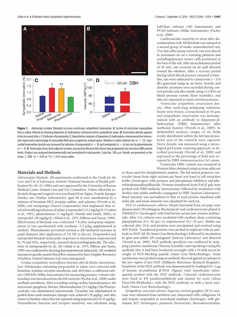
Figure 1. Adrenergic receptor blockade increases ventricular sympathetic innervation. A, Section of ventricular myocardiumfrom a saline-infused rat showing dopamine �-hydroxylase-immunoreactive sympathetic axons. B, Innervation density appearsto be increased after a 7 d infusion of propranolol. C, Quantitative analyses of dopamine �-hydroxylase-immunoreactive innerva-tion expressed as percentage of myocardial field area occupied by stained axons. Relative to saline-infused rats (n � 12), myo-cardial innervation density was increased by infusions of propranolol (n � 8) and metoprolol (n � 6) but not by phentolamine(n � 4). D, Ventricular tissue from adjacent sections assessed by Western blot shows that propranolol also increases DBH proteinlevels. Product was analyzed densitometrically and normalized to total protein. Scale bar, 100 �m. Results are presented as themean � SEM. *p � 0.05 or **p � 0.01 versus saline.
Clarke et al. • �-Blockers Induce Sympathetic Hyperinnervation J. Neurosci., September 15, 2010 • 30(37):12446 –12454 • 12447

and uridine, and 10 ng/ml NGF (Alomone Labs). Neurons were platedon glass coverslips (Bellco) coated with poly-D-lysine (MP Biomedicals)and laminin (Invitrogen) in control medium or medium containing pro-pranolol (10 �10 to 10 �8
M) or metoprolol (10 �8 to 10 �6M). Other
cultures were conducted with 10 �10–10 �6M dobutamine, with or with-
out 5 �M �-methyl-p-tyrosine (AMPT, Research Biochemicals Interna-tional) or with AMPT alone.
After 48 h, cultures were fixed in 4% parafomaldehyde and immuno-stained using a peripherin antibody (Millipore Bioscience Research Re-agents). Neurite outgrowth was measured blindly in six captured imagesdistributed equally within each well using a stereological grid withintersections at 25 �m intervals superimposed over each image. In-tersections overlying stained neurites were divided by total intersec-tions within the field (fraction of field occupied by neurites) andmultiplied by total field area (neurite area per field area). This wasdivided by the total number of neuronal somata within the field(neurite area per neuron), and values were normalized to 1 mm 2
(Chakrabarty et al., 2008). Cultures were also double stained for pe-ripherin and �1-AR (Santa Cruz Biotechnology).
Reverse transcriptase-PCR for �1-receptor mRNA. �1-AR gene expres-sion was assessed in SCG cultures by reverse transcriptase (RT)-PCR.Fresh heart tissue or SCG neurons were homogenized, and total RNAwas reverse transcribed (Wernli et al., 2009). Amplification of cDNAwas conducted using primers for �1-AR (5�-CAACGGCGGGAC-GACCACTG-3� sense; and 5�-ACACCTTGGACTCGGAGGAGAGC-3�antisense) and glyceraldehyde-3-phosphate dehydrogenase (GAPDH)(5�-CTCTACCCACGGCAAGTTC-3� sense; and 5�-CTCAGCACCAG-CATCACC-3� antisense), and analyzed by electrophoresis.
Myocardial infarction. To assess the effects of �-AR blockers in ratswith myocardial infarction, coronary artery ligation (CAL) was per-formed by ligating the left descending coronary artery (Wernli et al.,2009), or the artery was exposed but left unoccluded. Concurrently, ratsreceived pump infusions of AR antagonists or saline for 7 d. In CALhearts, images were obtained of six fields distributed evenly along the leftlateral posterior infarct border region where sympathetic hyperinnerva-tion occurs. In addition, images of six fields evenly distributed within thenonischemic posterior area of the left ventricle were also obtained. Pro-cedures and analyses were as described above.
Statistics. Data were analyzed using one-way ANOVA for multiplecomparisons followed by Newman–Keuls test. Data are expressed as themean � SEM. Statistical significance was accepted at p � 0.05.
ResultsAR blockade increases ventricular sympathetic innervationin vivoRats received 7 d infusions of saline, the nonselective �-blockerpropranolol, the selective �1-blocker metoprolol, or the nonse-lective �-blocker phentolamine. In rats receiving saline, quanti-tative analysis of DBH-ir sympathetic axons showed thatventricular innervation occurred at a density similar to that re-ported previously (Wernli et al., 2009) (Fig. 1A). DBH-ir fibersappeared more abundant in rats receiving propranolol (Fig. 1B)and quantitative analysis revealed a 48% increase in ventricularsympathetic axon density ( p � 0.05) (Fig. 1C). Analysis of adja-cent tissue by Western blot confirmed that DBH protein contentwas also significantly increased (Fig. 1D). Because propranololblocks both �1- and �2-ARs, we examined whether selectiveblockade of �1-AR alone increases innervation density. Metopro-lol was administered using a comparable regimen, and quantita-tive analysis showed a 44% increase in sympathetic innervationrelative to controls ( p � 0.01) (Fig. 1C). To assess whether otherAR antagonists with depressor effects also affect ventricular in-nervation, we administered the �-blocker phentolamine. DBH-irfiber density after phentolamine treatment was similar to that ofcontrols (Fig. 1C), showing that �-blockade and the associateddecrease in blood pressure does not modify cardiac sympatheticinnervation.
Ventricular sympathetic neuroeffector function andcardiovascular reflexes are enhanced after �-blockerdiscontinuationTo determine whether sympathetic hyperinnervation is associ-ated with altered ventricular sympathetic neuroeffector proper-ties, we assessed cardiac function 48 h after discontinuation ofsaline or propranolol infusions. Resting heart rate, ventricularpressures (end systolic and end diastolic pressures), and rates ofventricular contraction (dP/dtmax) and relaxation (dP/dtmin)were similar between groups (Table 1). Elimination of auto-nomic tone by the ganglionic blocker chlorisondamine reducedall values, and function was comparable between groups for allparameters except for modest increases in dP/dtmax and dP/dtmin
in rats receiving propranolol.
Figure 2. Cardiovascular responses to stressors are increased after �-blockade discontinu-ation. Arterial pressure was measured in awake, restrained rats 2 d after discontinuation ofpropranolol (n � 5) or saline (n � 4) infusion. Mildly restrained animals were subjected to stressinducedbyapuffofairtothefacialregion(Air)orsoundof�110dbgeneratedbyanairhorn(Sound).Peak values are presented as mean arterial pressure (MAP). *p � 0.05 versus saline.
Table 1. Cardiac function in response to isoproterenol and tyramine
Saline (n � 8) Propranolol (n � 8)
RestingHR, bpm 400 � 14 411 � 13ESP, mmHg 124 � 12 131 � 10EDP, mmHg 7.7 � 2.8 4.0 � 0.5Peak dP/dtmax , mmHg/s 11,808 � 1,275 14,497 � 825Peak dP/dtmin , mmHg/s 8,229 � 788 9,081 � 709
ChlorisondamineHR, bpm 342 � 10 350 � 8ESP, mmHg 60 � 3 68 � 4EDP, mmHg 6.9 � 0.9 4.8 � 0.4Peak dP/dtmax , mmHg/s 2,169 � 145 2,705 � 196*Peak dP/dtmin , mmHg/s 2,131 � 263 3,335 � 380*
IsoproterenolHR, bpm 461 � 12 479 � 11ESP, mmHg 88 � 5 93 � 5EDP, mmHg 3.9 � 0.7 2.5 � 0.4Peak dP/dtmax , mmHg/s 8,476 � 739 9,552 � 297Peak dP/dtmin , mmHg/s 4,894 � 476 5,731 � 779
TyramineHR, bpm 434 � 17 458 � 16ESP, mmHg 83 � 3 103 � 8*EDP, mmHg 4.9 � 0.7 2.9 � 0.5*Peak dP/dtmax , mmHg/s 8,081 � 688 10,922 � 581**Peak dP/dtmin , mmHg/s 5,427 � 622 6,881 � 696
Responses obtained 48 h after discontinuation of a 7 d infusion of saline or propranolol. Values were obtained in theresting state under urethane anesthesia, after ganglionic blockade with chlorisondamine, and after a maximallyeffective dose of the direct �-agonist isoproterenol, or displacement of sympathetic norepinephrine stores by amaximal dose of tyramine. Results are presented as the mean � SEM. HR, Heart rate; bpm, beats per minute; ESP,end systolic pressure; EDP, end diastolic pressure. *p � 0.05 versus saline infusion; **p � 0.01.
12448 • J. Neurosci., September 15, 2010 • 30(37):12446 –12454 Clarke et al. • �-Blockers Induce Sympathetic Hyperinnervation

We administered the �-AR agonist isoproterenol to assesswhether receptor sensitivity was altered at 48 h after propranololdiscontinuation, and whether cardiac function in response todirect �-AR activation was affected. To assess receptor sensitivity,incremental doses of isoproterenol were administered to achievemaximum values for heart rate and dP/dtmax. ED50 values werecomparable in saline- and propranolol-infused rats (0.07 � 0.02vs 0.07 � 0.02 mg/kg for heart rate, 0.09 � 0.02 vs 0.10 � 0.02�g/kg, respectively, for dP/dtmax in saline- or propranolol-infused rats), confirming prior reports that �-AR sensitivity inhumans and rats is normal at this time after discontinuation(Nattel et al., 1979; Hedberg et al., 1980). All values for heart rateand left ventricular function in response to maximally effectivedoses of isoproterenol were comparable in saline- and pro-pranolol-infused rats (Table 1).
To determine whether indices of sympathetic neuroeffectortransmission are enhanced in ventricles with increased sympa-thetic axon density, we administered maximally effective doses oftyramine, which acts by displacing sympathetic NE stores. Insaline- and propranolol-infused rats, maximum heart rate wascomparable, but rats after �-blockade showed significantlygreater ventricular end-systolic pressures (despite decreased end-diastolic pressure) and significantly increased rate of contraction(Table 1).
To determine whether cardiovascular reflexes that are nor-mally mediated by the sympathetic nervous system are also en-hanced after �-AR blocker discontinuation in unanesthetizedrats, we assessed the effects of two common stressors on bloodpressure. A puff of air delivered to the facial region of restrainedrats resulted in greater increase in blood pressure after propran-olol than after saline discontinuation. Similarly, loud noise alsoincreased blood pressure to a greater extent after propranololthan saline infusion (Fig. 2).
Adrenoceptors and cardiomyocyte NGF synthesis�-ARs modulate NGF synthesis in some cell types (Colangelo etal., 1998), and altered neurotrophin synthesis is a possible mech-anism for the observed increase in ventricular sympathetic axondensity. To determine whether �-ARs influence cardiac NGFsynthesis, we cultured ventricular cardiomyocytes in the presenceor absence of AR agonists and measured NGF protein producedand secreted in vitro. Cardiomyocytes in culture showed sponta-neous contractions and mature cytoskeletal features (Fig. 3A).These cells were immunoreactive for NGF protein (Fig. 3B), andthe endopeptidase furin (Fig. 3C), which converts pro-NGF tomature NGF, the pro-neuritogenic form of this neurotrophin. Toassess NGF secretion by these cells, we probed Western blotsusing an antibody to NGF. In characterization studies, this anti-body detected �10 ng of the 14 kDa mature recombinant humanNGF and higher molecular weight forms of NGF at higher con-centrations (Fig. 3D). Medium from cultured myocytes showed adominant band at �40 kDa, which was eliminated preabsorbingthe NGF antibody with an excess of blocking peptide (Fig. 3E).Probing membranes with an antibody directed to the non-NGFportion of pro-NGF confirmed that this band is pro-NGF (Fig.3F). To assess whether NGF-related protein secretion is regulatedby ARs, cardiomyocytes were incubated with 10�7–10�5
M of the�-agonist phenylephrine or the �-agonist isoproterenol. Isopro-terenol treatment did not appear to alter the amount or pattern ofsecreted protein. Similarly, phenylephrine did not have markedeffects on NGF-related proteins, although lower molecularweight forms may have been decreased at higher concentrations(Fig. 3G).
AR blockade increases sympathetic neuritogenesis in vitroTo determine whether �-AR blockers act directly on sympatheticneurons to increase axon outgrowth, we used an in vitro assay
Figure 3. Adrenergic receptors do not modulate cardiomyocyte NGF secretion. Dissociated cardiomyocytes were cultured for 3 d before treatment with phenylephrine or isoproterenol. A,Cardiomyocytes in culture show typical actin cytoskeleton after staining with phalloidin. B, C, Cardiomyocytes express NGF protein (B) and the pro-neurotrophin convertase furin (C). Scale bars, 50�m. D, Staining of Western blots loaded with different amounts of recombinant human �-NGF showed that antibody sensitivity is at least 10 ng/lane. E, After culture for 48 h, Western blot analysisof the culture medium revealed immunoreactive NGF, mainly as pro-NGF with a molecular mass of �40 kDa (left) and preincubation of the antibody with an NGF-blocking peptide resulted inabsence NGF detection (right). F, Probing with an antibody recognizing a non-NGF epitope in the pro region of the protein confirmed that the 40 kDa band is pro-NGF. G, Treatment of thecardiomyocyte cultures with 0.1–10 �M concentrations of the �-agonist phenylephrine (PE) or the �-agonist isoproterenol (IS) did not alter NGF content within the medium.
Clarke et al. • �-Blockers Induce Sympathetic Hyperinnervation J. Neurosci., September 15, 2010 • 30(37):12446 –12454 • 12449

system consisting of neonatal SCG sympa-thetic neurons cultured in defined media.After 48 h, SCG neurons in control cultureshad elaborated many neurites (Fig. 4A).Neurons cultured with 10�10
M proprano-lol appeared to have greater neurite out-growth (Fig. 4B), and quantitative analysisconfirmed a significant increase in neuritearea per neuron (Fig. 4C). However, cul-tures containing propranolol at the greaterconcentration of 10�8
M yielded outgrowthcomparable to controls (Fig. 4C).
To assess whether the propranolol-induced increase in outgrowth is attribut-able to blockade of �1-AR, we culturedSCG neurons in the presence of metopro-lol. Cultures containing 10�8
M metopro-lol showed substantial neurite outgrowth(Fig. 4D) similar to that of propranolol.Quantitative analysis confirmed that thisconcentration of metoprolol increasedneurite outgrowth by 29% (Fig. 4E). Cul-tures containing a higher concentration of10�6
M metoprolol again showed out-growth comparable to controls (Fig. 4E).
Sympathetic axons contain �2-ARsthat are known to enhance NE release(Deegan et al., 1995). We assessed whetherthese receptors play a role in modulatingsympathetic neurite outgrowth by incubat-ing SCG neurons with the �2-AR antagonistICI 118551. Sympathetic outgrowth wascomparable to control cultures at all con-centrations tested (Fig. 4F).
To confirm that outgrowth mediatedby �1 blockade is not caused by increasedneuronal survival, numbers of somas persquare millimeter were compared. We ob-served no differences in neuron numberswith different concentrations of propran-olol (Fig. 4G) or metoprolol (Fig. 4H).Similarly, ICI 118551 did not alter neuronnumbers (data not shown).
SCG neurons express �1-AR mRNA andprotein in cultureTo confirm that sympathetic neurons ex-press �1-AR mRNA, we performed RT-PCR analysis of SCG neurons after 48 h inculture. The sensitivity of our primers wasconfirmed using ventricular myocardiumas a control, which showed a strong bandat the predicted product size. RT-PCR ofSCG neurons also yielded PCR product ofthe correct size, albeit of less intensity thanthat of cardiac muscle (Fig. 5A). The pres-ence of the �1-AR protein in somata andneurites was evaluated in cultures stained for �1-AR protein (Fig.5B). Cultures were costained with the neuronal marker periph-erin (Fig. 5C), and merged images showed that �1-AR protein isdistributed widely throughout soma and neurites (Fig. 5D).�1-AR immunostaining of cells not known to express this proteinwas negligible (Fig. 5B, inset, glia).
Norepinephrine synthesis inhibits neurite outgrowthWe postulated that propranolol and metoprolol promote sympa-thetic axon sprouting by blocking �1 receptors that negativelyregulate outgrowth. Accordingly, we sought to determinewhether activating sympathetic axonal �1-ARs would suppressoutgrowth in culture. We cultured SCG neurons alone (Fig. 6A)
Figure 4. �1 adrenoceptors promote sympathetic neurite outgrowth. A, Superior cervical sympathetic ganglion neurons cul-tured without treatment extend many neurites. B, C, Addition of propranolol at a concentration of 10 �10
M appeared to increaseneurite outgrowth (B), and this was confirmed by quantitative analysis of peripherin-immunoreactive neurite area (C) (n � 12 percondition). D, E, Cultures treated in the presence of 10 �8
M metoprolol (D) showed increased neurite outgrowth similar topropranolol, and this was confirmed by quantitative analysis (E) (n � 12). F, Treatment with the �2-antagonist ICI 118551 had noeffect on neurite outgrowth (n � 3). G, H, Counts of neuronal somas per unit area showed no differences between control culturesand those treated with propranolol (G) or metoprolol (H ). Scale bar: (in D) A, B, D, 50 �m for all micrographs. Results are presentedas the mean � SEM. *p � 0.05 compared with control.
12450 • J. Neurosci., September 15, 2010 • 30(37):12446 –12454 Clarke et al. • �-Blockers Induce Sympathetic Hyperinnervation

or in the presence of the �1-agonist dobutamine (Fig. 6B, a rep-resentative concentration of 10�8
M is shown). Dobutamine didnot seem to modulate neurite outgrowth (Fig. 6B) and quantita-tive analysis confirmed that dobutamine was ineffective in de-creasing axonal outgrowth over a wide range of concentrations(Fig. 6C). However, because SCG neurons in culture express cat-echolamine biosynthetic proteins, it is likely that NE is synthe-sized and released in these cultures. If so, �1-AR activation may beongoing, thus masking effects of exogenously applied agonist.
To determine whether NE synthesized in culture tonically in-hibits axonal outgrowth, we blocked NE synthesis with AMPT, acompetitive inhibitor of tyrosine hydroxylase. Neurite outgrowthappeared to be greater in cultures containing AMPT (Fig. 6D),and quantitation confirmed a 35% increase relative to controls(Fig. 6F). To confirm that AMPT acted to increase neurite out-growth by eliminating tonic �1-AR activation, we now addeddobutamine to these cultures. This �1-agonist suppressed theAMPT-mediated increase in axon outgrowth to a level compara-ble to that of controls (Fig. 6E,F), confirming that locally synthe-sized NE inhibits sympathetic axon outgrowth in culture. Somacounts again confirmed that neurite changes are not the result ofdifferential neuronal survival (data not shown).
AR blockade increases ventricular sympathetic innervationafter myocardial infarctionAn important clinical application of �-blockers is to reduce com-plications after myocardial infarction (Cao et al., 2000). To de-termine whether this is associated with changes in ventricularsympathetic axon density, we induced myocardial ischemia byperforming CAL of the left anterior coronary artery (Hasan et al.,2006; Wernli et al., 2009) and infused saline or an AR antagonistfor 7 d; treatments did not affect infarct size (data not shown). Inintact myocardium adjacent to the infarct, sympathetic innerva-tion density was lower than that seen in uninjured subjects ( p �0.01), consistent with prior reports (Kaye et al., 2000). However,propranolol (but not phentolamine) increased sympathetic axondensity (41%, p � 0.05) (Fig. 7), restoring innervation toward
that of control animals (Fig. 1, compare with normative values).Consistent with previous reports (Cao et al., 2000; Hasan et al.,2006; Wernli et al., 2009), the infarct border region showedmarked sympathetic hyperinnervation in saline-infused rats, butthis was unaffected by either propranolol or phentolamine treat-ment (Fig. 7).
DiscussionOur findings provide evidence that �-blockers increase sympa-thetic target organ innervation, which contributes to exaggeratedsympathetic responses after �-blocker withdrawal. Chronicpropranolol administration increased numbers of ventricularDBH-ir axons consistent with sprouting of existing sympatheticprojections (Hasan et al., 2006; Wernli et al., 2009). Metoprolol wasequally effective, indicating that newer selective �-antagonists sharethis property. Therefore, ventricular sympathetic hyperinnervationappears to be a feature common to at least some �-AR blockers thatenjoy broad clinical use.
There is precedence for proposing that greater ventricularsympathetic axon density may contribute to enhanced target or-gan response. Increased sympathetic innervation is associatedwith smooth muscle hypertrophy and hyper-reactivity (Scott andPang, 1983; Kondo et al., 1996), and our findings indicate thatventricular neuroeffector function is also exaggerated in heartswith �-blocker-induced hyperinnervation. Indices of contractilefunction in response to tyramine were significantly greater in ratsafter chronic �-AR blockade. Because the magnitude of the tyra-mine response is determined by the amount of NE that can bedisplaced from intrinsic sympathetic nerves, this increased re-sponse is consistent with greater NE stores in hearts with elevatedinnervation density. Moreover, �-AR sensitivity was normal at48 h after withdrawal, as reported previously in rats and humans(Nattel et al., 1979; Hedberg et al., 1980), and responses to direct�-AR activation with isoproterenol were comparable, indicatingthat intrinsic ventricular properties during maximal �-AR acti-vation are largely unchanged. It is noteworthy, however, thatventricular function after complete ganglionic blockade did showmodest increases in the rate of contraction and relaxation, al-though no such differences were evident in the resting state orafter isoproterenol. Whether this reflects ventricular remodeling,or possibly altered preload and afterload induced by chronic�-blockade, remains to be determined. Nonetheless, thesechanges are unlikely to contribute substantially to the enhancedcardiac function observed with tyramine in chronically blockedsubjects. Accordingly, the increased tyramine response appearsto represent a functional correlate to ventricular sympathetic hy-perinnervation. Interestingly, no differences in heart rate weredetected in the resting or stimulated state. Because the sympa-thetic innervation density of the sinoatrial node is substantiallygreater than that of the ventricle, it may be the case that�-blockade was less effective in increasing axon density in thatregion, as we find to be the case for peri-infarct hyperinnervation.
Although the tyramine response is a reliable indicator of po-tential sympathetic neuroeffector efficacy, it does not provideevidence that sympathetically mediated behavioral responses arealso enhanced. To determine whether behavioral indices of sym-pathetic activation are altered, we assessed cardiovascular re-sponsiveness to stressors in awake rats. Rats show startleresponses when subjected to loud noise or a puff of air directed atthe face. In both cases, at a time when tyramine response wasenhanced but �-AR sensitivity was normal, mean arterial pres-sure was increased to greater levels in rats undergoing �-blockerwithdrawal. This indicates that sympathetic nervous system
Figure 5. �1-Adrenoceptor protein and gene expression in cultured sympathetic neurons.A, mRNA extracted from purified SCG neuronal cultures contains sequences corresponding tothe �1-adrenoceptor gene, as revealed by RT-PCR. Ventricular myocardium was used as apositive control, and GAPDH was used to show the relative level of receptor expression. B, C,Immunostaining for �1-adrenoceptor show that this protein is present in sympathetic neurons(B), as revealed by peripherin staining (C). Inset in B shows a glial cell that is negative for �1-ARimmunoreactivity. D, A merged image shows that �1-adrenoceptor protein is present through-out the soma and processes including axon terminations. Scale bar, 10 �m.
Clarke et al. • �-Blockers Induce Sympathetic Hyperinnervation J. Neurosci., September 15, 2010 • 30(37):12446 –12454 • 12451

responsiveness is exaggerated in unanesthetized rats, and thatrats, like humans, show increased cardiovascular lability after�-blocker withdrawal.
A central question concerns the mechanism by which�-blockers increase ventricular sympathetic axon density. Insome cell types, �-ARs regulate synthesis of NGF, a powerfulmediator of sympathetic sprouting (Dal Toso et al., 1987) thatcould initiate axon outgrowth. However, consistent with otherreports (Kaye et al., 2000), our culture studies show that �-ARsdo not appear to play a major role in regulating NGF synthesisand release by cardiac myocytes. Although treatment with an�-agonist has been reported to suppress cardiac NGF synthesis
(Kaye et al., 2000), it did not significantly modify NGF expressionin our hands, and our finding that ventricular sympathetic den-sity was unchanged by phentolamine argues against substantialmodulation of innervation by �-ARs. In any event, an increase inNGF synthesis by �-AR blockers is unlikely to contribute to sym-pathetic hyperinnervation.
An alternative mechanism is a direct effect of �-antagonistson the sympathetic neuron itself. We demonstrate that �1-ARmRNA and protein are present in sympathetic neuronal cellbodies and axons, which extends earlier pharmacologicalstudies inferring the presence of �1 receptors on cardiac sym-pathetic neurons (Butler et al., 1990; Watson-Wright et al.,1991). Hence, synthesis and distribution of �1-ARs in sympa-thetic neurons are consistent with their potential role as anautoreceptor.
Our culture studies provide evidence that �-blockers inducesprouting by interfering with activation of the sympathetic �1-autoreceptor. Thus, the nonselective �-antagonist propranololand the selective �1-AR antagonist metoprolol both increasedneurite outgrowth significantly in cultured sympathetic neurons,indicating direct growth-promoting effects on sympathetic neu-rons by �1-AR inhibition. Support for the selectivity of this effectcomes from the finding that blockade of �2 receptors by ICI118551 is ineffective in altering outgrowth. Interestingly, neu-ritogenic effects of both propranolol and metoprolol occurredonly at the lower concentrations, which most closely approx-imate estimates of in vivo therapeutic concentrations of thesedrugs (Abrahamsson et al., 1990; Takahashi et al., 1993), andwere lost when the concentration was increased 100-fold. Thismay not be surprising, as both agents at high concentrationshave membrane-stabilizing properties (Brunton et al., 2005),and membrane stabilization is known to inhibit axon out-growth (Ibarretxe et al., 2007).
Figure 6. Sympathetic neurite outgrowth in culture is regulated by locally synthesized norepinephrine. A–C, Compared with control cultures (A), dobutamine at a concentration of 10 �8M failed
to affect outgrowth (B), and this was confirmed by quantitative analysis of neurite area over a wide range of concentrations (C) (n � 3 per condition). D, When norepinephrine synthesis was blockedby AMPT, neurite outgrowth appeared to be enhanced. E, Addition of dobutamine to cultures in which norepinephrine synthesis was blocked led to diminished outgrowth. Scale bar, 50 �m. F,Quantitative analysis confirmed that inhibition of NE synthesis by AMPT increases neurite area, and that �1 activation by dobutamine is effective in reducing outgrowth only when NE synthesis isblocked. Results are presented as the mean � SEM. ***p � 0.001 versus control (n � 9) or versus AMPT � dobutamine (n � 6).
Figure 7. Adrenergic receptor blockade increases ventricular sympathetic innervationafter coronary artery ligation. Quantitative analyses of dopamine �-hydroxylase-immunoreactive innervation shows that regions of the ventricular myocardium remote tothe infarct (Non-ischemic) have relatively low innervation density in saline-infused rats(S; n � 4), and this was increased by propranolol (Pr; n � 4) but not by phentolamine (Ph;n � 4). Neither propranolol nor phentolamine altered the relatively high innervationdensity characteristic of the infarct border. Results are presented as the mean � SEM.*p � 0.05 versus saline.
12452 • J. Neurosci., September 15, 2010 • 30(37):12446 –12454 Clarke et al. • �-Blockers Induce Sympathetic Hyperinnervation
Findings thus far led us to hypothesize that sympathetic neu-rons possess �1-AR that negatively regulate axon outgrowth. Ac-cordingly, we attempted to demonstrate the activity of thesereceptors by adding the �1-agonist dobutamine to our cultures.However, despite using a wide range of concentrations, dobut-amine had no effect on neurite outgrowth. Since sympatheticneurons display features that suggest that they continue to syn-thesize NE in vitro (Landis, 1978), we postulated that culturedneurons may be releasing NE in quantities sufficient to maxi-mally activate �1-ARs, such that additional ligand is ineffective.To test this hypothesis, we used the tyrosine hydroxylase inhibi-tor AMPT to prevent catecholamine biosynthesis. In cultureswhere NE synthesis was inhibited, neurite outgrowth was in-creased to an extent similar to that seen with �-AR blockade,indicating that NE synthesized in culture does indeed act to in-hibit outgrowth. Now when dobutamine was added, outgrowthwas reduced to that of control cultures with intact NE synthesis.Collectively, these studies indicate that, under normal cultureconditions, NE tonically inhibits sympathetic neurite outgrowthvia �1-ARs. Consistent with this hypothesis, addition of dobut-amine to cultures where NE synthesis is blocked suppressed out-growth to levels typical of cultures in which NE is normallysynthesized.
Aside from a role in modulating NE release, presynaptic �1-ARs have not been implicated in influencing axonal outgrowth.However, there are several reports in other neural systems wheretransmitters do regulate axonal extension. For example, both do-pamine and serotonin suppress elongation of axons in the Heli-soma snail (Haydon et al., 1984; McCobb et al., 1988). At leastsome transmitter-mediated inhibition of axon growth appears tooccur via autoreceptors, as serotonin inhibits outgrowth of sero-toninergic axons from rat raphe neurons (Whitaker-Azmitia andAzmitia, 1986), and glutamate at high doses reduces axonal out-growth by immobilizing growth cones of glutaminergic pyrami-dal neurons (Mattson et al., 1988). Thus, precedents exist forsuggesting that sympathetic neurotransmitter autoreceptorscould play a critical role in regulating axon outgrowth.
Although enhanced sympathetic outgrowth is likely to con-tribute to �-blocker withdrawal syndrome, it may be relevant toother pathophysiological situations as well. �-Blockers are com-monly administered to patients with myocardial infarction be-cause of their ability to reduce cardiac excitability, myocardialoxygen consumption, plasma angiotensin II (Ichihara et al.,1995), and sudden death (Hunt, 2005). However, heart failure isaccompanied by diminished sympathetic innervation in non-necrotic regions of the ventricle (Himura et al., 1993; Li et al.,2004), which could contribute to cardiac dysfunction. It is inter-esting to speculate that in heart failure patients, �-blocker ther-apy could prove beneficial not only by directly altering cardiacproperties, but also by restoring cardiac sympathetic innervation.Our findings of normalization of axon numbers after proprano-lol infusion in the intact myocardium in this rat model of myo-cardial infarction suggest that this may be the case. Consistentwith the idea that sympathetic axon density may be restored inthe failing ventricle, 123I-metaiodobenzylguanidine scintigraphicstudies in patients receiving �-blockers show increased ventricu-lar catecholamine reuptake (Merlet et al., 1999; Toyama et al.,2003; Cohen-Solal et al., 2005). It is also of interest that, in ourmodel, hyperinnervation induced by infarction was unaffectedby propranolol administration. There is abundant evidence thatmyocardial ischemic injury is associated with proliferation ofsympathetic axons in the vicinity of the infarct (Cao et al., 2000;Hasan et al., 2006; Wernli et al., 2009). Somewhat surprisingly,
this hyperinnervation was not increased by �-blocker adminis-tration, a finding that may suggest an upper limit beyond whichadditional increases in innervation density are not apparent. Onthe other hand, it has been reported in rabbits that sympathetichyperinnervation in the infarct border region is reduced by meto-prolol (Jiang et al., 2007). Border region sprouting is likely toinvolve complex interactions among target-derived trophic fac-tors, inflammatory cells, and damaged and regenerating axons,and response variability may not be surprising. Further studiesare necessary to determine whether �-blockers alter postinfarctsympathetic innervation patterns in humans.
In summary, we propose that sympathetic neurons possesspresynaptic �1-ARs that, when activated by physiological levels ofNE or other agonists, inhibit axon extension. We further hypoth-esize that this is an important mechanism for establishing levelsof target innervation density. According to this concept, wheninnervation density is low, levels of NE will also be low and sym-pathetic axons will be encouraged to proliferate. This will con-tinue until local levels of NE are sufficient to activate presynaptic�1-ARs, at which point further axon extension would be pre-vented. Such a feedback system could provide a sensitive mecha-nism for establishing final levels of target innervation where theset point for innervation density is determined by the local con-centration of transmitter. In the presence of a drug that blocks�1-ARs, outgrowth inhibition is lost and target organ innervationdensity is therefore increased.
ReferencesAarons RD, Nies AS, Gal J, Hegstrand LR, Molinoff PB (1980) Elevation of
beta-adrenergic receptor density in human lymphocytes after proprano-lol administration. J Clin Invest 65:949 –957.
Ablad B, Borg KO, Carlsson E, Ek L, Johnson G, Malmfors T, Regardh CG(1975) A survey of the pharmacological properties of metoprolol in ani-mals and man. Acta Pharmacol Toxicol (Copenh) 36 [Suppl 5]:7–23.
Abrahamsson B, Lucker P, Olofsson B, Regårdh CG, Sandberg A, WieselgrenI, Bergstrand R (1990) The relationship between metoprolol plasmaconcentration and beta 1-blockade in healthy subjects: a study on con-ventional metoprolol and metoprolol CR/ZOK formulations. J Clin Phar-macol 30:S46 –S54.
Brunton L, Lazo J, Parker K (2005) Goodman and Gilman’s the pharmaco-logical basis of therapeutics. New York: McGraw-Hill.
Butler CK, Smith FM, Nicholson J, Armour JA (1990) Cardiac effects in-duced by chemically activated neurons in canine intrathoracic ganglia.Am J Physiol 259:H1108 –H1117.
Cao JM, Fishbein MC, Han JB, Lai WW, Lai AC, Wu TJ, Czer L, Wolf PL,Denton TA, Shintaku IP, Chen PS, Chen LS (2000) Relationship be-tween regional cardiac hyperinnervation and ventricular arrhythmia. Cir-culation 101:1960 –1969.
Chakrabarty A, Blacklock A, Svojanovsky S, Smith PG (2008) Estrogen elic-its dorsal root ganglion axon sprouting via a renin-angiotensin system.Endocrinology 149:3452–3460.
Cohen-Solal A, Rouzet F, Berdeaux A, Le Guludec D, Abergel E, Syrota A,Merlet P (2005) Effects of carvedilol on myocardial sympathetic inner-vation in patients with chronic heart failure. J Nucl Med 46:1796 –1803.
Colangelo AM, Follesa P, Mocchetti I (1998) Differential induction of nervegrowth factor and basic fibroblast growth factor mRNA in neonatal andaged rat brain. Brain Res Mol Brain Res 53:218 –225.
Conlon D, Johnston A, Turner P, O’Malley K, Kilfeather S (1991) Hepaticbeta-adrenoceptor adaptation during propranolol administration is im-paired in aging rats. Eur J Pharmacol 208:323–330.
Dal Toso R, De Bernardi MA, Costa E, Mocchetti I (1987) Beta-adrenergicreceptor regulation of NGF-mRNA content in rat C6 –2B glioma cells.Neuropharmacology 26:1783–1786.
Deegan R, He HB, Krivoruk Y, Wood AJ, Wood M (1995) Regulation ofnorepinephrine release by beta 2-adrenergic receptors during halothaneanesthesia. Anesthesiology 82:1417–1425.
DiBona GF, Sawin LL (1999) Effect of metoprolol administration on renalsodium handling in experimental congestive heart failure. Circulation100:82– 86.
Clarke et al. • �-Blockers Induce Sympathetic Hyperinnervation J. Neurosci., September 15, 2010 • 30(37):12446 –12454 • 12453

Dollery CT, Maling TJ (1979) Beta-blocker withdrawal syndrome. Br Med J2:1074 –1075.
Germino FW (2009) The management and treatment of hypertension. ClinCornerstone 9 [Suppl 3]:S27–S33.
Harrison DC, Alderman EL (1976) Editorial: Discontinuation of propran-olol therapy: cause of rebound angina pectoris and acute coronary events.Chest 69:1–2.
Hasan W, Jama A, Donohue T, Wernli G, Onyszchuk G, Al-Hafez B, BilgenM, Smith PG (2006) Sympathetic hyperinnervation and inflammatorycell NGF synthesis following myocardial infarction in rats. Brain Res1124:142–154.
Haydon PG, McCobb DP, Kater SB (1984) Serotonin selectively inhibitsgrowth cone motility and synaptogenesis of specific identified neurons.Science 226:561–564.
Hedberg A, Isaksson O, Lundgren B (1980) Sustained cardiac beta adreno-ceptor blockade in vitro and increased vulnerability to aconitine-inducedarrhythmias in vivo after propranolol withdrawal in rats. J Pharmacol ExpTher 214:664 – 669.
Heilbrunn SM, Shah P, Bristow MR, Valantine HA, Ginsburg R, Fowler MB(1989) Increased beta-receptor density and improved hemodynamicresponse to catecholamine stimulation during long-term metoprololtherapy in heart failure from dilated cardiomyopathy. Circulation79:483– 490.
Himura Y, Felten SY, Kashiki M, Lewandowski TJ, Delehanty JM, Liang CS(1993) Cardiac noradrenergic nerve terminal abnormalities in dogs withexperimental congestive heart failure. Circulation 88:1299 –1309.
Hoeks SE, Scholte Op Reimer WJ, van Urk H, Jorning PJ, Boersma E, Simo-ons ML, Bax JJ, Poldermans D (2007) Increase of 1-year mortality afterperioperative beta-blocker withdrawal in endovascular and vascular sur-gery patients. Eur J Vasc Endovasc Surg 33:13–19.
Hunt SA (2005) ACC/AHA 2005 guideline update for the diagnosis andmanagement of chronic heart failure in the adult: a report of the AmericanCollege of Cardiology/American Heart Association Task Force on Prac-tice Guidelines (Writing Committee to Update the 2001 Guidelines forthe Evaluation and Management of Heart Failure). J Am Coll Cardiol46:e1– 82.
Ibarretxe G, Perrais D, Jaskolski F, Vimeney A, Mulle C (2007) Fast regula-tion of axonal growth cone motility by electrical activity. J Neurosci27:7684 –7695.
Ichihara A, Suzuki H, Murakami M, Naitoh M, Matsumoto A, Saruta T(1995) Interactions between angiotensin II and norepinephrine on reninrelease by juxtaglomerular cells. Eur J Endocrinol 133:569 –577.
Jiang H, Lu Z, Yu Y, Zhao D, Jian X, Yang B, Huang C (2007) Effects ofmetoprolol on sympathetic remodeling and electrical remodeling at in-farcted border zone after myocardial infarction in rabbits. Cardiology108:176 –182.
Kaye DM, Vaddadi G, Gruskin SL, Du XJ, Esler MD (2000) Reduced myo-cardial nerve growth factor expression in human and experimental heartfailure. Circ Res 86:E80 –E84.
Kondo M, Fujiwara T, Tabei R (1996) Noradrenergic hyperinnervation inthe heart of stroke-prone spontaneously hypertensive rats (SHRSP). Hy-pertens Res 19:69 –73.
Landis SC (1978) Growth cones of cultured sympathetic neurons containadrenergic vesicles. J Cell Biol 78:R8 –R14.
Lefkowitz RJ (2004) Historical review: a brief history and personal retro-spective of seven-transmembrane receptors. Trends Pharmacol Sci25:413– 422.
Li W, Knowlton D, Van Winkle DM, Habecker BA (2004) Infarction alters
both the distribution and noradrenergic properties of cardiac sympa-thetic neurons. Am J Physiol Heart Circ Physiol 286:H2229 –H2236.
Mattson MP, Dou P, Kater SB (1988) Outgrowth-regulating actions ofglutamate in isolated hippocampal pyramidal neurons. J Neurosci8:2087–2100.
McCobb DP, Haydon PG, Kater SB (1988) Dopamine and serotonin inhi-bition of neurite elongation of different identified neurons. J Neurosci Res19:19 –26.
Merlet P, Pouillart F, Dubois-Rande JL, Delahaye N, Fumey R, Castaigne A,Syrota A (1999) Sympathetic nerve alterations assessed with 123I-MIBGin the failing human heart. J Nucl Med 40:224 –231.
Miller RR, Olson HG, Amsterdam EA, Mason DT (1975) Propranolol-withdrawal rebound phenomenon. Exacerbation of coronary events afterabrupt cessation of antianginal therapy. N Engl J Med 293:416 – 418.
Nattel S, Rangno RE, Van Loon G (1979) Mechanism of propranolol with-drawal phenomena. Circulation 59:1158 –1164.
Pacher P, Nagayama T, Mukhopadhyay P, Batkai S, Kass DA (2008) Mea-surement of cardiac function using pressure-volume conductance cathe-ter technique in mice and rats. Nat Protoc 3:1422–1434.
Rodriguez-Ospina L, Montano-Soto L (2008) Management of chronic sta-ble angina pectoris. Bol Asoc Med P R 100:39 – 47.
Scott TM, Pang SC (1983) The correlation between the development ofsympathetic innervation and the development of medial hypertrophy injejunal arteries in normotensive and spontaneously hypertensive rats. JAuton Nerv Syst 8:25–32.
Steinle JJ, Smith PG (2002) Role of adrenergic receptors in vascular remod-elling of the rat choroid. Br J Pharmacol 136:730 –734.
Takahashi H, Ogata H, Kashiwada K, Ohira M, Someya K (1993) Dosingrate-dependent relationship between propranolol plasma concentrationand beta-blockade. J Pharmacol Exp Ther 265:681– 689.
Toyama T, Hoshizaki H, Seki R, Isobe N, Adachi H, Naito S, Oshima S,Taniguchi K (2003) Efficacy of carvedilol treatment on cardiac functionand cardiac sympathetic nerve activity in patients with dilated cardiomy-opathy: comparison with metoprolol therapy. J Nucl Med 44:1604 –1611.
Vincent GM, Schwartz PJ, Denjoy I, Swan H, Bithell C, Spazzolini C, Crotti L,Piippo K, Lupoglazoff JM, Villain E, Priori SG, Napolitano C, Zhang L(2009) High efficacy of beta-blockers in long-QT syndrome type 1: con-tribution of noncompliance and QT-prolonging drugs to the occurrenceof beta-blocker treatment “failures.” Circulation 119:215–221.
Viskin S, Kitzis I, Lev E, Zak Z, Heller K, Villa Y, Zajarias A, Laniado S,Belhassen B (1995) Treatment with beta-adrenergic blocking agents af-ter myocardial infarction: from randomized trials to clinical practice.J Am Coll Cardiol 25:1327–1332.
Wang W, Zhu W, Wang S, Yang D, Crow MT, Xiao RP, Cheng H (2004)Sustained beta1-adrenergic stimulation modulates cardiac contractilityby Ca2�/calmodulin kinase signaling pathway. Circ Res 95:798 – 806.
Watson-Wright W, Boudreau G, Cardinal R, Armour JA (1991) Beta 1- andbeta 2-adrenoceptor subtypes in canine intrathoracic efferent sympa-thetic nervous system regulating the heart. Am J Physiol 261:R1269 –R1275.
Wernli G, Hasan W, Bhattacherjee A, van Rooijen N, Smith PG (2009) Mac-rophage depletion suppresses sympathetic hyperinnervation followingmyocardial infarction. Basic Res Cardiol 104:681– 693.
Whitaker-Azmitia PM, Azmitia EC (1986) Autoregulation of fetal seroto-nergic neuronal development: role of high affinity serotonin receptors.Neurosci Lett 67:307–312.
Yusuf S, Peto R, Lewis J, Collins R, Sleight P (1985) Beta blockade duringand after myocardial infarction: an overview of the randomized trials.Prog Cardiovasc Dis 27:335–371.
12454 • J. Neurosci., September 15, 2010 • 30(37):12446 –12454 Clarke et al. • �-Blockers Induce Sympathetic Hyperinnervation


![First draft prepared by Denis Hamilton, Animal and …...The Meeting received reports on studies on rats, lactating goats and laying hens. Rats. After the oral administration of [14C]flutolanil](https://img.dokumen.tips/doc/110x75/5fe09d66d9c73345665a01e1/first-draft-prepared-by-denis-hamilton-animal-and-the-meeting-received-reports.jpg)