Embed Size (px)
DESCRIPTION
Maturarea in vitro a ovocitelor de vaca
Citation preview

Please cite this article in press as: Sagirkaya, H. et al., Developmental potential of bovineoocytes cultured in different maturation and culture conditions, Anim. Reprod. Sci. (2006),doi:10.1016/j.anireprosci.2006.09.016
ARTICLE IN PRESS+ModelANIREP-3194; No. of Pages 16
Animal Reproduction Science xxx (2006) xxx–xxx
Developmental potential of bovine oocytes cultured indifferent maturation and culture conditions
Hakan Sagirkaya a,1, Muge Misirlioglu a, Abdullah Kaya c, Neal L. First b,John J. Parrish c, Erdogan Memili a,∗
a Department of Animal & Dairy Sciences, Mississippi State University, 4025 Wise Center, Box 9815, MS 39762, USAb Department of Biological Sciences, Mississippi State University, MS 39762, USA
c Department of Animal Sciences, University of Wisconsin-Madison, Madison, WI, USA
Received 9 May 2006; accepted 7 September 2006
Abstract
Diverse groups of chemicals in culture media are needed for successful bovine oocyte maturation andembryo development during which dramatic cytoplasmic and nuclear reprogramming events take place. Invitro embryo production (IVP) procedures frequently include supplements such as serum and/or co-culturewith various types of somatic cells. However, the presence of undefined serum in culture media introduces avariation from batch to batch, increases viral or prion contamination risk, and leads to problems during fetaldevelopment. The aim of the present study was to investigate the possibility of using chemically defined-synthetic serum substitute (SSS) in place of fetal calf serum (FCS) during maturation and long-term culture tostimulate in vitro maturation (IVM), fertilization (IVF) and subsequent embryo development. In ExperimentI, the effect of the protein source on in vitro maturation was tested by maturing oocytes in culture mediasupplemented with 10% FCS (Control Group), 10% SSS (Group I) and 10% SSS + 10 ng/ml epidermalgrowth factor (EGF) (Group II). In Experiment II, effects of SSS on both oocyte maturation and embryodevelopment during in vitro culture (IVC) were tested by maturing oocytes in media supplemented with10% FCS (FCS Group) or 10% SSS + 10 ng/ml EGF (SSS Group), followed by IVF and IVC in SOF mediasupplemented with 10% FCS and 10% SSS on day 4 for FCS and SSS Groups, respectively. Even thoughrates for cleavage and development to blastocyst stage were not different, blastocyst cell numbers werehigher in Group II containing SSS and EGF. The SSS supplementation group had higher apoptotic nucleias compared to the FCS Group in Experiment II. Transcripts for heat shock protein 70 (Hsp70), interferontau (IF-�), DNA methyltransferase 3a (Dnmt3a), desmosomal glycoprotein desmocollin III (DcIII) andinsulin-like growth factor II receptor (Igf-2r) were altered in different culture conditions in ExperimentI. However, only glucose transporter-1 (Glut-1) mRNA was different in the SSS and FCS Groups in the
∗ Corresponding author. Tel.: +1 662 325 2937; fax: +1 662 325 8873.E-mail address: [email protected] (E. Memili).
1 Present address: Uludag University, Veterinary Faculty, Gorukle, Bursa, Turkey.
0378-4320/$ – see front matter © 2006 Elsevier B.V. All rights reserved.doi:10.1016/j.anireprosci.2006.09.016

Please cite this article in press as: Sagirkaya, H. et al., Developmental potential of bovineoocytes cultured in different maturation and culture conditions, Anim. Reprod. Sci. (2006),doi:10.1016/j.anireprosci.2006.09.016
ARTICLE IN PRESS+ModelANIREP-3194; No. of Pages 16
2 H. Sagirkaya et al. / Animal Reproduction Science xxx (2006) xxx–xxx
second experiment. In summary, SSS and EGF in maturation medium and replacement of FCS with SSSalone in culture medium on day 4 of IVC support oocyte maturation and embryo development in vitro.However, significance of culture condition induced changes on the genome-wide abundance of messengerribonucleic acid and the significance of the apoptotic nuclei during fetal development still remain to bedetermined.© 2006 Elsevier B.V. All rights reserved.
Keywords: Early development; Gametogenesis; Oocyte development; Embryo; Developmental biology
1. Introduction
In vitro maturation of bovine oocytes, followed by in vitro fertilization for the productionof bovine embryos in the laboratory, is rapidly increasing (Zeuner et al., 2003). These in vitrotechniques have important values both in studying the basic biological events occurring duringoocyte maturation, fertilization and early embryonic development, and in providing an inexpen-sive and readily available source of preimplantation bovine embryos. Consistently successfuland reliable oocyte maturation (both cytoplasmic and nuclear maturation) would dramaticallyimprove the efficiency of preimplantation embryonic development as well as fetal development.However, current methods include supplements such as serum and/or co-culture with varioustypes of somatic cells because of the limited understanding of embryo metabolism and growthrequirements (Mastromonaco et al., 2004). This causes undefined and variable culture conditions.One of the most commonly used protein sources in the culture media has been serum, whichhas a biphasic effect. Including serum in the culture media can inhibit early cleavage divisions,while it can improve the development later in culture (Lonergan et al., 1999; Thompson et al.,1998). Its beneficial effect is especially important during the development of morulae into blas-tocyst (Gordon, 1994). In addition, it was reported that the effectiveness of serum in in vitroembryo production (IVP) might change considerably from one batch to another (Kane, 1987) andingredients of serum, such as amino acids, hormones, growth factors, cytokines, vitamins andmany other substances exhibit wide variations (Gordon, 1994). Even though a higher develop-ment rate to the blastocyst stage is obtained from media supported with serum, these variationscould cause some alterations in the ultrastructure of embryos, impaired compaction, abnormalblastulation, large calf syndrome, aberrant mRNA expression profiles, and greater incidences ofstillbirths and deaths after birth (Abe et al., 1999; Holm et al., 2002; Wrenzycki et al., 1999, 2004).Moreover, bovine-derived sera or proteins have recently been avoided especially in human IVPsystems because of the appearance of bovine spongiform encephalopathy (BSE) and a viral orprion contamination risk. Therefore, even more completely defined culture conditions supportinghigh developmental rates are important not only to obtain consistent results but also to eradicatethe contamination risk of BSE and other diseases. Because of these reasons, there has been a trendto use more defined proteins, such as bovine serum albumin (BSA), human serum albumin (HSA)and synthetic serum preparations instead of undefined natural serum preparations like fetal calfserum (FCS) and estrus cow serum (OCS) (Chanson et al., 2001; Russell et al., 1997; Sagirkayaet al., 2004).
Morphological characteristics such as appearance of cumulus cells and cytoplasm, oocyte sizeand time of polar body extrusion are related to the ability of oocytes to be fertilized and developinto viable embryos (Dominko and First, 1997). However, these are not reliable enough to actas the sole criteria for the evaluation of embryo developmental potential in vivo. Recently, there
Please cite this article in press as: Sagirkaya, H. et al., Developmental potential of bovineoocytes cultured in different maturation and culture conditions, Anim. Reprod. Sci. (2006),doi:10.1016/j.anireprosci.2006.09.016
ARTICLE IN PRESS+ModelANIREP-3194; No. of Pages 16
H. Sagirkaya et al. / Animal Reproduction Science xxx (2006) xxx–xxx 3
has been a tendency to use mRNA expression patterns as a criterion to predict the developmentalpotential of embryos in vivo (Rizos et al., 2002; Wrenzycki et al., 2004). Recent studies show thatthe relative abundance of several mRNA transcripts affected by the choice of culture medium andthe type of protein supplement played a critical role in preimplantation development (Rizos et al.,2002; Wrenzycki et al., 1999, 2001a, 2004; Sagirkaya et al., 2006). Knijn et al. (2002) comparedthe relative abundance of mRNA expressions for six developmentally important genes in bovineblastocysts produced in vitro from oocytes obtained from 3 to 8 mm follicles, preovulatory folliclesbefore LH surge and preovulatory follicles 24 h after the LH surge. While the first two groupswere matured in vitro in the presence of serum, the third group was an in vivo matured controlgroup. However, there is a need for a comparative study on development and mRNA patternsof embryos cultured in vitro in more defined media including the in vitro maturation (IVM),fertilization (IVF) and culture (IVC) steps.
The aim of this study was to investigate the effect of protein source (FCS versus serum substitutesupplement, SSS) during maturation and long-term culture on the development of in vitro producedembryos. Firstly, the effect of protein source on in vitro maturation was tested in Experiment I.After determining the effect on maturation, the effect of protein source supplementation on day4 of the culture was examined in Experiment II. The effect of protein source was determined bycomparing the rates of embryonic development (proportion of embryos developed to blastocyststage), mean cell number, percentages of apoptotic cells and mRNA expression patterns of apanel of developmentally important genes at the blastocyst stage. The genes assayed in the presentstudy are glucose transporter-1 (Glut-1), heat shock protein 70 (Hsp70), interferon tau (IF-�), DNAmethyltransferase 3a (Dnmt3a), desmosomal glycoprotein desmocollin III (DcIII) and insulin-likegrowth factor II receptor (Igf-2r). Expression patterns of these genes were determined relative tothat of housekeeping gene glyceraldehyde 3-phosphate dehydrogenase (GAPDH). These mRNAswere chosen to obtain the maximum possible understanding about important steps in early embryodevelopment and because the proteins of these genes are involved in metabolism (Glut-1), stress(Hsp70), maternal recognition of pregnancy (IF-�), DNA methylation (Dnmt3a), compaction andcavitation (DcIII) and growth factor signaling (Igf-2r).
2. Materials and methods
2.1. Reagents
In the present study, all chemicals were purchased from Sigma Chemical Company (St. Louis,MO) unless otherwise stated.
2.2. In vitro maturation
Bovine ovaries were obtained from a slaughterhouse and used for aspiration of follicles2–8 mm in diameter to collect oocytes. Only oocytes with several layers of cumulus cells andhomogenously granulated cytoplasm were used in this study. Selected oocytes were washedthree times in TL-HEPES. Immature bovine oocytes were matured in three different proteinsupplementation conditions. Tissue Culture Medium (TCM-199, Gibco/Invitrogen, Grand Island,NY) supplemented with pyruvate (0.2 mM), FSH (0.5 �g/ml) (Sioux Biochemicals, Sioux City,IA), LH (5 �g/ml) (Sioux Biochemicals), penicillin (100 U/ml) and streptomycin (100 �g/ml)(Gibco/Invitrogen) was used as the base medium for all of the groups. In Experiment I, oocyteswere matured in TCM-199 medium supplemented with 10% FCS (Gibco/Invitrogen) as a Control

Please cite this article in press as: Sagirkaya, H. et al., Developmental potential of bovineoocytes cultured in different maturation and culture conditions, Anim. Reprod. Sci. (2006),doi:10.1016/j.anireprosci.2006.09.016
ARTICLE IN PRESS+ModelANIREP-3194; No. of Pages 16
4 H. Sagirkaya et al. / Animal Reproduction Science xxx (2006) xxx–xxx
Group and Groups I and II were supplemented with 10% serum substitute supplement (SSS,Irvine Scientific, Santa Ana, CA), consisting of 6% total protein (84% human serum albumin,16% alpha and beta globulins) in physiological saline which is more defined than serum or 10%SSS + epidermal growth factor (EGF, 10 ng/ml), respectively. The EGF at 10 ng/ml concentrationhas previously been shown to improve embryonic development (Vigneron et al., 2004; Oyamadaet al., 2004). Following 4 days in culture, all treatments were supplemented with 10% FCS. InExperiment I, a total of 439, 395 and 415 oocytes were used for Control Group, Group I andGroup II, respectively. However, in Experiment II, oocytes were matured in TCM-199 mediumsupplemented with 10% FCS (FCS Group) or 10% SSS + EGF (10 ng/ml) (SSS Group). Thesetreatments were maintained with their respective protein supplementation on day 4 of in vitroculture. In Experiment II, a total of 574 and 644 oocytes were used for FCS and SSS Groups,respectively. For both experiments, 10 oocytes were matured in 50 �l drops covered with mineraloil for 24 h at 39 ◦C in 5% CO2 in a humidified tissue culture incubator. The same cultureconditions were used throughout all IVP procedures in Experiments I and II.
2.3. In vitro fertilization
After a 24 h maturation period, oocytes were rinsed twice in TL-HEPES and then groups of10 oocytes were transferred into 44 �l fertilization drops covered with mineral oil. The fertil-ization medium was glucose-free TALP supplemented with pyruvate (0.2 mM), fatty-acid-freeBSA (BSA-FAF, 6 mg/ml), penicillin (100 U/ml) and streptomycin (100 �g/ml). Frozen–thawedsemen from a single bull was used for the fertilization of oocytes. Percoll density gradient systemwas used for the separation of the motile fraction of the frozen–thawed semen (Parrish et al.,1986, 1995). Percoll (500 �l at 90% concentration) was transferred to the bottom of a 1.5 mlEppendorf centrifuge tube, and 0.5 ml of 45% Percoll was placed on top with a great caution.Frozen sperm was thawed at 35 ◦C for 1 min, and then carefully layered onto the Percoll gra-dient. The tube loaded with sperm sample and Percoll layers was then centrifuged at 700 × gfor 10 min at room temperature. The supernatant was removed carefully without disrupting thepellet containing live sperm cells. After the pellet was resuspended, sperm concentration was deter-mined using a hemocytometer. Sperm were then diluted to 50 × 106 sperm cells/ml in TL-HEPES,which would produce a 2 × 10⁶ spermatozoa/ml final concentration. The fertilization procedurewas completed by adding 2 �l of diluted sperm, 2 �l heparin (5 �g/ml) and 2 �l of PHE solution(20 �M penicillamine, 10 �M hypotaurine, 1 �M epinephrine in final concentration) into the 44 �lfertilization drops containing oocytes, respectively. Oocytes and sperm were cultured togetherfor approximately 18–19 h in the incubator (Leibfried and Bavister, 1982; Parrish et al., 1986,1995).
2.4. In vitro culture
Post-insemination (hpi 18 h), the cumulus cells were removed by vortexing the embryos in a1.5 ml Eppendorf tube at the highest speed for 3 min. Cumulus-free presumptive zygotes werewashed three times in TL-HEPES and transferred into 50 �l drops of embryo culture mediumknown as synthetic oviduct fluid, SOF (Specialty Media, NJ) under mineral oil (25–30 presumptivezygotes per each 50 �l drop). The SOF medium was supplemented with pyruvate (0.4 mM),BSA-FAF (8 mg/ml), 100× MEM (20 �l/ml), 50× BME (10 �l/ml), penicillin (100 U/ml) andstreptomycin (100 �g/ml) on the day of use. We previously demonstrated that proportion ofembryos developing to blastocyst stage were greater in SOF medium as compared to those in other
Please cite this article in press as: Sagirkaya, H. et al., Developmental potential of bovineoocytes cultured in different maturation and culture conditions, Anim. Reprod. Sci. (2006),doi:10.1016/j.anireprosci.2006.09.016
ARTICLE IN PRESS+ModelANIREP-3194; No. of Pages 16
H. Sagirkaya et al. / Animal Reproduction Science xxx (2006) xxx–xxx 5
common media such as KSOM and CR1 (Sagirkaya et al., 2006). In addition, supplementation onday 4 of culture until completion on day 8 of IVC were 10% FCS in Experiment I, and 10% FCSand 10% SSS (FCS and SSS Groups) in Experiment II. Developmental data were recorded after48, 96, 120 and 192 hpi culture periods for 2-cell, 8-cell, morulae and blastocyst stage embryos,respectively. Fertilization time was considered as 0 h. On day 8, after recording the blastocyststage embryos, they were either fixed in 4% formaldehyde for staining purposes or frozen insaline for the determination of amounts of mRNA of a panel of some developmentally importantgenes.
2.5. Blastocyst staining for total cell number
Day 8 blastocysts were fixed in 4% formaldehyde after washing three times in 1%polyvinylpyrrolidone (PVP) in PBS overnight. Embryos were placed in 1% Triton X-100 overnightand finally stained with Hoechst 33342. Embryos were then mounted on slides and covered with acover slip. The total cell numbers of blastocysts from three groups of Experiment I were determinedby counting the number of nuclei under an epifluorescent microscope. The total cell numbers ofblastocysts were visualized by a Nikon epifluorescent microscope with a 40× fluor objective(Tokyo, Japan) equipped with a 365 nm excitation filter, a 400 nm barrier filter, and a 400 nmemission filter.
2.6. Detection of DNA fragmentation by terminal deoxynucleotidyl transferase biotin-dUTPnick end labeling (TUNEL) assay
In Experiment II, day 8 blastocysts were used to determine the mean cell numbers and percent-ages of TUNEL-positive nuclei for FCS (n = 34) and SSS (n = 29) Groups. The TUNEL assay wasapplied according to the modified method of Fedorcsak and Storeng (2003) using the DeadEndTM
Fluorometric Apoptosis Detection Kit (Promega, Madison, WI). Briefly, blastocysts were removedfrom culture media and washed three times in 100 �l drops of 1% PVP in PBS, and then fixed with4% formaldehyde for approximately 1 h. Fixed embryos were stored at 4 ◦C until the TUNEL assaywas performed. On the day of the TUNEL assay, the embryos were transferred from 4 ◦C to roomtemperature and washed three times in 100 �l drops of 1% PVP in PBS and then permeabilized in50 �l of 0.5% Triton X-100 for 30 min at room temperature in a humidified chamber and rinsedtwice in PBS. Then, the embryos were incubated with 100 �l of DNase buffer for 5 min. Mean-while, 100 �l of DNase buffer containing DNase I (20 U/ml) was added to the embryos previouslyassigned as positive and negative controls for TUNEL assay, and 100 �l DNase buffer was addedto the other embryos. All embryos were incubated at 37 ◦C for 30 min, and then they were washedfour times with 100 �l of double distilled water by transferring embryos from one drop to another.Thereafter, the embryos were exposed to 100 �l equilibration buffer for 10 min. Subsequently,10 embryos were transferred into 50 �l of prepared rTdT reaction buffer (45 �l of equilibrationbuffer, 5 �l of nucleotide mix and 1 �l of rTdT enzyme). For negative control slides, rTdT enzymewas replaced with 1 �l autoclaved deionized water. All embryos were incubated for 1 h at 37 ◦C inthe dark. The reaction was terminated by incubating the embryos with 2XSSC solution for 15 minat room temperature and then they were washed twice in PBS. The embryos were then incubatedwith RNase A (50 �g/ml) in Tris (10 mM, pH 7.5) and NaCl (15 mM) for 40 min at 37 ◦C. Finally,embryos were stained with propidium iodide (1 �g/ml) for 15 min and washed with deionizedwater to remove excess stain. After the last wash, the embryos were mounted with an antifade solu-tion (DABCO) and covered with a coverslip for evaluation. Assessment of TUNEL-positive and
Please cite this article in press as: Sagirkaya, H. et al., Developmental potential of bovineoocytes cultured in different maturation and culture conditions, Anim. Reprod. Sci. (2006),doi:10.1016/j.anireprosci.2006.09.016
ARTICLE IN PRESS+ModelANIREP-3194; No. of Pages 16
6 H. Sagirkaya et al. / Animal Reproduction Science xxx (2006) xxx–xxx
total cell numbers was accomplished by evaluating each embryo under an epifluorescent micro-scope (Nikon, Japan) equipped with a 450–490 nm excitation filter, a 520 nm barrier emissionfilter and a 520 nm dichroic mirror, using a 40× objective. The apoptotic nuclei with frag-mented DNA were observed as yellowish green, while normal nuclei appeared as an orange-redcolor.
2.7. Total RNA isolation and cDNA synthesis
In vitro produced blastocysts were transferred into TL-HEPES. Then, groups of 10 blas-tocysts were washed three times in saline (0.9% NaCl) and transferred into 500 �l centrifugetubes with a minimum amount of saline. Following the transfer into centrifuge tubes, sam-ples were snap frozen and kept at −80 ◦C until the use for the RNA isolation. Total RNA wasisolated from two independent replicates of all in vitro produced blastocysts which were col-lected in a pool of 10 blastocysts using RNeasy Micro Kit (Qiagen, Valencia, CA) accordingto the manufacturer’s instructions. Quality and quantity of total RNA obtained from blasto-cysts were estimated using the Bioanalyzer 2100 RNA 6000 picochip kit (Agilent, Palo Alto,CA). Total RNA (8 ng) was used for cDNA synthesis using a first strand cDNA synthesis kitfor RT-PCR (AMV) (Roche Applied Sciences, IN, USA) according to the manufacturer’s pro-tocol. Samples were incubated at 25 ◦C for 10 min, 42 ◦C for 60 min and then at 99 ◦C for5 min.
2.8. Design of the primers and TaqMan probes
Primer Premier 5 software (Premier Biosoft International, Palo Alto, CA) was used to designall PCR primers and probes. The primers and probes were specifically designed to avoid anydetection of amplification from genomic DNA. The positions and sequences of the primers andTaqMan probes, fragment size and the sequence references of the expected PCR products areshown in Table 1.
2.9. Real-time quantitative PCR
Real-time quantitative PCR was performed to assess amounts of mRNA for Glut-1, Hsp70,IF-�, Dnmt3a, DcIII and Igf-2r. Each cDNA sample was analyzed in duplicate by using theLightCyclerTM instrument (Roche Applied Sciences) according to previously established meth-ods (Sagirkaya et al., 2006). Quantitative assessment of RNA amplification was detected withTaqMan probes, specific for the targeted genes. The RT-PCR reactions were performed in atotal volume of 10 �l according to the manufacturer’s manuals for Hybridization probes MasterMix (Roche Applied Sciences). The concentrations of TaqMan probes and primers (Tibmolbiol,Adelphia, NJ) were 0.2 and 0.3 �M, respectively. Probe and primer sequences used for RT-PCRare listed in Table 1. The cycling parameters were 2 min at 95 ◦C for denaturation, 50 cyclesof 5 s at 95 ◦C, 20 s at 60 ◦C for amplification and quantification. GAPDH mRNA was eval-uated to adjust the amount of total RNA in each sample with GAPDH probe and primer set.Amounts of mRNA for the developmentally important genes were determined relative to the tran-scripts of housekeeping gene GAPDH. In RT-PCR reactions the same initial amounts of targetmolecules were used, and the Cp values (22.80 ± 0.02) of GAPDH mRNA were constant in allgroups.

Please cite this article in press as: Sagirkaya, H. et al., Developmental potential of bovineoocytes cultured in different maturation and culture conditions, Anim. Reprod. Sci. (2006),doi:10.1016/j.anireprosci.2006.09.016
ARTICLE IN PRESS+ModelANIREP-3194; No. of Pages 16
H. Sagirkaya et al. / Animal Reproduction Science xxx (2006) xxx–xxx 7
Table 1Sequences of real-time PCR primers and TaqMan probes
Genes Primer and TaqMan probe sequences andpositions (5′ → 3′)
Fragmentsize (bp)
Sequence references(accession no.)
Glucose transporter-1(Glut-1)
CCAAggATCTCTCAgAgCACAg(1688–1709)
110 M60448
TTCTTCTggACATCACTgCTgg(1797–1776)FAM-gATAgATCTCAgCAgAgCCgggCCT-TAMRA(1734–1758)
Heat shock protein 70(Hsp70)
GACAAGTGCCAGGAGGTGATTT(1870–1891)
117 U09861
CAGTCTGCTGATGATGGGGTTA(1986–1965)FAM-AGCACAAGAGGAAGGAGCTGGAGCA-TAMRA(1934–1958)
Interferon tau (If-�) TCCATgAgATgCTCCAgCAgT (433–453) 103 X65539TgTTggAgCCCAgTgCAgA (535–517)FAM-AgCACTCgTCTgCTgCCTggAACA-TAMRA(475–498)
DNA methyltransferase 3a(Dnmt3a)
TgATCTCTCCATCgTCAACCCT(2062–2083)
124 AY271298
gAAgAAggggCggTCATCTC (2185–2166)FAM-TgAgTTCTACCgCCTCCTgCATgATg-TAMRA(2125–2150)
Desmocollin III (DcIII) CCTCTGTGATTGTACTAACCCCG(2006–2028)
92 L33774
GAAGTATGGCAAGGATCGCC(2078–2097)FAM-TGCTCGACGGAGTGCGGACGT-TAMRA(2042–2062)
Insulin-like growth factor 2receptor (Igf-2r)
CAggTCTTgCAACTggTgTATgA(4935–4957)
137 J03527
TTgTCCAgggAgATCAgCATg (5071–5051)FAM-AAgAgCgTCATCAgCTTCgTgTgCA-TAMRA(4998–5022)
Glyceraldehyde-3-phosphatedehydrogenase (GAPDH)
AAGGCCATCACCATCTTCCA (178–197) 76 U85042
CCACTACATACTCAGCACCAGCAT(253–230)FAM-AGCGAGATCCTGCCAACATCAAGTGG-TAMRA(200–225)
RT-PCR products of each gene were confirmed by sequencing (data not shown).

Please cite this article in press as: Sagirkaya, H. et al., Developmental potential of bovineoocytes cultured in different maturation and culture conditions, Anim. Reprod. Sci. (2006),doi:10.1016/j.anireprosci.2006.09.016
ARTICLE IN PRESS+ModelANIREP-3194; No. of Pages 16
8 H. Sagirkaya et al. / Animal Reproduction Science xxx (2006) xxx–xxx
2.10. Statistical analysis
The IVP trials were repeated at least three times. Developmental rates to different embryonicstages were calculated from the number of oocytes used for fertilization. Developmental rates offertilized oocytes to different developmental stages, cell numbers and the amount of apoptotic cellsper blastocyst were calculated as estimated marginal means using the SPSS statistical program(SPSS 10.0 for Windows; SPSS, Inc., Chicago, IL). Significant differences were evaluated byone-way ANOVA of SPSS program. In case of more than two groups, ANOVA was followedby multiple pair-wise comparisons using the LSD test. Differences at p < 0.05 were consideredsignificant.
Statistical analysis of mRNA expression patterns of the developmentally important genes wasperformed using a new software tool, relative expression software tool (REST©, 384-beta versionMay 2005), running in Microsoft Excel. The software combines mRNA quantification and nor-malization into a single computation. REST© is founded on an efficiency corrected mathematicalmodel for data analysis. It computes the relative expression ratio on the basis of the PCR efficiency(E) and crossing point deviation (�CP) of the investigated mRNAs. REST© uses the pair-wisefixed reallocation randomization test to compute significance of the results (Pfaffl, 2001; Pfafflet al., 2002). Differences at p < 0.001 were considered as significant. The software for statisticalanalyses is an established method and directly analyzes the RT-PCR results.
3. Results
3.1. Experiment I
3.1.1. Embryonic development and blastocyst cell numberDevelopmental rates to 2-cell, 8-cell, morulae and blastocyst stages are summarized in Table 2.
There was no significant difference among groups at any stage. On day 8 of IVC, 14, 10 and 16blastocysts were used to determine the mean cell numbers for Control Group, Group I and GroupII, respectively. Mean cell numbers for Control Group, Group I and Group II were 122.3, 90.0and 105.6, respectively (Table 3). There was only a significant difference between Control Groupand Group I (p < 0.05).
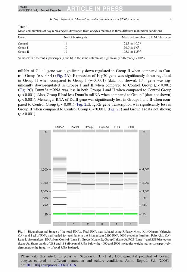
3.1.2. Gene expressionThe Bioanalyzer gel image of the total RNAs for Experiments I and II is shown in Fig. 1,
which indicates that the total RNA quality was sufficient to perform further analysis of mRNAs.Dynamics of mRNA patterns in Experiment I are shown in Fig. 2. In Experiment I, amount of
Table 2Development of embryos derived from oocytes matured in three different maturation conditions
Groupa No. of fertilizedoocytes
No. of 2-cellembryos (%)
No. of 8-cellembryos (%)
No. ofmorulae (%)
No. ofblastocyst (%)
Control 439 335 (76.15) 210 (48.40) 169 (39.09) 127 (29.10)Group I 395 296 (75.65) 185 (47.12) 146 (38.25) 110 (28.52)Group II 415 323 (78.39) 211 (51.03) 164 (39.75) 120 (29.15)
a Oocytes were matured in TCM-199 supplemented with 10% FCS (Control Group), 10% SSS (Group I) and 10%SSS + 10 ng/ml EGF (Group II). Developing embryos in SOF medium were supplemented with 10% FCS on day 4 ofIVC in all groups.
Please cite this article in press as: Sagirkaya, H. et al., Developmental potential of bovineoocytes cultured in different maturation and culture conditions, Anim. Reprod. Sci. (2006),doi:10.1016/j.anireprosci.2006.09.016
ARTICLE IN PRESS+ModelANIREP-3194; No. of Pages 16
H. Sagirkaya et al. / Animal Reproduction Science xxx (2006) xxx–xxx 9
Table 3Mean cell numbers of day 8 blastocysts developed from oocytes matured in three different maturation conditions
Group No. of blastocysts Mean cell number ± S.E.M./blastocyst
Control 14 122.3 ± 10.7a
Group I 10 90.0 ± 5.0b
Group II 16 105.6 ± 8.5a,b
Values with different superscripts (a and b) in the same column are significantly different (p < 0.05).
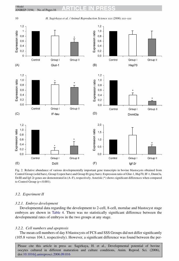
mRNA of Glut-1 gene was significantly down-regulated in Group II when compared to Con-trol Group (p < 0.001) (Fig. 2A). Expression of Hsp70 gene was significantly down-regulatedin Group II when compared to Group I (p < 0.001) (data not shown). IF-� gene was sig-nificantly down-regulated in Groups I and II when compared to Control Group (p < 0.001)(Fig. 2C). Dnmt3a mRNA was less in both Groups I and II when compared to Control Group(p < 0.001). Also, Group II had less Dmnt3a mRNA when compared to Group I (data not shown)(p < 0.001). Messenger RNA of DcIII gene was significantly less in Groups I and II when com-pared to Control Group (p < 0.001) (Fig. 2E). Igf-2r gene transcription was significantly less inGroup II when compared to Control Group (p < 0.001) (Fig. 2F) and Group I (data not shown)(p < 0.001).
Fig. 1. Bioanalyzer gel image of the total RNAs. Total RNA was isolated using RNeasy Micro Kit (Qiagen, Valencia,CA), and 1 �l of RNA was loaded for each lane in the Bioanalyzer 2100 RNA 6000 picochip (Agilent, Palo Alto, CA).Lane L size markers, RNA from Control (Lane 1), Group I (Lane 2), Group II (Lane 3), FCS (Lane 4) and SSS blastocysts(Lane 5). Sharp bands of 28S and 18S ribosomal RNA below the 4000 and 2000 molecular weight markers, respectively,demonstrate the integrity of total RNA isolated.
Please cite this article in press as: Sagirkaya, H. et al., Developmental potential of bovineoocytes cultured in different maturation and culture conditions, Anim. Reprod. Sci. (2006),doi:10.1016/j.anireprosci.2006.09.016
ARTICLE IN PRESS+ModelANIREP-3194; No. of Pages 16
10 H. Sagirkaya et al. / Animal Reproduction Science xxx (2006) xxx–xxx
Fig. 2. Relative abundance of various developmentally important gene transcripts in bovine blastocysts obtained fromControl Group (solid bars), Group I (open bars) and Group II (gray bars). Expression ratio of Glut-1, Hsp70, IF-�, Dnmt3a,DcIII and Igf-2r genes are demonstrated in (A–F), respectively. Asterisk (*) shows significant differences when comparedto Control Group (p < 0.001).
3.2. Experiment II
3.2.1. Embryo developmentDevelopmental data regarding the development to 2-cell, 8-cell, morulae and blastocyst stage
embryos are shown in Table 4. There was no statistically significant difference between thedevelopmental rates of embryos in the two groups at any stage.
3.2.2. Cell numbers and apoptosisThe mean cell numbers of day 8 blastocysts of FCS and SSS Groups did not differ significantly
(105.8 versus 104.1, respectively). However, a significant difference was found between the per-

Please cite this article in press as: Sagirkaya, H. et al., Developmental potential of bovineoocytes cultured in different maturation and culture conditions, Anim. Reprod. Sci. (2006),doi:10.1016/j.anireprosci.2006.09.016
ARTICLE IN PRESS+ModelANIREP-3194; No. of Pages 16
H. Sagirkaya et al. / Animal Reproduction Science xxx (2006) xxx–xxx 11
Table 4Development of embryos derived from oocytes matured in media supplemented with FCS or SSS
Groupa No. of fertilizedoocytes
No. of 2-cellembryos (%)
No. of 8-cellembryos (%)
No. of morulae (%) No. ofblastocyst (%)
FCS 574 393 (70.1) 222 (40.0) 193 (35.1) 145 (26.6)SSS 644 480 (75.6) 268 (41.8) 238 (36.9) 173 (27.8)
a Oocytes were matured in TCM-199 supplemented with 10% FCS and 10% SSS + 10 ng/ml EGF, and developingembryos in SOFaa medium were supplemented with 10% FCS and 10% SSS on day 4 of IVC for FCS and SSS Groups,respectively.
Table 5Mean cell numbers and percentages of apoptotic blastomeres of day 8 blastocysts from FCS and SSS Groups
Group n Mean cell num-ber ± S.E.M./blastocyst
Percentages of apoptotic blas-tomeres/blastocyst ± S.E.M.
FCS 34 105.8 ± 4.0 4.23 ± 0.6a
SSS 29 104.1 ± 6.0 8.67 ± 1.0b
Values with different superscripts (a and b) in the same column are significantly different (p < 0.01).
centages of TUNEL-positive nuclei of FCS and SSS Groups (4.23% versus 8.67%, respectively,p < 0.01) (Table 5).
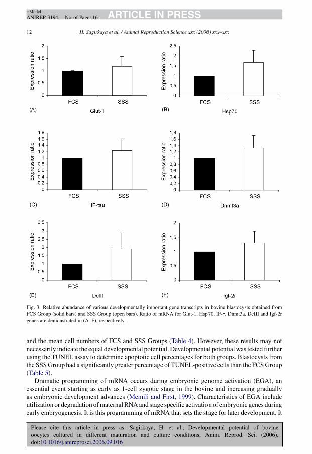
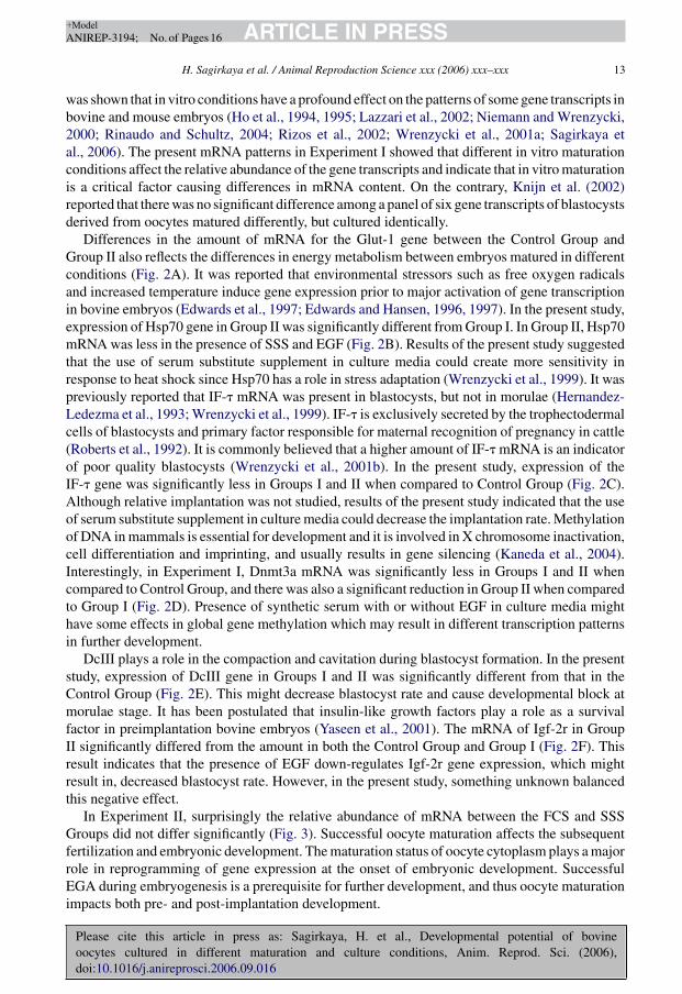
3.2.3. Gene expressionSurprisingly in Experiment II, there were no significant differences among the relative mRNA
abundance of the studied genes between FCS and SSS (Fig. 3A–F).
4. Discussion
In the present study, the effects of the FCS replacement with SSS in the maturation media alone,and in the maturation media as well as the culture media were investigated in terms of differentevaluation procedures for assessing the quality of developing blastocysts. In the present study theaim was to investigate the effects of replacement of FCS (Control Group) with SSS only (GroupI) or SSS + EGF (Group II) on in vitro maturation in Experiment I. Developing embryos were alltreated in the same way to observe the effect of serum replacement in the maturation medium andsupplemented with 10% FCS on day 4 of in vitro culture. Results of Experiment I showed that SSSand EGF combination might be successfully used instead of serum in the maturation medium.Then, in Experiment II embryos were produced by using FCS (FCS Group) or SSS + EGF (SSSGroup) in the maturation medium and developing embryos were, respectively, supplemented with10% FCS or 10% SSS on day 4 of in vitro culture to see the effect of FCS replacement in thematuration and culture media. In terms of cleavage rates results of the present study did not differsignificantly in both experiments (Table 2). However, mean cell number of blastocysts developedfrom Group I was significantly less than the Control Group in Experiment I (Table 3), whichsupported previous work showing the beneficial effect of EGF in the absence of serum (Lonerganet al., 1996; Mtango et al., 2003; Oyamada et al., 2004; Sagirkaya et al., 2004; Sakaguchi et al.,2000). In Experiment II, there was no significant difference between the developmental results
Please cite this article in press as: Sagirkaya, H. et al., Developmental potential of bovineoocytes cultured in different maturation and culture conditions, Anim. Reprod. Sci. (2006),doi:10.1016/j.anireprosci.2006.09.016
ARTICLE IN PRESS+ModelANIREP-3194; No. of Pages 16
12 H. Sagirkaya et al. / Animal Reproduction Science xxx (2006) xxx–xxx
Fig. 3. Relative abundance of various developmentally important gene transcripts in bovine blastocysts obtained fromFCS Group (solid bars) and SSS Group (open bars). Ratio of mRNA for Glut-1, Hsp70, IF-�, Dnmt3a, DcIII and Igf-2rgenes are demonstrated in (A–F), respectively.
and the mean cell numbers of FCS and SSS Groups (Table 4). However, these results may notnecessarily indicate the equal developmental potential. Developmental potential was tested furtherusing the TUNEL assay to determine apoptotic cell percentages for both groups. Blastocysts fromthe SSS Group had a significantly greater percentage of TUNEL-positive cells than the FCS Group(Table 5).
Dramatic programming of mRNA occurs during embryonic genome activation (EGA), anessential event starting as early as 1-cell zygotic stage in the bovine and increasing graduallyas embryonic development advances (Memili and First, 1999). Characteristics of EGA includeutilization or degradation of maternal RNA and stage specific activation of embryonic genes duringearly embryogenesis. It is this programming of mRNA that sets the stage for later development. It
Please cite this article in press as: Sagirkaya, H. et al., Developmental potential of bovineoocytes cultured in different maturation and culture conditions, Anim. Reprod. Sci. (2006),doi:10.1016/j.anireprosci.2006.09.016
ARTICLE IN PRESS+ModelANIREP-3194; No. of Pages 16
H. Sagirkaya et al. / Animal Reproduction Science xxx (2006) xxx–xxx 13
was shown that in vitro conditions have a profound effect on the patterns of some gene transcripts inbovine and mouse embryos (Ho et al., 1994, 1995; Lazzari et al., 2002; Niemann and Wrenzycki,2000; Rinaudo and Schultz, 2004; Rizos et al., 2002; Wrenzycki et al., 2001a; Sagirkaya etal., 2006). The present mRNA patterns in Experiment I showed that different in vitro maturationconditions affect the relative abundance of the gene transcripts and indicate that in vitro maturationis a critical factor causing differences in mRNA content. On the contrary, Knijn et al. (2002)reported that there was no significant difference among a panel of six gene transcripts of blastocystsderived from oocytes matured differently, but cultured identically.
Differences in the amount of mRNA for the Glut-1 gene between the Control Group andGroup II also reflects the differences in energy metabolism between embryos matured in differentconditions (Fig. 2A). It was reported that environmental stressors such as free oxygen radicalsand increased temperature induce gene expression prior to major activation of gene transcriptionin bovine embryos (Edwards et al., 1997; Edwards and Hansen, 1996, 1997). In the present study,expression of Hsp70 gene in Group II was significantly different from Group I. In Group II, Hsp70mRNA was less in the presence of SSS and EGF (Fig. 2B). Results of the present study suggestedthat the use of serum substitute supplement in culture media could create more sensitivity inresponse to heat shock since Hsp70 has a role in stress adaptation (Wrenzycki et al., 1999). It waspreviously reported that IF-� mRNA was present in blastocysts, but not in morulae (Hernandez-Ledezma et al., 1993; Wrenzycki et al., 1999). IF-� is exclusively secreted by the trophectodermalcells of blastocysts and primary factor responsible for maternal recognition of pregnancy in cattle(Roberts et al., 1992). It is commonly believed that a higher amount of IF-� mRNA is an indicatorof poor quality blastocysts (Wrenzycki et al., 2001b). In the present study, expression of theIF-� gene was significantly less in Groups I and II when compared to Control Group (Fig. 2C).Although relative implantation was not studied, results of the present study indicated that the useof serum substitute supplement in culture media could decrease the implantation rate. Methylationof DNA in mammals is essential for development and it is involved in X chromosome inactivation,cell differentiation and imprinting, and usually results in gene silencing (Kaneda et al., 2004).Interestingly, in Experiment I, Dnmt3a mRNA was significantly less in Groups I and II whencompared to Control Group, and there was also a significant reduction in Group II when comparedto Group I (Fig. 2D). Presence of synthetic serum with or without EGF in culture media mighthave some effects in global gene methylation which may result in different transcription patternsin further development.
DcIII plays a role in the compaction and cavitation during blastocyst formation. In the presentstudy, expression of DcIII gene in Groups I and II was significantly different from that in theControl Group (Fig. 2E). This might decrease blastocyst rate and cause developmental block atmorulae stage. It has been postulated that insulin-like growth factors play a role as a survivalfactor in preimplantation bovine embryos (Yaseen et al., 2001). The mRNA of Igf-2r in GroupII significantly differed from the amount in both the Control Group and Group I (Fig. 2F). Thisresult indicates that the presence of EGF down-regulates Igf-2r gene expression, which mightresult in, decreased blastocyst rate. However, in the present study, something unknown balancedthis negative effect.
In Experiment II, surprisingly the relative abundance of mRNA between the FCS and SSSGroups did not differ significantly (Fig. 3). Successful oocyte maturation affects the subsequentfertilization and embryonic development. The maturation status of oocyte cytoplasm plays a majorrole in reprogramming of gene expression at the onset of embryonic development. SuccessfulEGA during embryogenesis is a prerequisite for further development, and thus oocyte maturationimpacts both pre- and post-implantation development.
Please cite this article in press as: Sagirkaya, H. et al., Developmental potential of bovineoocytes cultured in different maturation and culture conditions, Anim. Reprod. Sci. (2006),doi:10.1016/j.anireprosci.2006.09.016
ARTICLE IN PRESS+ModelANIREP-3194; No. of Pages 16
14 H. Sagirkaya et al. / Animal Reproduction Science xxx (2006) xxx–xxx
In conclusion, SSS and EGF in maturation medium instead of FCS support oocyte matura-tion and embryo development in vitro. Linking morphological and developmental indicators ofoocyte and embryo viability with the expression and function of specific genes can provide valu-able insights into mechanisms contributing to embryonic mortality. However, perturbations ofglobal transcriptome in different culture conditions should be analyzed using DNA microarrays.Furthermore, correlations between amount of mRNA and developmental potential of in vitroderived embryos should be further studied by methods such as embryo transfer to recipient cows.
Acknowledgments
Authors would like to thank Brad Haley for help in obtaining bovine ovaries and KindraKelly-Quagliana for laboratory set up and preparations of the manuscript.
This project was supported by The Life Sciences and Biotechnology Institute, MississippiState University (Assignment number: J-10829).
References
Abe, H., Yamashita, S., Itoh, T., Satoh, T., Hoshi, H., 1999. Ultrastructure of bovine embryos developed from in vitro-matured and -fertilized oocytes: comparative morphological evaluation of embryos cultured either in serum-freemedium or in serum-supplemented medium. Mol. Reprod. Dev. 53, 325–335.
Chanson, A., Nocera, D., Senn, A., De Grandi, P., Germond, M., 2001. Development of a well-defined medium for the invitro maturation of immature bovine cumulus–oocyte complexes. J. Assist. Reprod. Genet. 18, 97–105.
Dominko, T., First, N.L., 1997. Timing of meiotic progression in bovine oocytes and its effect on early embryo development.Mol. Reprod. Dev. 47, 456–467.
Edwards, J.L., Ealy, A.D., Monterroso, V.H., Hansen, P.J., 1997. Ontogeny of temperature-regulated heat shock protein70 synthesis in preimplantation bovine embryos. Mol. Reprod. Dev. 48, 25–33.
Edwards, J.L., Hansen, P.J., 1996. Elevated temperature increases heat shock protein 70 synthesis in bovine two-cellembryos and compromises function of maturing oocytes. Biol. Reprod. 55, 341–346.
Edwards, J.L., Hansen, P.J., 1997. Differential responses of bovine oocytes and preimplantation embryos to heat shock.Mol. Reprod. Dev. 46, 138–145.
Fedorcsak, P., Storeng, R., 2003. Effects of leptin and leukemia inhibitory factor on preimplantation development andSTAT3 signaling of mouse embryos in vitro. Biol. Reprod. 69, 1531–1538.
Gordon, I., 1994. Laboratory Production of Cattle Embryos. CAB International, Wallington, pp. 30–142.Hernandez-Ledezma, J.J., Mathialagan, N., Villanueva, C., Sikes, J.D., Roberts, R.M., 1993. Expression of bovine
trophoblast interferons by in vitro-derived blastocysts is correlated with their morphological quality and stage ofdevelopment. Mol. Reprod. Dev. 36, 1–6.
Ho, Y., Doherty, A.S., Schultz, R.M., 1994. Mouse preimplantation embryo development in vitro: effect of sodiumconcentration in culture media on RNA synthesis and accumulation and gene expression. Mol. Reprod. Dev. 38,131–141.
Ho, Y., Wigglesworth, K., Eppig, J.J., Schultz, R.M., 1995. Preimplantation development of mouse embryos in KSOM:augmentation by amino acids and analysis of gene expression. Mol. Reprod. Dev. 41, 232–238.
Holm, P., Booth, P.J., Callesen, H., 2002. Kinetics of early in vitro development of bovine in vivo- and in vitro-derivedzygotes produced and/or cultured in chemically defined or serum-containing media. Reproduction 123, 553–565.
Kane, M.T., 1987. Culture media and culture of early embryos. Theriogenology 27, 49–57.Kaneda, M., Okano, M., Hata, K., Sado, T., Tsujimoto, N., Li, E., Sasaki, H., 2004. Essential role for de novo DNA
methyltransferase Dnmt3a in paternal and maternal imprinting. Nature 429, 900–903.Knijn, H.M., Wrenzycki, C., Hendriksen, P.J., Vos, P.L., Herrmann, D., van der Weijden, G.C., Niemann, H., Dieleman,
S.J., 2002. Effects of oocyte maturation regimen on the relative abundance of gene transcripts in bovine blastocystsderived in vitro or in vivo. Reproduction 124, 365–375.
Lazzari, G., Wrenzycki, C., Herrmann, D., Duchi, R., Kruip, T., Niemann, H., Galli, C., 2002. Cellular and moleculardeviations in bovine in vitro-produced embryos are related to the large offspring syndrome. Biol. Reprod. 67, 767–775.
Leibfried, M.L., Bavister, B.D., 1982. Effects of epinephrine and hypotaurine on in-vitro fertilization in the golden hamster.J. Reprod. Fertil. 66, 87–93.

Please cite this article in press as: Sagirkaya, H. et al., Developmental potential of bovineoocytes cultured in different maturation and culture conditions, Anim. Reprod. Sci. (2006),doi:10.1016/j.anireprosci.2006.09.016
ARTICLE IN PRESS+ModelANIREP-3194; No. of Pages 16
H. Sagirkaya et al. / Animal Reproduction Science xxx (2006) xxx–xxx 15
Lonergan, P., Carolan, C., Van Langendonckt, A., Donnay, I., Khatir, H., Mermillod, P., 1996. Role of epidermalgrowth factor in bovine oocyte maturation and preimplantation embryo development in vitro. Biol. Reprod. 54,1420–1429.
Lonergan, P., O’Kearney-Flynn, M., Boland, M.P., 1999. Effect of protein supplementation and presence of an antiox-idant on the development of bovine zygotes in synthetic oviduct fluid medium under high or low oxygen tension.Theriogenology 51, 1565–1576.
Mastromonaco, G.F., Semple, E., Robert, C., Rho, G.J., Betts, D.H., King, W.A., 2004. Different culture media require-ments of IVF and nuclear transfer bovine embryos. Reprod. Domest. Anim. 39, 462–467.
Memili, E., First, N.L., 1999. Control of gene expression at the onset of bovine embryonic development. Biol. Reprod.61, 1198–1207.
Mtango, N.R., Varisanga, M.D., Dong, Y.J., Rajamahendran, R., Suzuki, T., 2003. Growth factors and growth hor-mone enhance in vitro embryo production and post-thaw survival of vitrified bovine blastocysts. Theriogenology 59,1393–1402.
Niemann, H., Wrenzycki, C., 2000. Alterations of expression of developmentally important genes in preimplantationbovine embryos by in vitro culture conditions: implications for subsequent development. Theriogenology 53, 21–34.
Oyamada, T., Iwayama, H., Fukui, Y., 2004. Additional effect of epidermal growth factor during in vitro maturation forindividual bovine oocytes using a chemically defined medium. Zygote 12, 143–150.
Parrish, J.J., Krogenaes, A., Susko-Parrish, J.L., 1995. Effect of bovine sperm separation by either swim-up or Percollmethod on success of in vitro fertilization and early embryonic development. Theriogenology 44, 859–870.
Parrish, J.J., Susko-Parrish, J.L., Leibfried-Rutledge, M.L., Critser, E.S., Eyestone, W.H., First, N.L., 1986. Bovine invitro fertilization with frozen–thawed semen. Theriogenology 25, 591–600.
Pfaffl, M.W., 2001. A new mathematical model for relative quantification in real-time RT-PCR. Nucl. Acids Res. 29, e45.Pfaffl, M.W., Horgan, G.W., Dempfle, L., 2002. Relative expression software tool (REST) for group-wise comparison and
statistical analysis of relative expression results in real-time PCR. Nucl. Acids Res. 30, e36.Rinaudo, P., Schultz, R.M., 2004. Effects of embryo culture on global pattern of gene expression in preimplantation mouse
embryos. Reproduction 128, 301–311.Rizos, D., Lonergan, P., Boland, M.P., Arroyo-Garcia, R., Pintado, B., de la Fuente, J., Gutierrez-Adan, A., 2002. Anal-
ysis of differential messenger RNA expression between bovine blastocysts produced in different culture systems:implications for blastocyst quality. Biol. Reprod. 66, 589–595.
Roberts, R.M., Cross, J.C., Leaman, D.W., 1992. Interferons as hormones of pregnancy. Endocr. Rev. 13, 432–452.Russell, J.B., Knezevich, K.M., Fabian, K.F., Dickson, J.A., 1997. Unstimulated immature oocyte retrieval: early versus
midfollicular endometrial priming. Fertil. Steril. 67, 616–620.Sagirkaya, H., Yagmur, M., Nur, Z., Soylu, M.K., 2004. Replacement of fetal calf serum with synthetic serum substitute
in the in vitro maturation medium: effects on maturation, fertilization and subsequent development of cattle oocytesin vitro. Turk. J. Vet. Anim. Sci. 28, 779–784.
Sagirkaya, H., Misirlioglu, M., Kaya, A., Parrish, J.J., First, N.L., Memili, E., 2006. Developmental and molecularcorrelates of bovine preimplantation embryos. Reproduction 131, 895–904.
Sakaguchi, M., Dominko, T., Leibfried-Rutledge, M.L., Nagai, T., First, N.L., 2000. A combination of EGF and IGF-I accelerates the progression of meiosis in bovine follicular oocytes in vitro and fetal calf serum neutralizes theacceleration effect. Theriogenology 54, 1327–1342.
Thompson, J.G., Allen, N.W., McGowan, L.T., Bell, A.C., Lambert, M.G., Tervit, H.R., 1998. Effect of delayed sup-plementation of fetal calf serum to culture medium on bovine embryo development in vitro and following transfer.Theriogenology 49, 1239–1249.
Vigneron, C., Perrau, C., Dupont, J., Uzbekova, S., Prigent, C., Mermillod, P., 2004. Several signaling pathways areinvolved in the control of cattle oocyte maturation. Mol. Reprod. Dev. 69, 466–474.
Wrenzycki, C., Herrmann, D., Carnwath, J.W., Niemann, H., 1999. Alterations in the relative abundance of gene transcriptsin preimplantation bovine embryos cultured in medium supplemented with either serum or PVA. Mol. Reprod. Dev.53, 8–18.
Wrenzycki, C., Herrmann, D., Keskintepe, L., Martins Jr., A., Sirisathien, S., Brackett, B., Niemann, H., 2001a. Effects ofculture system and protein supplementation on mRNA expression in pre-implantation bovine embryos. Hum. Reprod.16, 893–901.
Wrenzycki, C., Herrmann, D., Lucas-Hahn, A., Lemme, E., Korsawe, K., Niemann, H., 2004. Gene expression patternsin in vitro-produced and somatic nuclear transfer-derived preimplantation bovine embryos: relationship to the largeoffspring syndrome? Anim. Reprod. Sci. 82-83, 593–603.
Wrenzycki, C., Wells, D., Herrmann, D., Miller, A., Oliver, J., Tervit, R., Niemann, H., 2001b. Nuclear transfer protocolaffects messenger RNA expression patterns in cloned bovine blastocysts. Biol. Reprod. 65, 309–317.

Please cite this article in press as: Sagirkaya, H. et al., Developmental potential of bovineoocytes cultured in different maturation and culture conditions, Anim. Reprod. Sci. (2006),doi:10.1016/j.anireprosci.2006.09.016
ARTICLE IN PRESS+ModelANIREP-3194; No. of Pages 16
16 H. Sagirkaya et al. / Animal Reproduction Science xxx (2006) xxx–xxx
Yaseen, M.A., Wrenzycki, C., Herrmann, D., Carnwath, J.W., Niemann, H., 2001. Changes in the relative abun-dance of mRNA transcripts for insulin-like growth factor (IGF-I and IGF-II) ligands and their receptors (IGF-IR/IGF-IIR) in preimplantation bovine embryos derived from different in vitro systems. Reproduction 122,601–610.
Zeuner, A., Muller, K., Reguszynski, K., Jewgenow, K., 2003. Apoptosis within bovine follicular cells and its effect onoocyte development during in vitro maturation. Theriogenology 59, 1421–1433.