Embed Size (px)
Citation preview

Development of a Model for the Valuation of Work Relative Value Units
Objective Service Time Task Status Report
June 30, 2014
Prepared for:
Centers for Medicare & Medicaid Services
Prepared by:
The Urban Institute Stephen Zuckerman, PhD
Robert Berenson, MD
Social & Scientific Systems, Inc. Katie Merrell, BA
Tyler Oberlander, BA
RTI International Nancy McCall, ScD
Rebecca Lewis, MPH Sue Mitchell, RHIA
Madhu Shrestha, BS
This project was funded by Centers for Medicare and Medicaid Services. The Urban Institute, Social & Scientific Systems, Inc., and RTI International assume responsibility for the accuracy and completeness of the information contained in this report.

[This page left intentionally blank]
ii

CONTENTS
Executive Summary .........................................................................................................................1
Section 1 Background ......................................................................................................................3 1.1 Developing Objective Service Time Data .....................................................................3 1.2 Report Overview ............................................................................................................6
Section 2 Study Services: Selection and Description ......................................................................7 2.1 Selecting Services for Inclusion .....................................................................................7
2.1.1 Potentially Misvalued Codes ................................................................................7
2.1.2 Importance to Medicare Payment Policy ..............................................................8
2.1.3 Project Considerations ..........................................................................................9
2.1.4 Final Study Services .............................................................................................9 2.2 Describing Service Elements for Data Collection .......................................................12
2.2.1 Pre-service, Intra-service, and Post-Service Definitions for Individual Services ...............................................................................................................12
2.2.2 Adapting the Definitions for this Project ............................................................15
2.2.3 Implications for Data Collection and Analysis ...................................................17
Section 3 Study Sites: Identification, Recruitment, Assessment, and Engagement ......................18 3.1 Identifying Potential Sites ............................................................................................18 3.2 Recruiting Sites ............................................................................................................18 3.3 Assessing Sites .............................................................................................................20 3.4 Engaging Sites .............................................................................................................21
Section 4 Data Collection ..............................................................................................................24 4.1 Data Collection Overview ............................................................................................24 4.2 Development of Study Protocols .................................................................................24 4.3 Direct Observation Approach ......................................................................................26 4.4 Other Site Data .............................................................................................................28 4.5 Implementation Challenges .........................................................................................28 4.5 Preliminary Data Validation ........................................................................................31
Section 5 Summary and Next Steps ...............................................................................................32
LIST OF TABLES
1 Distribution of Study Services’ Work RVUs and Service Times ........................................ 10 2 Study Service Types, with Number of Individual Codes .................................................... 10 3 Original and Short Descriptions of Service Elements of CT of the Head/Brain
(CPT® 70450) ....................................................................................................................... 15
iii

APPENDICES
A: Study Services with Narrative Descriptions .......................................................................... A-1
B: Study Services: Work RVUs, Times, and Other Characteristics ............................................B-1
C: Study Services: Shortened Service Element Descriptions ......................................................C-1
D: Recruitment Materials ............................................................................................................ D-1
E: Direct Observation Data Collection Protocol .......................................................................... E-1
F: Protocol for Supporting Data ................................................................................................... F-1
iv

EXECUTIVE SUMMARY
This pilot project is part of the Centers for Medicare & Medicaid Services’s (CMS) efforts to address potentially misvalued services in the Medicare Physician Fee Schedule, as identified in the Affordable Care Act. It aims to develop a validation process for the relative value units (RVUs) used in the fee schedule for both new and existing services to establish payment for the work of the physician or nonphysician practitioner. One of the key elements of the project is the development of objective time estimates based on data from several physician practices, health systems, or other appropriate entities. We are attempting to collect two types of data: administrative data from electronic health records and other sources, and direct observation, in which project staff will observe and document the time it takes to provide specific services to individual patients. This status report describes that task in detail, including selection of services to be studied, identification and engagement of sites for data collection, and development of data collection protocols and tools.
The research team worked closely with CMS to identify about 100 services for the study. We considered factors in three categories:
• Potentially misvalued services. The Affordable Care Act identifies several categories of services that are thought to be at risk for being misvalued1;
• Importance to Medicare. In addition to total spending level, services are important to Medicare for other policy reasons, including those with global periods and those serving as multiple points of comparison; and
• Project considerations. We need to select a mix of services that will allow us to test methods in a variety of settings, but need to limit the number of specialties that they involve because of the clinical panels that will examine the data later in the project.
Balancing these considerations, we developed a list of over 100 codes for the study. Collectively, they account for roughly 18 percent of total Medicare physician fee schedule spending. They include 29 services from CMS’s list of potentially misvalued services, 71 services with global periods of 0, 10, or 90 days, and 12 services that have been used as multiple points of comparison in the development and refinement of work relative values.
For the purpose of documenting the time associated with pre-service, intra-service, and post-service elements of care, we are using the definitions of these elements that are used by the American Medical Association/Specialty Society Relative Update Committee (RUC) in creating the current service time estimates. There are some potential challenges with these definitions, however, that may affect our measures. First, they differ with regard to the amount of detail about the specific tasks included in the service. Some services have very vague descriptions while others are quite detailed. As a result, observers may not consistently attribute tasks to the
1 The recently passed Protecting Access to Medicare Act of 2014 includes an expanded list of categories of services that may be misvalued.
1

pre, intra, or post period. The descriptions are also inconsistent about whether or not tasks performed by nonphysicians are included. As a result of this inconsistency, CMS has asked us to document tasks performed by both the physician and other personnel directly involved in providing the service. As a result, we have developed a data collection tool that allows observers to indicate which service elements from the RUC definitions are performed and by whom. It also allows observers to indicate that other tasks are performed as part of the service, even if they are not included in the RUC description. This approach will allow us to examine the specific elements provided and who provides them, which will help CMS understand how closely the RUC definitions relate to current service provision.
We also worked with CMS staff to develop a list of potential sites for data collection. In developing this list, we considered leaders’ interest in participating in studies like this, HIT capabilities, experience with direct observation, and the mix and volume of clinical services each site provides. The process of identifying, recruiting, assessing, and engaging sites has been much more complicated than originally anticipated. We have three sites currently involved, after having approached nearly 20 potential sites. As we prepare to do onsite observation at three sites, we will continue to try to recruit additional sites.
A number of unexpected issues have been raised by potential and participating sites, from concerns over union work rules and IRB requirements to low service volumes for study services and data system limitations. In response to the specific issues at the three sites currently involved, we have modified our data collection approach. For some services in some sites, our project staff will not actually perform the direct observation in patient care areas but instead will train site staff to do the observation, and will be onsite to oversee their work. We anticipate developing site-specific data collection plans for each site to reflect both IRB and other concerns as well as each site’s specific data systems and clinical organization. One issue that has come up is that the likely service volume for many of the services selected for this study appears to be lower at each study site than might have been expected based on the Medicare volume data used in the selection process.
We have developed data collection protocols for both direct observation and electronic time data. These protocols will serve both for training and for field reference. We have also developed an Access®-based data collection tool for direct observation and have developed a strategy for sites that prefer a pencil and paper approach.
At this point we are preparing for data collection at three sites, and we continue to try to engage additional sites. After we develop new objective time measures from participating sites, we will then move to the other project tasks: analyzing these new time estimates to develop alternative models of their implications for service work values; and reviewing the time data and work models with panels of physicians.
2

SECTION 1 BACKGROUND
This pilot project is part of CMS’s efforts to address potentially misvalued services in the Medicare Physician Fee Schedule.2 It aims to develop a validation process for the work relative value units (RVUs) used in the fee schedule for both new and existing services. It is designed to provide CMS with a process for reviewing proposed work RVUs, assessing how reasonable they are relative to external data, and assuring that the relativities within the fee schedule are internally consistent within families of services and specialties as well as across families.
Work RVUs reflect both the time it takes the clinician to provide a service and the intensity of the service (i.e., technical skill, physical effort, mental effort and judgment, and stress due to patient risk). Time is the component of the work RVU most amenable to objective measurement. Service time estimates are currently based on surveys conducted by specialty societies for the RUC. This project is focused on developing new sources of time data as a central element in our model to comprehensively validate the work RVUs for a selected set of services.
The project was designed with three key elements:
1. Obtain objective time estimates for a group of services from several physician practices or health care systems
2. Compare these objective time estimates with current fee schedule time data and develop alternative models of work values
3. Review objective time estimates and alternative work models with a series of clinical panels.
The approach to collecting objective time data was designed based on previous studies that, as summarized below, describe the types of administrative data that are available for some types of services, such as operating room logs, and the need for direct observation for other services. By acquiring administrative data and conducting direct observation at several sites, we aim to develop time estimates for each of the services selected for study. These estimates will then be compared to existing time values and work RVUs, and then provided to clinical panels for assessment. The object time data task is central to the project, and this status report describes our progress to date on this part of the project.
1.1 Developing Objective Service Time Data
The goal of the time collection task is to develop independent, objective measures of service-level times for fee schedule services. In the context of Medicare payment policy, service-level time estimates are used in two distinct ways:
2 A detailed description of the fee schedule’s background and related policies can be found in “Medicare Program; Revisions to Payment Policies Under the Physician Fee Schedule, Clinical Laboratory Fee Schedule & Other Revisions to Part B for CY 2014” , Centers for Medicare and Medicaid Services, Federal Register, Vol. 78, No. 237, Tuesday, December 10, 2013
3

• They are a key input into estimating work relative value units (RVUs). Work values, defined as the time, stress, expertise, and skill required to provide a service, are highly correlated with time, which explains between 75% and 90% of RVU variation within service families.3
• They are used in the practice expense relative value algorithm, with specialty-specific hourly cost data.
Prior research has shown that some of the Medicare fee schedule’s estimates of service time based upon surveys of physicians by specialty societies are considerably higher than estimates obtained from other data sources, such as operating room logs.4 That is, the survey times on which both the work and practice expense relative value units are based may diverge from objective measures of service time collected through administrative systems or direct observation.
These differences may be due in part to changes that have occurred since the physician fee schedule was introduced over 20 years ago. Through the substitution of new technologies, such as mastering one-time innovative new techniques such as endoscopy, the time for a particular service may be much less than what the fee schedule work relative value units reflect. Many such services have never been restudied or validated, other than by the RUC, which uses estimates of time obtained from surveys of physicians conducted by specialty societies as part of their request for changes in work RVUs. The differences may also indicate discrepancies in the time data for other reasons.
Because of the concerns that have been raised about the time estimates in the fee schedule, several years ago MedPAC funded a study assess the feasibility of using objective time data to establish or update work RUVs.5 The study had two key elements:
1. evaluate existing secondary databases to determine their usefulness for producing objective time estimates and
2. assess the feasibility of primary data collection.
The evaluation of secondary sources determined that none of the databases -- the Society of Thoracic Surgeons (STS), the National Surgical Quality Improvement Program (NSQIP), and four National Ambulatory Medical Care Survey (NAMCS) databases -- span all types of services or places of service. Intra-service time was most readily available for major surgical procedures
3 Medicare Payment Advisory Commission (MedPAC), “Report to the Congress: Medicare and the Health Care Delivery System ,” June 2011, p.15
4 McCall, N., Cromwell, J., Braun, P. Validation of physician survey estimates of surgical time using operating room logs. Medical Care Research and Review. 63: No. 6, 1-14, 2006
5 Braun, P. and McCall, N. Improving the Accuracy of Time in the Medical Physician Fee Schedule: Feasibility of Using Extant Data and of Collecting Primary Data. For MedPAC: 2011.
4

from the STS and NSQIP databases but only the NSQIP explicitly links service time to individual Current Procedural Terminology (CPT®) codes.6 NAMCS Office provides an estimate of intra-service time for E&M services, but is not linked to CPT® codes. This review of the secondary databases suggested that primary data collection would be required for most types of services, especially office-based procedures and diagnostic tests.
To assess the feasibility of primary data collection, the study team conducted key informant interviews with five organizations that represent a broad cross-section of sites and made site visits to two large multi-specialty group practices. The primary purpose of the interviews and site visits was to determine the feasibility of obtaining objective time data from their electronic data systems and conducting parallel direct observation studies to validate the electronic data. In the key informant interviews, the following challenges were noted as was the potential for overcoming them:
• A number of organizations had gathered extensive objective data on time, most notably for major surgical procedures by extracting data from electronic or paper operating room logs, or through direct observation. However, none of the interviewed organizations collected clinical service times at the CPT® code level. There was limited concern about their ability to link clinical service times to CPT® codes regardless of data collection method.
• Clinical time is best captured for major surgical procedures with most organizations capturing intra-service time electronically, if they are affiliated with a hospital setting. Pre- and post-service times are not well captured in electronic data.
• Most of the organizations were uncertain if their electronic data systems captured specific aspects of surgical time for ambulatory surgical centers or other types of procedures such as endoscopy, radiology, cardiac catheterization laboratory. Time stamps from EHRs, scheduled appointments, or provider work schedules were offered as potential anchors for clinical time.
• None of the organizations believed they capture clinical service time for office-based procedures, such as removal of lesions.
• Total work time of a clinician and total number of clinical services provided during that time is widely available although not necessarily linked with detailed information on the specific services to allow for direct calculation of time at the CPT® code level.
Interviewees noted the potential to overcome these challenges. In addition, they believed that the potential to use electronic data systems may be far greater than their organization had used to date and warranted further evaluation. Some seemed to have little direct knowledge of
6 CPT® codes and descriptions are copyright 2013 American Medical Association (AMA). All rights reserved. CPT is a registered trademark of the AMA.
5

the full capability of their electronic systems to capture clinical service time, and in many cases not all elements of their electronic data systems had been fully implemented.
The organizations that were interviewed had varying degrees of sophistication and experience with primary data collection of clinical service time through direct observation. Top policy leadership and principal researchers within the organizations were broadly supportive of the research objectives. One system had used direct observation to collect data on office-based services for the past ten years, with direct observation ingrained in their culture. Other organizations had varying degrees of direct observation experience with varying degrees of likely acceptance from their clinical staff. These organizations often conducted direct observation within the context of system redesign to improve the flow of care for their patients and providers. Although no organization had collected time for the purpose of payment under the fee schedule, all organizations felt that primary data collection of clinical service time through direct observation was feasible given their prior experience with collection clinical service time for other purposes.
The project team concluded that it is feasible to obtain objective measures of service time using multiple methods of data collection, and that in-depth interviews were needed to more fully understand the potential capabilities of electronic data systems and the method of direct observation for a fuller assessment of the feasibility of prospective primary data collection from a cohort of practices.
Our plan builds on this previous work by trying to develop time values from both administrative data and direct observation. Time estimates from direct observation will be used to both validate and augment estimates from administrative data. We will work with study sites to determine what types of administrative data are available to develop service time estimates and to develop a plan for direct observation.
1.2 Report Overview
This report presents the details of the objective service time task, including:
• Study service selection and descriptions;
• Study site identification, recruitment, and assessment; and
• Data collection.
The next three sections address each topic in turn, describing our original approach, issues and challenges that have arisen, and how we’ve modified our approach to address these challenges. A final section summarizes the key issues we have encountered and the next steps for this task. The other parts of the project – analyzing the time data and conducting clinical panels – are not discussed here but, as mentioned below, the plans for those components of the project have shaped the design of the data collection task.
6

SECTION 2 STUDY SERVICES: SELECTION AND DESCRIPTION
Although CMS is interested in ultimately validating all work values in the Medicare fee schedule, this pilot study was designed to focus on about 100 services. This section describes the factors we considered as we developed a list of study services and how we adapted the RUC’s detailed service element descriptions for use in the data collection process.
2.1 Selecting Services for Inclusion
A key decision for the project was the choice of services to be studied. This section describes the criteria that we used in selecting services and the final set of codes included in the study. Based on conversations with CMS staff, the project team went through a process of refining lists of candidate services based on three categories of criteria: potentially misvalued codes, importance to Medicare spending, and feasibility and appropriateness in the context of this project.
2.1.1 Potentially Misvalued Codes
As required by Sec. 3134 of the Affordable Care Act, HHS must periodically identify, review, and adjust values for potentially misvalued codes. The ACA specifies categories of codes that it recommends HHS focus on:
• codes (and families of codes, as appropriate) for which there has been the fastest growth;
• codes (and families of codes, as appropriate) that have experienced substantial changes in practice expenses;
• codes for new technologies or services within an appropriate period (such as 3 years) after the relative values are initially established for such codes;
• multiple codes that are frequently billed in conjunction with furnishing a single service;
• codes with low relative values, particularly those that are often billed multiple times for a single treatment;
• codes that have not been subject to review since the implementation of the RBRVS (the so-called ‘Harvard-valued codes’); and
• such other codes determined to be appropriate by the Secretary of HHS.7
7 U.S. Congress. Patient Protection and Affordable Care Act, H.R. 3590. Public Law 111–148. 111th Cong. (March 23, 2010). See Sec. 3134
7

CMS uses these categories to identify potentially misvalued codes for the RUC to review and has consolidated the misvalued codes initiative and the five-year review into an annual process.8 This project is a direct result of Sec. 3134’s requirement that CMS establish a formal process to validate RVUs.9 We flagged as potentially misvalued those codes that were included in the list CMS developed from these ACA criteria.10 The recently-passed Protecting Access to Medicare Act of 2014 expanded the list of categories to be considered as potentially misvalued.
In addition to these groups of codes, we discussed other groups of potentially misvalued codes with CMS, including those that have had potential changes in service time due to computerization and automation, those with relatively recent shifts in the typical site of service, services with recent changes in personnel as suggested in the direct practice expense data, and recently adopted services whose work RVUs may not yet exemplify “learning curve” efficiencies.
2.1.2 Importance to Medicare Payment Policy
We considered several Medicare-specific aspects of services while identifying study services: their importance to program spending; role of the service in the work valuation process; and services that are covered by a global period. CMS uses the fee schedule to determine payment for covered services provided by physicians and non-physician practitioners, such as podiatrists, who can bill Medicare directly. Therefore, program spending includes payments to both physicians and non-physician practitioners.11
First, we calculated total Medicare spending for each service and focused on those that account for significant levels of spending, either because of high volume or high expenditures. CMS data categorize services through the Healthcare Common Procedure Coding System (HCPCS), Level 1 of which is comprised of CPT® codes. We used 2011 volumes mapped to 2013 codes and RVUs to calculate each code’s total work and total spending rank. For the most part, studied services are among the top 400 services when ranked by total work.
Second, services can reflect specific aspects of payment policy development that are important in the context of a project like this that aims to model work values based on new time data. Some of these considerations, like codes that retain “Harvard values”, are already included
8 Department of Health and Human Services, Centers for Medicare and Medicaid Services. 2011. “Medicare Physician Fee Schedule: Payment System Fact Sheet Series.” (https://www.cms.gov/MLNProducts/downloads/MedcrePhysFeeSchedfctsht.pdf )
9 Department of Health and Human Services, Centers for Medicare and Medicaid Services, “Medicare Program; Five-Year Review of Work Relative Value Units Under the Physician Fee Schedule; Proposed Rule,” 42 CFR Part 414, Federal Register vol. 76, no. 108, June 6, 2011.
10 Federal Register, Vol. 76, no. 228, page 73066-73067, November 28, 2011
11 Throughout the report, “provider” and “clinician” are used to refer to physicians and non-physician providers who can bill Medicare directly.
8

in the potentially misvalued categories. We flagged codes that were used as multiple points of comparison in the process used to establish work values, since any errors in their time (and work) values may have a cascade effect on other services’ values.
Finally, as mentioned earlier, some services have a global period defined, during which additional evaluation and management (E&M) services are provided, either on the same day as the procedure or in a defined post-operative period of time. These services’ work values reflect both the index service as well as these included E&M services. We recorded whether services were part of a global period and made sure that the final study service universe included a number of these codes.
2.1.3 Project Considerations
The project itself raises a number of factors that needed to be taken into consideration when selecting services. For example, we wanted to select services that would provide a range of clinical contexts in which to test our administrative and direct observation data acquisition approaches. At the same time, we needed to limit the number of specialties encompassed by the universe of study services, so that we can cover them with a manageable number of clinical panels later in the project. We will rely on single-specialty panels, reflecting the specialty most likely to provide these services, to validate and begin work on extrapolation to other services within the same family. As a result, we did not include some services that met several of the other criteria but were provided by a specialty that does not provide other candidate study services. We also included some services that are commonly provided by more than one specialty, permitting us to examine inter-specialty variation in time estimates.
Project constraints also led us to exclude certain services that may merit study in a subsequent project. Most importantly, we have not included E&M services in this pilot study of methods for collecting time data, even though collection of time data on E&M services is important for several reasons. Stand-alone E&M services comprise more than 40 percent of the spending under the MPFS Although studies suggest that there may be time errors in E&M services, the issues associated with determining empirically derived times for E&M services are different and, in some ways, more complex than those posed by obtaining objective time data for procedures, imaging, and tests. E&M services are unique, complex, and important enough to justify a separate and detailed examination of the time issues associated with these services.
We also excluded services that are performed primarily by nonphysician practitioners and those that present unique data acquisition problems, such as Moh’s surgery, radiation therapy, and chemotherapy. In the case of Moh’s surgery, for example, the fact that the patient waits for a pathologist’s report between surgical steps makes direct observation infeasible.
2.1.4 Final Study Services
The final group of study services that we arrived at represents a broad range of non-E&M services that vary across the characteristics listed above. These services permit a broad test of the ability to use administrative data systems efficiently to contribute reliable time data and of the ability to determine service time by direct observation. As a group, they represent spending of nearly $15 billion, or about 17 percent of total Medicare fee schedule spending in 2011.
9

As shown in Table 1, the study services represent a broad range of work RVU and service time values, with a median of 2.87 work RVUs and 30 minutes of intra-service time. They include 29 services from CMS’s list of potentially misvalued services, 71 services with global periods of 0, 10, or 90 days, and 12 services that have been used as multiple points of comparison. Table 2 below summarizes the services; see Appendix A for list of all study services with description and Appendix B for work RVU, current time values, and other characteristics of each service.
Table 1 Distribution of Study Services’ Work RVUs and Service Times
Distributional Properties Work RVUs Intra Time (mins)
Total Time (mins)
Minimum Value 0.07 2 2 1st Quartile 0.87 15 25 Median 2.87 30 65 3rd Quartile 12.56 90 236 Maximum Value 50.93 259 913
Table 2 Study Service Types, with Number of Individual Codes
Type of Service Number study codes CPT® 10000-19999
Debridement 1 Paring or cutting lesions 2 Biopsy skin 2 Destruction, benign or premalignant lesions 4 Destruction, malignant lesions 3
CPT® 20000-29999 Injection tendons, trigger points 1 Arthrocentesis 2 Arthrodesis 4 Spinal Instrumentation 4 Repair rotator cuff, open 1 Shoulder joint reconstruction 1 Hip reconstruction 2 Treat hip fracture 3 Knee arthroplasty 1 Arthroplasty shoulder 1
(continued)
10

Table 2 (continued) Study Service Types, with Number of Individual Codes
Type of Service Number study codes CPT® 30000-39999
Pacemaker insertions 2 Cardiac valve procedures 2 Coronary artery surgery 4 Thromboendarterectomy 1
CPT® 40000-49999 UGI endoscopy (excluding ERCP) 2 Resection small bowel 1 Colectomy 4 Laparoscopy, small and large bowl surgery 3 Colonoscopy 5 Laparoscopic liver and gall bladder procedures 2 Repair inguinal hernia 1 Lithotripsy 1
CPT® 50000-59999 Cystourethroscopy procedures of urethra and bladder 3 Cystourethroscopy prostate 1 Biopsy prostate 1 Laparoscopic radical prostatectomy 1
CPT® 60000-69999 Decompression lumbar spine
1
Injection epidural lumbar or sacral 1 Discission membranous cataract by laser 1 Lens procedures 2 Intravitreal eye injection 1 Destruction retina or choroid 2
CPT® 70000-79999 CT head
1
CT face 1 MRI brain 2 Chest, thorax plain x-rays 2 CT thorax 2 CT angiography non-coronary 1 MRI spine 4 CT abdomen and pelvis 3
(continued)
11

Table 2 (continued) Study Service Types, with Number of Individual Codes
Type of Service Number study codes DXA bone density 1 Myocardial perfusion imaging 1 Surgical pathology 3
CPT® 80000-89999 Special stains
1
Pathology consultation during surgery 1 Immunohistochemistry 1
CPT® 90000-99999 Opthalmic computerized scanning
2
Audiometry 1 Coronary revascularization 3 Electrocardiogram 2 Cardiovascular stress test 3 Transthoracic echocardiography 1 Cardiac catheterization 3 Cerebrovascular arterial studies 1 Injection, SQ or IM 1
HCPCS “G Codes” Mammography
3
2.2 Describing Service Elements for Data Collection
The goal of the time collection task is to develop independent, objective measures of service-level times. Services are considered to have three distinct parts – pre-service, intra-service, and post-service. In general, the intensity of intra-service tasks is higher than that of pre-service and post-service tasks. As a result, the data collection task was designed to allow for collecting time associated with each of the three parts separately.
2.2.1 Pre-service, Intra-service, and Post-Service Definitions for Individual Services
The RUC maintains a database for each CPT® code that includes detailed descriptions of the elements of the pre-service, intra-service, and post-service parts of each service. This information is used as part of the work valuation process. For comparability with current values and to take advantage of these existing definitions, we intend to use them as the basis for our analysis of administrative time data and for direct observation. This section describes these data and how they were adapted for use in this project.
12

The RUC descriptions of pre, intra, and post service elements have some inconsistencies that present potential challenges for the project: level of detail, inclusion (or not) of non-practitioner tasks, and in which part of the service (pre, intra, or post) specific tasks are placed. While we discuss the implications of these factors for the direct observation task, all three are also likely to affect practitioners who respond to the RUC surveys.
With regard to level of detail, some definitions include many very specific details in the description of intra-service work while others are fairly general. This difference in specificity may have two different implications on time measurement. First, the chance that time for a specific task is included in estimates of either pre-, intra-, or post-time by some observers and in a different part by another observer may be higher for those services with relatively vague, broad descriptions. To the extent that the descriptions reflect a clear, discernable delineation between the three parts, this may not be a problem. As mentioned, work intensity is thought to vary across the three different parts, so attributing tasks to the correct part of the service is important and could affect work values. The second issue is that, from a cognitive processing perspective, observers may assume that codes with more tasks detailed in the description take longer than those with fewer tasks. As a result, the differing level of detail may be a source of bias. If two codes have roughly similar key elements and take the same amount of time, the code with the longer, detailed list of elements could be perceived as taking longer. This is likely a bigger problem for the current survey data than this project’s direct observation, where objective measurement should help insulate from potential cognitive bias.
Another inconsistency is the inclusion (or not) of tasks performed by non-practitioner providers in the definition of pre-service, intra-service, and post-service elements, and lack of clarity of who performs what tasks. For example, Audiometry (CPT® 92557) has the following intra-service description:
The patient is seated in the booth, headphones placed and the test explained. The ear canals are checked for obstruction. Each ear is tested at 250Hz, 500Hz, 1000Hz, 2000Hz, 4000Hz and 8000Hz for air conduction and bone conduction with masking as indicated. Speech reception threshold and speech discrimination is then determined for each ear using a forty word list.
The tasks described are all usually performed by a technician, not a clinician, and the one task – test interpretation – that is done by the clinician is not mentioned. There is no mention in the description of who is performing each task.
The service has intra-service time of 20 minutes recorded in the fee schedule time file, raising the question of whether there are other tasks involved that are not listed here.
Conversely, the intra-service description of Ophthalmic Computerized Scanning (CPT® 92133) includes detail about both the technologist’s and practitioner’s tasks:
The scanning head is aligned with the eye to be examined and the other eye instructed to follow a fixation light in order to bring the optic nerve of the eye being examined into position. Focus and brightness are then properly adjusted. Three scans are obtained. Once the images have been processed by the computer, and examined for quality by the
13

physician, they are stored to the hard disc cache. A mean image is displayed and the operator uses the computer pointing device to mark the edge of the optic nerve allowing determination of nerve fiber layer parameters and comparison of the age-corrected normative database. The mean image is then stored to the disc. A printout is then obtained. The physician then evaluates the printouts for the quality of the study, interpretation of the printed data looking for areas of nerve fiber layer loss, significance of differences form the normal database and the clinical correlation with the patient's other data. If the current study is a follow-up of a previous one, evaluation and change over time and correlation with the new clinical course is done. A report is dictated.
At the other extreme, the intra-service description of Therapeutic, Prophylactic, or Diagnostic Injection (CPT® 96372) says in its entirety:
Physician provides direct supervision and is immediately available in office. Physician assesses patient's response to treatment.
Like the level of detail discussed above, the inclusion of many tasks not provided by the physician may lead physicians to overstate time in a survey, but this effect should be minimized with objective measurement. However, general or vague descriptions raise the possibility that tasks are misallocated among the three parts of the service during direct observation.
The lack of clarity about who does what led the research team to include technician and other personnel time, as feasible, in the direct observation exercise, in addition to practitioner time. This time is presumably included in the clinical labor measures in the Direct Practice Expense Input (DPEI) database, used to calculate practice expense relative values. As a result, we modified our data collection to allow us to capture all time aspects of the service, regardless of who performs them.
A final inconsistency across services is in which part of the service specific tasks are included. For example, chart and history review is typically included as pre-service work, but for pathology tissue exam (CPT® 88309) this review is included as intra-service elements. Similarly, report preparation or dictation is typically included as an intra-service element but several services (e.g., ECG Complete (CPT® 93000) and ECG Report (CPT® 93010), among others) include this as post-service. This may be substantively appropriate or it may reflect different decisions made by different specialty societies when they develop these data elements to survey their members. In either case, this may be confusing to service observers, since there’s a strong norm that they have to remember to ‘override’ for these exceptions. We will emphasize this in our training to try to minimize this potential problem. While maintaining these exceptions for this study will allow us to develop time estimates that are directly comparable to those currently in use, it means that we cannot develop new estimates if it is subsequently determined that these elements should always be in the same part of the service. Since we are not recording times for each individual element of each part of the pre-, intra-, and post- service time, it will not be feasible to examine the effect of ‘moving’ an element, such as report preparation for ECG Report (CPT® 93010), from post-service to intra-service.
14
2.2.2 Adapting the Definitions for this Project
When available, we will use the pre-service, intra-service, and post-service definitions from the RUC database for planning and training purposes and to aid in direct observation. For 23 study services (including “G codes”) without available descriptions of pre, intra, and post elements, we adapted them from closely related services, e.g., with biopsy descriptions adapted from closely related biopsy services, CT descriptions from closely related CT services, and so forth. There are four services with no definitions available or adapted from related services and so have no elements listed in the data collection tool.
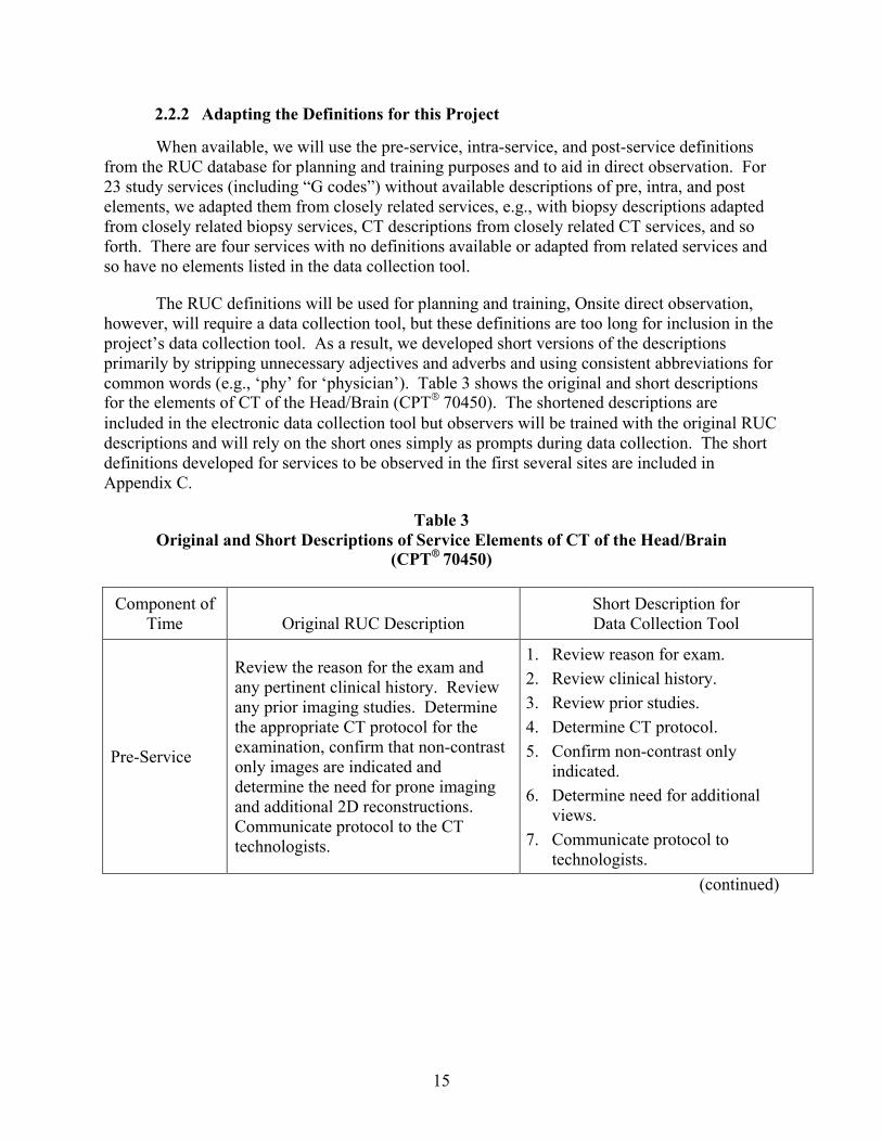
The RUC definitions will be used for planning and training, Onsite direct observation, however, will require a data collection tool, but these definitions are too long for inclusion in the project’s data collection tool. As a result, we developed short versions of the descriptions primarily by stripping unnecessary adjectives and adverbs and using consistent abbreviations for common words (e.g., ‘phy’ for ‘physician’). Table 3 shows the original and short descriptions for the elements of CT of the Head/Brain (CPT® 70450). The shortened descriptions are included in the electronic data collection tool but observers will be trained with the original RUC descriptions and will rely on the short ones simply as prompts during data collection. The short definitions developed for services to be observed in the first several sites are included in Appendix C.
Table 3 Original and Short Descriptions of Service Elements of CT of the Head/Brain
(CPT® 70450)
Component of Time Original RUC Description
Short Description for Data Collection Tool
Pre-Service
Review the reason for the exam and any pertinent clinical history. Review any prior imaging studies. Determine the appropriate CT protocol for the examination, confirm that non-contrast only images are indicated and determine the need for prone imaging and additional 2D reconstructions. Communicate protocol to the CT technologists.
1. Review reason for exam. 2. Review clinical history. 3. Review prior studies. 4. Determine CT protocol. 5. Confirm non-contrast only
indicated. 6. Determine need for additional
views. 7. Communicate protocol to
technologists. (continued)
15

Table 3 (continued) Original and Short Descriptions of Service Elements of CT of the Head/Brain
(CPT® 70450)
Component of Time Original RUC Description
Short Description for Data Collection Tool
Intra-Service
Supervise acquisition of scout views, prescribe area of coverage and supervise acquisition of axial source image sections. Review initial and subsequent series of CT image data to assure adequacy of anatomic coverage and assess need for repeat sections or reconstruction of thin sections in specific locations. Supervise reconstruction of coronal and/or sagittal 2D multiplanar reformatted (MPR) images; assess need for oblique or other 2D images. Evaluate and interpret findings related to head/brain. Compare current findings to previous studies. Dictate report for medical record.
1. Supervise acquisition of scout views.
2. Prescribe coverage area. 3. Supervise acq of axial image. 4. Review adequacy of coverage. 5. Assess need for repeat sections. 6. Supervise reconstruction of MPR
images. 7. Assess need for other 2D images. 8. Interpret findings. 9. Compare to previous studies. 10. Dictate report.
Post-Service Review, edit, and sign report for medical record. Discuss findings with referring physicians.
1. Review, edit, sign report. 2. Discuss findings with referring
phys.
One issue we cannot address is whether or not the RUC definitions apply to current practice. During preliminary site visits, the project team observed multiple non-practitioner staff performing work tasks as defined by the RUC, most notably in the pre-service component. In discussions with clinical leaders, all three sites voiced the perception that the clinical processes of care have changed over time and that now most staff work at the top of their license. For example, during Skin Biopsy (CPT® 11100), the technician or nurse may perform certain elements of pre-service work, such as preparing instruments and taking vital signs, while the physician or physician assistant (PA) will administer anesthesia. The physician then performs all intra-service work tasks, i.e., debridement, but a PA applies the dressing. In the post-service period, the nurse may perform elements of post-service work, such as providing instructions on wound or home care, and discussion of follow-up activities, while the physician will complete the medical record and communicate with the primary physician if necessary.
Thus, there may be substantial work performed by medical assistants, LPNs or RNs that are included by the RUC as work in these descriptions. In fact, some clinical leaders at sites we visited stated that for some services, physician activities listed by the RUC are not performed by
16

physicians. Since we are recording, to the extent feasible, who is performing what tasks within a service, we may be able to analyze the extent to which this occurs. This information may be helpful to the practice expense relative value process, which accounts for the non-practitioner clinical labor costs of each service.
2.2.3 Implications for Data Collection and Analysis
As a result of variability in the RUC descriptions of pre, intra, and post service elements, we have developed a data collection tool that allows the observer to indicate what elements are being performed during the recorded period and who is doing them. The RUC descriptions are included in the training materials for observers so they are aware of the elements that they are likely to observe, and the data collection tool includes a list of these elements – shortened – for each service. By allowing the observer to verify expected items and add additional items, we will be able to analyze the extent to which the RUC definitions – both what elements are performed and who performs them -- conform to service delivery in practices where direct observation data are collected.
We will compare the pre-service, intra-service, and post-service time values from the direct observation and, as feasible, from administrative data, with current time values, and we will examine the mix of tasks included in each to see whether there are elements in the RUC definition not performed or elements performed not included in the definition. It may also be feasible to test the hypothesis suggested above that detailed descriptions affect time values, particularly when obtained by survey.
17
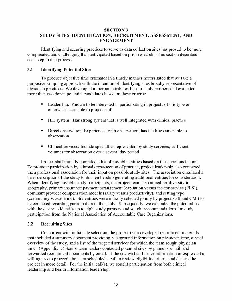
SECTION 3 STUDY SITES: IDENTIFICATION, RECRUITMENT, ASSESSMENT, AND
ENGAGEMENT
Identifying and securing practices to serve as data collection sites has proved to be more complicated and challenging than anticipated based on prior research. This section describes each step in that process.
3.1 Identifying Potential Sites
To produce objective time estimates in a timely manner necessitated that we take a purposive sampling approach with the intention of identifying sites broadly representative of physician practices. We developed important attributes for our study partners and evaluated more than two dozen potential candidates based on these criteria:
• Leadership: Known to be interested in participating in projects of this type or otherwise accessible to project staff
• HIT system: Has strong system that is well integrated with clinical practice
• Direct observation: Experienced with observation; has facilities amenable to observation
• Clinical services: Include specialties represented by study services; sufficient volumes for observation over a several day period
Project staff initially compiled a list of possible entities based on these various factors. To promote participation by a broad cross-section of practice, project leadership also contacted the a professional association for their input on possible study sites. The association circulated a brief description of the study to its membership generating additional entities for consideration. When identifying possible study participants, the project team also aimed for diversity in geography, primary insurance payment arrangement (capitation versus fee-for-service (FFS)), dominant provider compensation models (salary versus productivity), and setting type (community v. academic). Six entities were initially selected jointly by project staff and CMS to be contacted regarding participation in the study. Subsequently, we expanded the potential list with the desire to identify up to eight study partners and sought recommendations for study participation from the National Association of Accountable Care Organizations.
3.2 Recruiting Sites
Concurrent with initial site selection, the project team developed recruitment materials that included a summary document providing background information on physician time, a brief overview of the study, and a list of the targeted services for which the team sought physician time. (Appendix D) Senior team leaders contacted potential sites by phone or email, and forwarded recruitment documents by email. If the site wished further information or expressed a willingness to proceed, the team scheduled a call to review eligibility criteria and discuss the project in more detail. For the initial call(s), we sought participation from both clinical leadership and health information leadership.
18

Sites interested in proceeding after this initial call were then sent a second set of recruitment materials: a detailed health information technology (HIT) worksheet and a set of HIT questions to assist the team in collecting more detailed information on the volume of services provided by the system as well as to better understand the sources of electronic data and the components of time that may be captured electronically.
Our goal was to recruit up to eight sites participate in the study. At this point, the project team has made an initial recruitment contact with nearly 20 potential sites. Three are moving forward as study participants. Recruitment for additional study partners continues. This rest of this section describes some of the key recruitment challenges encountered by the project team.
Of the entities that have not moved forward to study participation, a number of themes emerged for non-participation:
• Non-response. After initial expression of interest, a number of organizations did not respond to follow-up email or telephone contact. It seems as though, in these cases, the original contact who wanted to participate in the study was unable to generate internal interest.
• Pre-existing HIT staff commitments. In a number of sites, clinical leadership were very supportive of participating in the study but subsequent engagement of the Chief Information Officer or other senior leaders with responsibility for implementing and maintaining internal health information systems revealed pre-existing demands of HIT staff that precluded participation. Several potential sites cited meeting Meaningful Use requirements and ICD-9 to ICD-10 conversion as major ongoing activities that make it impossible for them to commit to this project. These two activities were in addition to other internal projects, such as an enterprise-wide electronic medical record upgrade or moving clinical departments from paper records to the system’s electronic systems.
• Obtaining physician buy-in. Several organizations expressed initial interest but, after learning more about the study and in particular the need to gain support across many clinical departments, declined to participate. Most notable were organizations that do not employ physicians. Senior clinical leaders felt that gaining support of private-practice physicians would be an enormous logistical and potential political challenge. Organizations with employed physicians were in a better position to garner broad clinical support.
• Other reasons. A number of sites had unique reasons for non-participation. For example, one organization declined participation at the request of their legal department because of institutional risk in light of an ongoing legal review. And, although we promised anonymity, another expressed concern about being publically associated with the project and the potential negative reaction of clinical staff to a project that could be perceived as leading to fee reductions.
19
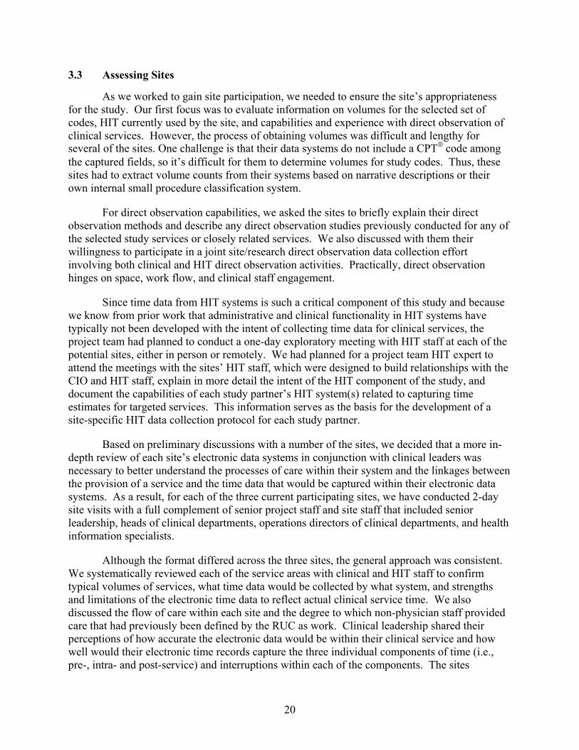
3.3 Assessing Sites
As we worked to gain site participation, we needed to ensure the site’s appropriateness for the study. Our first focus was to evaluate information on volumes for the selected set of codes, HIT currently used by the site, and capabilities and experience with direct observation of clinical services. However, the process of obtaining volumes was difficult and lengthy for several of the sites. One challenge is that their data systems do not include a CPT® code among the captured fields, so it’s difficult for them to determine volumes for study codes. Thus, these sites had to extract volume counts from their systems based on narrative descriptions or their own internal small procedure classification system.
For direct observation capabilities, we asked the sites to briefly explain their direct observation methods and describe any direct observation studies previously conducted for any of the selected study services or closely related services. We also discussed with them their willingness to participate in a joint site/research direct observation data collection effort involving both clinical and HIT direct observation activities. Practically, direct observation hinges on space, work flow, and clinical staff engagement.
Since time data from HIT systems is such a critical component of this study and because we know from prior work that administrative and clinical functionality in HIT systems have typically not been developed with the intent of collecting time data for clinical services, the project team had planned to conduct a one-day exploratory meeting with HIT staff at each of the potential sites, either in person or remotely. We had planned for a project team HIT expert to attend the meetings with the sites’ HIT staff, which were designed to build relationships with the CIO and HIT staff, explain in more detail the intent of the HIT component of the study, and document the capabilities of each study partner’s HIT system(s) related to capturing time estimates for targeted services. This information serves as the basis for the development of a site-specific HIT data collection protocol for each study partner.
Based on preliminary discussions with a number of the sites, we decided that a more in-depth review of each site’s electronic data systems in conjunction with clinical leaders was necessary to better understand the processes of care within their system and the linkages between the provision of a service and the time data that would be captured within their electronic data systems. As a result, for each of the three current participating sites, we have conducted 2-day site visits with a full complement of senior project staff and site staff that included senior leadership, heads of clinical departments, operations directors of clinical departments, and health information specialists.
Although the format differed across the three sites, the general approach was consistent. We systematically reviewed each of the service areas with clinical and HIT staff to confirm typical volumes of services, what time data would be collected by what system, and strengths and limitations of the electronic time data to reflect actual clinical service time. We also discussed the flow of care within each site and the degree to which non-physician staff provided care that had previously been defined by the RUC as work. Clinical leadership shared their perceptions of how accurate the electronic data would be within their clinical service and how well would their electronic time records capture the three individual components of time (i.e., pre-, intra- and post-service) and interruptions within each of the components. The sites
20

confirmed that it was feasible to capture time data for the codes of interest from the sites’ HIT system(s), for those services for which the site had electronic data. Each site visit concluded with an in-depth discussion of organizational requirements for the conduct of the study at that site and logistical next steps.
3.4 Engaging Sites
From identifying candidate sites through planning site visits, the process of engaging sites has been much more time consuming and challenging than anticipated. Even after a site has fully committed to participating, we have had to go through a number of additional steps to fully engage each site. These may reflect the likely challenges that will be faced with other potential study partners given the nature of this study, which requires considerable buy-in from a large number of departments within the organization or across a distributed network of providers and the desire to conduct research in the clinical areas with direct observation of patient care. Some of the key challenges to engaging sites include:
• Timing for effective engagement of a large number of key stakeholders. It took a considerable amount of time for the sites (both participating and ultimately non-participating organizations) to identify and communicate with key stakeholders in their organizations. Staff vacations during the months of July and August when recruitment activities were at a peak at a number of the sites were a factor. Not unexpectedly, schedule demands of busy key contacts (chief medical officer, chief information officer, chief finance officer, department chairs, etc.) were also a factor; however, it was only necessary to have the primary point of contact be a very senior member of the organizational leadership team until sufficiently broad buy-in had occurred. At that point, it became possible to identify a less senior person to be the primary contact who would have the time for ongoing communication with the project team as well as time to devote to efforts in moving the project forward (such as identify applicable IRB requirements and processes), coordinate data gathering, plan pilot visits, ongoing communication, etc.
• Competing organizational priorities. Not unexpectedly, there are many competing demands of staff that are required to be active participants in this study. In addition to the issues noted above, preparation for provisions of the Affordable Care Act’s implementation, such as the new insurance marketplaces, presented challenges that limited active engagement of the HIT department at the outset for one organization in particular.
• Completion of the HIT worksheet proved challenging. As originally envisioned, potential study partners were asked to complete the HIT spreadsheet which was designed to collect estimates of the typical number of services provided per week for each of the study codes as well as to better understand the sources of electronic data and the components of time that may be captured electronically. For most sites, this request proved to be challenging because our main points of contact were typically clinical staff. We found that if volume estimates were obtained from clinical systems, in contrast to billing systems, the services were often not defined by CPT® code but by narrative descriptions or internally developed service codes. Thus, there was not
21

always a good crosswalk between the institution’s definition and the CPT® code definition. This led to very low reported volume for the targeted codes, and hindered primary data collection planning. In addition, the organizations had to do considerable work to provide the volume estimates. Future efforts to obtain service-level volumes for specific services should involve the billing offices at a much earlier stage.
We also encountered difficulties in obtaining detailed information on the sources of electronic time data and elements of time captured by electronic data systems within the organizations. Although efforts were made to provide the information, often the information provided was too generic, such as the name of the EMR. This led us to conclude that we needed to make in-person site visits to each potential study partner to meet in person with the cadre of staff that has responsibility for and a deep understanding of the capabilities and limitations of their electronic data systems to provide the necessary elements of time. Further, we found that it was also necessary to jointly review the electronic data systems with clinical staff from each site to fully understand how the processes of care would be captured electronically and what limitations might exist from a clinical service perspective.
• Organizational requirements for conducting the study in patient care areas. Although RTI’s IRB found that this study is exempt [45CFR46.101(b)] from IRB review based upon a category 5 reason12, all study partners needed to go through their own IRB process. In many cases, these were lengthy and time-consuming, requiring substantial input from the project team (responding to questions, reviewing multiple drafts) and numerous revisions by staff at the site in response to questions from their respective IRBs. One study partner has completed the IRB process and a second is complete pending the signature of the IRB Chairman. Both processes took approximately five months. The third site is seeking an expedited IRB review and the process is ongoing. During the IRB process at one organization, it was determined that both patient and physician consent would be required thereby necessitating the development of a more elaborate primary data collection plan for that organization to ensure compliance with the IRB.
Throughout the recruitment and engagement processes, we encountered a number of additional requirements at each site that had to be dealt with before project staff would be able to conduct onsite direct observation. One organization required Collaborative Institutional Training Initiative (CITI) training prior to IRB approval. Another organization’s IRB ruled that project staff could not directly observe patient care, which led us to redesign direct observation data collection plan limiting the
12 Specifically, Category 5 includes: “Research and demonstration projects which are conducted by or subject to the approval of Department or Agency heads, and which are designed to study, evaluate, or otherwise examine: (i) Public benefit or service programs; (ii) procedures for obtaining benefits or services under those programs; (iii) possible changes in or alternatives to those programs or procedures; or (iv) possible changes in methods or levels of payment for benefits or services under those programs.”
22
project team’s role to observing services such interpretation of radiology services and clinical pathology services and training site staff to observe other services. One site also required negotiations with the nurses union to allow direct observers on site. Other organizations have fitness for duty requirements that include evidence of immunizations, criminal background checks, attending a 40-hour new employee training, credentialing for staff that have professional licenses, (e.g., M.D., R.N.), and other requirements. We have been able to develop strategies to address these various requirements. For example, limiting on-site hours to less than 40 reduced some of the requirements and limiting the direct observation staff to non-medical professionals negated the need to go through the lengthy credentialing process without compromising the integrity of the data collection process
23

SECTION 4 DATA COLLECTION
Once all necessary paperwork (IRB, contracts, etc.) is in place, we will either do direct observation on site or train staff to do the observation. This section describes the actual data collection process.
4.1 Data Collection Overview
We will do direct observation for services that have no electronic data available and for services that do. The primary purpose for parallel HIT and direct observation for some services is to determine if the HIT times are validated or need to be modified to account for interruptions. As necessary, we will calculate an allocation algorithm across a group of services to reconcile electronic measures to those obtained through direct observation. Our intent is to develop a ratio for other services in the same small clinical family based those services that we do observe. So, for example, if we observe colonoscopy with polyp removal and we do not observe colonoscopy without polyp removal but sites provide electronic time for both, then we would use the ratio of HIT time to direct observation time for the first service to adjust the HIT time for the second service to develop a measure comparable to those obtained through direct observation. We will perform fewer direct observations for services with electronic data than for the services for which no HIT time is available, to leverage project resources as effectively as possible across study services.
We are finalizing the data collection plans for the current three study sites. Based upon volume statistics provided by the three sites, it is likely that the number of services for which we can conduct direct observation during a one-week timeframe may be very small. In general, the volumes of study services at sites we will observe are lower than we had originally anticipated. These lower-than-expected volumes may result in small samples of time observations that could make some of the study’s time estimates less robust than intended. As noted above, in some sites we will have to rely on site staff to do the direct observation for many services. In these instances, project staff will train the data collectors and then observe them at work, where feasible.
The general approach to finalizing the data collection plan is to (1) identify the one-week data collection period, (2) review upcoming appointments for the selected study procedures through the site’s scheduling software, (3) determine the services for which direct observation only will be conducted versus direct observation validation of electronic data, (4) assign project staff or site staff for the direct observation of services, and (5) coordinate the receipt of the electronic time data for the observed services. For radiology and pathology services that do not involve direct patient care, project staff will be assigned a window of time to observe the selected services in each of those departments, rather than specific patient appointments.
4.2 Development of Study Protocols
We have developed two data collection protocols reflecting lessons learned from the first three site visits: one for direct observation data collection (Appendix E) and one for the collection of HIT data (Appendix F). Each data collection protocol is being modified to reflect the individual features of electronic data or care processes within the participating sites. To the
24

extent possible, we will seek to have consistency in collection of data across the sites recognizing that there are unique aspects within each. The detailed information gathered from the pre-implementation conference calls and site visits is being used to tailor the general HIT protocol to each system and will allow us to understand where differences in collected time may arise due to system differences. The HIT protocol specifies the administrative, clinical or EHR systems that store time data and the components of time that we will capture (i.e., intra-service, pre- and post-time, total time). The HIT protocol will also detail what will take place during the HIT observation component of the primary data collection phase. The draft protocols are being shared with each of the sites and a one-on-one conference call will be held with appropriate HIT staff to review and finalize.
A second data collection protocol has been developed to conduct the direct observation studies. We have held numerous conversations with the study partners to discuss how the organization will gain participation from each of the necessary clinical services, the number of direct observations that will be required for each service, logistical considerations, and their procedures for conducting direct observations. Using this information, draft data collection protocols have been developed incorporating, to the extent possible, validated direct observation methods from each of the systems or building a “best practices” approach to direct observation should a site have limited experience. We are working with the sites to identify and minimize in the protocol potential observer (Hawthorne effect), clinical (clinician’s number of years in practice), or environment (service volume or day of week) confounding factors that may influence the observed time. Lastly, the protocols will specify the procedures that will be used to obtain permission from all clinicians and patients that will be observed, if required by a site’s IRB. The draft clinical protocols will be shared with each of the sites and a one-on-one conference call will be held with appropriate clinical staff to review and finalize.
As part of the direct observation protocol development, we developed a uniform data collection Microsoft Access® database. As originally envisioned the team planned to start with the publicly available Time and Motion Study Tool: Ambulatory Practice (TMS-AP), Version 1.0 database that was developed by Partners Healthcare System with a grant from the Agency for Healthcare Research and Quality (AHRQ). The AHRQ/Partners tool can be run on a laptop and had been used successfully in a prior collection of physician work time.13 However, as development of the protocol proceeded, it became apparent that the AHRQ/Partners tool was unsuited to project needs. For example, it was not designed to capture pre-, intra- and post-service time, interruptions, and note special circumstances expediently.
The project team then evaluated Excel® and Access®-based tools that were more closely suited project needs. Advantages of these tools included:
• Recording of pre-, intra- and post-service time
• Documentation of interruptions
25
13 Pizziferri L, Kittler AF, Volk LA, Honour MM, Gupta S, Wang S, Wang T, Lippincott M, Li Q, Bates DW. Primary care physician time utilization before and after implementation of an electronic health record: A time-motion study. J Biomed Inform. 2005 Jun;38(3):176-88. Epub 2004 Dec 14.

• Documentation of non-physician providers
• Automatically computes duration of time components and total time
• Permits immediate correction of input errors
There were drawbacks to the Excel®-based tool, including time delays in moving from one section of the tool to another in order to record interruptions and notes. Another was that the tool could not be used for paper data collection, a requirement of some study partner’s staff who felt they were not facile enough to use an electronic data collection tool. The most significant drawback of the Excel®-based tool was the inability to capture the performance of specific elements involved in each component of time, as well as the provider type performing the task.
After initial pilot visits to two sites, the project team agreed that we needed a tool that could also capture the performance of those elements that differ by CPT® code, as well as the provider type performing the element/task as discussed above in Section 2. The final Access®-based tool we created is similar in concept to the AHRQ/Partners tool but has the capability to capture this level of detail and has the following features:
• Electronic or paper recording of observation
• Edit or delete observation
• Manual entry for paper recordings
• Capture time and description of interruptions
• Record time for multiple providers and provider description (physician, nurse practitioner, physician assistant, registered nurse, technician, licensed practical nurse, medical assistant, other), as well as the elements/tasks specific to the components (pre-, intra- and post-service), of each service.
Additionally, the tool permits output of the data in Excel® or tab delimited flat file, all data, by date or session, and prints quality assurance (QA) reports. The tool is described in Appendix E.
4.3 Direct Observation Approach
As described above, all data sites have or are in the process of attaining IRB approval. Once IRB and other administrative steps are complete, we will schedule a week for data collection at each site. This section describes the data collection process.
During primary data collection, clinical time data will be directly inputted into laptops that will be both encrypted and password protected, or directly entered at the end of day if paper and pencil time data were obtained. No personal identifying information of either the clinician or patient will be maintained in the time database; rather an encrypted identifier will be used for patient and service identification and a log will be maintained by each of the sites to ensure
26

correct linking of electronic time to the observed time. Each night an upload of the data will be made to RTI’s secure data system. Once data for the observed service are available in the site’s database, transfer of the electronic HIT data will be made to RTI’s secure data system based upon the agreed transfer procedures in the HIT protocol.
In our original project plan, we assumed 1 HIT and 2 clinical observers from the project team would be on-site at each practice for three days observing both clinical and HIT activities. The direct clinical observation would be done in collaboration with staff from each of the sites. We have since expanded the time to 5 days that the project clinical observers will be onsite and increased the number of clinical observers to 3. Based on what we have learned during the site visits to the three sites and additional requirements imposed by the sites’ IRB or leadership, we have modified our original plan for data collection as follows:
• Site 1: Given the study site’s restrictions on non-employees in clinical rooms, site 2 will provide four staff persons to perform the direct observation of clinical services. Project staff will assist in direct observation data collection for services where the procedure can be observed without being present in the clinical room (e.g., reading of radiology services) and will be available to answer any questions that may arise during the direct observation activities.
• Site 2: We will follow our plan of having three project clinical observers on-site for up to 39 hours observing clinical services. Project staff will restrict their on-site time to less than 40 hours per person during the one-week data collection period due to the study site’s fit-for-duty requirements that apply to study personnel on-site for 40 hours or more.
• Site 3: The project team is currently developing plans for direct observation of clinical services. Originally, the training requirements shared by site 3 for any outside researchers were onerous and the project team was planning to use personnel from the site to perform direct observation data collection activities. However, during the pilot visit we received revised training requirements for outside researchers that were much more manageable. The project team is continuing to work with site 3 to formulate plans for direct observation data collection.
After contracts with the sites have been finalized, primary data collection will occur at each site. Prior to any study staff engaging in direct observation activities, project staff will provide training on: (i) the study’s direct observation protocol, and (ii) the electronic and paper data collection forms designed to capture activity and time data for observed services.
Because of the extensive pilot activities that have been conducted at each of the sites, the project HIT staff person will not make another in-person visit to the site. Rather, the project HIT staff person will work with each of the sites to finalize the HIT data collection protocol and to coordinate the extraction of historical time data for study services. We will request each site to extract, in an agreed upon data format, relevant time data for observed clinical services. In addition, we will request each site to extract, in an agreed upon data format, historical time data for previously performed targeted services. The time period covered (e.g., 6 months, 1 year) and volume of cases for the historical data extraction will be jointly determined with each site.
27

4.4 Other Site Data
It is difficult to know if the data we collect will be representative of physician practices nationwide. Throughout the project, we will keep track of site characteristics that will help us understand any potential bias. Some factors that might introduce bias include:
• Use of Residents. The presence of residents is generally thought to increase service time, but the effect is likely to differ depending on whether the senior, billing physician has to be present throughout the service or not. The effect of residents may also depend on their year of training. For example, one surgical leader suggested that residents in their first two years may add to service time while those in their final year may have little or no effect on time. The effect may also depend on whether residents are regularly onsite working with site physicians or if they rotate through intermittently or work with a teaching professor who does not regularly practice on site. These nuanced differences suggest that the effect of residents could vary across specialties within the same site. We will document the role of residents, if any, at all direct observation sites.
• Team-based Care. As the result of a number of factors, many practices have adopted strong team-based models of care, in which many traditional physician functions are now done by others. This is likely most relevant for evaluation and management services, which are NOT being studied. We will document the mix of clinical staff involved at direct observation sites.
• Cultural Differences. Factors such as payment arrangement (FFS v. salaried) and setting type (community v. academic/referral) may affect the complexity and acuity of cases. For instance, one clinical leader suggested that her radiology department in a referral center had a much higher share of patients with positive findings than she had experienced in a previous community setting so that the average time for a specific service would likely be higher. FFS settings and community settings may have a larger share of simpler cases with more “normal” test results. Similarly, FFS may inherently lead to lower acuity levels, since it is thought to generate more volume, with the marginal volume including cases less likely to have “abnormal” results. There may be offsetting effects, so that the higher acuity cases in settings where they are the more typical cases leads to systems and experience that reduce the effect of acuity on service time. We will document the compensation model and type of setting for each site.
We will document these characteristics, and others that might help understanding how the service times at each site are likely to compare to other practices.
4.5 Implementation Challenges
Over the course of recruiting sites to participate in the study, we encountered a number of challenges moving forward on our original project plan. These considerations have affected details in how we will collect the data at the three sites and may inform future efforts to do this
28

type of data collection. Some of these challenges could affect the reliability of the study findings as they relate to the time measures for specific CPT codes.
• Low volumes of services for many codes at participating sites. As discussedearlier, one criterion used to select the study codes was high frequency amongMedicare FFS beneficiaries. A review of typical weekly volumes of services for ourselected codes across the three sites shows lower than expected volumes requiring usto adjust the frequency of direct observation events per code:
Site 1: Using retrospective and prospective scheduling data, we estimated that 15 of the study codes will have a weekly volume of 10 or more; 31 codes will have a weekly volume of 3 or more. The 31 codes are in 22 of the service groups in Table 2 (Section 2 above), reflecting the following broad clinical families: dermatology, gastroenterology, orthopedics, urology, ophthalmology, radiology, podiatry, and vascular lab services. Eighteen minor procedures and 13 diagnostic services are represented among these services. Determination of availability of electronic data is still ongoing but we have confirmation that surgical procedures performed in an ambulatory surgical center have electronic data. There are also electronic data available for technicians for most radiology procedures as well.
Site 2: Using billing data, we estimate that 56 of the targeted study services have a weekly volume of 10 or more; 72 codes have a volume of at least 3 per week. We believe that this site can provide electronic data for 53 of the study services. These 53 services reflect the following 7 broad clinical service categories: orthopedics, general surgery, cardiac surgery, back and neck surgery, gastroenterology (including colonoscopy and colorectal screening), cardiac catheterization, and urology. Twenty-two minor procedures, 4 major procedures, and 30 diagnostic services are represented among these services.
Site 3: Using data obtained from clinical departments that we believe are significantly lower than volume estimates we would obtain from billing data (which has been requested), we estimated that 6 of the targeted codes have a weekly volume of 10 and another 13 of the targeted codes have a typical weekly volume of 3 or more. These volume estimates are from three service sites within this project site, and include predominantly diagnostic and minor procedures/services. Minor procedures/ services showing volumes of 3 or more include GI procedures (i.e., colonoscopies and upper GI endoscopies), surgical pathology services, and therapeutic/prophylactic/ diagnostic injections. Diagnostic procedures/services showing volumes of 3 or more include radiology/imaging (including CT), vascular lab and audiometry services. It is likely that this site can provide electronic time data for major surgical procedures, other procedures (with implementation of an outpatient package specifically for this project), and radiology.
Based upon the planned 1-week direct observation period and likely volumes of services, our primary data collection plan is focusing upon the collection of at least 3 observations for services for which there is electronic data and between 3 and 10
29

observations for which there is no electronic data. Thus, we would not be observing services for which the typical weekly volume is less than 3.
• Identifying specific CPT® codes for scheduled procedures presents a significantchallenge for direct observation. Discussion with clinical leaders within each of thestudy sites confirmed that for some services, the scheduled procedure description willlikely be the final performed procedure, e.g., total hip replacement. However, forother services such as screening colonoscopy it is likely that there will a degree ofuncertainty as to whether or not the colonoscopy will include removal of a polyp(s).Thus, we may not always be conducting direct observation for one of the targetedservices reducing the number of direct observations for the study. For validation ofthe electronic time data, we do not believe this presents a problem within smallclinical families. Discussion with clinical leaders at the participating sites yielded acollective belief that within small clinical families the ratio of observed time toelectronic time is likely to be invariant to individual procedure codes.
• Select high volume procedures are heterogeneous. During pilot site visits, clinicalleaders expressed concern to the project team that median time estimates derived for afew of the selected codes is not representative of time necessary to perform allservices within the service due to extreme clinical heterogeneity. Two examples werecited: Cystourethroscopy (CPT® 52000) could be a short uncomplicated follow-upprocedure after treatment for cancer taking very few minutes, or a lengthy initialdiagnostic procedure for a newly diagnosed cancer patient; Level IV surgicalpathology (CPT® 88305) can be performed on one or multiple tissue samples, and theelements can differ depending on the nature of the request and tissue source(s).
• The site visits provided additional insight into the likely availability of electronicdata across a broad range of sites. In more detailed discussions with HIT staff atthe three sites, we received confirmation that the strongest electronic data are forsurgical procedures. This finding is consistent with that of the MedPAC report14 thatservice time, in particular intra-service time, is most available for major surgicalservices. And, we received confirmation that pre- and post-service times for most ofthe services are not well captured in electronic data. Electronic time data are oftennot available, most notably for office-based procedures and dermatology services.While time data is generally available for radiology services, there are substantialinterruptions during the intra-service period of interpretation and reporting whichreflect pre- and post-service work for other services to other patients, i.e., consultingwith a technician on set-up or consulting with the ordering physician on theinterpretation. These confirmatory findings have led to modification of the directobservation data collection protocol and tool.
30
14 Braun, P. and McCall, N. Improving the Accuracy of Time in the Medical Physician Fee Schedule: Feasibility of Using Extant Data and of Collecting Primary Data. For MedPAC, 2011.

4.6 Preliminary Data Validation
At the completion of data collection at each site, we will perform basic data validity checks and construct an analytic file of direct observation and HIT times. We will produce distributional properties of time (median, mean, standard error) for each service and by type of data collection method (i.e., direct observation, HIT data, or both). As noted above, the number of observations for which we will have time measurements will vary by service and method and will depend upon the volume performed by the participating sites during the data collection period. For some service codes, the likely volume available for direct observation may be low. We will send the time estimates to the sites and request that they review their respective data for reasonableness, noting any areas of concern with potential outlier time measurements. Final determinations will be made jointly with the sites and CMS on how to handle potential outlier data, if necessary.
After they are incorporated into the materials being prepared by the analysis team, these time estimates will be submitted to the clinical panel for review. For services for which only direct observation time was collected, the median or typical time and the distributional properties of the time estimate will be sent to the clinical panels for review. For services with direct observation and HIT times, we will calculate median times for each method and develop a ratio of observed to electronic data.
31

SECTION 5 SUMMARY AND NEXT STEPS
As described above, our experience identifying services, recruiting sites, and developing data collection protocols has led us to modify our original project plan in a number of ways. While the service selection and site recruitment steps both took much longer than anticipated and we are still trying to recruit additional sites, the three developments that most affected our approach are the pre-, intra-, and post-service element descriptions, low expected service volumes for many of the selected study services, and some of the various site rules that limited our ability to have project staff conduct direct observation. These developments have added complexity and time to the project.
As discussed, we also have concerns about the completeness, comparability, and relevance to current practice of the RUC service element descriptions. As a result, we developed an Access® data collection tool that allows the service observers to indicate which elements are included in the measured time and who is doing them.
The various issues that were raised about having project staff conduct direct observation led us to develop a second approach to direct observation. Whether for IRB or other considerations, in these instances we have developed an approach that has project staff train site staff to conduct the observations under project staff supervision.
At this point, we are preparing to conduct direct observation and acquire other data from two sites, with the third site pending IRB and other clearances. At the same time, we continue to try to recruit and engage additional partners that are willing to participate in the data collection process. As we go through the site engagement process with new prospective entities, we are using our experiences to date to make the process smoother and quicker. From encouraging involvement of key internal leaders to learning about IRB requirements and processes, we have become more adept at working this this process with potential sites.
After we develop new objective time measures from participating sites, we will then move to the other project tasks, namely analyzing these new time estimates to develop alternative models of their implications for service work values and then reviewing the time data and work models with panels of physicians.
32

A-1
APPENDIX A STUDY SERVICES, WITH NARRATIVE DESCRIPTION
Healthcare Common Procedure
Coding System (HCPCS) Code
Narrative Description
11042 Debridement, subcutaneous tissue (includes epidermis and dermis, if performed); first 20 square centimeters or less
11056 Paring or cutting of benign hyperkeratotic lesion (e.g., corn or callus); two to four lesions
11057 Paring or cutting of benign hyperkeratotic lesion (e.g., corn or callus); more than four lesions
11100 Biopsy of skin, subcutaneous tissue and/or mucous membrane (including simple closure), unless otherwise listed (separate procedure); single lesion
11101 Biopsy of skin, subcutaneous tissue and/or mucous membrane (including simple closure), unless otherwise listed (separate procedure); each separate/additional lesion (list separately in additional to code for primary procedure)
17000 Destruction (e.g., laser surgery, electrosurgery, cryosurgery, chemosurgery, surgical curettement), premalignant lesion(e.g., actinic keratoses); first lesion
17003 Destruction (e.g., laser surgery, electrosurgery, cryosurgery, chemosurgery, surgical curettement), premalignant lesion(e.g., actinic keratoses); 2nd through 14 lesions, each (list separately in additional to code for first lesion)
17004 Destruction, (e.g., laser surgery, electrosurgery, cryosurgery, chemosurgery, surgical curettement), premalignant lesions(e.g., actinic keratoses), 15 or more lesions
17110 Destruction, (e.g., laser surgery, electrosurgery, cryosurgery, chemosurgery, surgical curettement), of benign lesions other than skin tags or cutaneous vascular proliferative lesions; up to 14 lesions
17262 Destruction, (e.g., laser surgery, electrosurgery, cryosurgery, chemosurgery, surgical curettement), trunk, arms or legs; lesion diameter 0.5 cm or less
17281 Destruction, (e.g., laser surgery, electrosurgery, cryosurgery, chemosurgery, surgical curettement), face, ears, eyelids, nose, lips, mucous membrane; lesion diameter 0.6 cm to 1.0 cm
17282 Destruction, (e.g., laser surgery, electrosurgery, cryosurgery, chemosurgery, surgical curettement), face, ears, eyelids, nose, lips, mucous membrane; lesion diameter 1.1 cm to 2.0 cm
(continued)

A-2
Healthcare Common Procedure
Coding System (HCPCS) Code
Narrative Description
20550 Injection(s); tendon sheath, or ligament, aponeurosis (e.g., plantar "fascia")
20605 Arthrocentesis, aspiration and/or injection; intermediate joint or bursa (e.g., temporomandibular, acromioclavicular, wrist, elbow or ankle, olecranon bursa)
20610 Arthrocentesis, aspiration and/or injection; major joint or bursa (e.g., shoulder, hip, knee joint, subacromial bursa)
22551 Arthrodesis, anterior interbody, including disc space preparation, discectomy, osteophytectomy and decompression of spinal cord and/o nerve roots; cervical below c2
22612 Arthrodesis, posterior or posterolateral technique, single level; lumbar (with lateral transverse technique, when performed)
22614 Arthrodesis, posterior or posterolateral technique, single level; each additional vertebral segment (list separately in addition to code for primary procedure)
22633 Arthrodesis, combined posterior or posterolateral technique with posterior interbody technique including laminectomy and/or discectomy sufficient to prepare interspace (other than for decompression), single interspace and segment; lumbar
22840 Posterior non-segmental instrumentation (e.g. harrington rod technique, pedicle fixation across 1 interspace, atlantoaxial transarticular screw fixation) (list separately in addition to code for primary procedure)
22842 Posterior segmental instrumentation (e.g., pedicle fixation, dual rods with multiple hooks and sublaminal wires); 3 to 6 vertebral segments (list separately in addition to code for primary procedure)
22845 Anterior instrumentation; 2 to 3 vertebral segments (list separately in addition to code for primary procedure)
22851 Application of intervertebral biomechanical device(s) (e.g., synthetic cage(s), threaded bone dowel(s), methylmethacrylate) to vertebral defect or interspace (list separately in addition to code for primary procedure)
23412 Repair of ruptured musculotendinous cuff (e.g., rotator cuff) open; chronic
23472 Arthroplasty, glenohumeral joint; total shoulder (glenoid and proximal humeral replacement (e.g., total shoulder))
27130 Arthroplasty, acetabular and proximal femoral prosthetic replacement, with or without autograft or allograft
(continued)

A-3
Healthcare Common Procedure
Coding System (HCPCS) Code
Narrative Description
27134 Revision of total hip arthroplasty; both components, with or without autograft or allograft
27236 Open treatment of femoral fracture, proximal end, neck, internal fixation or prosthetic replacement
27244 Treatment of intertrochanteric, pertrochanteric, or subtrochanteric femoral fracture; with plate/screw type implant, with or without cerclage
27245 Treatment of intertrochanteric, pertrochanteric, or subtrochanteric femoral fracture; with intramedullary implant, with or without interlocking screws and/or cerclage
27447 Arthroplasty, knee, condyle and plateau; medial and lateral compartments with or without patella resurfacing (total knee arthroplasty)
29827 Arthroscopy, shoulder, surgical; with rotator cuff repair 33208 Insertion of new or replacement of permanent pacemaker with
transvenous electrode(s); atrial and ventricular 33249 Insertion or replacement of permanent pacemaker with transvenous
lead(s); single or dual chamber 33405 Replacement, aortic valve, with cardiopulmonary bypass; with prosthetic
valve other than homograft or stentless valve 33430 Replacement, mitral valve, with cardiopulmonary bypass 33518 Coronary artery bypass, using venous graft(s) and arterial graft(s); two
venous grafts (list separately in addition to code for primary procedure) 33519 Coronary artery bypass, using venous graft(s) and arterial graft(s); three
venous grafts (list separately in addition to code for primary procedure) 33533 Coronary artery bypass, using arterial graft(s); single arterial graft 33536 Coronary artery bypass, using arterial graft(s); four or more coronary
arterial grafts 35301 Thromboendarterectomy, including patch graft if performed; carotid,
vertebral, subclavian, by neck incision 43235 Upper gastrointestinal endoscopy including esophagus, stomach, and
either the duodenum and/or jejunum as appropriate; diagnostic, with or without collection of specimen(s) by brushing or washing (separate procedure)
43239 Upper gastrointestinal endoscopy including esophagus, stomach, and either the duodenum and/or jejunum as appropriate; with biopsy, single or multiple
(continued)

A-4
Healthcare Common Procedure
Coding System (HCPCS) Code
Narrative Description
44120 Enterectomy, resection of small intestine; single resection and anastomosis
44140 Colectomy, partial; with anastomosis 44143 Colectomy, partial; with end colostomy and closure of distal segment
(hartmann type procedure) 44145 Colectomy, partial; with coloproctostomy (low pelvic anastomosis) 44160 Colectomy, partial, with removal of terminal ileum with ileocolostomy 44204 Laparoscopy, surgical; colectomy, partial, with anastomosis 44205 Laparoscopy, surgical; colectomy, partial, with removal of terminal ileum
with ileocolostomy 44207 Laparoscopy, surgical; colectomy, partial, with anastomosis, with
coloproctostomy (low pelvic anastomosis) 45378 Colonoscopy, flexible, proximal to splenic flexure; diagnostic, with or
without collection of specimen(s) by brushing or washing, with or without colon decompression (separate procedure)
45380 Colonoscopy, flexible, proximal to splenic flexure; with biopsy, single or multiple
45384 Colonoscopy, flexible, proximal to splenic flexure; with removal of tumor(s), polyp(s), or other lesion(s) by hot biopsy forceps or bipolar cautery
45385 Colonoscopy, flexible, proximal to splenic flexure; with removal of tumor(s), polyp(s), or other lesion(s) by snare technique
G0105 Colorectal cancer screening; colonoscopy on individual at high risk 47562 Laparoscopy, surgical; cholecystectomy 47563 Laparoscopy, surgical; cholecystectomy with cholangiography 49505 Repair initial inguinal hernia, age 5 years or over; reducible 50590 Lithotripsy, extracorporeal shock wave 52000 Cystourethroscopy (separate procedure) 52224 Cystourethroscopy, with fulguration (including cryosurgery or laser
surgery) or treatment of minor (less than 0.5 cm) lesion(s) with or without biopsy
52281 Cystourethroscopy, with calibration and/or dilation of urethral stricture or stenosis, with or without meatotomy, with or without injection procedure for cystography, male or female
(continued)

A-5
Healthcare Common Procedure
Coding System (HCPCS) Code
Narrative Description
52601 Transurethral electrosurgical resection prostate, including control of postoperative bleeding, complete (vasectomy, meatotomy, cystourethroscopy, urethral calibration and/or dilation, and internal urtherotomy are included)
55700 Biopsy, prostate; needle or punch, single or multiple, any approach 55866 Laparoscopy, surgical prostatectomy, retropubic radical, including nerve
sparing, includes robotic assistance, when performed 63047 Laminectomy, facetectomy and foraminotomy (unilateral or bilateral with
decompression of spinal cord, cauda equina and/or nerve root(s), (e.g., spinal or lateral recess stenosis), single vertebral segment; lumbar
64483 Injection, anesthetic agent and/or steroid, transforaminal epidural with imaging guidance (fluroscopy or computed tomography); lumbar or sacral, single level
66821 Discussion of secondary membranous cataract (opacified posterior lens capsule and/or anterior hyaloid); laser surgery (e.g., yag laser) (one or more stages)
66982 Extracapsular cataract removal w insertion of intraocular lens prosthesis (1 stage procedure), manual or mechanical technique (complex, requiring devices or techniques not generally used in routine cataract surgery (e.g. iris expansion device, suture support for intraocular lens, or primary posterior capsulorhexis) or performed on patients in the amblyogenic developmental stage)
66984 Extracapsular cataract removal with insertion of intraocular lens prosthesis (one stage procedure), manual or mechanical technique (e.g., irrigation or aspiration or phacoemulsification)
67028 Intravitreal injection of a pharmacologic agent (separate procedure) 67210 Destruction of localized lesion of retina (e.g., macular edema, tumors), one
or more sessions; photocoagulation 67228 Treatment of extensive or progressive retinopathy, 1 or more sessions;
(e.g., diabetic retinopathy), photocoagulation 70450 Computed tomography, head or brain; without contrast material 70486 Computed tomography, maxillofacial area; without contrast material 70551 Magnetic resonance (e.g., proton) imaging, brain (including brain stem);
without contrast material (continued)

A-6
Healthcare Common Procedure
Coding System (HCPCS) Code
Narrative Description
70553 Magnetic resonance (e.g., proton) imaging, brain (including brain stem); without contrast material, followed by contrast material(s) and further sequences
71010 Radiologic examination, chest; single view, frontal 71020 Radiologic examination, chest, two views, frontal and lateral; 71250 Computed tomography, thorax; without contrast material 71260 Computed tomography, thorax; with contrast material(s) 71275 Computed tomographic angiography, chest (noncoronary), without
contrast material(s), followed by contrast material(s) and further sections, including image post processing
72125 Computed tomography, cervical spine; without contrast material 72141 Magnetic resonance (e.g., proton) imaging, spinal canal and contents,
cervical; without contrast material 72148 Magnetic resonance (e.g., proton) imaging, spinal canal and contents,
lumbar; without contrast material 72158 Magnetic resonance (e.g., proton) imaging, spinal canal and contents,
without contrast material, followed by contrast material(s) and further sequences; lumbar
74176 Computed tomography, abdomen and pelvis; without contrast material 74177 Computed tomography, abdomen and pelvis; with contrast material 74178 Computed tomography, abdomen and pelvis; without contrast material in
one or both body regions, followed by contrast material(s) and further sections in one or both body regions
77080 Dual-energy x-ray absorptiometry (DXA), bone density study, 1 or more sties; axial skeleton (e.g., hips, pelvis, spine)
78452 Myocardial perfusion imaging, tomographic (SPECT) (including attenuation correction, qualitative or quantitative wall motion, ejection fraction by first pass or gated technique, additional quantification, when performed); multiple studies, at rest and/or stress (exercise or pharmacologic) and/or redistribution and/or rest reinjection
88305 Level iv-surgical pathology, gross and microscopic examination 88307 Level v - surgical pathology, gross and microscopic examination 88309 Level vi - surgical pathology, gross and microscopic examination bone
resection breast, mastectomy - with regional lymph nodes colon, segmental resection for tumor
(continued)

A-7
Healthcare Common Procedure
Coding System (HCPCS) Code
Narrative Description
88312 Special stain including interpretation and report; group I for microorganisms (e.g. acid fast, methananmine silver)
88331 Pathology consultation during surgery; first tissue block, with frozen section(s), single specimen
92133 Scanning computerized ophthalmic diagnostic imaging, posterior segment, with interpretation and report, unilateral or bilateral; optic nerve
92134 Scanning computerized ophthalmic diagnostic imaging, posterior segment, with interpretation and report, unilateral or bilateral; retina
92557 Comprehensive audiometry threshold evaluation and speech recognition (92553 and 92556 combined)
92920 Percutaneous transluminal coronary angioplasty; single major coronary artery or branch
92928 Percutaneous transcatheter placement of intracoronary stent(s), with coronary angioplasty when performed; single major coronary artery or branch
92941 Percutaneous transluminal revascularization of acute total/subtotal occlusion during acute myocardical infarction, coronary artery or coronary artery bypass graft, any combination of intracoronary stent, atherectomy and angioplasty, including aspiration thrombectomy when performed, single vessel.
93000 Electrocardiogram, routine ECG with at least 12 leads; with interpretation and report
93010 Electrocardiogram, routine ECG with at least 12 leads; interpretation and report only
93015 Cardiovascular stress test using maximal or submaximal treadmill or bicycle exercise, continuous electrocardiographic monitoring, and/or pharmacologic; with physician supervision only, with interpretation and report
93016 Cardiovascular stress test using maximal or submaximal treadmill or bicycle exercise, continuous electrocardiographic monitoring, and/or pharmacologic; with physician supervision only, without interpretation and report
93018 Cardiovascular stress test using maximal or submaximal treadmill or bicycle exercise, continuous electrocardiographic monitoring, and/or pharmacologic; interpretation and report only.
(continued)

A-8
Healthcare Common Procedure
Coding System (HCPCS) Code
Narrative Description
93306 Echocardiography, transthoracic, real-time with image documentation (2d), includes m-mode recording, when performed, complete, with spectral doppler echocardiography, and with color flow doppler echocardiography
93458 Catheter placement in coronary artery(s) for coronary angiography, including intraprocedural injections(s) for coronary angiography, imaging supervision and interpretation; with left heart catheterization including intraprocedural injection(s) for left ventriculography, when performed
93459 Catheter placement in coronary artery(s) for coronary angiography, including intraprocedural injections(s) for coronary angiography, imaging supervision and interpretation; with left heart catheterization including intraprocedural injection(s) for left ventriculography, when performed catheter placement(s) in bypass graft(s) (internal mammary, free arterial, venous graft(s) with bypass graft angiography
93460 Catheter placement in coronary artery(s) for coronary angiography, including intraprocedural injections(s) for coronary angiography, imaging supervision and interpretation; with right and left heart catheterization including intraprocedural injection(s) for left ventriculography, when performed
93880 Duplex scan of extracranial arteries; complete bilateral study 96372 Therapeutic, prophylactic, or diagnostic injection (specify substance or
drug); subcutaneous or intramuscular G0202 Screening mammography, producing direct digital image, bilateral, all
views G0204 Diagnostic mammography, producing direct digital image, bilateral, all
views G0206 Diagnostic mammography, producing direct digital image, unilateral, all
views
Note: The short descriptions are based on those usually used by the Centers for Medicare & Medicaid Services. However, for clarity, words are spelled out completely in the version shown here.

B-1
APPENDIX B STUDY SERVICES WITH WORK RELATIVE VALUE UNITS (RVU), TIME VALUES,
MEDICARE SPENDING, GLOBAL PERIOD, CENTER FOR MEDICARE & MEDICAID SERVICES AFFORDABLE CARE ACT (ACA) LIST, AND MULTIPLE
POINTS OF COMPARISON (MPC) STATUS
HCPCS Work RVU
Service Time Medicare Spending ($1,000)
Global Period
CMS ACA List MPC
Intra Service
Total Service
11042 1.01 15 36 112 000 — * 11056 0.50 10 19 104 000 — — 11057 0.65 15 24 23 000 — — 11100 0.81 12 22 267 000 — * 11101 0.41 10 10 39 ZZZ — — 17000 0.65 3 25 335 010 — * 17003 0.07 2 2 103 ZZZ — — 17004 1.85 20 46 129 010 * — 17110 0.70 7 29 167 010 — — 17262 1.63 20 50 32 010 — — 17281 1.77 17 47 20 010 — — 17282 2.09 25 55 20 010 — — 20550 0.75 5 20 41 000 — — 20605 0.98 5 21 30 000 — — 20610 0.79 5 21 343 000 * — 22551 25.00 120 395 50 090 — — 22612 23.53 150 482 73 090 * — 22614 6.43 40 40 39 ZZZ — — 22633 27.75 200 565 74 090 — — 22840 12.52 60 60 26 ZZZ — — 22842 12.56 105 105 34 ZZZ — — 22845 11.94 90 90 26 ZZZ — — 22851 6.70 90 90 44 ZZZ * — 23412 11.93 100 287 19 090 — — 23472 22.13 140 448 42 090 — — 27130 21.79 135 478 180 090 * — 27134 30.28 240 617 23 090 — —
(continued)

B-2
HCPCS Work RVU
Service Time Medicare Spending ($1,000)
Global Period
CMS ACA List MPC
Intra Service
Total Service
27236 17.61 90 433 81 090 * — 27244 18.18 75 438 25 090 — — 27245 18.18 80 443 94 090 — — 27447 23.25 124 469 429 090 * — 29827 15.59 120 334 78 090 — — 33208 8.77 60 236 59 090 — — 33249 15.17 120 249 47 090 — * 33405 41.32 197 768 72 090 * — 33430 50.93 232 913 24 090 * — 33518 7.93 50 112.6 13 ZZZ — — 33519 10.49 70 139.8 11 ZZZ — — 33533 33.75 158 682 131 090 * * 33536 48.43 259 783 1 090 — — 35301 19.61 144 431 61 090 * — 43235 2.39 20 63 52 000 * — 43239 2.87 34 84.5 217 000 — — 44120 20.82 134 611 32 090 — — 44140 22.59 150 480 32 090 — — 44143 27.79 150 607 22 090 — — 44145 28.58 180 615 15 090 — — 44160 20.89 120 551 20 090 — — 44204 26.42 180 455 19 090 — — 44205 22.95 165 428.5 14 090 — — 44207 31.92 195 560 13 090 — — 45378 3.69 30 75 156 000 * — 45380 4.43 51.5 118.5 209 000 — — 45384 4.69 42 73 42 000 — — 45385 5.30 43 74 211 000 — — 47562 10.47 80 228 77 090 * — 47563 11.47 90 238 43 090 * — 49505 7.96 70 198 40 090 * —
(continued)

B-3
HCPCS Work RVU
Service Time Medicare Spending ($1,000)
Global Period
CMS ACA List MPC
Intra Service
Total Service
50590 9.77 60 207 32 090 * — 52000 2.23 15 42 166 000 — * 52224 4.05 30 79 26 000 — — 52281 2.75 20 46 24 000 — — 52601 15.26 75 355 39 090 — — 55700 2.58 15 65 36 000 — — 55866 32.06 210 512 27 090 — — 63047 15.37 90 362 76 090 * — 64483 1.90 15 49 130 000 — * 66821 3.42 11 82 183 090 — — 66982 11.08 33 165 125 090 * — 66984 8.52 21 147 1,080 090 — — 67028 1.44 5 22 206 000 * — 67210 6.36 15 106 56 090 — — 67228 13.82 60 208 86 090 — — 70450 0.85 17 17 231 XXX * — 70486 1.14 22 22 48 XXX — — 70551 1.48 25 45 140 XXX * — 70553 2.36 43 43 233 XXX * — 71010 0.18 3 5 184 XXX — * 71020 0.22 3 5 213 XXX — * 71250 1.02 15 25 121 XXX — — 71260 1.24 15 23 163 XXX — — 71275 1.92 30 49.5 74 XXX — — 72125 1.07 15 25 42 XXX — — 72141 1.60 30 30 115 XXX * — 72148 1.48 28 28 266 XXX * — 72158 2.36 43 43 75 XXX — — 74176 1.74 22 32 192 XXX — — 74177 1.82 25 35 265 XXX — — 74178 2.01 30 40 139 XXX — —
(continued)

B-4
HCPCS Work RVU
Service Time Medicare Spending ($1,000)
Global Period
CMS ACA List MPC
Intra Service
Total Service
77080 0.20 5 9 88 XXX — — 78452 1.62 20 40 800 XXX — — 88305 0.75 25 25 973 XXX — * 88307 1.59 47 47 93 XXX — — 88309 2.8 90 90 28 XXX — — 88312 0.54 24 24 89 XXX * — 88331 1.19 25 25 40 XXX — — 92133 0.50 10 13 89 XXX — — 92134 0.50 10 17 168 XXX — — 92557 0.60 20 28 41 XXX — — 92920 10.1 68 137 14 000 — — 92928 11.21 76 145 129 000 — — 92941 12.56 70 149 52 000 — — 93000 0.17 5 7 214 XXX * — 93010 0.17 4 5 161 XXX — * 93015 0.75 20 26 121 XXX * * 93016 0.45 15 19 25 XXX — — 93018 0.30 5 11 19 XXX — — 93306 1.30 20 31.5 812 XXX — — 93458 5.85 45 123 172 000 — — 93459 6.60 50 133 46 000 — — 93460 7.35 50 128 39 000 — — 93880 0.60 18 18 421 XXX * — 96372 0.17 3 7 234 XXX — — G0105 3.69 30 75 49 000 — — G0202 0.70 5 12 375 XXX — — G0204 0.87 10 23 54 XXX — — G0206 0.70 7 19.5 55 XXX — —

C-1
APPENDIX C SHORTENED DESCRIPTION OF PRE-SERVICE, INTRA-SERVICE,
AND POST-SERVICE ELEMENTS OF STUDY SERVICES
These shortened descriptions of service elements are based on the full descriptions in the Relative Value Update Committee (RUC) database. They have been developed for use in the data collection tool that will be used for direct observation. In the data collection tool, common words are truncated – e.g. “phys” for “physician” – but they are written out in table below for clarity
Pre-Service Intra-Service Post-Service 11042 BIOPSY SKIN
Review chart Relevant physical examination Review treatment options Consult referring physician, if needed Explain procedure Other
Inspect ulcer Debride ulcer Ensure hemostasis Measure, record size Medicate and dress Choose and apply padding Other
Instruct on post-procedure care Discuss future management Discuss appropriate footwear Discuss concomitant conditions Complete medical record Communication to primary physician Other
11056 PARING OR CUTTING LESIONS
Review chart Update history Perform physician exam Discuss procedure with patient Verify supplies are available Position patient Prep foot Other
Inspect and palpate lesion Pare lesion Apply antiseptic Determine type of padding Cut pad to fit toe Remove pressure point Other
Instruct on post-procedure care Discuss condition Instruct on footwear Discuss concomitant conditions Complete medical record Communication to primary physician Other
(continued)

C-2
Pre-Service Intra-Service Post-Service 11057 PARING OR CUTTING LESIONS
Pre-service evaluation Review medical records Update medical records Review treatment options Discuss procedure with patient Discuss possible complications Prepare instruments Communicate with family Other
Inspect and palpate lesion Pare lesion Apply antiseptic Determine type of padding Cut pad to fit toe Remove pressure point Other
Instruct on post-procedure care Discuss condition Instruct on footwear Discuss concomitant conditions Complete medical record Communication to primary physician Other
11100 BIOPSY SKIN
Review chart Evaluate patient status Dermatologic exam Explain treatment Prepare instruments Take vital signs Prepare patient Administer anesthesia Other
Debride infected skin Flush with saline Apply dressing Other
Fit surgical shoe, if indicated Instruct re: wound care Instruct re: home care Prescribe antibiotics Discuss follow-up activities Complete medical record Communication with referring physician Other
11101 BIOPSY SKIN ADD-ON
N/A Other
Debride infected skin Flush with saline Apply dressing Other
N/A Other
(continued)

C-3
Pre-Service Intra-Service Post-Service 17000 DESTRUCTION OF BENIGN OR PREMALIGNANT LESIONS
Review medical records Review treatment options Review medical risks Obtain consent Instruct patient and staff Prepare equipment Communication with patient family Prep liquid nitrogen Obtain shield Perform clean preparation Other
Inspect lesion Administer anesthetic Destroy lesion Apply curette Control bleeding Additional destruction, as needed Other
Apply antibiotic Apply dressing Instruct re: wound care Instruct re: risks, follow-up Schedule follow-up Complete medical record Communication with referring physician Other
17003 DESTRUCTION OF BENIGN OR PREMALIGNANT LESIONS 2-14 ADD-ON
N/A Other
Inspect lesion Administer anesthetic Destroy lesion Control bleeding Other
N/A Other
(continued)

C-4
Pre-Service Intra-Service Post-Service 17004 DESTRUCTION OF BENIGN OR PREMALIGNANT LESIONS
Pre-operative evaluation Review medical records Discuss choice of treatment Review risks Obtain consent Instruct patient and staff Prepare for procedure Transfer liquid nitrogen Obtain protective shields Perform clean preparation Other
Inspect and palpate lesion Administer local anesthetic Destroy lesions Freeze/thaw cycles Control bleeding Other
Apply antibiotic Apply dressing Instruct patient and staff Schedule follow-up Communication with patient and family Complete medical record Dictate report Communication with referring physician Other
17110 DESTRUCTION OF BENIGN OR PREMALIGNANT LESIONS 15/> ADD-ON
Review medical records Review treatment options Review medical risks Obtain consent Instruct patient and staff Prepare equipment Communication with patient family Prep liquid nitrogen Obtain shield Perform clean preparation Other
Inspect lesion Administer anesthetic Destroy lesion Apply curette Control bleeding Additional destruction, as needed Other
Apply antibiotic Apply dressing Instruct re: wound care Instruct re: risks, follow-up Schedule follow-up Complete medical record Communication with referring physician Other
(continued)

C-5
Pre-Service Intra-Service Post-Service 17262 DESTRUCTION OF MALIGNANT LESIONS
Pre-operative evaluation Review medical records Discuss choice of treatment Review risks Obtain consent Instruct patient and staff Prepare for procedure Prepare equipment Communicate with family Inspect and palpate lesion Mark clinical margins Administer local anesthetic Wait for anesthesia Other
Apply dermal curette Spot hemostasis Apply additional curette(s) Apply cautery Electrosurgical fulgration Electrosurgical coagulation Inject anesthetic Repeat curettements Repeat electrosurgery Repeat anesthesia Control bleeding Other
Apply antibiotic Instruct patient and staff Instruct staff Schedule follow-up Communication with patient and family Complete medical record Dictate report Communication with referring physician Other
17281 DESTRUCTION OF MALIGNANT LESIONS
Pre-operative evaluation Review medical record Discuss treatment options Discuss risks Obtain consent Instruct patient and staff Perform procedural preparation Prepare equipment Communicate with family Inspect and palpate lesion Mark clinical margins Administer anesthetic Wait for anesthesia Other
Apply curette Spot hemostasis Apply smaller curette Apply true cautery Inject anesthetic, as needed Repeat curettements Repeat electrosurgery Repeat anesthesia Control bleeding, as needed Other
Apply antibiotic Apply dressing Instruct patient and staff Communication with patient and family Complete medical record Dictate report Communication with referring physician Other
(continued)

C-6
Pre-Service Intra-Service Post-Service 17282 DESTRUCTION OF MALIGNANT LESIONS
Pre-operative evaluation Review medical record Discuss treatment options Discuss risks Obtain consent Instruct patient and staff Perform procedural preparation Prepare equipment Communicate with family Inspect and palpate lesion Mark clinical margins Administer anesthetic Wait for anesthesia Other
Apply curette Spot hemostasis Apply smaller curette Apply true cautery Inject anesthetic, as needed Repeat curettements Repeat electrosurgery Repeat anesthesia Control bleeding, as needed Other
Apply antibiotic Apply dressing Instruct patient and staff Communication with patient and family Complete medical record Dictate report Communication with referring physician Other
20550 INJECTION TENDONS AND TRIGGER POINTS
Communicate with other professionals Review options Review x-rays Discuss procedure with patient Discuss complications Obtain consent Communicate with family Prepare injection Place patient on barrier Physician gloves Other
Locate injection site Give injection Other
Cleanse area Apply bandage Monitor patient Instruct re: home care Discuss condition Complete medical record Other
(continued)
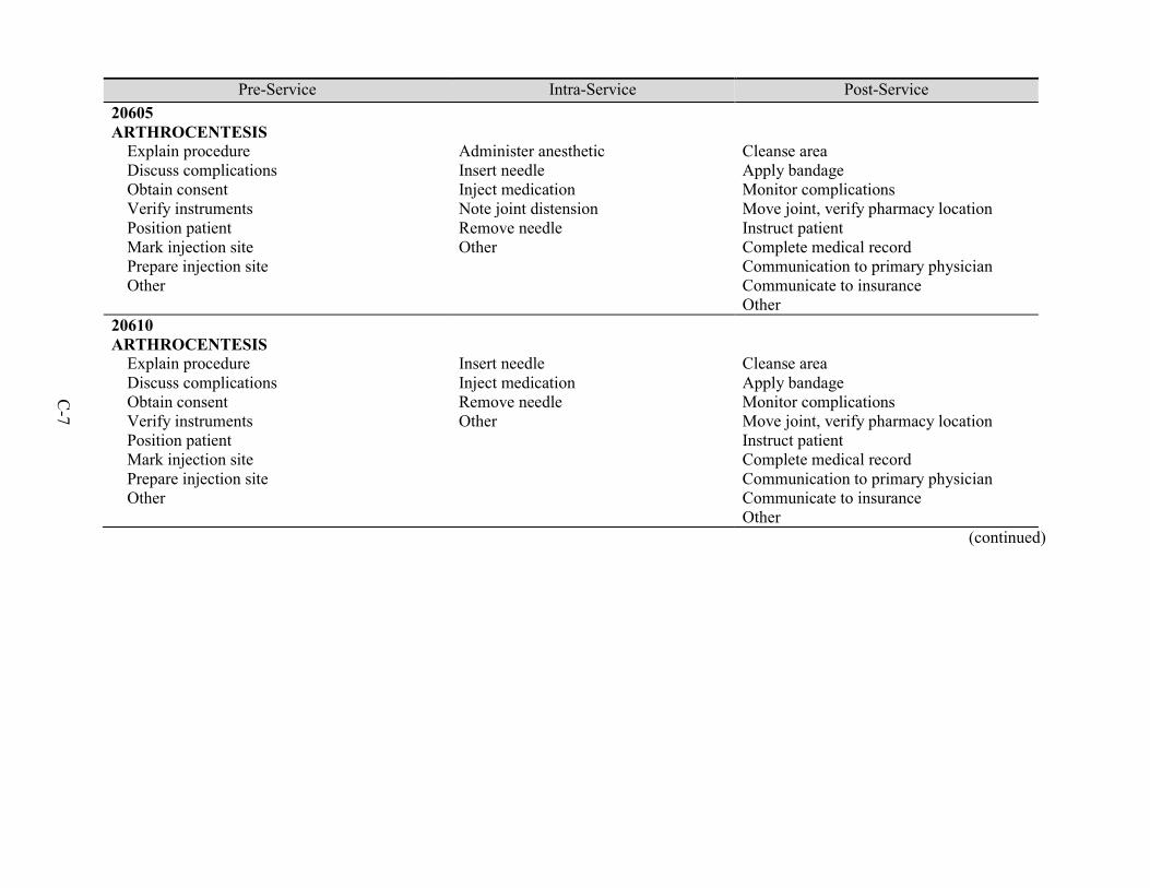
C-7
Pre-Service Intra-Service Post-Service 20605 ARTHROCENTESIS
Explain procedure Discuss complications Obtain consent Verify instruments Position patient Mark injection site Prepare injection site Other
Administer anesthetic Insert needle Inject medication Note joint distension Remove needle Other
Cleanse area Apply bandage Monitor complications Move joint, verify pharmacy location Instruct patient Complete medical record Communication to primary physician Communicate to insurance Other
20610 ARTHROCENTESIS
Explain procedure Discuss complications Obtain consent Verify instruments Position patient Mark injection site Prepare injection site Other
Insert needle Inject medication Remove needle Other
Cleanse area Apply bandage Monitor complications Move joint, verify pharmacy location Instruct patient Complete medical record Communication to primary physician Communicate to insurance Other
(continued)

C-8
Pre-Service Intra-Service Post-Service 23412 REPAIR ROTATOR CUFF, OPEN
Order antibiotics Confirm selections and timing Write preadmission orders Review test results Perform History and Physical Meet with patient and family Review procedure with patient and family Review post-op care Review consent Verify supplies are available Monitor patient positioning Pad bony prominences Apply drapes Assess patient position Position shoulder on surgery table Place bolster Mark incisions Prep skin Prep and drape shoulder Scrub and gown Take "time out" Other
Make incision over shoulder Place retractors Excise bursa Expose rotator cuff Avulse supraspinatus Avulse infraspinatus Attend to tear Resect tendon Apply sutures Abrade bone Place sutures Place bone anchors Re-approximate tendon Inspect security of tendon Apply additional anchors Repair deltoid fascia Inspect wound Irrigate wound Close wound Other
Apply dressing and sling Monitor patient Assist in patient transfer Monitor transport Discuss post-op with staff Communication with patient and family Write operative note Place note in chart Dictate report Communication with referring physician Assess operated extremity Discuss post-op with patient Write prescriptions Complete medical record Examine patient Answer questions Remove dressing and sling Assess wound Remove sutures Assess extremity Redress wound Order therapy Supervise rehabilitation Discuss progress with Primary care physician Write prescriptions Dictate notes Other
(continued)

C-9
Pre-Service Intra-Service Post-Service 27130 HIP RECONSTRUCTION
Review imaging, pathology, studies Review radiographs, if necessary Review preoperative lab test Consult with referring physician Communication with patient and family Explain risks and benefits Obtain consent Template the case Measure leg Assess center of rotation Assess proper height for cut Assess sizing of components Scrub Arrange intraoperative cell saver Position patient Assess leg length Mark incision Supervise prep and draping the pat Ensure instruments available Other
Incise skin and fascia Take glutei off femur Complete leg length assessment Dislocate femoral head Perform femoral neck osteotomy Find femoral canal Perform sequential raspings Achieve rotation and stability Use calcar planer Perform dissection and releases Expose socket Place retractors Remove excess tissue and bone Find base of acetabulum Place reamers Seat trial implants Measure stability and length Place drill and insert screws Place central hole sealer Clean soft tissue Place and tap lining Irrigate copiously Assess leg length Place head/neck Reduce hip Take x-ray Verify position of components Count sponges and needles Place deep drain Other
Splint Monitor stabilization Communicate with family Communication with other professionals Hospital visits and services Monitor lab reports Remove of drains and dressings Supervise therapy Order and review x-rays Other Perform medication management Arrange discharge Instruct re: continuing care Prepare discharge records Post-discharge office-visits, 90 days Remove sutures Evaluate imaging and lab reports Review lab values Adjust medication Attend to hip dislocation Supervise care Direct physical therapy Other
(continued)
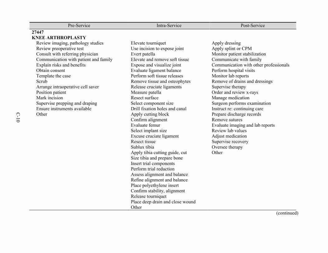
C-10
Pre-Service Intra-Service Post-Service 27447 KNEE ARTHROPLASTY
Review imaging, pathology studies Review preoperative test Consult with referring physician Communication with patient and family Explain risks and benefits Obtain consent Template the case Scrub Arrange intraoperative cell saver Position patient Mark incision Supervise prepping and draping Ensure instruments available Other
Elevate tourniquet Use incision to expose joint Evert patella Elevate and remove soft tissue Expose and visualize joint Evaluate ligament balance Perform soft tissue releases Remove tissue and osteophytes Release cruciate ligaments Measure patella Resect surface Select component size Drill fixation holes and canal Apply cutting block Confirm alignment Evaluate femur Select implant size Excuse cruciate ligament Resect tissue Sublux tibia Apply tibia cutting guide, cut Size tibia and prepare bone Insert trial components Perform trial reduction Assess alignment and balance Refine alignment and balance Place polyethylene insert Confirm stability, alignment Release tourniquet Place deep drain and close wound Other
Apply dressing Apply splint or CPM Monitor patient stabilization Communicate with family Communication with other professionals Perform hospital visits Monitor lab reports Remove of drains and dressings Supervise therapy Order and review x-rays Manage medication Surgeon performs examination Instruct re: continuing care Prepare discharge records Remove sutures Evaluate imaging and lab reports Review lab values Adjust medication Supervise recovery Oversee therapy Other
(continued)

C-11
Pre-Service Intra-Service Post-Service 29827 ARTHROPLASTY SHOULDER
Review imaging studies Review lab studies Consult with referring physician Consult with other professionals Apprise patient and family Discuss risks Obtain consent Go to operating room Verify equipment available Bring patient to operating room Position patient Pad bony prominences Place axillary roll Apply drapes Check patient position Perform exam under anesthesia Prep and drape arm Place sterile sleeve Place shoulder holder Palpate and mark landmarks
Make incision Insert arthroscope Identify surfaces and structures Create anterior portal Insert probe Confirm pathology Note tendons Mark tendons Resect tendons, if necessary Address retraction Perform repairs Move arthroscope Evaluate tear Create lateral portal Ensure adequate release Create shallow trough Place sutures Place bone anchors Pass and tie sutures Reinspect cuff Remove arthroscope Close portals Inject Marcaine Apply dressing Transfer patient
Apply dressing and sling Monitor stabilization Consult with patient and family Discuss post-op regimen Communication with other professionals Complete reports and orders Coordinate care Check vital signs Assess extremity Evaluate and review medications Review procedure with patient Review restrictions with patient Assess extremity Remove sling Evaluate range of motion Assess wound Redress wound Order therapy Clarify restrictions Supervise rehabilitation Order and review radiographs Manage antibiotics Manage pain medication Remove sutures Evaluate lab reports Communication with other professionals Communication with patient and family Monitor patient follow-ups
(continued)

C-12
Pre-Service Intra-Service Post-Service 33208 PACEMAKER INSERTION
Review history Review prior studies Review physical exam Determine prior procedures Note medications Review pre-op tests Consider renal insufficiency Ensure renal protection and hydration Estimate equipment range Ensure equipment available Review procedure with patient and family Obtain consent Assess airway Assess respiratory status Evaluate required sedation Arrange anesthesia, if necessary Check interventional suite Check equipment function Ensure safety compliance Ensure personnel familiarity Supervise equipment selection Assure equipment available Review prior studies Don protection Position patient Other
Ensure ECG and monitor function Administer sedation Position patches Prep and drape patient Perform under flouroscopic guidance Direct technical personnel Infiltrate skin with anesthetic Incise and dissect skin Produce a pocket Use electrocautery Obtain vein access Avoid punctures / complications Position J-wire Repeat for second venous access Introduce ventricular lead Shape lead stylet Position lead Exchange stylet Assess lead position Position suture tie down sleeve Suture sleeve to lead and fascia Repeat for atrial lead Test stimulation of leads Inspect and irrigate pocket Interrogate and program pacemaker Place leads and tighten screws Wrap leads Place generator in pocket Close pocket and apply dressing Reinterrogate pacemaker Other
Ensure blood pressure and heart rate are stable Write postoperative orders Communicate with family Communication with referring physician Review results with patient Dictate note Interpret lead, pulse, rhythm Review and sign report Send report to primary care physician and referring physician Review post-op PA and x-ray Review telemetry or holter monitor Test pacemaker function Check incision and pocket Communication with patient and family Discuss follow-up Discuss post-op care Other
(continued)

C-13
Pre-Service Intra-Service Post-Service 33249 PACEMAKER INSERTION
Perform history Perform physical exam Review lab tests Discuss risks with patient Obtain consent Document history and exam Consult primary physician Decide to proceed with surgery
Perform procedure under monitoring Prep patient Give antibiotic prophylaxis Place under sedation / anesthesia Inject anesthetic Make incision Perform dissection Insert leads Position leads Confirm characteristics Reposition leads Attach pacemaker/generator Place generator and leads in pocket Assess characteristics of leads Induce fibrillation Determine defibrillation threshold Reposition leads, if necessary Close incision Dress wound Document services Generate report Communication with patient and family Communication with referring physician
Monitor vital signs Review x-ray and ECG Assess characteristics of leads Determine final settings Assess wound Instruct re: post-operative care Determine pacemaker settings Interrogate ICD Exercise patient Assess settings Assess wound Remove dressing Prescribe medications
(continued)

C-14
Pre-Service Intra-Service Post-Service 33405 CARDIAC VALVE PROCEDURE
Perform history Perform physician exam Identify contraindications Assess patient Write preoperative orders Review preoperative work-up Review reports Assess cardiology, pulmonary Review plan Other Review procedure with patient and family Review expected outcome Discuss risks Obtain consent Confirm start time Notify patient and family Arrange for surgical assist Change into scrubs Identify patient Other
Make incision, divide sternum Cauterize edges Place wax or paste Inspect aorta Give heparin Place sutures Insert cannulas Initiate bypass Place vent, initiate cooling Place cannulas Clamp aorta Administer cardioplegia Administer hypothermia Open aorta Remove native aortic valve Debride calcium Measure annulus Select prosthesis Other
Apply dressings Remove drapes Move to ICU Monitor patient Observe drainage Sign OR forms Indicate diagnosis and procedure Obtain x-ray Write postoperative orders Write operative note Establish short term goals Review procedure with patient and family Dictate report Communication with referring physician Communicate to insurance Coordinate care Evaluate neurological state Monitor anesthesia Other
(continued)

C-15
Pre-Service Intra-Service Post-Service 33405 (continued) CARDIAC VALVE PROCEDURE
Mark incision Answer concerns Check with lab Review anesthesia Plan monitoring Review positioning Verify drug administration Review ECG Place catheter Other Position patient Scrub, gown, glove Prep and drape patient Other
Place sutures Remove clamp Resuscitate heart Remove air monitoring Place pacing wires Remove vent Discontinue bypass Remove cannulas Repair site Other Irrigate wound Place drains Re-approximate sternum Close incision Other
Administer sedatives as needed Repeat neurological evaluation Assess hemodynamic condition Evaluate cardiac rhythm Consider pacemaker Assess arrhythmias Correlate pressures Administer fluids Adjust agents Other Evaluate perfusion Monitor bleeding Assess drainage Other
(continued)

C-16
Pre-Service Intra-Service Post-Service 33518 CORONARY ARTERY SURGERY
Discuss grafts Discuss incisions Discuss potential problems Other
Identify target vessels Open artery Tailor vein end Construct anastomoses Probe anastomoses Ensure patency Perform aortotomy Tailor vein ends Ensure graft lengths Construct anastomoses Inspect anastomoses Assess inflowithoutflow Terminate bypass Conclude operation Close incisions Suture/drain as needed Other
Intensive care Ventilator hours Hospital stay Additional office work Other
(continued)

C-17
Pre-Service Intra-Service Post-Service 43235 UGI ENDOSCOPY DIAGNOSTIC
Review symptoms Review history Assess need for antibiotics Review allergies Note medications Assess airway Perform cardiopulmonary evaluation Review lab studies Review x-rays Review procedure with patient Obtain consent Other
Start IV access Administer sedation Insert endoscope Examine duodenal mucosa Withdraw endoscope to stomach Retroflex endoscope Examine gastric fundus Straighten endoscope Examine gastric mucosa Withdraw endoscope to esophagus Measure gastro-esophageal junction Assess presence of hernia Examine esophageal mucosa Withdraw endoscope Other
Transfer patient to recovery Assess vital signs Dictate report Review findings, when stable Other
(continued)

C-18
Pre-Service Intra-Service Post-Service 43239 UGI ENDOSCOPY BIOPSY
Review symptoms Review history Assess need for antibiotics Review allergies Note medications Assess airway Perform cardiopulmonary evaluation Review lab studies Review x-rays Review procedure with patient Obtain consent Other
Start IV access Administer sedation Insert endoscope Examine duodenal mucosa Withdraw endoscope to stomach Retroflex endoscope Examine gastric fundus Straighten endoscope Examine gastric mucosa Perform biopsy(ies) Withdraw endoscope to esophagus Measure gastro-esophageal junction Assess presence of hernia Examine esophageal mucosa Withdraw endoscope Other
Transfer patient to recovery Assess vital signs Dictate report Review findings, when stable Other
45378 DIAGNOSTIC COLONOSCOPY
Review symptoms Review history Assess need for antibiotics Note medications Assess airway Perform cardiopulmonary evaluation Review lab studies Review x-rays Review procedure with patient Obtain consent Other
Start IV access Administer sedation Insert colonoscope Identify ileo-cecal valve Intubate terminal ileum Withdraw through colon Examine colon mucosa Retroflex colonoscope Examine rectal mucosa Straighten colonoscope Withdraw colonoscope Other
Transfer patient to recovery Assess vital signs Dictate report Review findings, when stable Other
(continued)
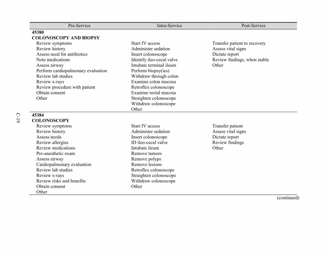
C-19
Pre-Service Intra-Service Post-Service 45380 COLONOSCOPY AND BIOPSY
Review symptoms Review history Assess need for antibiotics Note medications Assess airway Perform cardiopulmonary evaluation Review lab studies Review x-rays Review procedure with patient Obtain consent Other
Start IV access Administer sedation Insert colonoscope Identify ileo-cecal valve Intubate terminal ileum Perform biopsy(ies) Withdraw through colon Examine colon mucosa Retroflex colonoscope Examine rectal mucosa Straighten colonoscope Withdraw colonoscope Other
Transfer patient to recovery Assess vital signs Dictate report Review findings, when stable Other
45384 COLONOSCOPY
Review symptoms Review history Assess needs Review allergies Review medications Pre-anesthetic exam Assess airway Cardiopulmonary evaluation Review lab studies Review x-rays Review risks and benefits Obtain consent Other
Start IV access Administer sedation Insert colonoscope ID ileo-cecal valve Intubate ileum Remove tumors Remove polyps Remove lesions Retroflex colonoscope Straighten colonoscope Withdraw colonoscope Other
Transfer patient Assess vital signs Dictate report Review findings Other
(continued)

C-20
Pre-Service Intra-Service Post-Service 45385 LESION REMOVAL COLONOSCOPY
Review symptoms Review history Assess need for antibiotics Note medications Assess airway Perform cardiopulmonary evaluation Review lab studies Review x-rays Review risks and benefits Obtain consent Other
Start IV access Administer sedation Insert colonoscope Identify ileo-cecal valve Intubate terminal ileum Remove tumor, polyp, or lesion Withdraw through colon Examine colon mucosa Retroflex colonoscope Examine rectal mucosa Straighten colonoscope Withdraw colonoscope Other
Transfer patient to recovery Assess vital signs Dictate report Review findings, when stable Other
(continued)

C-21
Pre-Service Intra-Service Post-Service 47562 LAPAROSCOPIC LIVER OR GALL BLADDER PROCEDURE
Select and order antibiotics Confirm timing and administration Assure appropriate DVT prophylaxis Assess need for beta-blockers Order beta-blockers as required Review work-up Reexamine patient Update history and physical Review test results Review procedure with patient and family Mark incision Confirm with patient Obtain consent, including witness Review anesthesia Verify equipment available Transfer patient Position patient Install Foley catheter Assist anesthesia team Place line Induct anesthesia Intubate Prep skin Mark incisions Scrub and gown Take "time out" Other
Make incision Expose and incise fascia Incise midline Place sutures Open peritoneum Place trocar Attach sutures Insufflate abdomen Monitor changes Insert camera Instruct assistant Explore abdominal cavity Inject anesthesia Place trocar Place ports Retract liver Perform laparoscopy Lyse adhesions Grasp gall bladder Fix gall bladder in place Dissect cystic duct Dissect triangle of Calot Identify anomalies Place clips Reconfirm cystic artery Dissect cystic artery Dissect gallbladder free Place gallbladder over liver Achieve hemostasis Other
Apply Steristrips and dressings Protect wound Transfer patient Discuss care with staff Review post-operative labs Communicate with family Write operative note Write post-operative note Dictate report Communication with referring physician Discharge patient from recovery Write orders Auscultate patient Manage dressing Continue prophylaxis Order beta-blockers as required Order antibiotics as required Chart progress and assess pain Manage drain Write prescriptions and orders Discuss home care Discharge patient Complete medical records Remove staples or sutures Monitor healing Communication with patient and family Write prescriptions Review labs and films Communication with referring physician Other
(continued)

C-22
Pre-Service Intra-Service Post-Service 47563 LAPAROSCOPIC LIVER OR GALL BLADDER PROCEDURE
Order antibiotics Confirm timing Administer DVT prophylaxis Assess beta-blocker need Review work-up Review results Meet with patient and family Review procedure with patient and family Review post-op care Re-examine patient Update H&P Mark incision Confirm with patient Obtain consent Review anesthesia Verify supplies are available Transfer patient Position patient Scrub and gown Take "time out" Other
Induce pneumoperitoneum Monitor changes Insert camera Ensure adequate image Explore abdominal cavity Retrace liver View organs Lyse adhesions Decompress gallbladder Dissect cystic duct Identify junction with gallbladder Expose triangle of Calot Occlude systic duct Position patient Maneuver equipment Perform cholangiography Inject contrast Interpret images Free gallbladder Store gallbladder Irrigate field Aspirate debris Inspect fossa Achieve hemostasis Inspect arteries and stumps Place drain Ensure no bleeding Other
Apply dressing Monitor patient Protect wound Transfer patient Discuss post-op care Write operative note Write post-operative note Dictate report Call referring physician Discharge patient Write orders Examine patient Auscultate Manage dressings Continue prophylaxis Assess beta-blocker need Assess antibiotic need Document progress Other Assess pain scores Monitor renal function Review notes Write orders Chart progress Answer questions Advance diet Manage drain Write prescriptions Other
(continued)

C-23
Pre-Service Intra-Service Post-Service 47563 (continued) LAPAROSCOPIC LIVER OR GALL BLADDER PROCEDURE
Aspirate carbon dioxide Close defects Confirm supply count Other
Review post-op care Remove staples Dictate notes Other
49505 REPAIR IGUINAL HERNIA
Administer prophylaxis Write orders Administer antibiotics Review work-up Re-examine patient Review results Meet with patient and family Review procedure with patient and family Review post-op care Mark incision Confirm with patient Obtain consent Review anesthesia Verify supplies are available Transfer patient Assist anesthesia Install Foley catheter Prep skin Clip hair Scrub and gown
Make incision Divide tissues Secure hemostasis Identify fascia Incise and grasp fascia Extend incision Develop flaps Expose canal Insert retractor Mobilize nerve Employ vessel loop Dissect spermatic cord Explore for hernial sac Dissect hernial sac Inspect hernial sac Transfix with suture Approximate fascia ID conjoint tendon Place mesh Assess internal ring Ensure hemostasis
Apply dressings Monitor anesthesia Transfer patient Transport patient Discuss post-op care Write operative note Write post-operative note Dictate report Call referring physician Discuss procedure with family Answer questions Write orders Write prescriptions Discuss restrictions Discharge patient Complete medical record Examine patient Remove sutures Review activity Review restrictions Monitor healing
(continued)

C-24
Pre-Service Intra-Service Post-Service 49505 (continued) REPAIR IGUINAL HERNIA
Take "time out" Other
Return ilioinguinal nerve Infiltrate with anesthetic Close fascia Evaluate external ring Re-approximate fascia Examine operative site Irrigate with antibiotic Conduct supply count Place sutures Re-approximate skin Other
Answer questions Write prescriptions Order labs/films Discuss progress Dictate notes Other
50590 LITHOTRIPSY
Review prior studies Communication with other professionals Communicate w patient Explain risks Obtain consent Dress for surgery Wait for anesthesia Position patient Prep patient Drape patient Scrub and gown Prepare equipment Other
Check stone position Adjust anesthesia Determine treatment energy Commence treatment Monitor treatment Fragmentation occurs Other
Post-operative care Stabilize patient Write postoperative orders Communicate with family Communication with referring physician Discharge day management Post-operative hospital visits Post-discharge office visits Post-operative care Other
(continued)

C-25
Pre-Service Intra-Service Post-Service 52000 CYSTOSCOPY
Check video equipment Check schedule Ensure instruments available Ensure personnel available Notify patient Make images available Change into scrubs Discuss with anesthesiologist Review procedure with patient and family Review post-op care Answer questions Obtain consent Evaluate need for antibiotics Identify patient Position patient Prep patient Other
Inject anesthetic Apply penile clamp Assemble equipment Connect video system Apply defogger Connect irrigation source Connect life source Insert endoscope Inspect patient Identify and assess Other
Transfer patient from table Transfer patient to recovery Conduct pain assessment Write orders and prescriptions Discuss procedure and findings Call referring physician Diagram bladder Dictate narrative Schedule appointment Other
(continued)

C-26
Pre-Service Intra-Service Post-Service 52224 CYSTOURETHROSCOPY PROCEDURES OF URETHRA AND BLADDER
Check video equipment Check schedule Ensure instruments available Ensure personnel available Patient notification Change into scrubs Discuss anesthesia Review procedure with patient and family Answer questions Obtain consent Take "time out" Position patient Prep and drape Other
Inject anesthetic Assemble equipment Connect video Apply defogger Connect irrigation source Connect light source Insert endoscope Inspect patient Assess bladder neck Inspect trigone Identify urethral orifices Check for efflux of urine Inspect bladder mucosa Assess bladder Treat lesions Retroflex scope Inspect dome of bladder Drain bladder Disconnect equipment Remove endoscope Other
Transfer patient Conduct pain assessment Write orders Write prescriptions Discuss procedure Discuss findings with patient Call referring physician Complete mapping diagram Dictate narrative Schedule follow-up Other
(continued)

C-27
Pre-Service Intra-Service Post-Service 52281 CYSTOURETHROSCOPY PROCEDURES OF URETHRA AND BLADDER
Review records Communicate with other professionals Communicate w patient Explain procedure Obtain consent Dress for surgery Prep and drape Scrub Prep equipment Other
Position patient Prep perineum Drape perineum Perform meatotomy Insert cystoscope Insert guidewire Remove cystoscope Pass dilator over guidewire Utilize Heyman dilators Pass cystoscope Inspect prostatic channel Inspect bladder neck Inspect bladder Remove cystoscope Remove guidewire Other
All post-op care Stabilize patient Post-op orders Communicate with patient Call referring physician Other follow-up care Write prescriptions Other
(continued)

C-28
Pre-Service Intra-Service Post-Service 52601 CYSTOURETHROSCOPY
Review lab results Review medical record Write pre-op orders Change into scrub clothes Communication with patient and family Answer questions Obtain consent Communication with anesthesiologist Position patient Verify equipment available Take "time out" Wait for anesthetic Other
Position patient Prepare genitalia Apply drapes Place lubricating jelly Assemble equipment Connect irrigation source Connect light Insert endoscope Inspect patient anatomy Check for efflux of urine Inspect bladder mucosa Assess bladder Inspect dome of bladder Drain bladder Disconnect equipment Remove endoscope Dilate urethra Perform meatomy, if necessary Perform urethrotomy, if necessary Introduce resectoscope Remove obturator Drain bladder Place working element Assemble equipment Resect tissue Coagulate bleeders Irrigate bladder Remove equipment Insert catheter Inflate catheter Other
Awaken patient Attach irrigation Irrigate Foley catheter Transfer patient Review care with staff Communicate with family Communication with patient Discuss procedure with patient Conduct pain assessment Write orders Dictate report Communication with referring physician Examine patient Chart progress and assess pain Irrigate clots Review medical record Communication with patient and family Answer questions Write orders Write note Check lab values Write prescriptions Dictate discharge summary Remove Foley catheter Assess voiding pattern Reinsert catheter, if necessary Schedule office visit Dictate notes Communication with referring physician Other
(continued)

C-29
Pre-Service Intra-Service Post-Service 55700 BIOPSY OF PROSTATE
Check schedule Ensure equipment available Check with staff Notify patient Review lab studies Ensure studies available Change into scrubs Review procedure with patient and family Review post-op care Answer questions Obtain consent Check pre-op procedures Check patient Position patient Other
Take "time out" Position patient Repeat rectal exam Dilate anus Introduce probe Infiltrate with anesthetic Take biopsies Remove biopsy gun Remove specimens Recock and insert gun Repeat biopsy Place samples Remove probe Apply pressure Other
Transfer patient Review care Review medications Meet with patient Discuss procedure Discuss outcome Discuss post-op care Conduct pain assessment Discuss pathologist Call referring physician Dictate report Other
(continued)

C-30
Pre-Service Intra-Service Post-Service 55866 LAPAROSCOPIC RADIAL PROSTATECTOMY
Review lab results Review medical records Write orders Change into scrubs Review procedure with patient and family Review post-op care Answer questions Speak to anesthesiologist Position patient Verify instruments available Other
Position patient Prep lower abdomen Drape lower abdomen Achieve pneumoperitoneum Place four trocars Other
Examine patient Check wound(s) Check progress Review medical records Answer questions Write orders Write note Write prescriptions Discuss post-op care Dictate discharge summary Review bladder exercises Remove sutures Schedule follow-up Mark diagnosis Dictate notes Dictate letter to referrer Other
(continued)

C-31
Pre-Service Intra-Service Post-Service 63047 DECOMPRESSION LUMBAR SPINE
Review work-up Write orders Locate imaging studies Place imaging studies Review planned incision Review procedure with patient and family Greet patient Review procedure Review outcomes Mark operative site Obtain consent Verify instruments available Review anesthesia Position patient Induce anesthesia Reposition patient Pad patient Scrub and gown Mark incisions Supervise prepping and draping Other
Make incision Expose spinous process Expose lamina Remove process/lamina Remove flavum Expose thecal sac Expose nerve roots Remove facets Expose nerve roots Perform foraminotomy Perform discectomy Irrigate wound Close wound Apply dressings Other
Turn patient Reverse anesthesia Transfer patient Write note Examine patient Check wound(s) Check progress Monitor patient Sign forms Discuss outcome Dictate report Communication with referring physician Dictate outcome Order and review films Write orders Examine patient Chart progress Discuss progress Answer questions Review post-op care Write prescriptions Order films Review films Perform exams Monitor wounds Remove sutures Review brace Review progress Dictate notes Communication with referring physician Other
(continued)

C-32
Pre-Service Intra-Service Post-Service 64483 TRANSFORMINAL LUMBAR INJECTIONS
Review records and studies Communicate with other professionals Communication with patient and family Obtain consent Dress and scrub Prepare patient Prepare equipment Position patient Drape injection site Other
Identify affected foramen Infiltrate skin with anesthetic Direct needle Obtain necessary views Perform contrast injection Confirm needle tip location Inject anesthetic Inject steroid Remove needle Apply dressing Other
Take patient to recovery Observe for neurological deficits Review procedure with patient Review procedure with professionals Evaluate neurological condition Discharge from recovery area Other
66821 DISCISSION MEMBRANOUS CATARACT BY LASER
Complete ophthalmological exam Note visual acuity Note opacification Examine macula Rule out diseases Obtain consent Review chart, history, exams Instill medications, anesthetic Other
Seat patient Apply contact lens Deliver laser energy Apply test burns Confirm adequacy of energy Adjust laser Other
Monitor patient Treat pressure Follow-up in 1-2 weeks Follow-up in 1-3 months Other
(continued)

C-33
Pre-Service Intra-Service Post-Service 66982 LENS PROCEDURE
Review history Review exam Review pre-op tests Counsel family Document patient record Other
Prep and drape Place microscope Perform flap Make groove Make incision Fix pupil Make incisions Insert hooks Retract iris Photoemulsification Remove hooks Check wound(s) Dress eye Alternative: capsulotomy Other
See patient Give instructions Arrange follow-up See patient periodically Ensure wound stability Obtain visual rehab Commence therapy Other
(continued)

C-34
Pre-Service Intra-Service Post-Service 66984 LENS PROCEDURE
Prepare patient Ascertain correct eye Examine eye Rule out changes since last exam Check adequacy of dilation Administer antibiotics Greet patient Address questions and concerns Check chart and patient data Perform history and physical Perform anesthetic block Give anesthesia Monitor vital signs Guard against emergency Place compressive device Other
Transport patient to suite Position patient Prep and drape operative site Check adequacy of anesthetic Apply anesthetic Perform scrub, gown Check devices Enter parameters Check machine operation Position microscope Assure adequate reflex Communication with anesthesiologist Place speculum and bridle-suture Raise conjunctiva Make side-port incision Insufflate anterior chamber Enter anterior chamber Fashion opening in capsule Loosen lens Perform hydrodissection Check handpiece function Carry out phacofragmentation Rotate and disassemble lens Remove cataractous material Remove phacoemulsification tip Introduce irrigation-aspiration tip Clean residual material Vacuum and polish lens Open larger incision Deliver intraocular lens Other
Speak with patient and family Give instructions Address questions Give prognosis Ascertain pat ability to leave See patient next day Follow-up exam Take history of post-op course Examine eye Instruct re: medication use Instruct re: activity Instruct re: follow-up Perform keratometry, topography Ascertain stigmatism Perform spectacle examination Address complications Other
(continued)

C-35
Pre-Service Intra-Service Post-Service 67028 INTRAVITREAL EYE INJECTION
Review visual acuity Review intraocular pressure Confirm eye to be treated Obtain consent Answer questions Review studies Re-confirm eye to be treated Examine eye Position patient Other
Apply anesthetic Apply antisepsis solution Draw drug into syringe Place lid speculum Measure injection location Instruct pat to position eye Pass needle through pars plana Confirm needle position Inject drug Remove needle Control bleeding Confirm nerve perfusion Measure intraocular pressure Irrigate eye Remove residual iodine Other
Instruct re: post-operative care Review potential complications Complete operative note Other
67210 DESTRUCTION RETINA OR CHOROID
Measure vision Dilate pupils Apply anesthesia Review angiograms Place in monitoring position Other
Perform photocoagulation Use angiogram as guide Close microaneurysms Attend to capillary dropout Attend to diffuse leakage Assess patient response Assess tissue reaction Assess opacity Assess lesion Pause frequently Ensure steadiness Other
Assess treatment adequacy Check for complications Counsel patient Review post-op care Schedule follow-up Administer laser therapy Other
(continued)

C-36
Pre-Service Intra-Service Post-Service 67228 TREATMENT OF RETINAL LESION
Review medical records Obtain history Review testing Discuss procedure Obtain consent Other
Measure visual acuity Dilate pupil Examine fundus Ensure laser appropriateness Apply anesthetic Administer laser Apply contact lens or anesthetic Other
Irrigate eye Apprise progress Instruct re: complications Instruct re: post-op Write orders Prepare operative note Schedule follow-up Notify referring physician Other
70450 CT HEAD OR BRAIN WITHOUT CONTRAST
Review reason for exam Review history Review previous studies Determine CT protocol Confirm non-contrast needed Deter need for Additional images Communicate protocol Other
Supervise scout views Prescribe area of coverage Supervise acquisition of source images Review image data Assure adequacy of coverage Assess need, repeats or reconstruction Supervise reconstruction of images Assess need for Additional images Interpret findings Compare to previous studies Dictate report Other
Review, edit, sign report Discuss findings with referring physician Other
(continued)

C-37
Pre-Service Intra-Service Post-Service 70486 CT FACE WITHOUT CONTRAST
Review reason for exam Review history Review previous studies Determine CT protocol Confirm non-contrast needed Communicate protocol Other
Interpret scout topograms Review image data Assure adequacy of coverage Assess need for repeat sections Review axial images Assess maxillofacial area Review area for injury or process Interpret examination Compare to previous studies Dictate report Other
Review, edit, sign report Discuss findings with referring physician Other
70551 MRI BRAIN WITHOUT CONTRAST
Discuss exam Review history Set protocol Select pulse sequences Review prior studies Other
Review images Determine if additional sequences Inform technician, if necessary Interpret study Prepare report Other
Review and sign report Direct copies to referring physician Talk to referring physician Other
70553 MRI BRAIN WITHOUT CONTRAST
Discuss exam Review history Set protocol Select pulse sequences Review prior studies Other
Review unenhanced exam Acquire pre-contrast sequences Review enhanced portion of exam Assure needed sequences obtained Interpret exam Compare unenhanced and enhanced Deter pathologic enhancement Compare to prior exams Interpret study Prepare report Other
Review and sign report Direct copies to referring physician Talk to referring physician Other
(continued)

C-38
Pre-Service Intra-Service Post-Service 71010 CHEST X-RAY, SINGLE VIEW
Review reason for exam Review history Review prior studies Other
Supervise technician performing exam Interpret exam Compare to previous studies Dictate report Other
Review and sign report Discuss findings with referring physician Other
71020 CHEST X-RAY, TWO VIEWS
Review reason for exam Review history Review prior studies Other
Supervise technician performing exam Interpret exam Compare to previous studies Dictate report Other
Review and sign report Discuss findings with referring physician Other
71250 CT THORAX WITHOUT CONTRAST
Review reason for exam Review history Review prior studies Determine appropriate protocol Confirm non-contrast needed Deter need for Additional images Communication protocol to techs Other
Supervise acquisition of scout views Prescribe area of coverage Supervise acquisition of source images Review image data Assure adequacy of coverage Assess need, repeats or reconstruction Supervise reconstruction of images Assess need for other images Interpret findings Compare to previous studies Dictate report Other
Review and sign report Discuss findings with referring physician Other
(continued)
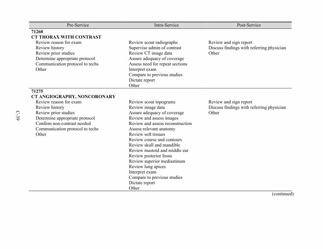
C-39
Pre-Service Intra-Service Post-Service 71260 CT THORAX WITH CONTRAST
Review reason for exam Review history Review prior studies Determine appropriate protocol Communication protocol to techs Other
Review scout radiographs Supervise admin of contrast Review CT image data Assure adequacy of coverage Assess need for repeat sections Interpret exam Compare to previous studies Dictate report Other
Review and sign report Discuss findings with referring physician Other
71275 CT ANGIOGRAPHY, NONCORONARY
Review reason for exam Review history Review prior studies Determine appropriate protocol Confirm non-contrast needed Communication protocol to techs Other
Review scout topograms Review image data Assure adequacy of coverage Review and assess images Review and assess reconstruction Assess relevant anatomy Review soft tissues Review course and contours Review skull and mandible Review mastoid and middle ear Review posterior fossa Review superior mediastinum Review lung apices Interpret exam Compare to previous studies Dictate report Other
Review and sign report Discuss findings with referring physician Other
(continued)

C-40
Pre-Service Intra-Service Post-Service 72125 CT SPINE
Review reason for exam Review history Review prior studies Determine exam protocol Confirm non-contrast needed Communication with technologists Other
Review topograms Review CT image data Assure coverage Assure adequacy of coverage Review axial images Review reconstructions Review soft tissues Review vascular structures Review skull and mandible Review mastoid and ear Review posterior fossa Review superior mediastinum Review lung apices Interpret exam Compare to previous studies Dictate report Other
Review and sign report Communication with referring physician Other
72141 MRI SPINE
Discuss exam Review history Set protocol Select pulse sequences Review prior studies Other
Review images Determine if additional sequences are needed Inform tech, if necessary Interpret study Prepare report Other
Review and sign report Direct copies to referring physician Direct verbal report to referring physician Other
(continued)

C-41
Pre-Service Intra-Service Post-Service 72148 MRI SPINE AND LUMBAR WITHOUT CONTRAST
Discuss exam Review history Set protocol Select pulse sequences Review prior studies Other
Review images Determine if additional sequences are needed Inform tech, if necessary Interpret study Prepare report Other
Review and sign report Direct copies to referring physician Talk to referring physician Other
72158 MRI SPINE AND LUMBAR WITH AND WITHOUT CONTRAST
Discuss exam Review history Set protocol Select pulse sequences Review prior studies Other
Review unenhanced exam Acquire pre-contrast sequences Review enhanced portion of exam Assure needed sequences obtained Interpret exam Compare enhanced and unenhanced Deter pathologic enhancement Compare to prior exams Interpret study Prepare report Other
Review and sign report Direct copies to referring physician Talk to referring physician Other
72171 MRI SPINE WITHOUT CONTRAST
Discuss exam Review history Set protocol Select pulse sequences Review prior studies Other
Review images Determine if additional sequences are needed Inform tech, if necessary Interpret study Prepare report Other
Review and sign report Direct copies to referring physician Talk to referring physician Other
(continued)

C-42
Pre-Service Intra-Service Post-Service 74176 CT ABDOMEN AND PELVIS WITHOUT CONTRAST
Review reason for exam Review history Review prior studies Determine appropriate protocol Confirm non-contrast needed Communication protocol to techs Other
Supervise acquisition of scout views Prescribe area of coverage Supervise acquisition of axial sections Review image data Assure adequacy of coverage Assess need for other images Supervise reconstruction of images Interpret soft-tissue window Interpret bone window images Interpret liver window images Interpret lung window images Compare to previous studies Dictate report Other
Review and sign report Discuss with referring physicians Other
(continued)

C-43
Pre-Service Intra-Service Post-Service 74177 CT ABDOMEN AND PELVIS WITH CONTRAST
Review reason for exam Review history Review prior studies Determine appropriate protocol Confirm non-contrast needed Communication protocol to techs Other
Supervise insertion of catheter Supervise setup of injector Supervise acquisition of scout views Prescribe area of coverage Supervise use of injector Supervise acquisition of axial sections Monitor contrast Review image data Assure adequacy of coverage Assess need for additional sections Supervise reconstruction of images Interpret soft-tissue window Interpret bone window images Interpret liver window images Interpret lung window images Compare to previous studies Dictate report Other
Review and sign report Discuss with referring physicians Other
(continued)

C-44
Pre-Service Intra-Service Post-Service 74178 CT ABDOMEN AND PELVIS WITH AND WITHOUT CONTRAST
Review reason for exam Review history, contraindications Review prior studies Determine appropriate protocol Communication protocol to techs Other
Supervise insertion of catheter Supervise setup of injector Prescribe area of coverage Supervise acquisition of axial sections Review image data Assure adequacy of coverage Supervise use of injector Supervise acquisition of axial sections Monitor contrast Review image data Assure adequacy of coverage Supervise reconstruction of images Interpret soft-tissue window Interpret bone window images Interpret liver window images Interpret lung window images Compare to previous studies Dictate report Other
Review and sign report Discuss with referring physicians Other
77080 DXA BONE DENSITY STUDY
Review reason for exam Review history Review prior studies Other
Review scout images Assure satisfactory technique Interpret scan data Compare to established norms Compare to previous studies Dictate report Other
Review and sign report Discuss with referring physician Other
(continued)

C-45
Pre-Service Intra-Service Post-Service 78452 MYOCARDIAL PERFUSION IMAGING
Deter study appropriateness Prescribe IV dose Oversee IV dose Adjust protocol as necessary Assess cardiac rhythm Direct lead placement Answer questions Review components of study Other
Acquire stress and rest data Verify adequacy of data sets Order additional imaging, if necessary Review images Review tomographic datasets Re-reconstruct, if necessary Compare tomographic datasets Score wall function and perfusion Score wall motion Attend to myocardial function Judge myocardial scar Note perfusion defects Note geographic territories Generate and review data Compare data to images Generate wall motion analysis Note ejection fraction Confirm correctness of calc Review data Refine qualitative impression Compare to prior studies Dictate report Other
Review and sign report Discuss results with referring physician Discuss results with pat and family Provide regulatory oversight Other
(continued)

C-46
Pre-Service Intra-Service Post-Service 88305 TISSUE EXAM BY PATHOLOGIST
N/A Other
Review history Review previous studies Examine specimen Interpret test result Compare to previous studies Consider variations Identify findings Review literature Research Prepare report Communication with other professionals Other
N/A Other
88307 SURGICAL PATHOLOGY
N/A Other
Review clinical history Review diagnostic studies Examine previous reports Communicate with other professionals Perform exam Interpret test result Compare to previous studies Consider statistical variations Identify findings Review literature Research test result Dictation and pathology report Report sign-out Communication with other professionals Other
N/A Other
(continued)

C-47
Pre-Service Intra-Service Post-Service 88309 SURGICAL PATHOLOGY
N/A Other
Review clinical history Review diagnostic studies Examine previous reports Communicate with other professionals Perform exam Interpret test result Compare to previous studies Consider statistical variations Identify findings Review literature Research test result Dictation and pathology report Report sign-out Communication with other professionals Other
N/A Other
88312 SPECIAL STAINS
N/A Other
Examine positive control Verify structures are stained Examine patient sample Examine presence of organisms Interpret, morphologically Correlate findings with pat history Compose and dictate report Edit and sign report Communication results to caregivers Other
N/A Other
(continued)

C-48
Pre-Service Intra-Service Post-Service 88331 PATHOLOGY CONSULTATION DURING SURGERY
N/A Other
Receive specimen Review information from surgeon Query medical record Determine original diagnosis Examine specimen Maintain orientation Ink margins, as appropriate Section specimen Embed tissues Freeze tissue Section tissue in a cryostat Capture sections on glass slides Evaluate stained slides Formulate an interpretation Present verbal report to surgeon Record written report Other
N/A Other
88342 IMMUNOHISTOCHEMISTRY
N/A Other
Perform immunohistochemistry Other
N/A Other
(continued)

C-49
Pre-Service Intra-Service Post-Service 92133 OPHTHALMIC COMPUTERIZED SCANNING
Boot computer Enter data Examine patient Other
Align scanning head Adjust focus and brightness Obtain three scans Examine for quality/adequacy Obtain printout of mean image Interpret printout/findings Compare to previous studies Dictate report Other
N/A Other
92134 OPHTHALMIC COMPUTERIZED SCANNING
Prepare instrument Enter patient identifiers Assess fixation ability Describe procedures Other
Position patient Align scanner Process images Display output Ensure image adequacy Test fellow eye Evaluate study quality Interpret findings Assess change, if follow-up Correlate with clinical course Prepare report Send report to requesting physician Other
N/A Other
(continued)

C-50
Pre-Service Intra-Service Post-Service 92557 COMPREHENSIVE HEARING TEST
Greet patient Review background Record demographics Other
Seat patient Place headphones Explain test Check ear canals Test each ear at multiple Hz Deter speech reception threshold Deter speech discrimination Other
Compile results Generate report for referring physician Review findings, when stable Other
93000 ELECTROCARDIOGRAM COMPLETE
Review request Review history Other
Review tracing Make measurements Make overall interpretation Compare tracing to previous Observe potential etiologies Other
Prepare report Sign report Transmit to medical record Other
93010 ELECTROCARDIOGRAM REPORT
Review request Review history Other
Review tracing Make measurements Make overall interpretation Compare tracing to previous Observe potential etiologies Other
Prepare report Sign report Transmit to medical record Other
(continued)

C-51
Pre-Service Intra-Service Post-Service 93015 CARDIOVASCULAR STRESS TEST
Review medical records Review chart Determine clinical questions Other
Assess new symptoms Assess pharmacological stress Answer questions Obtain consent Supervise stress infusion Assess adequacy of data Treat untoward medical events Treat side effects Interpret resting ECG Interpret serial ECGs Review and analyze results Dictate report Other
Review, edit, sign report Discuss values with patient Discuss with referring physician Other
93016 CARDIOVASCULAR STRESS TEST
Review medical records Review chart Other
Assess new symptoms Assess stress versus treadmill Answer questions Obtain consent Supervise infusion, if necessary Assess adequacy of data Treat untoward medical events Treat side effects Reviewithanalyze hemodynamics Review and analyze arrhythmias Review and analyze symptoms Review functional capacity Other
N/A Other
(continued)

C-52
Pre-Service Intra-Service Post-Service 93018 CARDIOVASCULAR STRESS TEST
Review medical records Review chart Deter questions to be answered Other
Assess adequacy of data Interpret resting ECG Interpret serial ECGs Interpret timing, morphology, changes Review and analyze hemodynamics Review and analyze arrhythmias Review and analyze symptoms Review functional capacity Other
Review, edit, sign report Discuss values with patient Discuss with referring physician Other
(continued)

C-53
Pre-Service Intra-Service Post-Service 93306 TRANSTHORACIC ECHOCARDIOGRAPHY
Review information Review records Clarify indications Deter questions to be answered Other
Obtain structure and dynamics Record on videotape or digitally Make time-motion recordings View blood flow patterns Record flow velocity Verify suitability of images Record additional views, if necessary Review recorded views of heart Analyze heart structure, dynamics Note abnormalities in flow Review velocity recordings Verify quantitative measures Assess heart function Calculate pressure gradients Calculate valve orifice areas Calculate regurgitant volumes Calc regurgitant fractions Calc regurgitant orifice areas Develop complete interpretation Make anatomic, functional measures Document sizes Review tomographic views Review hemodynamic data Compare to previous studies Deter if changes have occurred Other
Prepare report Review and correct report Sign report Review findings with requesting physician Facilitate patient management Other
(continued)

C-54
Pre-Service Intra-Service Post-Service 93458 CARDIAC CATHETERIZATION
Prepare report Review and correct report Sign report Review findings with requesting physician Facilitate patient management Other
Administer conscious sedation Verify sedation monitoring Insert needle Insert guidewire Remove needle Insert sheath/dilator Remove dilator Flush sidearm Insert catheter Attach catheter to manifold Measure pressure Check pressure and waveform Test contrast injections Perform injections with diff views Remove catheter Introduce additional catheters Measure pressure Attach catheter to power injector Perform power injection Disconnect power injector Attach catheter to manifold Perform pullback Monitor pat throughout procedure Remove catheter Review images Perform angiography Treat side effects Remove catheter and sheath Achieve hemostasis Other
Discuss care with staff Monitor patient Ensure hemostasis Discuss precautions with patient Examine patient prior to discharge Review films and hemodynamics Generate formal report Send report to referring physician Discuss findings with patient Recommend follow-ups Other
(continued)

C-55
Pre-Service Intra-Service Post-Service 93880 CEREBROVASCULAR ARTERIAL STUDIES
Review equipment set-up Review history Review risk factors Other
Evaluate arteries for flow Evaluate arteries for velocity Evaluate arteries for stenosis Review previous studies Look for interval changes Interpret study Prepare report Other
Review and sign report Direct copies to referring physician Prep report for referring physician Other
96372 INJECTION SQ OR IM
Provide and confirm orders Interact with staff Review plan Other
Provide direct supervision Be available in office Assess response to treatment Other
Instruct re: immediate care Instruct re: ongoing care Interact re: pat monitoring Other
G0105 COLORECTAL CANCER SCREENING
Review symptoms Review history Assess need for antibiotics Review allergies Note medications Examine airway Perform cardiopulmonary evaluation Review lab studies Review x-rays Review procedure with patient Obtain consent Other
Start IV access Administer sedation Insert colonoscope Identify ileo-cecal valve Intubate terminal ileum Withdraw through colon Examine colon mucosa Retroflex colonoscope Examine rectal mucosa Straighten colonoscope Withdraw colonoscope Other
Transfer patient to recovery Assess vital signs Dictate report Other
(continued)

C-56
Pre-Service Intra-Service Post-Service G0202 SCREENING MAMMOGRAPHY
Review clinical information Discuss exam Other
Review images Interpret images Determine significant findings Formulate recommendations Code using BIRADS terminology Dictate report for referring physician Make follow-up recommendations Second review of images Other
Discuss findings with patient Review and authenticate report Complete second interpretation Review films Review disc with referring physician Prepare report in lay language Call results to referring physician Re-review images, if necessary Review statistics Review quality assurance data Oversee mammography certification Prep for annual MQSA inspection Document MQSA compliance Other
G0204 DIAGNOSTIC MAMMOGRAPHY
Review screening mammography Decide on additional views Other
Review requested views Compare with screening study Request additional views, if necessary Evaluate asymmetric density Render interpretation Dictate report for referring physician Code using BIRADS terminology Dictate report in lay language Send report to patient Other
Call results to referring physician Discuss results with patient Other
(continued)

C-57
Pre-Service Intra-Service Post-Service G0206 DIAGNOSTIC MAMMOGRAPHY
Review history Review previous mammograms Other
Evaluate mammogram Obtain additional views Evaluate palpable mass Interpret findings Compare to previous studies Dictate report for referring physician Code using BIRADS terminology Dictate report in lay language Send report to patient Other
Call results to referring physician Discuss results with patient Other

D-1
APPENDIX D RECRUITMENT MATERIALS
This appendix includes various materials that were shared with potential and participating sites during the recruitment and engagement process:
• Overview of Project
• Overview of Primary Data Collection Process
• HIT Questionnaire and Worksheets

D-2
THE URBAN INSTITUTE 2100 M STREET, N.W. / WASHINGTON D.C. 20037
Objective of Study
We would like to inquire about your interest in taking part in an information-gathering study of Medicare’s Physician Fee Schedule (PFS). The Centers for Medicare & Medicaid Services (CMS) has contracted with the Urban Institute (UI), Research Triangle Institute International (RTI), and Social and Scientific Systems (SSS) to collect data on the amount of physician time involved in the provision of a range of services, defined by CPT code. These time estimates will be based on reliable health information technology (HIT) administrative and clinical data sources (such as operating room logs, appointment schedules, time stamps from electronic health records (EHRs), etc.) and/or through direct observation. Previous work conducted by the UI/RTI/SSS team for CMS, ASPE, and MedPAC has shown that objective time data at the CPT code level are not readily available from secondary data sources for many types of services.
Physician time is an important part of the resource inputs to physician services. Original analysis by Harvard investigators, including Peter Braun, M.D., a member of our research team, found that when physicians in all specialties discussed their work, what was important to them was the time required, the knowledge and judgment they needed, the technical skill and physical effort, and the stress due to risk. Time was also the single most important factor in physicians’ ratings of the work of surgical procedures. Further, time is a critical factor in the allocation of practice expenses within the PFS.
Prior research suggests that current estimates of time used by the American Medical Association (AMA) Relative Value Update (RUC) committee-based on physician surveys as opposed to more direct measures – may overstate time for many services. For example, survey reports of surgery times are significantly greater than the times taken from operative logs and other more objective sources. This may be partly the result of major changes that have occurred since the PFS was introduced almost 20 years ago. Through the substitution of new technologies, such as computerized interpretation of bone scans, the substitution of non-physician personnel, and the mastering a new techniques such as endoscopy, the physician time for a particular service may be much less than what the fee schedule work relative value units reflect. Many such services have never been restudied or validated, other than by the RUC.
Identification of Five Health Systems
We are seeking the participation in our study of five to eight health systems whose mission is to provide efficient, high-quality health care and that make use of information technology. Ideally, participating health systems will have
• experience with the extraction of clinical service time from electronic data systems,
• the ability to link CPT codes to clinical service time estimates, and

D-3
• a culture that would be receptive to direct observation to allow for validation of the time estimates developed from the electronic data or direct collection of time for a small set of services.
Overview of Primary Data Collection Process
Determination of Health System Capabilities. Our first step will be to schedule a conference call with your health system to obtain three types of preliminary information: (1) volume of services provided per week for the CPT codes of interest contained in the attached document; (2) Health Information Technology (HIT) system data; and (3) direct observation capabilities and experience. For HIT system information, we would like to discuss the degree to which you feel that your HIT systems can capture data on service time, the burden associated with extracting concurrent and retrospective time per service, and attaching CPT codes. We would also like to discuss whether your organization has conducted direct observation of clinical services and whether you have conducted any direct observation studies for any of the study services or related services, and your willingness to participate in a joint direct observation data collection effort involving both clinical and HIT direct observation activities.
Data Collection Plan. Following collection of service volumes and discussion of your willingness to jointly conduct direct observation, we will develop a data collection plan for a subset of the study services that require parallel HIT and direct observation and a separate data collection plan for a smaller set of services that will require direct observation only. The primary purpose of the parallel HIT and direct observation studies will be to determine if the HIT times need to be modified to account for interruptions or other factors that occur during the provision of the service that would over- or understate the time data from the HIT sources.
Because HIT information is such a critical component of this study and we know from prior work that administrative and clinical systems have not been developed uniformly across vendor systems to collect clinical service time, RTI will conduct a one-day on-site meeting with HIT staff at each of the finalist locations. There are two primary purposes to this meeting: (1) to build a relationship with the Chief Information Officer (CIO) and HIT department staff by explaining the intent of the HIT component of the study and to gain their active support; and (2) to document in a more detailed fashion the capabilities of their systems for capturing time estimates. This information will serve as the basis for the development of the HIT data collection protocol.
Development of Study Protocols. The UI/RTI/SSS team will develop two sets of data collection protocols in collaboration with your health system (one set for the collection of HIT data and a second set for direct observation data), working collaboratively with staff within your health system to determine the best approach.
Primary Data Collection. Over a subsequent 2 to 3 month period, primary data collection will occur across the participating health system. We plan on having 1 RTI HIT staff person and 2 to 3 UT/RTI/SSS clinical observers on-site at each health system for three to five days observing both clinical and HIT activities. The direct clinical observation would be done in collaboration with staff from each of the health systems. We will also require the receipt of HIT time data for the services for which concurrent direct observation is occurring and a larger set of times for

D-4
previously provided services by your health system. The number of time estimates and relevant time period for extracting the time data will be developed jointly with your health system. We are able to offer up to $50,000 as compensation for staff time during the planning and data collection phases.
Primary Data Analysis. At the completion of data collection at each health system, basic data validity checks will be made and an analytic file constructed of direct observation and HIT time estimates. We will send the derived average/median time estimates to your health system for review and use.
In summary, we are seeking objective time data for a set of clinical services that span the PFS. We will provide compensation to your health system for participation. If you are interested in learning more about this study, please contact Dr. Nancy McCall at 202-728-1968 or via email at [email protected].

D-5
HIT QUESTIONNAIRE AND WORKSHEETS
As described in the introductory document, this project seeks to identify components of physician work time and sources of data available in electronic or manual (e.g., hardcopy operating room or scheduling records) systems in your organization for select procedures and services. We would like to spend some time discussing the portfolio of electronic and manual systems (e.g., EHR, surgical software suite, diagnostic software suite, practice management suite, audit logs, or hard copy records) that explicitly capture or support the calculation of time data for services under study, obtain estimates of the volume of services you provide during a typical week, discuss work flow processes that may affect the time estimates, and briefly discuss the processes that will be used and resources needed to obtain the time data.
We are providing an Excel workbook with several spreadsheets that define the type of information that we would like to discuss with your organization in the near future during an in-person meeting, webinar, or conference call depending upon your preference. We would like to ask that you provide preliminary information to help guide the discussion. You may return the completed Excel workbook to Rebecca Lewis at [email protected]. If you have any questions regarding this request, please contact Sue Mitchell at [email protected], 919-597-5190 or Nancy McCall at [email protected], 202-728-1968.
We thank you in advance for your assistance.
Excel Workbook 1. Availability of Physician Work Time (Spreadsheet: Physician Time Data Availability) The first spreadsheet in the Excel workbook provides a listing of CPT codes within 61 clinical families and requests information for each of the clinical families regarding the systems which you will need to access to provide physician time data.
Table 1: Instructions for Physician Time Data Availability
Column/Description Instructions Column A: CPT Code Range Range of CPT Codes for clinical family of service.
Click on in column header to filter list for specific code range(s) (e.g., select all codes in the 33000s)
Column B: Clinical Family of Service Descriptive name of clinical family of service. Note: Services currently listed within the clinical families are preliminary assignments that will be refined during the course of the project.
Click on in column header to filter list for specific clinical family of service(s) (e.g., select all orthopedic clinical families of service)
Column C: Procedure Category Assigned category that correlates to definition of time components Time components are defined for each procedure
category (see Appendix C - Components of Clinical Service Time)
Click on in column header to filter list for specific procedure category(ies) (e.g., select all items assigned to the “imaging” category)
(continued)

D-6
Table 1: Instructions for Physician Time Data Availability (continued)
Column/Description Instructions Column D: Site of Service Setting(s) in which service is performed: ambulatory surgery center (ASC), hospital, and/or office.
Click on the cell to display the icon () for the cell’s dropdown list.
Click on to select the service setting (ASC, hospital, or office) from the dropdown list. o If the service is performed in more than one
setting, please insert additional rows beneath the clinical family to capture all settings for the clinical family of service if the system to capture the information or the components of time differ by site of service.
Column E: Process Used to Record Time Data Process used in capture of time data regarding the services in the clinical family: Manual system(s) (e.g., Operating Room log), Electronic system(s) (e.g., electronic health record,
lab information system), or Both manual and electronic systems
Click on the cell to display the icon () for the cell’s dropdown list.
Click on to select the method used to record time data (electronic, manual or both) from the dropdown list.
Column F: Systems Electronic and/or manual system(s) that capture time data on the services in the clinical family.
If “electronic” was selected in Column E, specify the system vendor/ developer, product name and version. o Please insert additional row(s) as needed to
capture more than one electronic system If “manual” was selected in Column E, describe
briefly the type of hardcopy data repository or records in which the time data resides o Please insert additional row(s) as needed to
capture more than one manual system If “both” was selected in Column E, please insert
additional row(s) as needed to capture relevant electronic and manual systems.
Columns G – J: Types of Time Data Available Components of physician work time available for services in each clinical family.
Click on the cell to display the icon () for the cell’s dropdown list.
Click on to select the appropriate response from the dropdown list to indicate availability of: o Total time only o Pre-service time o Intra-service time o Post-service time
Please see Appendix C for time component definitions for the procedure category assigned to the clinical family.
(continued)

D-7
Table 1: Instructions for Physician Time Data Availability (continued)
Column/Description Instructions Column K: Variances from RTI Definitions of Time Components Variances in components of time captured by facility systems that deviate substantively from the definitions provided by RTI.
Please note if there are any variances in how your systems capture the components of time that deviate substantively from the definitions provided by RTI.
Please note if any of the information (on systems to be accessed or components of time available) vary within the small clinical families
Column L: Format for Submitting Data to RTI Preferred format for submitting data to RTI
Please record preferred data submission format
Column M: Comments Note if any of the information (on systems to be accessed or components of time available) vary within the small clinical families, or other issues.
Please record any variances WITHIN the small clinical families or any other issues of note.
2. Availability of Technician Work Time (Spreadsheet: Technician Time Data Availability)
The second spreadsheet in the Excel workbook provides a listing of CPT codes within 11 clinical families and requests information for each of the clinical families regarding the systems which you will need to access to provide time data for technicians performing imaging services. The organization and functionality of this tab is identical to the spreadsheet described above for physician time data availability. Please follow the spreadsheet instructions found in section 1 when completing this tab.
3. Volume of Services (Spreadsheet: Service Volume) The third spreadsheet in the Excel workbook provides a listing of CPT codes within 61 clinical families and requests an estimate of the volume of each service that you provide during a typical week.
Table 2: Instructions for Service Volume
Column/Description Instructions Column A: CPT Code CPT code of services in clinical family of services
Click on in column header to filter list for specific CPT code(s)
Column B: Narrative Description Narrative description for CPT code
Click on in column header to filter list for specific narrative description(s)
Column C: Procedure Category Assigned category that correlates to definition of time components
Click on in column header to filter list for specific procedure category(ies) (e.g., select all items assigned to the “imaging” category)
Column F: Typical Weekly Volume Typical weekly volume of service
Please provide a count of a typical weekly volume of service
(continued)
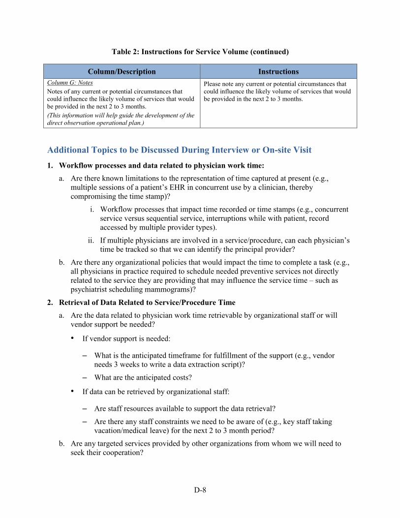
D-8
Table 2: Instructions for Service Volume (continued)
Column/Description Instructions Column G: Notes Notes of any current or potential circumstances that could influence the likely volume of services that would be provided in the next 2 to 3 months. (This information will help guide the development of the direct observation operational plan.)
Please note any current or potential circumstances that could influence the likely volume of services that would be provided in the next 2 to 3 months.
Additional Topics to be Discussed During Interview or On-site Visit 1. Workflow processes and data related to physician work time:
a. Are there known limitations to the representation of time captured at present (e.g., multiple sessions of a patient’s EHR in concurrent use by a clinician, thereby compromising the time stamp)?
i. Workflow processes that impact time recorded or time stamps (e.g., concurrent service versus sequential service, interruptions while with patient, record accessed by multiple provider types).
ii. If multiple physicians are involved in a service/procedure, can each physician’s time be tracked so that we can identify the principal provider?
b. Are there any organizational policies that would impact the time to complete a task (e.g., all physicians in practice required to schedule needed preventive services not directly related to the service they are providing that may influence the service time – such as psychiatrist scheduling mammograms)?
2. Retrieval of Data Related to Service/Procedure Time a. Are the data related to physician work time retrievable by organizational staff or will
vendor support be needed?
• If vendor support is needed:
– What is the anticipated timeframe for fulfillment of the support (e.g., vendor needs 3 weeks to write a data extraction script)?
– What are the anticipated costs?
• If data can be retrieved by organizational staff:
– Are staff resources available to support the data retrieval?
– Are there any staff constraints we need to be aware of (e.g., key staff taking vacation/medical leave) for the next 2 to 3 month period?
b. Are any targeted services provided by other organizations from whom we will need to seek their cooperation?

E-1
APPENDIX E: DIRECT OBSERVATION DATA COLLECTION PROTOCOL

RELATIVE VALUE UNIT (RVU) WORK TIME
DIRECT OBSERVATION DATA COLLECTION PROTOCOL
Part of a Centers for Medicare & Medicaid Services (CMS) Sponsored Study: Medicare Physician Fee Schedule (PFS): Development of a Model for the Valuation of Work Relative Value Units (RVUs)

ii
CONTENTS
1. Introduction and Overview of the Project ...............................................................................1 1.1 Data Collection within Your Health System .................................................................1 1.2 De-identifying Patient Information ................................................................................1
2. Overview of the Direct Observation Data Collection Process................................................2
3. Time Information ....................................................................................................................4 3.1 Time Components ..........................................................................................................4
3.1.1 Pre-Service Time ..................................................................................................4
3.1.2 Intra-Service Time ................................................................................................4
3.1.3 Post-Service Time .................................................................................................4
3.1.4 Elements within Each Time Component ..............................................................4
3.1.5 Recording Time for Radiology/Imaging Technicians ..........................................5 3.2 Interruptions during Observed Time ..............................................................................5 3.3 Physician Observed Time: other physicians and non-physicians .................................5
4. Data Collection .......................................................................................................................5 4.1 Direct Observation Data Collection Access Tool Overview .........................................5 4.2 Instructions for Using the Direct Observation Data Collection Access Tool –
Live Observation of Physician and Radiology/Imaging Technician Time ....................6
4.2.1 Prior to Direct Observation Data Collection activity ............................................6
4.2.2 Recording Direct Observations in the Access Tool ..............................................6 4.3 Direct Observation Data Collection Paper Form .........................................................10
4.3.1 Capturing time with the stop watch ....................................................................10
4.3.2 Printing the data collection form .........................................................................10
4.3.3 Instructions: Completing the Direct Observation Data Collection Paper Form–Physician and Radiology/Imaging Technician Time ...............................11
Appendix A – Study Services: Pre-Service, Intra-Service, and Post-Service Elements ...............12

1
1. INTRODUCTION AND OVERVIEW OF THE PROJECT
Thank you for participating in the Centers for Medicare & Medicaid Services (CMS) project entitled, “Medicare Physician Fee Schedule (PFS): Development of a Model for the Valuation of Work Relative Value Units (RVUs).
1.1 Data Collection within Your Health System
We will be collecting physician and radiology/imaging technician clinical service time data on a set of services performed in your hospital, physician offices, clinics, surgical centers, or other facilities appropriate to the services under study. Clinical service time data will be collected through direct observation, as well as from various electronic (and paper) systems within your organization.1 The volume of targeted services your health system performs will determine which of the services in our study are observed.
This document is the direct observation data collection protocol. A separate protocol for data collection from your electronic (and paper) systems will be delivered to your information technology (IT) department and/or other department(s) as appropriate.
This direct observation data collection protocol will be customized based on feedback from your health system on how to best observe clinicians and technicians performing services included in this study. Direct observation will be done in collaboration with staff at your facility and our study team, and conducted over a mutually convenient, pre-scheduled 5-day period. The following sections describe your roles and responsibilities, as well as specific instructions, methods, and services for which clinical service time data through direct observation will be collected.
1.2 De-identifying Patient Information
No patient identifying information will be sent to RTI. The Study Coordinator (SC) will be responsible for maintaining a log to keep track of all patient unique study ID’s. The SC will be provided a Patient Privacy and Confidentiality Crosswalk (see Figure 1) to use in maintaining patient study IDs. This is for your internal tracking purposes only - RTI will not see this form. The unique study ID assigned to each observed patient will be the only patient identification provided to RTI with the extracts of time data captured in your organization’s electronic data systems, and will be used by the SC to verify the electronic records in the file prepared for RTI. Also, if there are any follow-up questions regarding a particular patient’s data, RTI will reference the study ID when contacting the SC to obtain information.
1 The data from direct observation and your various information systems will be compared. We fully understand
that the majority of information systems were never intended to capture clinical service time as a primary purpose. Consequently, this study is not intended to assess the adequacy of your individual information systems, nor your implementation of them. The objectives for comparing time data from your information systems against direct observation data are to identify if, and how well, current information systems capture clinical service time data.
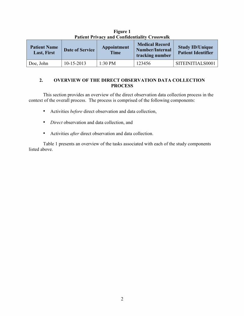
2
Figure 1 Patient Privacy and Confidentiality Crosswalk
Patient Name Last, First Date of Service Appointment
Time
Medical Record Number/Internal tracking number
Study ID/Unique Patient Identifier
Doe, John 10-15-2013 1:30 PM 123456 SITEINITIALS0001
2. OVERVIEW OF THE DIRECT OBSERVATION DATA COLLECTION PROCESS
This section provides an overview of the direct observation data collection process in the context of the overall process. The process is comprised of the following components:
• Activities before direct observation and data collection,
• Direct observation and data collection, and
• Activities after direct observation and data collection.
Table 1 presents an overview of the tasks associated with each of the study components listed above.

3
Table 1 Direct Observation Process
Step # Task Description Responsible Entity
Dir
ect O
bser
vatio
n &
Dat
a C
olle
ctio
n A
ctiv
ities
Steps Related to Direct Observation and Data Collection: Pre-Service Time 1 Review Appendix A (Components of Clinical Service Time) to verify clinical
service elements included in pre-service time for the service to be observed. Note: these elements are listed in the direct observation data collection tool for each service (see Section 4.2.)
2 Observe and time the duration of: pre-service elements for assigned service AND any interruptions or exceptions to pre-service time
3 On the Direct Observation electronic or paper form , document: each activity performed as part of pre-service (e.g., obtaining patient
consent, reviewing patient chart, consulting with other providers, etc.), the type of provider performing the activity (e.g., physician, NP, PA, etc.) the time it took for the activity to be performed a description of any interruptions or exceptions to pre-service time the duration of the interruption(s)
Steps Related to Direct Observation and Data Collection: Intra-Service Time 1 Review Appendix A (Components of Clinical Service Time) to verify clinical
service elements included in intra-service time for the service to be observe. Note: these elements are listed in the direct observation data collection tool for each service(see Section 4.2)
2 Observe and time the duration of: intra-service elements for assigned service AND any interruptions or exceptions to intra-service time
3 Document intra-service time on Direct Observation spreadsheet or form and describe any interruptions or exceptions to intra-service time
Steps Related to Direct Observation and Data Collection: Post-Service Time 1 Review Appendix A (Components of Clinical Service Time) to verify clinical
service elements included in post-service time for the service to be observe. Note: these elements are listed in the direct observation data collection tool for each service (see Section 4.2)
2 Observe and time the duration of: post-service elements for assigned service AND any interruptions or exceptions to post-service time
3 Document post-service time on Direct Observation spreadsheet or form and describe any interruptions or exceptions to post-service time
Act
iviti
es A
fter
D
irec
t Obs
erva
tion
&
Dat
a C
olle
ctio
n
Steps Related to Collected Data 1 Provide Direct Observation electronic and/or paper forms completed by
facility staff to RTI researchers
2 Provide unique study identifiers for patients observed by RTI researchers and facility staff to IT department, and other departments as necessary, to use in compiling and reporting clinical service time data captured in electronic (and paper) systems
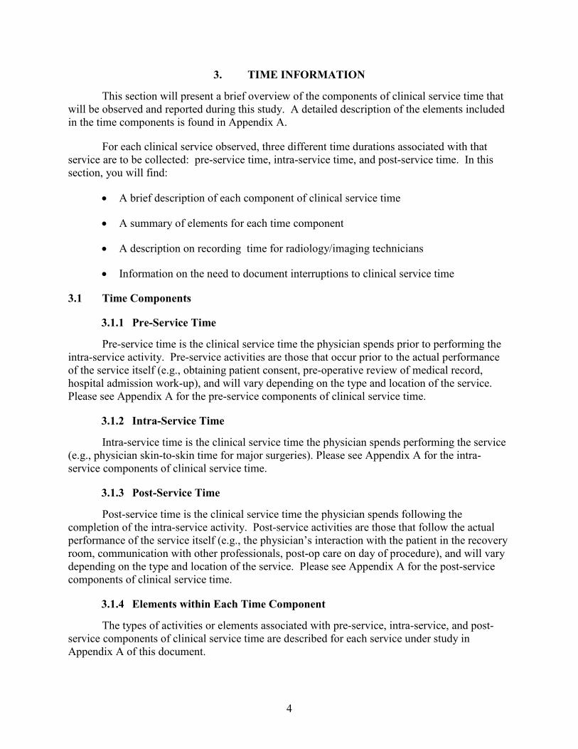
4
3. TIME INFORMATION
This section will present a brief overview of the components of clinical service time that will be observed and reported during this study. A detailed description of the elements included in the time components is found in Appendix A.
For each clinical service observed, three different time durations associated with that service are to be collected: pre-service time, intra-service time, and post-service time. In this section, you will find:
• A brief description of each component of clinical service time
• A summary of elements for each time component
• A description on recording time for radiology/imaging technicians
• Information on the need to document interruptions to clinical service time
3.1 Time Components
3.1.1 Pre-Service Time
Pre-service time is the clinical service time the physician spends prior to performing the intra-service activity. Pre-service activities are those that occur prior to the actual performance of the service itself (e.g., obtaining patient consent, pre-operative review of medical record, hospital admission work-up), and will vary depending on the type and location of the service. Please see Appendix A for the pre-service components of clinical service time.
3.1.2 Intra-Service Time
Intra-service time is the clinical service time the physician spends performing the service (e.g., physician skin-to-skin time for major surgeries). Please see Appendix A for the intra-service components of clinical service time.
3.1.3 Post-Service Time
Post-service time is the clinical service time the physician spends following the completion of the intra-service activity. Post-service activities are those that follow the actual performance of the service itself (e.g., the physician’s interaction with the patient in the recovery room, communication with other professionals, post-op care on day of procedure), and will vary depending on the type and location of the service. Please see Appendix A for the post-service components of clinical service time.
3.1.4 Elements within Each Time Component
The types of activities or elements associated with pre-service, intra-service, and post-service components of clinical service time are described for each service under study in Appendix A of this document.

5
The Access direct observation data collection tool (described in Section 4.1) will present the different activities or elements defined in Appendix A as being associated with pre-service, intra-service and post-service time components for each specific CPT code. Note, however, that not all of the activities/elements listed for a service may be performed during the observed procedure. It is also possible that other, additional activities/elements not listed for a service may be performed during an observed procedure. Finally, note that one or more of the listed activities/elements may be performed by a non-physician provider (technician, physician’s assistant, nurse practitioner, RN, LPN, medical assistant, or other). All of these permutations will be captured in the direct observation process.
3.1.5 Recording Time for Radiology/Imaging Technicians
Time (for radiology/imaging technicians) is the time the technician spends in performing a radiological service (e.g., technician time to perform a head CT).
3.2 Interruptions during Observed Time
There may be instances during the observation of clinical services where a physician’s, technician’s, or other provider’s engagement in service activity is interrupted. To obtain more accurate estimates of actual time that it takes to perform the service, the number and duration of interruptions will need to be deducted from the reported time for the service.
The Access Direct Observation Data Collection tool forms are designed to accommodate the capture of interruptions to clinical service time. Instructions on use of these tools are found in Section 4.2 (Access tool instructions) and Section (4.3) (paper form instructions).
3.3 Physician Observed Time: other physicians and non-physicians
There may be instances during the observation of clinical services where other physicians (such as residents or fellows as part of a teaching program) or other clinical staff (such as physician assistants (PAs)) performs elements of the pre-service, intra-service, and/or post-service components of clinical service time. If a physician such as a resident or fellow performs an element(s) of the service, this activity should be documented in the notes section of the Access tool, but there should be no change to the timing. If a non-physician (such as a PA or nurse practitioner (NP)) performs some of the service, this should be recorded as a second provider in the appropriate screen of the data collection tool.
Instructions on use of these tools are found in Section 4.2 (Access tool instructions) and Section 4.3 (paper form instructions).
4. DATA COLLECTION
4.1 Direct Observation Data Collection Access Tool Overview
The Access tool for Physician and Radiology/Imaging Technician Time captures the start and end times of pre-service, intra-service and post-service time components for services under study. The duration of time components are automatically calculated. In addition, the Access tool provides the ability to capture both interruptions and exceptions to components of clinical service time for physicians and radiology/imaging technicians.

6
The Access Tool has a variety of features designed to capture the start and end times of pre-service, intra-service, and post-service time components for services under study. The duration of time components is automatically calculated using the “Start” and “End” buttons. The Tool has a feature to identify the specific elements associated with pre-, intra-, and post-service time for each procedure (CPT code). In addition, the Tool provides the ability to capture both interruptions and exceptions to components of clinical service time for physicians and non-physician providers including technicians.
When recording technician time, the time is recorded in the same manner as for physicians, with the following exceptions: 1) the element(s) performed by the technician are documented in the Notes section (such as setting up equipment, bringing the patient back to the room, giving instructions to the patient, walking the patient to the imaging room, positioning the patient, taking view(s), checking the image quality, etc.); and 2) the second provider section of the Tool will not be used. The functionality of the Tool, including further detail on recording Technician Time, is described below.
4.2 Instructions for Using the Direct Observation Data Collection Access Tool – Live Observation of Physician and Radiology/Imaging Technician Time
4.2.1 Prior to Direct Observation Data Collection activity
1. Review the instructions on using the stop-watch (4.2.2 Capturing time with the stop watch).
2. Note the following provider types: Physician PA (physician assistant) NP (nurse practitioner) RN LPN Medical Assistant Technician Other
3. Next, familiarize yourself with the Activities listed on the form. These are possible activities the provider will perform during pre-service activity.
4.2.2 Recording Direct Observations in the Access Tool
I. Open the Access direct observation data collection tool. Click on Main Menu: Select New Observation. Next select an Activity to observe: click on a procedure in the box. If performing a live observation, select Live Observation. The Live Observation screen will open.
II. Live Observation Screen. At the top of the page is a dark green banner with text that indicate “Live Observation,” a brief description of the procedure, the CPT code, the date of data entry, and an auto-generated Subject ID.

7
III. The Live Observation Screen is divided into 3 sections: Pre-Service, Intra-Service and Post-Service. Each of the three sections is divided into two sub-sections: for recording the time of the first provider and (if there is one) recording the time of the second provider.
Note: the basic steps to recording time are the same for Pre-Service, Intra-Service or Post-Service Time.
When recording time for a radiology/imaging technician performing the technical component of a radiological procedure, time is recorded in the same manner as for physicians, with the following exceptions: 1) the element(s) for any pre-, intra-, and post-service time performed by the technician are noted in the Notes section (such as setting up equipment, bringing the patient back to the room, giving instructions to the patient, walking the patient to the imaging room, positioning the patient, taking view(s), checking the image quality after taking views, etc.); and 2) the second provider section in the data collection Tool will not be used.
If the unique study patient identifier has not been pre-populated for you, please enter the unique study patient identifier into the Live Observation screen of the data collection tool, or, if recording time on paper, enter the unique study patient identifier onto the paper form.
IV. Recording Pre-Service Time: Before beginning timing, select the provider type of the person performing the activity. 1. Put the cursor in the drop-down menu just below the header “Pre-
service – first provider.
2. Select the provider type from the drop-down menu (physician, technician, PA, RN, LPN, etc.): prior to the start of pre-service.
3. At the start of the pre-service activity, click on the “Start” button. The time will display in the format of hh:mm:ss AM or PM
a. The duration of pre-service will display as 0 in the third box until a pre-service end time is entered. A red highlighted message “Timing!” will appear in the third column until the End time is recorded.
b. While timing the procedure, select the specific element/task(s) that are performed which are listed in the box on the left hand side of the screen just below the provider type drop-down box. This is done by putting the cursor on the element (numbered 1-Other) in the box. The element will be highlighted. If selecting an “Other” activity, please provide a definition of this activity in the Notes section. To unselect an element, just click again on the activity.

8
V. Recording Interruptions Please document any interruptions to physician or other provider Pre-service time.
Note: one interruption can be captured for the first and second provider for each of the service components of clinical service time: Pre-Service, Intra-Service and Post-Service.
1. See the row marked as “Interruption.” Note if there is an interruption to the Pre -Service activity, it must be recorded prior to clicking on the “End” of the Pre-Service time, or prior to completing Step III, below.
2. Begin to time the interruption:
a. At the start of the pre-service interruption, click on the “Start” button. Time will display in the format of hh:mm:ss AM or PM.
b. The duration of the pre-service interruption will display as 0 in the third box until an end time for the interruption is entered. A red highlighted message “Timing!” will appear in the third column until the End time is recorded.
c. While timing the interruption, note the reason(s) for the interruption in the Notes section at the bottom of the sub-section.
d. At the end of the interruption, click on the “End” button in the same row. The time will display in the format of hh:mm:ss AM/PM. To the right of the “End” time display box, the total time for the interruption in minutes, now labeled “Complete” will display. The time will display in the format of “mm” for minutes.
e. Continue timing the remainder of the Pre-Service activity. When that is complete, record the End time as instructed in Step III, below.
3. Please document a brief description of the Interruption in the Notes section.
VI. Recording Pre-Service End Time: At the end of the pre-service activity, click the "End” command button
1. The end time will display in the format of hh:mm:ss AM/PM.
2. To the right of the “End” time display box, the total times in minutes, now labeled “Complete” will display.
3. The time will display in the format of “mm” for minutes.
VII. Recording Pre-Service: Second Provider 1. If a second provider performs Pre-Service activities in addition to the first
provider, record their time in the section on the right-hand side of the screen titled “Pre-Service – second provider”

9
2. Follow Steps IV.1-.3 and Step VI.1-.3, above
3. If there is an interruption to the Pre-Service Second Provider, please follow steps V.1-.3 above, Recording Interruptions.
Note: first and second providers may perform tasks simultaneously; if so, the observer needs to record their time simultaneously.
VIII. Recording Intra-Service Time: Please see the section of the screen titled “Intra-Service”
1. Follow instructions in Step IV.1-.3 and Step VI.1-.3 above
2. If there is an interruption to the Intra-Service first provider, please follow steps VI.1-.3 above, Recording Interruptions.
3. If a second provider performs Intra-Service activities in addition to the first provider, record their time in the section on the right-hand side of the screen titled “Intra-Service – second provider,“ following Step IV.1-.3 and Step VI.1-.3 above.
IX. Recording Post-Service Time: Please see the section of the screen titled “Post-Service”
1. Follow instructions in Steps I.1-I.3 – Step III.1 – III.3 above
2. If there is an interruption to the Post-Service first provider, please follow steps II.1 – II.3 above, Recording Interruptions.
3. If a second provider performs the Post-Service activities in addition to the first provider, record their time in the section on the right-hand side of the screen titled “Post-Service – second provider,“ following Step IV.1-.3 and Step VI.1-.3 above.
X. Recording Radiology/Imaging Technician Time Note: when recording time for a radiology/imaging technician to perform the technical component of a radiological procedure, the element(s) for any pre-, intra-, and post-service time performed by the technician are noted in the Notes section (such as setting up equipment, bringing the patient back to the room, giving instructions to the patient, walking the patient to the imaging room, positioning the patient, taking view(s), checking the image quality after taking views, etc.). The second provider section in the data collection Tool will not be used when recording time for a radiology/imaging technician.
1. Go to the “Intra-Service” section of the screen.
2. Prior to the start of the activity(ies) by the Technician, see the provider type drop-down menu just below the header “Intra-service – first provider.”
3. Select “Technician” from the menu.
4. At the start of the activity by the technician, click on “Start” button. The time will display in the format of hh:mm:ss AM/PM.

10
5. The duration of pre-service will display as 0 in the third box until a pre-service end time is entered. A red highlighted message “Timing!” will appear in the third column until the End time is recorded.
6. At the end of the activity by the technician, click the "End” command button
a. The end time will display in the format of hh:mm:ss AM/PM.
b. To the right of the “End” time display box, the total times in minutes, now labelled “Complete” will display.
c. The time will display in the format of “mm” for minutes.
Please type any notes or comments and/or document any exceptions in the Notes section.
4.3 Direct Observation Data Collection Paper Form
If facility staff assigned to perform direct observation and data collection do not have access to the Access Direct Observation Data Collection tool, or devices to run Access, data capture can be performed by a manual process that uses stopwatches (see Figures 5 and 6). This section explains how time is captured with a stopwatch, how to print out the form for recording data, and then how to fill out the form appropriately.
4.3.1 Capturing time with the stop watch
SPLIT/ RESET
MODE
START/ STOP
1. At start of service (pre, intra or post), Hit START button
2. At first interruption:
a) Hit SPLIT button at start of interruption
b) Hit SPLIT button at end of interruption
c) Quickly record time displayed on watch
3. Time will be recording on the watch as the physician/technician/other provider continues the service.
4. Time will be recording on the watch as the physician/technician/other provider continues the service.
5. Time will record on the watch as the physician/technician/other provider continues the service.
6. At conclusion of service, hit STOP button to conclude the recording of time.
7. Record the final time on the stopwatch.
4.3.2 Printing the data collection form
<<Insert Print Instructions Once This Functionality Is Available>>

11
4.3.3 Instructions: Completing the Direct Observation Data Collection Paper Form–Physician and Radiology/Imaging Technician Time
1. If service tracking information has not been pre-filled for you, please print the following information on the form:
a) Your name b) The pre-assigned unique study ID for the patient c) The date of the service. d) The appointment time.
2. Please record the start and end times, as captured by your stopwatch(es) (see stopwatch instructions at 4.3.1), for each observed component of time:
a) Pre-service b) Intra-service c) Post-service
Note: if recording time for a radiology/imaging technician to perform the technical component of a radiological procedure, the times on whatever component(s) the technician performs: pre-, intra-, and post-service time, should be collected.
3. Please record the provider type (physician, technician, PA, nurse practitioner, RN, LPN, medical assistant, or Other)
4. Please record the start and end times for any interruptions to physician or other provider pre-service, intra-service, or post-service time and provide a brief description of the interruption.
5. There may be instances during the observation of clinical services where other physicians (such as residents or fellows as part of a teaching program) or other clinical staff (such as PAs) perform elements of the pre-service, intra-service, and/or post-service components of clinical service time. If a physician such as a resident or fellow performs an element(s) of the service, this activity should be noted in the notes section, but there should be no change to the timing. If a non-physician (such as PAs or NPs) performs some of the service, this should be recorded as a second provider in the appropriate screen of the data collection tool.
6. Please record any element(s) performed by the radiology/imaging technician in the Notes section.
7. Please use the Notes section to capture any issues or questions encountered during the observation of pre-service, intra-service, or post-service activities.

12
Appendix A
STUDY SERVICES: PRE-SERVICE, INTRA-SERVICE, AND POST-SERVICE ELEMENTS
[The table includes the pre-, intra-, and post- service element descriptions from the
RUC database. For reference, see the Main Report Appendix C.]

F-1
APPENDIX F: PROTOCOL FOR SUPPORTING DATA

RELATIVE VALUE UNIT (RVU) WORK TIME:
HIT PROTOCOL FOR SUPPORTING AND HISTORIC TIME DATA
Part of a Centers for Medicare & Medicaid Services (CMS) Sponsored Study:
Medicare Physician Fee Schedule (PFS): Development of a Model for the Valuation of Work Relative Value
Units (RVUs)
March 4, 2013

ii
CONTENTS
1. Introduction and Overview of the Project ...............................................................................1 1.1 Data Collection within Your Health System .................................................................1
2. Roles and Responsibilities ......................................................................................................2 2.1 Site Coordinator .............................................................................................................2 2.2 RTI Staff ........................................................................................................................2
3. Overview of the Process for Collecting Supporting and Historic Time Data .........................3
4. De-identifying patient information .........................................................................................4
5. Time Information ....................................................................................................................5 5.1 Time Components ..........................................................................................................5
5.1.1 Pre-Service Time ..................................................................................................5
5.1.2 Intra-Service Time ................................................................................................5
5.1.3 Post-Service Time .................................................................................................5
6. Data Collection .......................................................................................................................5 6.1 Data Mapping.................................................................................................................5 6.2 Electronic Data...............................................................................................................6 6.3 Data from Paper-based Systems ....................................................................................7
Appendix A – Components of Clinical Service Time .....................................................................8

1
1. INTRODUCTION AND OVERVIEW OF THE PROJECT
Thank you for participating in the Centers for Medicare & Medicaid Services (CMS) project entitled, “Medicare Physician Fee Schedule (PFS): Development of a Model for the Valuation of Work Relative Value Units (RVUs).
1.1 Data Collection within Your Health System
We will be collecting physician and radiology/imaging technician clinical service time data on a set of services performed in your hospital, physician offices, clinics, surgical centers, or other facilities appropriate to the service under study. Clinical service time data will be collected through direct observation, as well as from various electronic (and paper) systems within your organization.2 The volume of targeted services your health system performs, as well as your procedures and systems for capturing clinical service time, will determine which of the services in our study will be
• Observation Only – Time data will be collected by direct observation only
• Observation & Supporting Data – Time data will be collected both through observation and supporting time data from electronic (and paper) systems will be submitted to the research team, and
• Historical Data - Time data on previously performed services, captured in electronic (and paper) systems, will be submitted to the research team.
This document is the HIT Protocol for Corroborative and Historic Time Data which addresses data collection from your electronic (and paper) systems. A separate Protocol for Direct Observation Data Collection will be delivered to your clinical site coordinator.
This Protocol will be customized based on feedback from your health system regarding the electronic (and paper) systems that contain information that support the clinical service time for services under study. The following sections describe your roles and responsibilities, as well as specific instructions, methods, and services for which corroborative clinical service and historic time data will be collected.
2 The data from direct observation and your various information systems will be compared. We fully understand
that the majority of information systems were never intended to capture clinical service time as a primary purpose. Consequently, this study is not intended to assess the adequacy of your individual information systems, nor your implementation of them. The objectives for comparing time data from your information systems against direct observation data are to identify if, and how well, current information systems capture clinical service time data.

2
2. ROLES AND RESPONSIBILITIES
2.1 Site Coordinator
One staff person at your facility will be identified as the site coordinator (SC). The SC:
• Serves as the primary contact person
• Assists in obtaining your facility’s IRB approval, if necessary
• Assists in securing a Limited Data Set Agreement and/or a Business Associate Agreement, if necessary
• Coordinates dates and times for RTI staff to conduct on-site visit
• Works with RTI researchers to identify services to be observed by the RTI team and/or facility staff
• Maintains the Patient Privacy and Confidentiality Crosswalk (see Section 4), which enables your facility to accurately identify patients involved in this study, while not disclosing personally identifiable health information to RTI
• Provides unique identifiers of the patients—for which clinical service time data has been collected through direct observation by RTI’s or your facility’s staff—to your health system’s IT department, and other departments as necessary, so that RTI can be provided with corroborative time data found in electronic (and paper) systems on those patients
• Coordinates with your health system’s IT department, and other departments as necessary, to provide RTI with historical clinical service time data – for services and a time period as negotiated with your organization – found in electronic (and paper) systems
• Coordinates actions between your health system’s IT department, other departments as necessary, and RTI to ensure that RTI has received accurate clinical service time data from your electronic (and paper) systems
• Ensures the destruction of the Patient Privacy and Confidentiality Crosswalk upon completion of the study
2.2 RTI Staff
RTI staff will work with the SC to coordinate study activities. The following staff will be available to answer any questions you may have regarding the electronic (and paper) system data component of the study.
Sue Mitchell [email protected] 919-597-5190
Don Mon [email protected] 312-777-5228

3
3. OVERVIEW OF THE PROCESS FOR COLLECTING SUPPORTING AND HISTORIC TIME DATA
This section provides an overview of the process for collecting corroborative and historic time data in the context of the overall process. The process is comprised of the following components:
• Activities before collecting corroborative and historic time data,
• Collection of corroborative and historic time data
• Activities after collection of corroborative and historic time data.
Table 1 presents an overview of the tasks associated with each of the study components listed above.
Table 1 Direct Observation Process
Step # Task Description Responsible
Entity
Steps Related to Identifying Services/Patients for Study
1 Work with RTI researchers to identify targeted services with sufficient volume for study
Act
iviti
es P
rior
to C
olle
ctin
g C
orro
bora
tive
and
Hist
oric
al T
ime
Dat
a 2 Work with RTI researchers to determine volumes of services to be studied: By observation only By observation and supporting data By historical data
3 Work with RTI researchers to identify: electronic (and paper) systems that capture time data for services under
study type(s) of time data available (i.e., total time and/or components of time
such as pre-service/intra-service/post-service) (see Appendix A for definitions of components of time)
resources needed to retrieve time data from electronic (and paper) systems (e.g., electronic data retrievable by staff, electronic data requires vendor support to retrieve)
(continued)

4
Table 1 (continued) Direct Observation Process
Step # Task Description Responsible
Entity
Col
lect
ing
Cor
robo
rativ
e an
d H
isto
rica
l Tim
e D
ata
Steps Related to Collecting Supporting Time Data –Direct Observation Patients
1 Receive unique study identifiers for patients observed by RTI researchers and facility staff
2 Compile clinical service time data captured from electronic (and/or paper) systems
3 Report clinical service time data, in file format as agreed upon with RTI researchers, using unique study identifiers (RTI should NOT receive personally identifiable health information)
Steps Related to Collecting Historical Time Data – Previously Performed Services
1 Assign unique study identifiers to patients for whom time data on previously performed services will be captured from electronic (and/or paper) systems
2 Compile clinical service time data captured from electronic (and/or paper) systems
3 Report clinical service time data, in file format as agreed upon with RTI researchers, using unique study identifiers (RTI should NOT receive personally identifiable health information)
Act
iviti
es A
fter
D
ata
Com
pile
d Steps Related to Collected Data
1 Work with RTI to ensure that accurate clinical service time data from electronic (and paper) systems has been received
2 Destroy the Patient Privacy and Confidentiality Crosswalk upon completion of the study
4. DE-IDENTIFYING PATIENT INFORMATION
No patient identifying information will be sent to RTI. The SC will be responsible for maintaining a log to keep track of all patient unique study ID’s. The SC will be provided a Patient Privacy and Confidentiality Crosswalk (see Figure 1) to use in maintaining patient study IDs. This is for your internal tracking purposes only - RTI will not see this form. If there are any follow up questions regarding a particular patient’s data, RTI will reference the study ID when contacting the SC to obtain information.
Figure 1 Patient Privacy and Confidentiality Crosswalk
Patient Name Last, First Date of Service Appointment
Time
Medical Record Number/Internal tracking number
Study ID/Unique Patient Identifier
Doe, John 10-15-2013 1:30 PM 123456 SITEINITIALS0001

5
5. TIME INFORMATION
This section will present a brief overview of the components of clinical service time that will be reported during this study. A list of the elements of pre-, intra- and post-time components is found in Appendix A.
For each clinical service, three different time durations associated with that service are to be collected: Pre-service time, Intra-service time, and Post-service time. In this section you will find:
• A brief description of each component of clinical service time
• A summary of elements for each time component
• A brief description of Total Time for radiology/imaging technicians
5.1 Time Components
5.1.1 Pre-Service Time
Pre-service time is the clinical service time the physician spends prior to performing the intra-service activity. Pre-service activities are those that occur prior to the actual performance of the service itself (e.g., obtaining patient consent, pre-operative review of medical record, hospital admission work-up), and will vary depending on the type and location of the service. Please see Appendix A for the pre-service components of clinical service time.
5.1.2 Intra-Service Time
Intra-service time is the clinical service time the physician spends performing the service (e.g., physician skin-to-skin time for major surgeries). Please see Appendix A for the intra-service components of clinical service time.
5.1.3 Post-Service Time
Post-service time is the clinical service time the physician spends following the completion of the intra-service activity. Post-service activities are those that follow the actual performance of the service itself (e.g., the physician’s interaction with the patient in the recovery room, communication with other professionals, post-op care on day of procedure), and will vary depending on the type and location of the service. Please see Appendix A for the post-service components of clinical service time.
6. DATA COLLECTION
6.1 Data Mapping
Prior to commencing direct observation and data collection activities, RTI staff will work with appropriate facility personnel (clinical and IT) to carefully document what IT (or paper) systems and data elements are used to record the performance of each procedure/service targeted for study. Information sources for this process will include screen shots, data dictionaries,

6
procedures, training materials, staff interviews and/or other materials. This analytic activity will produce a data mapping that inventories the systems and data elements that capture components of time for targeted procedures/services. This data mapping will describe data:
• usage (e.g., recording activities during diagnostic cardiac procedures, time stamp showing record access)
• pairings (e.g., event activity description and time stamp), and
• idiosyncrasies (e.g., radiologist time stamps for access and closure of a record may reflect a file that sat open all day and not reflect actual “work time”)
Particular emphasis will be placed on carefully identifying and describing the data elements that will be used as anchors for pre, inter, and post service time (see Figure 2).
Figure 2 Sample Data Map
CPT Code & Description
Service Time Component
System Specify software app or manual process used to document
performance of the procedure/ service
Usage Data Element
Pairing Start of Service Anchor
End of Service Anchor
Other Comment
49505 – Prp i/hern init reduc >5 yr
Pre-service XYZ Application Procedure event log
Event w/time stamp
N/A N/A 1) Times re pre-Op are not completed consistently
2) Physician pre-Op activities are not recorded
49505 – Prp i/hern init reduc >5 yr
Intra-service
XYZ Application Procedure event log
Event w/time stamp
Value = Procedure Start/ Incision
Value = Procedure Finish/ Close
—
49505 – Prp i/hern init reduc >5 yr
Post-service
XYZ Application Procedure event log
Event w/time stamp
N/A N/A 1) Times re post-Op are not completed consistently
2) Physician post-OP activities are not recorded
93458 – Cardiac Cath
Pre-service XYZ Vascular Lab Application
Procedure event log
Event w/time stamp
N/A N/A 1) Some pre-service activities are listed (e.g., informed consent signed by patient, but service provider is not identified and duration cannot be computed
93458 – Cardiac Cath
Intra-service
XYZ Vascular Lab Application
Procedure event log
Event w/time stamp
Value = “Access xxx” (e.g., Access 5 fr sheath into Rt radial artery. Single wall stick - uncomplicated
Value = “xxx catheter removed” (e.g., 5Fr JR 4.0 catheter removed)
—

7
6.2 Electronic Data
For each patient identified for the study groups “Observation and Supporting Data” and “Historical Data”, we are requesting the following data from your organization.
• Unique facility study identifier
• Unique patient study identifier
• Date of service
• Time of service
• CPT code for service performed
• Provider type for reported time data (i.e., physician time or radiology/imaging technician time), if available
• Data elements, at the most granular level, that capture and support the components of clinical service time (pre, inter and post-service) as identified in the data mapping document. These data elements have a many to one relationship with the data elements above.
6.2 Data from Paper-based Systems
Facility specific processes will be developed with the organization for instances where information supporting clinical service time is captured in paper-based systems.

8
Appendix A
STUDY SERVICES: RUC PRE-SERVICE, INTRA-SERVICE, AND POST-SERVICE ELEMENTS
[The table includes the pre-, intra-, and post- service element descriptions from the
RUC database. For reference, see the Main Report Appendix C.]