Embed Size (px)
Citation preview

Loyola University Chicago Loyola University Chicago
Loyola eCommons Loyola eCommons
Master's Theses Theses and Dissertations
1989
Dentofacial Development in Children with Chronic Nasal Dentofacial Development in Children with Chronic Nasal
Respiratory Obstruction -- a Cephalometric Study Respiratory Obstruction -- a Cephalometric Study
Tai-Yang Hsi Loyola University Chicago
Follow this and additional works at: https://ecommons.luc.edu/luc_theses
Part of the Dentistry Commons
Recommended Citation Recommended Citation Hsi, Tai-Yang, "Dentofacial Development in Children with Chronic Nasal Respiratory Obstruction -- a Cephalometric Study" (1989). Master's Theses. 3577. https://ecommons.luc.edu/luc_theses/3577
This Thesis is brought to you for free and open access by the Theses and Dissertations at Loyola eCommons. It has been accepted for inclusion in Master's Theses by an authorized administrator of Loyola eCommons. For more information, please contact [email protected].
This work is licensed under a Creative Commons Attribution-Noncommercial-No Derivative Works 3.0 License. Copyright © 1989 Tai-Yang Hsi

DENTOFACIAL DEVELOPMENT IN CHILDREN
WITH CHRONIC NASAL RESPIRATORY OBSTRUCTION
-- A CEPHALOMETRIC STUDY
by
TAI-YANG HSI B.D.S.
A Thesis Submitted to the Faculty of the Graduate School
of Loyola University of Chicago in Partial Fulfillment
of the Requirements for the Degree of
Master of Science
December
1989

ACKNOWLEDGEMENTS
I would like to express my sincere gratitude and appreciation to the
following people:
To Dr. Lewis klapper, Chairman of Orthodontics, thesis director, for his
support, guidance and instruction through this investigation.
To Dr. Richard Port, assistance professor of Orthodontic department, for
passing his original study to me and his instruction and assistance.
To Dr. Michael Kiely, Professor of department of Anatomy, for his
instruction and assistance.
To Delia Vazquez, clinic coordinator of Orthodontic department, for her
assistance to take all the head x-ray film of all the patients in this study.
Special thanks would like to be given
To Dr. Richard Bulger, the Ear, Nose.and Throat specialist, for his
continuous support all the patients as experimental group make this study
possible.
To Bolton Growth Study Center of Case Western Reserve University, for
supplying their head x-ray film as control group in this study.
ii

VITA
Tai Yang Hsi was born in Taipei, Taiwan, Republic of China, on November
11, 1950.
After graduation from high school, he enter Taipei Medical College, School
of Dentistry and received his degree of Bachelor of Dental Science in June 1975.
Upon graduating from Dental school, he served in the Army as reserved
Dental Officer for two years.
After discharging from the Army, he went to LuKu Health Center, the so
called "No formal Health Provider - Village, as dentist for one year and then back
to his hometown Taipei to open his own dental office.
During those years of practicing as general practitioner, he has been the
Broad of Director of Taipei Dental Association for four years, also been the
Delegate of National Dental Association for two years devoting himself to dental
public health and education.
In September 1985, he entered the School of Public Health of Columbia
University in New York city and major in dental public health for one year.
In July 1989, he went to Loyola University, School of Dentistry as a post
graduate student in the Orthodontic Department and also enrolled in the
graduate program of Oral Biology.
He received a certificate of specialty in Orthodontics . in December 1988.
iii

DEDICATION
To my wife, my mother-in-law, and my brother
whose supports to my family and the off ice
plus encouragement
made my study overseas possible.
iv

TABLE OF CONTENTS
PAGE
ACKNOWLEDGEMENTS ................................................................................ ii
VITA ................................................................................................................. iii
DEDICATION .................................................................................................. iV
TABLE OF CONTENTS .................................................................................. V
UST OF ILLUSTRATIONS .............................................................................. VI
CHAPTER
I. INTRODUCTION .......................................................................... 1
II. REVIEW OF UTERATURE ........................................................... 4
(A}. ANATOMY, PHYSIOLOGY AND GROwrH .............................. 4
(B}. ETIOLOGY, SYMPTOMS OF AIRWAY OBSTRUCTION .......... 14
(C}. AIRWAY OBSTRUCTION RELATED TO MOUTH BREATHING .. 16
(D}. RESPONSE CHAIN: TONGUE,NEUROMUSCUL.AR FUNCTION,.
AND MANDIBLE POSTURE .......................... 20
(E}. CEPHALOMETRIC STUDIES ................................................ 29
(F). TREATMENT ..................•.........•..........................•................. 41
Ill. MATERIALS AND METHODS ..................................................... .46
IV. RES UL TS ............................................................................ "' ......... 66
V. DISCUSSION ............................................................................. 79
VI. SUMMARY .................................................................................. 84
BIBLOGRAPH ................................................................................................ 86
v

LIST OF ILLUSTRATIONS
R~r~ P~e
1 Anatomy of upper airway ........................................................................... 4
2 Anatomy of adenoid of adenoid tissue ..................................................... 5
3 Growth of nasopharynx in height and depth ........................................... 7
4 Growth of nasopharynx area .................................................................... 8
5 Scammon•s curve of growth of the lymphoid tissue ............................... 9
6 Curve of growth of adenoid tissue .......................................................... 1 O
7 Growth of Adenoid tissue, from infancy to adol~cence ........................ 11
a Growth cycle of adenoid tissue ............................................................... 11
9 Development of structure contigous to adenoid tissue .......................... 12
1 o Development of excessive adenoid tissue .............................................. 12
11 Adenoid face ............................................................................................ 15
12 Adenoid face ............................................................................................ 15
13 Clinic test for nasal airway obstruction ................................................... 17
14 Antroadenoid and superioinferior diameter on lateral radiograph .......... 20
15 l\Jrway dimension measurement by Undr-Aronson/ Henrikson ............. 33
16 Diagram of airway dimension measurement by Linde-Aronson /
Henrikson .................................................................................................. 33
17 Diagram of airway dimension measurement by Under-Aronson /
Henrikson .................................................................................................. 34
18 Diagram of airway dimension measurement by Under-Aronson /
Henrikson .................................................................................................. 34
19 l\Jrway dimension measured by Cohn and Konak52 ............................... 35
vi

20 Grapgic synopsis of six methods for measuring the size of adenoid .... 36
21 Ajrway measurement by Hibbert and whitehouse34 •••••••••••••••••••••••••••••••• 37
22 Adenoid measurement by Fujioka, Young, and Girdany54 •••••••••••••••••••••• 39
23 Nasopharynx measurement by Fujioka, Young, and Girdany54 ••••••.••••• .40
24 Adenoid/ Nasopharynx ratio by Fujioka, Young and Girdany54 •••••.•••• .40
25 History form used in the Ear, Nose and Throat clinic ........................... .47
26 Examination form used in the Ear, Nose and Throat clinic .................. .48
27 Case form used in the Ear, Nose and Thraot clinic .............................. .49
28 Sample of experimental group ................................................................. 51
29 Sample of control group .......................................................................... 51
30 Standard cephalometric machine: X-Ray beam source .......................... 52
31 Standard cephalometric machine: headstate .......................................... 52
32 Example of tracing of the head X-Ray plate ........................................... 53
33 Definition of anatomical landmarks for Ricketts facial pattern analysis .. 55
34 Definition of cephalometric planes for Ricketts facial pattern analysis .. 56
35 Definition of cephalometric measurement for
Ricketts facial pattern analysis ................................................................. 56
36 Definition, clinic norm, and clinic deviation of facial axis and
facial depth used in Ricketts facial pattern analysis ............................... 57
37 Definition, clinic norm, and clinic deviation of mandibular plane angle .. 57
38 Definition, clinic norm, and clinic deviation of mandibular arc ............... 58
39 Definition, clinic norm, and clinic deviation of lower facial height.. ......... 58
40 Tracing of the five measurements for facial pattern analysis in
Ricketts facial pattern analysis ................................................................. 58
vii

41 Determination of Ricketts facial pattern analysis ..................................... 59
42 Example of determination of Ricketts facial pattern analysis .................. 60
43 Example of calculation of Ricketts facial pattern analysis ...................... 61
44 Growth chart, female, from National Center for Health Statistics .......... 62
45 Growth chart, male, from Natonal Center for Health Statistics ............. 63
46 Life size growth chart from National Center for Health Statistics ........... 64
47 Statistical calculation of experimental group ........................................... 67
48 Statistical calculation of control group ..................................................... 68
49 Table of Mann-Whitney U. test.. .............................................................. 70
50 Statistical analysis of facial pattern .......................................................... 71
51 Statistical analysis of facial axis ............................................................... 72
52 Statistical analysis of facial depth ............................................................ 73
53 Statistical analysis of mandibular plane angle ......................................... 7 4
54 Statistical analysis of mandibular arc ...................................................... 75
55 Statistical analysis of lower facial height ................................................. 76
viii

Chapter I
INTRODUCTION
since the first study1 showed a relationship between
nasal airway obstruction and the development of facial growth
patterns, many attempts have been made to establish a
relationship between nasal airway obstruction and dentof acial
abnormalities. The relation between nasorespiratory function
and craniof acial morphology has a long and contentious history
in orthodontics. It was based on the premise that restricted
nasal airway function leads to "mouth breathing," which in
turn results in a lowered tongue position and depressed
mandibular posture2 • If this altered posture was sufficiently
prolonged during active growth, the result may be a narrowed
maxillary dental arch, an increased lower facial height, an
increased mandibular angle and an incompetent lip morphology.
These features were often called long face syndrome or
"adenoid face". Ricketts3 described this condition as
"Respiratory obstruction syndrome"
There has been some disagreement between groups : who
exclusively support the functional matrix theory, that is,
that function dictates form, and others who believe that
facial structure was governed strictly by heredity.
The differing views on the relation between mouth
breathing and a specific type of facial structure and
l
2
malocclusion fell into the following main groups:
1. Mouth breathing gave rise to a specific type of facial
structure and malocclusion.
2. No relation exists between these phenomena.
3. Mouth breathing was a secondary phenomena to a
specific hereditary pattern of facial structure.
Ranly4 proposed a composite view. She stated that the
chondrocranium was influenced by both intrinsic genetic and
local environmental factors. These theories were relevant to
the
controversy regarding the effects of altered respiration on
facial structures.
Though there were some controversial points of view5 678
9 a number of studies confirmed a relationship between
nasopharyngeal airway obstruction and abnormal craniof acial
development. 10 11 12 13 14 15 16 11 11
Several articles suggested a direct cause-and-effect
relationship between nasal airway obstruction and altered
dentofacial morphology. Further well-controlled studies
designed to quantify the relative amounts of oral versus nasal
respiration were necessary before airway obstruction could
be implicated as a significant etiologic factor in the
development of any specific dentofacial deformity.
Within the field of orthodontics it has recently become
apparent that nasal respiratory function played a significant
role in the development of the face and occlusion. For this

3
reason, it was important to be able to determine whether or
not there exists a reduced capacity for nasal breathing.
The purpose of this study was to identify the effect of
chronic nasopharyngeal obstruction on the growth of facial
pattern in children between ages three and seven years old.

Chapter II
REVIEW OF LITERATURE
(A}. ANATOMY, GROWTH, AND PHYSIOLOGY
ANATOMY
The nasopharynx was a musculomembranous tube serving as
a portal between the nasal chamber anteriorly and the oral
pharynx inferiorly. The roof and posterior wall made a
continuous curve downward upon the body of the sphenoid bone,
th• baailar part of the occipital bone, the arch of atlas and
the body of axis. Its primary biologic function was to provide
a pasaageway for air from the nasal chamber to the oral
pharynx and ultimately to the lunga.(fig.l}
acrimal duct g into inferior meatus
I Glossopharyngeal nerve
Stylohyoid ligament '
Thyro-hyoid mcmhrum:
Tensor pa la ti and ptcrygoid hamul us
. uirut lube
al pin
up. c
"tyio-p
Middle
~------ Epiglot
Inf.
Fig. 1 Anatomy of upper airway 4
5
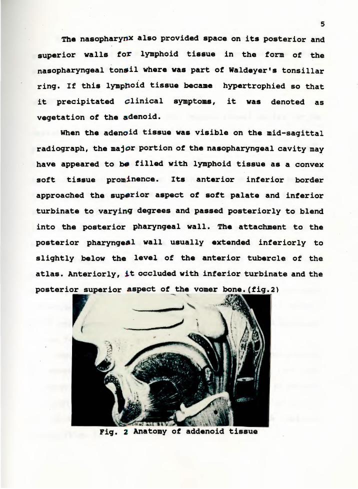
Th• nasopharynx also provided apace on its posterior and
superior walls tor lymphoid tissue in the form of the
nasopharyngeal ton•il where was part of Waldeyer•s tonsillar
ring. If this lymphoid tissue became hypertrophied so that
it precipitated clinical symptoas, it waa denoted as
vegetation of the adenoid.
When the adenoid tissue waa visible on the mid-sagittal
radiograph, the major portion of the nasopharyngeal cavity may
have appeared to ~ tilled with lymphoid tissue aa a convex
soft tissue promi nence. Its anterior inferior border
approached the sup•rior aspect of soft palate and inferior
turbinate to varyinq degrees and passed posteriorly to blend
into the posterior pharyngeal wall. The attachment to the
posterior pharynge•l wall usually extended inferiorly to
slightly below the level of the anterior tubercle of the
atlas. Anteriorly, i t occluded with inferior turbinate and the
poster ior superior a spect of the vomer bone.(fig.2)
r. ------~ ~i
-
Fig. 2 Anatomy of addenoid tissue

6
The enlargement of the adenoid pad may have led to
partial or total blockage of the nasopharyngeal passage making
nasal respiration either inefficient or impossible.
GROWTH
The shape and size of the nasopharyngeal cavity can be
defined in terms of depth and height in the median sagittal
plane and width in the frontal plane. According to Brodie19,
King7, Handelman & Osbornes' study~, the total depth of the
nasopharynx was established in the first or second year of
life. King further stated that the increase of the depth of
the nasopharynx was by the growth at the spheno-occipital
junction. Ricketts21 and Bergland22 demonstrated that the more
obtuse the cranial base, the greater the depth.
In contrast to the early stabilization of depth, King7
demonstrated continued increase in nasopharyngeal height until
maturity by the descent of the hard palate and cervical
vertebrae from the cranium. Bergland22 demonstrated a thirty
eight percent increase in nasopharyngeal height from six years
of age to maturity.
Subtelny23 demonstrated the width of the nasopharynx may
be established early in life. The volume of the bony
nasopharynx increased from six years to maturity by 80 percent
in Bergland's~ skull material. This increase was primarily
due to changes in height and width, while depth remained
stable. (Fig. 3)

"s 60ll 'i.~
a ~ 400 .. !i JOO
200
100
100
•oo
"'"
Ou'htfl\H)fl~ of l~ t-.a~o.pOQt)'nA fn o L.unQilud1no1 M•,Jlr So.mplt ( N • 6 J
•···•···• ......... •· .............. :_:_:_;._:.;.;...: ........... --------· -·- - ..,,,.,,,u"P Oa111111 •
I 2 l 4 ~ i 7 8 9 iO II 12 tl '4 0 6 11 • Att IA U•11
01mtn110n• of 1M l\l:OiOJ.oA011na. 1n o L.on\j1 f\11J11u;u F tmolr Som pie (hi• 6 )
I 4l ) 4 ~ il I • ) j(j l I I~ •l •• ·~ •• 17 '8
.... ·'• f•<lf\
Fig. 3 Growth of Nasopharynx in height and depth
Subtely23
7
In Handelman and Osborne's study20 of the growth of the
nasopharynx and adenoid development using lateral head films
in patients from one to eighteen years of age. Four skeletally
defined lines are used to measure the airway area and adenoid
area. The nasopharyngeal area was defined as a
trapezoid.(Fig.4) The nasopharyngeal area was divided into an
adenoid-pharyngeal wall and an airway areas which were
measured using a polar planimeter. The trapezoid analysis
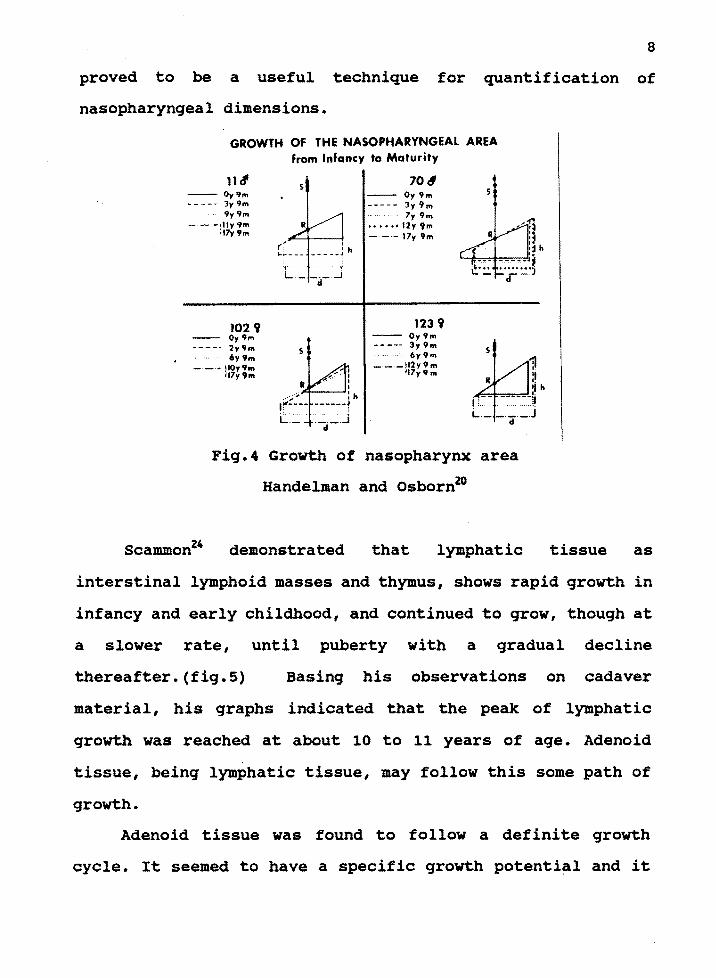
8
proved to be a useful technique for quantification of
nasopharyngeal dimensions.
GROWTH OF THE NASOPHARYNGEAL AREA from Infancy to Maturity
11d' -- Oy9m •---- 3y9m
.. 9y 9m - - -•lly9m
;17y9m
102 9 Oy9m
---· 2y9m 6y9m
-·-·• JIOy9m 17y9m
s
s
70d -- Oy9m ----- ly 9m
7y 9m ••••u12y9m - --17y 9m
123 9 -- Oy9m ----- 3y 9m
6y9m -·-·-ll2y9m
117y'lm
s
Fig.4 Growth of nasopharynx area
Handelman and Osborn20
Scammon24 demonstrated that lymphatic tissue as
interstinal lymphoid masses and thymus, shows rapid growth in
infancy and early childhood, and continued to grow, though at
a slower rate, until puberty with a gradual decline
thereafter.(fig.5) Basing his observations on cadaver
material, his graphs indicated that the peak of lymphatic
growth was reached at about 10 to 11 years of age. Adenoid
tissue, being lymphatic tissue, may follow this some path of
growth.
Adenoid tissue was found to follow a definite growth
cycle. It seemed to have a specific growth potential and it

9
was on this potential that the hypertrophic reactions to
nasorespiratory infections and allergies may be superimposed.
200
CP N c;; :; 'O < 0 100
c CP 0 ... CP ll.
Birth 10 Years 20 Years
Fig.5 Scammon's curve of growth of the lymphoid tissue
Subtelny and Baker's radiographic study25 indicated that
the adenoids attained its maximum bulk between the ages of
nine and fifteen years, and showed subsequent atrophy. They
also point out that at age four to six the growth of the
adenoids and the contiguous nasopharynx were largely related
to each other in a delicate balance if the airway was to . be
maintained. The adenoids usu~lly peaked in their growth prior
to the adolescent spurt of the skeleton. If they increased in
mass faster than the nasopharynx increased in size, proper
nasorespiratory function was impeded and mouthbreathing may
have developed. They concluded that the adenoids led to

10
mouthbreathing primarily in children with a small nasopharynx.
Johannesson26 believed the roentgenographic evaluation of
adenoid size was reliable and used it to investigate the
nasopharyngeal tonsil in children of different ages.
Only minor changes in size were observed between the ages
of 2 to 15 years. The means. for these age groups ranged from
12.0 to 14.3 mm. It was reported that the increase of the size
of adenoid occurred during the first two years of life and
thereafter remained unchanged.
Generally, most subjects demonstrated minimal adenoid
tissue at one year of age, adenoid hypertrophy evident by two
years,a maximum amount of adenoid tissue during the early
school years.(Fig.6,7,8,9,10)
Fiir. 2. Graph dt"picts the greatt"~t width of the ~t tissue in thl" nasophanmgeal roof in relation to· age. Each po.int of tr~ 10lid cun·e is the mean of mras· UrmMmU made in 10 children. The ranps are indicated b)· broken cur'\'e1.
....
::j 161
~! ~ 13.
I 12 ~
' ·'
,~~ '
•• ! a• 7J
6~ ~~
' ,
., 1
'' ,, I \ ",, ... ,
I
I . I
I
,r-·• -,, ,,.'·--"""', I • '
'
2 J • s· 6 7 a I 10 11 12 14 Ate 1J 1~ 1•
Fig.6 curve of growth of adenoid tissue

"' \ ( ' I \ " °"
' > -...... . ~I ,,., );
~,01 A \.. ~~ "~~ . ~ 1 \ ,, ~
\ ~ ~ ~ J Cj AG[ '2 A'iE 3
)~ AOUL THOOO
~
~.'iJ: Ii Sni11l • PJ1huh111wtri1 hf':t•lrluft' tr:will)C!I depirth1Jl tht• full ICrm,th 1•.n•lf' of 1tdt"uoi.J flll'IUt• 1;r11wth tu 1t1U'll:l11111111 hulk a11d ~11h1trq1w11t :1lruphy ht ~\·i1IP11t.
Fig.7 Growth cycle of adenoid tissue
GROWTH OF ADENOID TISSUE - INFANCY TO ADOLESCENCE
AG£ 6 rnoa.
J.G.
li'iK. :! Serial trul'lDgM ot ccphulo111ctrit• headplates re,·eali11g change. in the adeaoid t!aaue ma8 !1 with age. The stippled area represents ndenoid tiuue aa well a1 the •ft tiasue underlying the root ot the bony nasopharyu.
11
Fig.a Growth of adenoid tissue, from infancy to adolescence

Fig.9
DEVELOPMENT OF STRUCTURES TISSUE
~ ,~ GROWTH Of SPMlNOID DOWNWARD 8 FORWAJm MAXI.LARY
MINIMAL ADENOID CHANGE GROWTH - DROP Of THE PALATE
VERTICAL GROWTH ()IF *SA&. CAlllTY
1',itc. 3 !Seri.al t racing~ of. cephnlomctric headplatcs revealing ,.._.,... growth, bel\\cen rufonutd ttssu£> nnd !'ontiguous 11tru('turea, _____ ,.
12
Development of structures contigous to adenoid tissue
-- - -- --. DEVELOPMENT OF EXCESSIVE ADENOID TISSUE
Fig. 4 Serial tracings ot cephalometric headplates depicting an over·abundant develop· ment ot adenoid ti•ue. Note the change in poaitional relationships between the "tongue and aoft. palate.
Fig.10 Development of excessive adenoid tissue

13
PHYSIOLOGY
Miller,et al27 tried to test the traditional concept that
newborn inf ants were unable to breathe through the mouth and
were thus obligatory nasal breathers.
The conditions under which oral breathing could occur and
the contribution of oral ventilation to total ventilation were
studied in 30 healthy term infants (aged 1 to 3 days). Nasal
and oral airflow were measured using two resistance-matched
pneumotachometers. The findings were as follows:
1. Spontaneous oronasal ventilation occurred during sleep.
2. Oronasal ventilation was also observed after crying.
J. oral airway may be used effectively by infants in response
to complete nasal occlusion.
These findings considerably alter the previous concept of the
newborn infant as an obligatory nasal respiration.
Rodenstein & Stanescn28 investigated the ability of the
soft palate to direct airflow during breathing. They found the
soft palate closed the oropharyngeal isthmus during quiet
breathing(resulting in pure nasal breathing) and closed the
nasopharynx during FVC effort (Forced Vital Capacity), which
resulted in mouth breathing. During oronasal breathing, the
soft palate was positioned between the tongue and the
posterior pharyngeal wall.

14
(B)· ETIOLOGY, SYMPTOMS OF AIRWAY OBSTRUcrION
Nasal obstruction that led to an alteration in mode of
breathing can be caused by a variety of factors such as
allergic rhinitis, adenoid hypertrophy, nasal polyps,
congenital nasal deformities, neoplasms,and recurrent upper
respiratory infections. 29 Perennial allergic rhinitis with
accompanying nasal edema was the most common cause of nasal
obstruction in children.
Weimert30 ,an Ear,Nose,Throat Specialist, emphasized
the function of the nose and role of the nares. The most
critical area to the nose with regard to obstruction was the
laminae valve area,located just inside the nares anteriorly.
This is the smallest cross-sectional area of the nose.
Relatively minor changes in nasal architecture in this area
resulted in a significant increase in nasal airway resistance.
It was the inferior turbinate responsible for airway
obstruction. When there was inferior turbinate hypertrophy,
choanal atresia, vasomotor rhinitis and polyps were other
frequent etiology of nasopharyngeal airway obstruction.
Adenoids have long been regarded as one of the chief
causes of mouth breathing, and this hypothesis recurs in many
textbooks. 1 Several authors have stressed the importance of
adenoids as the primary cause· of mouth breathing. 1 31 The
relative size of the nasopharynx as a cause of mouth breathing
has also been cited. 32 33 34 35 Linder-Aronson13 found that the

15
adenoids led to mouth breathing primarily in children with a
small nasopharynx.
Adenoidal hypertrophy was the most common source of chronic
airway obstruction in patients screened by the orthodontist. 36
It was accompanied by a description of a particular facial
expression, which was typical of individuals with adenoids,
Le. the adenoid facies(Fig.11,12). Individuals that exhibited
this f acies were characterized by enlarged tonsils and had
most or all of the following characteristics in common: the
mouth stays open,a long narrow face with increased anterior
vertical facial height in the lower third of the dentofacial
skeleton, a flattened nose, small and underdeveloped nostrils,
a short hypotonic upper lip, a thick and exerted hypertonic
lower lip. The bite was also stated to be of a special type. 37
,.. ... , 1/
"AdEti"loid facies" aooearance.
Fig.11 12 Adenoid facies

16
The "Allergic shiners" described by Weimert38 were
darkened areas below the eyes that were seen in people with
allergies or in any patient with significant nasal
obstruction. They are caused by venous congestion due to
swelling in the nasal tissues.
(C). Airway Obstruction related to mouth-breath
Dr.Weimert38 , evaluated his young otolaryngeal patients
and found that patients who were observed to mouth-breathe
"all of the time" had an 85-percent incidence of demonstrable
airway compromise.
There have been a number of studies correlating airway
obstruction symptoms with various diagnostic techniques and
the conclusions were that direct clinical examination of the
nasal chambers using anterior and posterior rhinoscopy
correlated best with patient symptomatology.
Galen Quinn39 stated a practical clinical approach to
identifying and evaluating nose breathing capabilities.It was
whether or not the individual could comfortably inspire air
through both nasal cavities without effort. Resistance in
inspiration was greater in the child than in the adult.
Patient position for the breathing test was shown. Nose
breathing capability was first tested by gently closing the
lips together with light pressure of thumb and middle fingers

17
for 2 to 5 minutes(Fig.13). It was important that the patient
not be informed of the purpose of this act.
Fig S Bkwk ~ hdt of th< now to tn1 rhr oppoeirr litk
I • I ~
Fig.13 Clinic test for nasal airway obstruction
In a multi-dimensional study, Linder-Aroson13 evaluated
the relationship between adenoids and mode of breathing.
Experimental and control groups were evaluated biometrically,
rhino-manometrically,and cephalometrically.
The results showed that the size of adenoid and the nasal
· airflow resistance was essentially determined by the
relationship between size of adenoid and the size of

18
nasopharynx. The nasopharyngeal airway was important for the
mode of breathing and large adenoids lead to mouth breathing
primarily in children with a small nasopharynx. In these
children, adenoidectomy was indicated as a means of promoting
a change to nasal breathing.
Hibbert and Tweedie40 investigated the relationship
between preoperative signs and symptoms and the actual size
of the adenoid found at the time of operation in a group of
children listed for adenoidectomy.
A series of 80 children was the sample of the this study.
The day before the operation the parents of the children were
interviewed and questioned as to the presence of nasal
obstruction with mouth breathing, snoring, rhinorrhoea, cough,
headache and hyponasal speech. The children were then examined
and assessed for evidence of mouth breathing. They were
examined by anterior and posterior rhinoscopy.
The following day an adenoidectomy and bilateral antral
lavage were performed. The removed adenoid was washed in
saline, dried with gauze and weighed, and its volume was also
measured.
The result of this study showed that in children under
7 the signs and symptoms usually attributed to adenoid
hypertrophy have no statistical significance in the prediction
of the size of adenoid.In children aged 7 and over, a history
of snoring or clinical evidence of mouth breathing was ~elated

19
to the weight of the adenoid and statistically significant at
the 5% level.This would also suggest that in the younger age
group adenoidectomy has little place in the management of most
cases of nasal obstruction,nasal discharge and snoring.
Crepeau, et al 41 did a study on
evaluation of the symptom-producing
the radiographic
adenoid. Adenoid
hypertrophy had several variable symptoms. In this study,
symptoms were divided into minor and major. A lateral
radiograph of the nasopharynx was performed in 114 patients
to study the superior and anterior adenoid diameters(Fig.14).
A correlation was made between the various clinical groups and
the adenoid measurements. Their result support Hibbert' s 40
finding that the anterior adenoid width was a better indicator
of the symptom-producing adenoid than adenoid mass
measurements with their loosely defined norms. A through
history and physical examination remained paramount in the
diagnosis and management of adenoid hypertrophy.

Fig.14 Antroadenoid and superioinferior diameter
on lateral radiograph
20
(D). Response Chain (Tongue, Neuromuscular function, Mandible
posture and head position)
Hannuksela42 and Shapiro and Shapiro43 have demonstrated
that children with allergic hypertrophy of the faucial
tonsils, adenoidal pad, and later, the inferior turbinates
would develop the long-face syndrome. Conversely, the child
with a normal upper airway was much less likely to develop
this syndrome.
The question whether adenoids were associated with a
special facial type was also evaluated by Linder-Aronson12 • In

21
that study, photographs were observed by two observers
independently and it was found that 75% of all of the children
who underwent adenoidectomy were classified as having adenoid
facies. Furthermore, adenoid facies was judged to be present
in about only 4% of the controls. It followed that in a
screening based on facial type alone, many cases requiring
adenoidectomy would be missed at the same time as some cases
would not need surgery. The facial characteristics of the
group of children who underwent adenoidectomy showed a large
facial height, high mandibular plane angle, small sagittal
nasopharyngeal depth and small width/height facial ration. He
concluded that adenoids occur in children of various facial
types and obstructed nose breathing due to adenoids appeared
to be most common among children with a leptoprosopic type of
face and a small nasopharynx.
The upper airway may play a primary role in the
generation of a secondary tongue dysfunction. 44 A close
interaction between airway and tongue dysfunction may present
many different aspects that enable a variety of clinical
situations to occur. These differences in the morphogenic
effect of a few basic and common etiologic factors may have
been related to the timing at which an anatomic discrepancy
occurs during growth.
The forward pressure from the alteration of
proprioception of inflamed upper airways caused protraction

22
of the tongue. By acting during growth, these factors may
change the growth pattern of the bony architecture to which
the neuromusculatures to tongue were related.
considerable hypertrophy of the tonsils and adenoids may
push forward a normal tongue and transform it into a
pathogenic factor acting to create a skeletal discrepancy. A
simple volumetric correction of the hypertrophied tissues,
when effected early, may be sufficient to deactivate the
pathogenicity of the tongue and normalize the growth patterns
of the face.
Thus, the pathogenicity of any given tongue was related
to the status of the airways at a given time. Therefore, when
abnormal growth and development at the level of the
stomatognathic system was recognized at an early stage, and
was related to a large tongue with upper airway obstruction.
Vargervik, et al45 evaluated monkeys to test whether
specific recordable changes in the neuromuscular system could
be associated with specific alterations in soft and hard
tissue morphology in the craniofacial region.
The neuromuscular changes were triggered by complete
nasal airway obstruction and the need for an oral airway.
Statistically, significant morphologic effects of the induced
changes were documented in several of the measured variables
after the 2-year experimental period.
They concluded that the changes in neuromuscular

23
recruitment patterns, which were necessary to establish and
maintain an oral airway, resulted in altered soft-tissue and
skeletal morphology. The extent of the skeletal changes
appeared to depend on the degree of soft-tissue alterations.
The degree of morphologic change, therefore, does not depend
on the amount of air that flows through the mouth or nose, as
has been stated by some authors. Rather, it depended on the
nature of the neuromuscular and soft-tissue adaptations.
The other findings were as follows:
1. The anterior face height increased more in the experimental
animals than in the control animals,
2. The occlusal and mandibular plane angles measured to the
sella - nasion line increased,
3. The anterior crossbites and malposition of teeth occurred.
The experimental use of silastic plugs to create nasal
obstruction in the rhesus monkey has clearly demonstrated that
nasal obstruction with open-mouth posturing recruits accessory
respiratory muscles around the mouth and jaws and led to the
same clinical facial deformity and malocclusion.
Harvold10 has produced increased anterior face height,
narrowing of the maxilla, steeper mandibular phase angles,
narrower thinly pointed tongues and larger gonial angles in
monkeys by obstruction of air flow with nasal plug. He
concluded that specific changes in jaw positioning could cause
corresponding bone remodeling, but this should not be

24
correlated with a particular type of malocclusion.
Another animal experiment11 determined if lowered tongue
position caused by mouthbreathing can affect the craniofacial
morphology. The lowered tongue position was induced by tactile
stimulation to tongue from an acrylic block positioned in the
palatal vaults of three groups of monkeys.
In group I all the experimental monkeys with the insert
in the posterior part of the palate developed an open bite and
significant changes in the dental arch. In group II and III
with the insert in the anterior part of the palate, all
animals manifested malocclusion and significant changes in the
dental arches • The face height increased significantly in all
experimental animals.
This study showed that any consistent changes affecting
the relative tonus in the muscle groups suspending the
mandible influences the extrusion of the teeth and the
establishment of face height.
The findings of Drs.• Vargervik and Harvold animal study
suggested that the position of the chin and the inclination
of the mandibular plane were controlled by the balance between
the hyoid and the orofacial muscles. 21 The morphology of the
ramus appeared to be primarily controlled by the masticatory
muscles. They also concluded that the changes in neuromuscular
recruitment patterns were necessary to establish and maintain
an oral airway and resulted in altered soft-tissue and
skeletal morphology. The extend of the skeletal . changes

25
appeared to depend on the degree of soft-tissue alteration
present.
The nose and nasopharynx were the primary airway. Under
normal circumstances, nasal breathing did not require
recruitment of accessory respiratory muscles. When mouth
breathing was forced by obstructions in the nasal airway or
by increased oxygen demands, accessory respiratory muscles
were recruited. These included craniofacial muscles involved
in formation of an oral airway. They may include neck muscles
that extend the head and neck. If the mouth-breathing was
temporary, such as during catching a cold or during exercise,
the neuromuscular change would fluctuate and would not produce
dental or skeletal changes. If mouth-breathing persisted and
became a habitual pattern during those periods of normal
whole-body growth, the associated changes in the position and
shape of the tongue with lowering of the mandible may have
certain effects on dentoalveolar and skeletal morphology. It
was that the child's neuromuscular adjustments to and impaired
nasal airway were the determining factors in the effects on
developing facial and dental structures.
Changes in mandibular morphology will only occur when
lowering of the mandible was sufficiently consistent. Downward
displacement of the maxilla and excessive extrusion of teeth
~- or may not have occurred in response to a lowered - -- ·-·~----,.,._-
mandibular posture. The maxillary response was mainly
determined by tongue posture and movements. Lower face height

26
was measured with the teeth in occlusion and increased
significantly when a downward displacement of the maxilla or
excessive molar extrusion occurred. Increased molar extrusion
would be expected to occur most rapidly during eruption of the
first and second molars.
Chronic mouth-breathing called forth the recruitment of
perioral and suprahyoid muscles~. The increased tonicity and
rhythmicity of these muscle groups often produced a negative
effect on dentofacial form and function. Often, the long-face
syndrome developed as a result.
~ Children with a genetic proclivity for dolichocephalic
dentofacial development were at higher risk, as were children
with neuromuscular dysfunctions~. Allergic hypertrophy of the
tonsils, adenoid pad, and inferior turbinate, when combined
with neuromuscular dysfunction and a genetic predisposition
for the dolichocephalic face, placed that child in the highest
risk group of all.
The causal relationship between adenoid vegetation
associated with mouth breathing and increased lower facial
height may be due to a rotation downward and backwards of the
mandibular symphysis. 47
The head posture was investigated by Linder-Aronson in
16 patients who had undergone adenoidectomy due to
difficulties in nose breathing. A comparison was made with a
similar number of controls in the same age group without
impeded nose breathing. Inclination of SN line was measured

27
relative to a vertical reference line included in the lateral
skull radioqraphs. A small value of the SN/vert. angle
expressed extended head posture. Measurement was made
initially and 1 month after adenoidectomy.
A significant difference was noted. In order to increase
the respiratory passage, the head was extended forward with
an increase in lower facial height and a resultant increased
retrusive pressure from the facial musculature on the
underlying skeleton.
Bosma~ has stated that one important function of head
posture was to maintain an adequate naso-oro-pharyngeal
airway, In patients with morphologic disturbances which impede
and adequate airflow one can expect to find an extended head
posture. The Pierre Robin syndrome was an example of such a
morphological disturbance.
Solow and Greve61 studied head posture and its relation
to nasal respiratory resistance.It confirmed the results of
Linder-Aronson's work. They examined 24 children ages 4 to 12
years before and after adenoidectomy. Cephalometric recordings
of the natural head position and rhinomanometric readings of
nasal resistance were obtained for each child.Before
adenoidectomy, a large craniocervical anqulation was seen in
relation to large nasal respiratory resistance and narrow
airway. After adenoidectomy,reduction of craniocervical

28
anqulation resulted in children who had received adenoidectomy
and nasal resistance was reduced.
The findings confirm predictions of soft tissue stretch
hypothesis and provide an explanation for the reversibility
of craniofacial morphology previously observed.
Bibby49 stated that in mouth breathers one might have
expected a different head posture to be adapted to facilitate
breathing especially where the mouth breathing was due to an
obstructed nasopharynx.
Individual Variance
In one of Vargervik' s animal studies45, silicon plugs
were formed to fit the individual nares to obstruct
inspiration but allowed some air to escape during expiration.
The changes observed in the middle and front of the tongue
showed considerable variation in tongue adaptations. This was
reflected in the individual animal's optimal adjustment to the
experimental condition present. This study demonstrated a wide
individual variation in response to an identical stimulus.
For this reason Or.Meredith46 suggested that a detailed
history and physical examination should be complemented by
serial cephalometric x-ray studies, PA tomograms of the nasal
vault ,rhinomanometric studies and, in selected cases, sleep
laboratory studies.

29
(E).CEPHALOMETRIC STUDIES
The nasal passages and nasopharyngeal airway can be
clinically assessed by the ear, nose, and throat specialist
using anterior and posterior rhinoscopy. The sagittal depth
of the nasopharynx can also be evaluated on lateral skull
radiographs. There were differing opinions, however,
concerning the accuracy of this method in view of the fact
that these radiographs can reflect the nasopharynx in only two
dimensions. A number of authors on the other hand, have found
this type of radiographic examination to be practical, having
satisfactory results in children of all ages.
An investigation was carried out by Linder-Aronson50 in
an attempt to clarify the value of lateral skull and frontal
radiographs as a means of evaluating nasal respiratory
function. The following factors were selected for evaluation:
1. The relationship between the size of the adenoids as
measured on lateral skull radiographs and judged clinically
following posterior rhinoscope examination.
2. The relationship between the size of the adenoids as
measured on lateral skull radiographs and nasal airflow
measured in liters per minute.
3. The relationship between the size of the nasal airway
as measured on frontal radiographs and nasal airflow measured
in liters per minute.
4. The degree of nasal obstruction as judged on.visual

30
examination of frontal radioqraphs compared with the nasal
airflow measured in liters per minute.
Subsequent correlation analysis qave the following
results:
1. A siqnificant relationship between the size of the adenoids
as measured on lateral skull radioqraphs and assessed
clinically.
2. A neqative relationship between the size of the adenoids
as measured on lateral skull radioqraphs and the nasal
airflow.
3. A siqnif icant relationship between the capacity of the
nasal airway as measured on frontal radiographs and the nasal
airflow.
4. A reasonable assessment of the nasal airflow by subjective
evaluation of airway capacity from frontal radioqraphs.
He made the conclusion that lateral and frontal skull
radioqraphs provided a satisfactory means of evaluating the
dimensions of the nasopharynx and the capacity of the nasal
airway, respectively.
Bresolin,et al51 completed a cephalometric investiqation
of thirty allerqic children, aged 6 to 12 years who had
moderate-to-severe nasal mucosal edema on physical examination
and who appeared to breathe predominantly throuqh the mouth.
They compared them to 15 children without allergy who had
normal f indinqs from nasal examination and who appeared to

breathe predominantly through the nose. The
31
facial
characteristics of children who were mouth breathers were as
follows:
1. They had longer faces.
2. The faces were more retrognathic in lateral profile.
3. The mandibles had more obtuse gonial angles.
4. The palates were higher and narrower.
s. They were more likely to have posterior dental crossbites
than children who breathed through the nose.
In Trask's stud~ they analyzed the effect of perennial
allergic rhinitis on dental and facial skeletal
characteristics. Twenty-five allergic children who were
apparent mouth breathers, their 25 siblings who did not have
the disease and were apparent nose breathers, and 14 nasal
breathing control subjects were used in this study in an
attempt to differentiate the facial characteristics most
strongly determined by heredity from the facial structures
more vulnerable to environmental influence-specifically, mode
of breathing. A control group of nasal breathers was used to
determine whether the sibling pairs had genetic
predispositions to specific facial and skeletal
characteristics.
overall, the allergic children had longer, more retrusi ve
faces than controls.These results confirm earlier reports that
allergic rhinitis may be associated with mouth breathing and

32
altered facial qrowth.
Linder-Aronson and Henr ikson50 compared the
anteroposterior nasopharyngeal dimensions cephalometrically
of 6 to 12 year old mouth breathers to nose breathers. The
purpose of the study was to calculate the average
anteroposterior size of the nasopharynqeal airway in children
of this age group in order to obtain cephalometric standards.
From these standards, it is possible to judge the extent by
which mouth breathing may be obstructed.
Lateral radiographs were taken and evaluated by two
independent examiners. Measurements were made to assess
airway dimension and a test was used for calculating
statistical differences between the groups.(Fiq.15) The
result showed that variable Al and A2 gave a good indication
of the anteroposterior size of the nasopharyngeal airway. This
gave a more reliable indication of the need for an otologic
examination. The standard values obtained in this study showed
that an otoloqic examination of the nasopharyngeal space was
to be recommended if the measured distance pm-adl or pm-ad2
was less than the present mean minus 1 SD for mouth breathers
in the appropriate age group.(Fig.16,17,18).
The results also showed that when planning orthodontic
therapy, in which it was desirable to assess the ability of
the patient to breathe through the nose, a clinical record of
the mode of breathing can be supplemented with

33
radiocephalometric data on the anteroposterior size of the
nasopharyngeal airway. Furthermore, they found that the contour
of the posterior nasopharyngeal wall could be satisfactorily
assessed on lateral skull radiographs of children.
• Refcrcn.:e point,. pm •· ptcrn:<>ma.\illary; ' ,_ >clla tur.:i.:a; t>;a • t>.nn>n . .. ~ the mid·point on the line joining sand t>a; ad1 = 1he intcn.c.:tion uf 1he JX"lerior nuopharyn41eal wall and the line pm-t>a; ad, the intcri.c.:tiun of the posteriur n11i.c:r
pharyn41eal wall and the line pm~.
Fig.15 Airway dimension measurement by
mm
20
15
10
5
6
Linder-Aronson/Henrikson
7
Radiocephalometric Analysis
I Nost' bre•ther
ti Mouth brt'•tht'r
·-·Borderline
......... ;_ II I -
8 9 10 11 Age
Diagram of means and SD for variable Al (the distance pm-ad mouth breathers and nose breathers aged 6-12 years.
Fig.16 Diagram of airway dimension measurement
by Linder-Aronson/Henrikson

mm 20
10
1-:::::L--- _'_ -_ _J r-- I II 1
7 8 9 10
I NOS.C' br.-•th.-r II Mouth brC"•th.-r
·-·Bord.-rlin.-
11 Ag.,
Diagram of means and SD for variable A2 (the dislance pm-ad1) for .moutl oreamers and nose brealhers aged 6-12 years.
mm2
125 I No!.eo tlrr•theor II Mouth breo•theor
100
75
{~ so II
' 25 ...
7 8 9 10 11 Ag.-
Diagram of means and SD for variable A3 (the area pm-ad1-ad,-pm) fo ~outh breathers and nose breathers aged 6-12 years.
Fig.17,18 Diagram.of airway dimension measurement
by Linder-Aronson/Henrikson
34
Radiographs of the nasopharynx were sometimes
misinterpreted because of poor technical quality. A simple
method of interpretations suggested by Cohen and Konak52 was
based upon measuring the airway immediately behind the upper
part of the soft palate(Fig.19).If it was narrower than the
width of the soft palate it was considered as markedly
obstructed. When narrower than half of the soft palate, it was
severely obstructed. When it was the same width as the soft
palate, it is not narrowed.

35
( b)
....... . _....-., 1111 IOft ...., . Fig. 19 Airway dimension measurement by Cohn and K6n.ak52
In this study, he also showed six other methods of
nasopharyngeal airway evaluation. (Fig.20) All methods showed
good correlation and the present method was easy to use and
has proven to be useful even in radiographs which other
methods fail to interpret. This study also stressed the
importance of evaluating the airway instead of the adenoidal
thickness.

a1GraplucaJ IJllOPlil or 6 methods cited (or .._ 1ize of adenoids: Johanncuon, 1
-..-:· ...... 1 Hibbtrt. • s.remco' A
36
Fig.20 Graphic synopsis of six methods for measuring
the size of adenoid
Hibbert and Whitehouse34 evaluated the accuracy of
radiology in the assessment of both adenoidal size and the
size of the nasopharyngeal airway.
Seventy-six consecutive children who subsequently
underwent adenoidectomy were reviewed. A lateral radiograph
of the postnasal space was taken on the day before surgery.The
area of the adenoid shadow on the radiograph was traced onto
graph paper. (Fig.21). It has been observed that the posterior

37
wall of the maxillary antrum was in close approximation to the
plane of the posterior choana. A line drawn at right angles
to the adenoid shadow will intersect the line of the
posterior wall of the antrum. The shortest line between these
2 points was considered to be the width of the nasopharyngeal
airway.
' .
-,.
Fig.21 Airway measurement by Hibbert and whitehouse34
The adenoids were removed by a standard technique and
they
were washed, dried weighed, and their volume was measured by
displacement.
The study showed that radiograms were an accurate method
of assessing the size of the adenoid mass, in contrast to
preoperative signs and symptoms which were poor predictors of
adenoid weight. This study also indicated that it was . the size

38
of the adenoid rather than the size of the nasopharynx which
was important of the impairment of the airway.
Hibbert and Stell~3 \~ompared the adenoid of two groups of
children: those selected for adenoidectomy and those who
presented as normal control group.
The method to evaluate the size of adenoid and
nasopharyngeal airway was previously described.
This study showed that in a series of children selected
for adenoidectomy the radiographic area of the adenoid did not
differ significantly from that in normal children. That meant
the adenoid in children selected for adenoidectomy was no
larger than in normal children. However, the children selected
for adenoidectomy have a significantly smaller nasopharyngeal
airway than the same measurement in normal children.
In this series of normal children studied it was shown
that the radiographic area of the adenoid does not increase
with age, though the nasopharyngeal airway does. The increase
of the nasopharyngeal airway must therefore be due to an
increase in the anterior-posterior dimension of the
nasopharynx as the child grows. It was suggested that in
children below 70 months an airway of 2 mm or less can be
considered abnormal and in children over 70 months an airway
of 3 mm or less can be considered abnormal.
'' Fujioka, Young & Girdany~ thought the absolute size of
the adenoids and the size and shape of the nasopharyngeal

39
space were major factors that determine nasopharyngeal
obstruction. They described an adenoidal-nasopharyngeal ratio
(AN ratio) derived from linear measurements on lateral
radiographs of the nasopharynx. The ratio of these two sizes
can provide a simple arithmetic measure of nasopharyngeal
obstruction.
Lateral radiographs of the nasopharynx of 1,398 children
between ages 1 month and 16 years were reviewed and the AN
ratio were calculated, tabulated, and statistically
analyzed(Fig.22,23). The 143 lateral nasopharyngeal
radiographs of 92 patients and their adenoidal size and
nasopharyngeal air patency had been estimated visually by
experienced observers and classified to the AN ratio (Fig.24) .
..4deMidarmeasurement:i• .. ,,. .. represents distance from A.' point of maximal c:oo .. eiury, alonc;i interior margin of aaeooia sll~ctow to hoe B. arawn alonc;i s1ra1gnt part ol aoteraor marc;iin o~ basiocc:iput. .. ,,. .. i• meuurect alonc;i hoe perpeoa1cular trom point A
to its 111terwc:taoo w11n 8.
Fig.22 Adenoid measurement by Fujioka, Young and Girdany54

1.00
1.20
J.10
• -- -(Nasopn4rvn-!l;al-;;;;;:-- · j .. C . - - - z..,;. ..-dsuu:m~n N •S d.1s1ance bet
. '"'li)Sltrr1or s'-'pt:r ior ~09e oJ n. •t:"t:n eOqu of ~pnenoDaStOCC plat dlO palate. an<J 0'. dOleromftrflOI
1 1 syncnono1os•s When n nor clearly w•~uah.r:t:d, pc-mt 0 , can sync onoras1' 11 POSh:rom1e11or marg.1n 01 ldlt=r
1 1 Ott O~lt:rmineo .ts siltt ol eto:..~ng
n•~'-'Qnaryna. d P t'f)'QOul p1a11:s P an'-1 floor of Dc.nW'
Fig.23 Nasopharynx measurement by
Fujioka, Young and Girdany54
ADENOIDAL SIZE IN Crt1LUHfN
• z so
•I 50
Mll.N
LOO
o.~o
a so
0.70
Q 0.l;i.O:
.. "'o.~o ~ c
0.40
..
• I Ill II I 1 I
I .... :' I 1:11 e\ .--..... · . ••
. . O.lO /,,-·· ·- -.:··· ·---~--~ 0
/ .................... ... 0.20 ,' ......
- I SO
-zso 0.10
....... , '"'··-250
I 2 l 4 ~ • 1 8 '!t 10 11 12 ll 14 I~ 16 AGE
I 2 .) 4 ~ i 1 8 9 10 II I.I il 14 I~ 19 AGE
Fig.24
-'V1au.IACIH ... l~-rt 4"fell01r1 l•lt.oioutAQftlOlilJ~
.n tne Pal'lllOoM 1IW'r ~~-.. • Wftwlv .. ~ lelge ·~· .,_.,. ,._,_
n•~fR(fHll • aieec•.<A. • "Df"'-1 ~-"'"',.,. ...........,..., -..I iMll9nooGI. M*3 _,.. • "'•• Q' ... l.J .. ~I.·--~-· •l....W,. .,.._Jlil\111---·----
Adenoid/Nasopharynx ratio
by Fujioka, Young and Girdany54
40

41
The results were as follows:
1. The frequency distribution of the AN ratios for each gender
and in each age group followed the expected curvature of a
normal distribution.There were no statistically significant
differences on AN ratio for gender in any age group.
2. The assessment of visualized classification of the size of
the adenoid and nasopharyngeal space was in general agreement
with the statistical analysis.
3. For Practical purposes, a value of the AN ratio greater
than Q.80 may be considered indicative of enlarged adenoids.
(F). Treatment, effect of adenoidectomy.
Tonsillectomy and adenoidectomy play a certain role in
the treatment of certain infectious and inflammatory diseases
of the upper airway. 55
Linder-Aronson et,al56 did a study on the mandibular
growth direction following adenoidectomy. The adenoidectomy
sample initially showed significantly longer lower face
heights, steeper mandibular plane angles, and more
retrognathic mandibles than the matched controls.
Analysis showed the following results:
l. During the 5 years after adenoidectomies, the girls had a
more horizontal mandibular growth direction than did the

42
female controls.
2. A corresponding but not significant trend was found for the
boys.
J. The growth directions were significantly more variable for
both boys and girls after adenoidectomies than for controls
during the five-year growth period.
4. The mean airflow through the nose increased for both sexes
1 year after adenoidectomy to values equal to the initial
values for the control.
Respiratory function and its effects on craniof acial
growth was evaluated by Linder-Aronson47 • Longitudinal results
of five years post adenoidectomy were presented to examine the
effects on the dentition and facial skeleton with a change in
the mode of breathing.
The sagittal depth of the bony nasopharynx, as measured
from the pterygomaxillary point to basion, changed in children
who became mouth breathers after removal of their adenoids as
well as that in the control children. The greatest change
occurred in the first year post-operatively in the group of
children whose adenoids had been removed, During the following
four years, the
increase in this group was similar to the controls.
The angle between the mandibular plane and the palatal
plane changed due to the change of the mode of breathing. The
change during the first year was not signif icarit but by the

43
fifth year post-operatively, a significant change was noted.
A correlation analysis between reductions in the ML/N angle
and lower facial height was found to be significant at the
• 001 level.
Linder-Aronson57 in the study of the effects of
adenoidectomy on mode of breathing stated that the multiple
regression analysis clearly supported the hypothesis that
enlarged adenoids give rise directly or indirectly to mouth
breathing and that in most cases the individual changes to
nasal breathing after adenoidectomy. The multiple regression
analysis also showed that the size of the nasopharynx was of
importance in this respect.
He concluded that in any case, improved adeno-tonsillar
function and lessened inferior-turbinate hypertrophy will
improved the upper airway and further reduced the effect of
a large tongue on the developing tissues or structures.
Linder-Aronson & Lindgren58 stated that the narrow
maxilla may be treated by surgical or orthodontic expansion.
The mid-palatal suture split will decrease the higher to
normal nasal resistance.
Guenthner,et al59 studied the effect of Le Fort I
maxillary impaction on nasal airway resistance. The nasal
airway resistance was determined by means of a universal

44
active rhinomanometric technique. Contrary to the predicted
negative effects of maxillary superior movement on nasal
airway function, there was a statistically significant
improvement in nasal airway resistance after maxillary
superior movement.
When abnormal growth and development at the level of the
stomatognathic system was recognized at an early stage, and
is related to a large tongue with upper airway obstruction«.
It may be wise to act medically or surgically to normalize the
enlarged tissue mass of the tongue before its full
development(at about age eight years).
Improved adenotonsillar function and lessened inferior
turbinate hypertrophy improved the upper airway and further
reduce the effect of a large tongue on the developing tissues
or structures of the oral cavity. Thus, the pathogenicity of
any given tongue was related to the status of the airways at
a given time. A simple volumetric correction of the
hypertrophied tissues, when effected early, may be sufficient
to deactivate the pathogenicity of the tongue and normalize
the growth patterns of the face.
It was well established that the jaws were vulnerable to
environmental factors that may have detrimental effects. 60
Hypertrophic tonsils causing forward tongue displacement may
have similar effects. The tendency for self-correction of
dental irregularities after removal of various detrimental
45
factors can be interpreted as a definite indication that a
cause-and-effect relationship may exist. Similarly, elimination
of nasal airway interferences followed by a change from oral
to nasal respiration may result in improvement of certain
aspects of facial and dental deviations.
Because of the individual variation response, adeno
tonsillectomy or other airway surgery should not be done in
a very young child to prevent future unfavorable craniofacial
development because this may never ensue. 60 Moreover, we
believe that surgery should be considered only when the
characteristic deviations are manifested. Hoverer. they stress
that children who demonstrate features associated with open
mouth posture should receive appropriate airway treatment and
facial growth management to prevent undesirable growth
patterns from persisting and progressing.

MATERIALS:
Chapter III
MATERIAL AND METHOD
A. Experimental group:
1. There were nineteen subjects in this study. They were
all ref erred to one Ear, Nose and Throat specialist by
physicians on the basis of a history of persistent nasal
respiratory obstruction which was confirmed by physical
examination. Obstruction was still present after
administration of vasoconstrictor spray. All of them had
obstructive adenoids and were scheduled for adenoidectomy
after the study records were obtained. The subjects' general
medical histories, physical conditions and mouthbreathing
situations were evaluated and understood employing the history
form, examination form, and case form used in this Ear, Nose
and Throat clinic.(Fig.25,26,27)
All subjects were caucasians. There were four subjects
in three age group- two were males and two were females; three
in four age group- two males and one female; one in five age
group- one male; four in six age group- one male and three
females; and seven in seven age group- three males and four
females.(Fig.28)
2. The subjects of this research were referred to an ENT
specialist from physicians and pediatricians to eliminate any
bias for certain facial characteristics that might influence
46

47
the results. Because general dentists and pedodontist were
aware of the association between airway obstruction and facial
deformity, referral by them was excluded.
FAMILY HISTORY ~ Sutt of Health
Father Mother l>OOUH
Brothen !Bl
S11ten !SI
Children Sons !SI
Daughters !01
Check dise.r.er. blood rel&ti•es have had. Ill c;hecked-state rel111onshipl
0 High Blood Preuure 0 Hurt Disuse 0 Stroke 0 Kidney 01SHst
0 Ep1ltPlY IConvulsionsl ----------
0 011bl!tl 0 Hay Fever
0 C.nc11r 0 T ublrculotil
0 ~"'°"'"'"
If deCNMd. cause of duttl.
0 Jaundice 0 Ne1Yous Breakdown 0 Migraine 0 TenQl!ncy 10 Bleed
Pleue hn any iUnes1..i you have had and give the dates:
0 Ulctn 0 Ollwr
Age at death
Oat•-----------------Oate-----------------
Dn•------------------
Type
Pica.~ hit any allergies or reac:t1ons vou nan had to mf'd1ut1oni.. food!., c;or.metic;s, pl1nu. etc:
Please tilt any med1e1tions that you are uiking including aspirin, lpativtl, hormones, 1r1nquilian. c:ortilOnt, blood pmsurt
pills, or other: -------------------
Habits
Coff•------------------~ T••-------------------Tobleco------------------Alcohol ------------------BH•-------------------Wint-------------------Whiskey _________________ _
REPORT OF MEDICAL HISTORY
How much ptr dlov or per wHkl
Fig.25 History form used in the Ear,Nose and'Throat clinic

Pleu• check 1nv of the following complaint$ wtuc:h presently trouble you:
o Hud cold' D Ct>e\! cold' Q Sort lhfOIU
O Sonul trouble 0 NOH blted' 0 Cough D Hav Fever. Auhm1 O Asthm1 0 E•cm1v1 penplfll!on D Lou of -1ght 0 01:>n11y D Joint pain$ D Blut mood' O ln1b1lity to conun1J1te O Lick of hlf confidence
0 Nt!NOUl...U
0 SleepleH1wtU D Back trouble 0 Abdominal pain D Painful unn1tion 0 Shortneu of btuth 0 Ht111 pain 0 Skin trouble 0 Vllual d1ll1cult1e1 D Earache 0 D1Khlfgt from tars 0 Dufneu 0 Poor 1ppe111e D 01$comfort afler mHll D N1u,tt·vom1t1ng
D Connipat1on 0 Diarrhea 0 $pft(:h d1fficulty 0 Convulsions. t1u 0 Fainting spells D Headaches D D•uinen 0 Thyroid d11turblnces D Fn9T 0 Anemia 0 Aec11I bleeding D Frequent unn1t1on D E11y f111gue D Htari pounding 0 Hives
Pu1 medical illnem11··g1ve appro•1m11e 419' 11 which you had any of the following 1Unnu1:
German measles Mea1le1 Mumps Clucken po• Scarlet lev•r Whooping cough O•ptheria T ypho1d fevtt Auhm1 D11bettl Stom.c:h trouble Appendic111s Herni1 lrupturel Coli ti$ Concuu1on
Kidney trouble Albumin 1n unM Sugar in urine M1laria Undulant fever Hepatitis Pol1omyelit11 Influenza f'MUmon11 Pleu111y T ubercul~11 Tumor or etnc:er Alld1oact•ve e•posure Nen1ou1 btnkdown Epilepsy
Venere1I d11ea1e Jaundice Dvientery
---- Tendency to bind Infectious mononud•ot11 A heum1t1c fewer St. Vitus dance
----- Tonsillit•' Discharve from un Unto•d infection
----- Sinu' troubl• ----- Hurt trouble
High blood preuure
----- H1yfevet
Are you subiect to d1mn11ng perioOs of mental dep<euion? -------------------------H••• you ever been treaied for 1 rwrvou1 or menlll disorder? ________________________ _
Do you cons1dtl1 vounelf more nervous thin the IVtltge person1-----------------------
H•ve you any 1W1thension in 1t111rd to your health?----------------------------Ha.e you hid psycholhertpy? ___________________________________ _
Hav• you lived with anyone with luberculosis? _____________________________ _
List anv """and count11t1 in which you hive liveel ___________________________ _
WomenchKk.
Mt:n1111.111ton: Age at onaet __ h1iom reeula1 fYery ___ dlys lrregi•I••--- Durllion - Divs Amount: Smtll __ Medium __ Profuae __ P•in ___ dlys Char.:111 of ptin __ Crlll'IP"ll- Dull
8.-:kache-- Go io bed-- Stay l\ol'M __ Vlgif\11 dtlChargt?_Color ----------
Prl:Mftt or p.nt trutment of menatrual disordtl1? If so, what -------------------------
Pre;ntnelft: D1tA-------------------------------------
Complic;at1ons ----------------------------------------
Pluw uate the ••non why vou 1'9QUirt medictl cart and include any additiontl information that -Id be helpful in the
diagnosis or management of thi1 problem.------------------------..... -------
REPORT OF MEDICAL HISTORY
Page 2
48
Fig.26 Examination form used in the Ear,Nose and Throat clinic

49
~I
ALLERGIES or SENSITIVITIES: --------------- Hone lCnown:
RINNE Norul
D D
L
RINN£
NOSE
0 D
NASOPHARYNX
D D
D
PHARYNX and ORAL CAVITY No Exu
LARYNX
AUDIO
IMPRESSION:
PLAN:
Noriul
D 0
D D
D c
Richard F. Bulger. M.D James E. Rejo~·sk.i. M.D.
lleXC V1siC ------
Fig.27 case form used in the Ear, Nose and Throat clinic

50
J. subjects were qiven a vasoconstrictinq spray to differ
between mucosal vs adenoid blockaqe in Ear, Nose and Throat
examination. It could be assumed that the subjects had
persistent and obliqatory oral respiration rather than
temporary and transitory mouthbreathinq histories because
nearly all subjects had adenoids and were scheduled for
adenoidectomy soon afterwards. From parents• anamnestic
information and the lateral head X-Ray films, it was also
confirmed that most subjects were sufferinq from
mouthbreathinq due to adenoids.
B. Control group:
Nineteen lateral head X-Ray films from the Broadbent
Brush Growth study Center in Case Western Reserve University
were chosen to match the experimental qroup in race, age, and
sex. Due to the fact that adenoidectomy was very prevalent,
even as a routine surqery for younq children when the
Broadbent-Brush Growth study was beinq conducted in the
nineteen thirties, all the subjects in the control group were
chosen by their medical histories of either those who had
their adenoids removed very early at ages of three or four;
or those who had never had adenoidectomy. For those who had
early adenoidectomy it is presumed they did not have problems
of chronic nasal respiratory obstruction later on as well as
those who had never had adenoidectomy. Subjects havinq
adenoidectomies after six or seven years of aqe were excluded
from this control qroup due to the consideration that they

51
might have had adenoidal obstruction and mouthbreathing
problems during their early ages but waited until later to
have adenoide.ctomy.
EXperi11ental qroup
Source: Sa~ples were referred to one E.N.T •pecialiat,
and then referred to Orthodontic department
for takinq lat!='ral h0
ead X-Ray plate.
NUJDber: 19 Race: Caucasian
Male Female
Aqa 3
5
7
History: 1, Peraiatent nasal respiratory obstruction.
2, Obstructive adenoid• and scheduled for
adenoidectomy.
Fig. 28 Sample of the experimental group
Control Group
Source: Lateral head X-Ray tilm11 trom the Broadbent-Brush
Growth study Center in case Western Reserve u.
Number: 19 Race: Caucasian
Male Female
Age 3 2 2
4 2 1
5 1 0
6 1 3
7 3 4
History: 1, Early removed adenoids at age three or tour.
2, Never had adenoid removed.
Fig.29 Sample of the control group

52
METHODS:
A. Experimental group:
l. Cephalometric radioqraphs for all subjects were taken .·
on a standard cephalometer in the Orthodontic
Department. (Fiq.30,31) The saqittal plane of the head was five
feet from the X-Ray source and 15 centimeter to the X-Ray film
cassette. The X-Ray machine was set at seventy-sev~n KVP, l/6
second, and 4.5 milliamperaqe. The radioqraphs were taken with
the subjects' heads in upriqht natural position, their teeth
in centric occlusion and their lips at rest.
Fiq. 30 Fiq. 31
Standard cephalometric machine ·

53
2. Tracings of the radiographs were made on 0.003-inch
matte acetate paper with an 0.5-mm pencil. Soft-tissue
outlines were excluded to eliminate measurement bias created
by lip posture.
Fig. 32 Example of tracing of the head X-Ray plate

54
3. Skeletal landmarks (Fig.33) and Planes (Fig.34)
necessary for Ricketts facial pattern analysis were
identified, and selected by two orthodontists to produce five
angular measurements i.e. facial axis, facial depth,
mandibular plane angle, mandibular arc and lower facial
height. (Fig.35) Those radiographic and skeletal landmarks
needed for the Ricketts facial pattern analysis are
illustrated. (Fig.36,37,38,39,40)
4. Those cephalometric measurements for the facial
pattern analysis were calculated for each individuals' facial
pattern according to Ricketts' facial pattern analysis method.
(Fig.41,42,43) The norm of each of the above measurements for
each age group were extrapolated from original Ricketts• norms
due to the fact that the stature growth rate is almost
constant from young age to puberty according to growth studies
from the National Center for Health Statistics,1979.
(Fig.44,45,46) The standard deviations of these five
measurements were kept the same as in the Ricketts' facial
pattern analysis.

Point•
Na
Or
Pr
Gni
Me
Ba
Ptv
An•
Sn
Ra
De
Xi
DEFINITIONS OF ANATOMIC LANDMARXS
( USED IN RICKETTS FACIAL PA'l"l'ER.N ANALYSIS
Definition
The •uture between the frontal and nasal bones.
Tb• loweat point on the average of left and riqht infraorbital aarqin.
The hiqheat point on the averaqe of the left and right •uperior surface of the external auditory meatus.
The aost anterior point on the mandible in the midline, determined by a tangent through naaion.
a point at the intersection of the facial and aandibular planes.
Tb• aoat inferior point on the syaphyaeal outline.
Tb• aost inferior poaterior point on the anterior border of the foraaen aaqnwa.
Intersection of inferior border of foramen rotundu:.m with posterior wall of pterygomaxillary fossa.
The aost anterior point on the aaxilla at the level of the palate.
Point on the anterior border of the syaphysis between B point and Poqonion where the curvature changes from concave to convex.
A point located at the center and aost inferior aspect of the sigmoid notch of the ramus of the mandible.
The deepes't;_,point on the curve of the anterior border of the ramua.
Tb• aidpoint between the anterior and posterior border of the condyle intersected with Na-Ba line.
The geometrical center of the ramus. Lacated as •hown on fig._.
Fig.33 Definition of anatomical landmarks for
Ricketts facial pattern analysis
55

Line
Po-Or
Me-tangent
N-Ba
Xi-De
Xi-Pm
Measurement
Facial a.xi•
Facial depth
DEFINITION OF CEPHALOHETRIC PLANES
USED IN RICKETTS FACIAL PATTERN ANALYSIS
De&cription
Frnakfort Horizontal : a horizontal plane running through the right and luft porion and orbitale.
Mandibular plane ; A line at the olwer border of the mandible tangent to the gonion angle and Me.
Facial plane : A line from na&ion to pogonion.
Dividing line between the face and the craniWll.
condylar axis to de&cribe the morphology of the mandible.
corpus axis to evaluate the morphology of thP mandible.
Fig. 34
DEFINITION OF CEPHALOHETR.IC MEASUREMENTS
USED IN RICKETTS FACIAL PATTERN ANALYSIS
Description
The angle between the Facial axi& and BasionNasion plane.
The angle between the Frankfort plane and the facial plane N-Po.
Mandibular plane angle
The angle betwen the Mandibular plane and the Frankfort plane.
Lower facial Height
Mandibular arc
The angle from Ans to Xi To Pm.
The angle between the Condylar axi& and the Corpus axi&.·
Fig.35
56

FIELD V - CRANIO·FACIAL RELATION @ FACIAL DEPTH; Tt,e <1ngle between the foc1ol pl.in1: .. no Frilnklort pliin1. OowM lar.ial .. ogle .
....
M·1
CLINICAL NORM: 57• "'age 9. lnr.re .. ses 0.33' per yHr.
CLINICAL DEVIATION: !3"
INTERPRETATION: L"'-Jles 1h11 chm hor1Lontally. Dci~rmme) 11 the skeleul ci .. ss II or Class 111 IS dli'l to the mandible.
FACIAL AXIS3 The dllqle bct...,,ecn the lac1iil aus and Bc1s1011 N.:is1on.
CLINICAL NORM: 90'
CLINICAL DEVIATION: 3.5"
INTERPRETATION: The d1r1:1:11on of yrowih ol the chm and the mo1 .. 11. ExpresM:s me r<illo of facial he19h1 10 depth.
35. FACIAL TAPER:· Tht INl'Klibular pt..0. meas.urea to tile fKi.tl pl<tne.
' CLINICAL NORM: 68"
CLINICAL DEVIATION: 3.5'
MANQf8ULAR PLANE ANGLE: Mea$111ed 1u Frani.f,}11 horilOntal.
CLINICAL NORM: 26' at .. ge 9. o~'(;r1111i.c5 O.J• per yeai.
CLINICAL OEV14TION: 4.!i"
INTERPRETATION: "H1yh'' mand1buliit µl.im: implies 1n .. 1 si..clctal open blle is due 10th? 111.ind1bh:. "Low": maou1· uul.ir j1la1 .e 1mphC) ~• eh:t•d d~p b11e is !Jue tu 1h11 n>.ioddlh:.
57
Fiq.36, 37, Definition, clinic norm and clinic deviation cf
facial axis, facial depth and mandibular plane
anql used in Ricketts facial pa.ttern analysis

Fig.40
Fig.38,39
~ .
5
-,,.
MANDIBULAR ARC · Th : th . e angle between
. e corpuse and condyle axes.
CLINICAL NORM : 26" at a e 8' . . g Y. . Increases O.s· per year .
CLINICAL DEVIATION: 4•
INTERPRET_A TION: High angles are squdrt mandibles - deep bites. some times prognath1c patterns. low angles tenc to open bites. retrogna th ic.
LOWER FACE HEIGHT : T111, oniilc from
dlllt: r 1c)r ' ' · •~J' ")111nr tu tilt' cenrt:t of
,,, .. r,1r•1u~ IXll to Po~ ff· ··
CLINICAL NORM : 4 7'. St.1ys consr;,n1 w111
ogC.
CLINICAL DEVIATION : ·4 Q
INTERPRETATION · D · . t!>Ctobes ltw d1v1:r y<•nc of lht: oral ~.1;ity \\ 111; gru.\·th H1yh
VJlut~ arc.: 01
lJjU•n ll1h :" !t ~ .t.•lelalJy -
lo,\ \' i1ltic' .. det•µ hire... ·
mandibular arc and lower facial height
58
used in Ricketts facial pattern analysis
Tracing of the five measurements for facial patt~rn analysis

Determlnatton of Faclal Pattern
As described there are three basic facial patterns, dollchofaclal (vertical>, mesofaclal (normal) and brachyfaclal (horizontal>. Facial pattern 1 ! an Important factor In growth prediction and In treatment planelag. The f lrst step In cranlofaclal diagnosis Is classltlcatlon of the patient's facial type. The following diagram Illustrates the ma~ner In which the magnitude of these measurements helps to classify the patient's facial type.
This table can be used to develop a scheme tor describing the facial pattern of the patient more precisely.
F~~ each of the five facial classlflcatlon measurements, the number • oi clinical deviations from the norm Is calculated. All
measurements which are more dollchofaclal than the norm are given a minus sign. All measurements which are more brachyfaclal are assigned a plus. The five signed clinical deviations are then averaged. The resultant number Is called Vertical Description (amount of vertical growth>. If Vertical Description ls slgnltlcantly negative, the patient Is dol!chofaclat. The larger the negative number. the more dollchofaclal the patient. Similarly, a high positive number Indicated and extremely brachyfaclal patient. A useful descriptive guideline for using Vertical Description appears below.
Facial Pattern
Cl lnlcal · Deviation
Severe Doi lcho Dollcho
-2.0 -t .o
Hiid Meso Brachy Doi lcho
-o.s o o.5
Severe Br achy
t.o
59
Fig.41 Determination of Ricketts facial pattern analysis

fACTOI •1.uuq.
MIMI MIJIT DOUOIO llUO llMCllf
-... fAmllll ..... .. , IOUCllll MUI IUCAJ
I ftall Alll .,.., . .. 2f-~ 1r.3" II" ----3--Melt211':4' 13" 4.._, ___
.,. -=·· w ,_.,, ..... ..
8EU.CURYI
+1CO +2CO
Together, these rive angles determine whether the facial pattern la Meso-, Brachy·,or Oolichofaclal. On a Bell curve, the middle section (representing one clinical (or standard) deviation on either side of the mean) ii the range of Mesofacial patterns. Approximately 70% of the malocclusions that we treat fall In the Mesofacial range. Approximately 12¥.1% fall on the Brachyfacial side and 12¥.1% on the Oolichofacial side, one additional clin· lcaJ deviation from the mean. This leaves approximately 2¥.1% on each side, which are extreme Brachy· or ex· treme Oolicholacial, more than two clinical deviations lrom the mean.
Three different laces are preaented to demonstrate how the five factors are used to describe the face. 1. MG Is a Mesofaclal pattern with a Brachyf acial mandible. 2. AP is a severe Oollchofacial pattern or vertical grower. 3. SK is an extreme Brachylacial or horizontal growth pattern.
It Is important to establish what the facial type la, because the reaction to treatment mechanics and the stability of the denture ii dependent UP,On the analysis of the facial pattern. For example Brach)'facja! pattarns show a resjstance to mandjby!ar rgtalion dye· jog treatment and can accept a more pmtru5hce den!yce, whereas Qo!jcho· facial patterns tend IQ open ducjng treatment and regyjre a more retracteg denture In order to assure posttreat· ment stability. Thu:a, certain expecta• tions from treatment may be modified with reference to facial type.
60
Fig.42 Example of determination of Ricketts facial pattern analysis

Facial pattern analysis calculation
Example:
case 2, (AP) Measure-
Factor Mean .ant cal.culation
Facial Axis 900+-30 850 (85-90) • c-f' i 3 - 1.67
2 Facial Angle 870+-30 820 (82-87) .
3 - l.67 (-) i'
J Mandibular plane 260+-40 370 (37-26) • 4 - 2.75 (-) T angle
4 Lower facial 470+-40 570 (57-47) • 4 - 2.5 (-) T Height
5 Mandibular Arc 260+-40 190 (19-26) .! 4 - 1.75 (-) . Total • 10.34 (-)
Devided by 5
- 2.06 (-)
( On the Dolicofacial pattern) * E1,ck -- • 1n&1111 tile 1n&Uure111e11t it 011 de Dol•c..flci1/ tile •
61
Fig.43 Example of calculation of Ricketts facial pattern analysis

62
--. -~
Fiq.44 Life size growth chart from Nation Center for Health Statistics

.._.,,.~·c.u....c a.t\~l'W' •,., NCH8 PERC!NTILE&"
"'
ii
Fil. 2-4 • Growth of a nonnal 1-irl ploncJ nn th1: km..t.I hu1 Ni-.c 1ha1 cht" [!:irt remained al about lhc seventy·llf1h pen:cn11le forht1f!:hl ,.c1 hi 1) lh1 .. ...-n1in: rt"rlnd ol 1>h-<rvatton. 1Da1a
~r;~.~um1ll ct al., Nauonal Center for I hh S1at1'11"· J \117~ h..1n 'opynghl Ro'' Laboratones . ..
63
Fig.45 Growth chart,female, National Center for Health Statistics

64
77 ,.. ,. 74
73
72
" • f 70 .. ··~ .. lh .. .. 63 62 .. ~
Fiq.46 Growth chart,male,National Center for Health Statisti cs

65
B. control group:
1. By using a set of preformed templates supplied by the
Broadbent-Brush Growth Center to cover on the original lateral
head plates of the control group, the machine porions were
converted to anatomical porions which would be used in the
Ricketts' facial pattern analysis. Each different templet was
fabricated according to the different types of ear rods and
head fixer poles of the X-Ray machines used in different years
during that Growth Study.
2. The facial patterns of all the tracings of the lateral
head plates in the control group were also calculated
following the same method as the experimental group.
3. The standard templates of the Broadbent-Brush Growth
study for each age group from three to seven were also traced
and the facial patterns were calculated following the same
method as above.

CHAPTER IV
RESULT
statistical analysis:
This study used Ricketts• facial pattern analysis as a
method to calculate the facial patterns of the experimental
samples, the control samples and the Broadbent-Brush standard
templates from aqe three to seven.
The means of the facial patterns of each aqe qroup for
both the experimental and the control groups were calculated.
The means of the facial patterns for all of the experimental
group, the control group and the Broadbent-Brush standard
templates group were also calculated.(Fiq. 47 and 48)
66

67
FACIAL FACIAL HAND. MAND. 1.0WD FACIAL BOLTON Factor AXIS DEPTH ANG LB ARC FACB ll'l'. PATTERN FACE P.
A NORM S.D. NORM S.D. NORM S.D. NORM S.D. HORN S.D, G E go• :t3.5 as• :t 3 29• :t4.5 23• :t 4 47• :t 4 I 3, I 1 86.5 - 1 84 -o.3 31 -.9 30.5 +1.9 45 +0.5 +0.1
I 2 86.5 - 1 82 - 1 27 +0.2 30 +1.8 41 -1.2 +0.2 I 3 90 0 88 + 1 25 +0.7 31 + 2 42 +1.3 + 1
N I 4 90 0 83.5 -o.s 27 +0.2 30.5 +1.9 41.5 +1.4 +0.6
I Mean 88.3 -o.s 84.4 -0.2 27.5 +0.1 30.5 +l.9 42.4 +1.2 + 0.5
4 Bltn 91 +0.3 82.5 -o.a 26 +0.4 2!1 +1.5 45 +0.5 + 0.4
A !10 :t3.5 85.3 :t 3 27.7 :t4.5 23.5 :t 4 47 :t 4 G E I 5 83 .. 2 82 -1.1 31.5 -o.a 27 +0.9 47 0 -0.6 I I 6 87.5 -0.1 81 -1.4 30 -o.s 25 +0.4 48 -o.3 -0.5 4, I 7 90 0 88 + 1 19 +1.!I 37 +:1.5 41 +2.5 +1.8
N Mean 86.8 - 1 86.3 -o.5 26.8 +0.3 29.6 +1.5 45,3 +0.5 + 0.2
3 Bltn 93 +o.a 84 -o.4 23.5 +o.t 32 +2.l 41 +1.5 + 1.0
90 :t3.5 85.7 :t 3 27,3 :t4. 5 24 :t 4 47. :t 4
AGE I 8 92.5 +0.7 86 +0.1 17 +2.3 37 +3.3 40 +1.8 +1.6 u 5, Mean +1.6 N 1 Btln 91.5 0.4 84 0.6 H 0.7 29 1.:1 ... o.8 + o.5
90 :t3.5 86 :t 3 27 :t4. 5 24.5 :t 4 47 :t 4
AGE I 9 8!I -o.3 81 -1.7 33 -1.3 28 +q...t 50 -o.8 - 0.6
I I 10 95 +1.4 91 +1.7 19 +1.8 25 -+;O.l 38 +2.3 + 1.5 I 11 87 -o.t 84.5 -o.s 28 -0.2 27.5 ..:o.8 46 +0.3 - 0.1
6, I 12 88.5 -0.4 87 +0.3 27 0 35 +2.!I u +1.3 + 0.8 N
II Mean 8!1.9 -0.1 85.9 -0.1 24.3 +0.1 28.9 +1.2 ... +0.8 + 0.4
4 Btln 91 +0.3 84 -o.6 25 +0.4 30 +1.4 42 +1.J + 0.6
90 :t3.5 86.3 :t 3 26.7 :t4.5 25 .:t 4 47 t 4
AGE I 13 86 -1.1 84.5 •0.6 29 -o.5 31.5 +1.6 46 +0.3 - 0.1
r I 14 85 -o.J 85 -o.4 21 +1.3 34 +2.3 42 +1.3 + 0.8 I 15 86.5 - 1 87 +0.4 24.5 +0.5 29 + 1 45 +0.5 + O.J
7, I 16 90 0 91 +l.6 23 +0.8 30 +1.3 41 +1.5 + 1.1 I 17 80 -2.g 82.5 -1.:i 32 -1.2 25 0 48 -o.3 + 1.6
H I 18 !10 0 88 +0.6 26 +0.2 23.5 -o.4 46.5 +0.2 + 0.2
I I 19 85 -1.4 88 +0.6 25 +0.4 35 +2.5 " +o.a + 0.5
7 Mean 86 -1.1 86.6 +0.1 25.8 +0.2 29.7 +1.2 44.6 +0.6 + 0.2
Btln 91 +0.J 85 +0.4 25 +o.• 31 +1,5 43 + 1 + o.6
All M•an 87.5 -0.6 85.4 -0.1 26.5 +0.2 29.7 +1.4 u.1 +0.8 + 0.2
All Btln 91.5 +0.4 83.!I -0.6 25 +0.5 30.2 +1.6 43.2 +0.7 + o.5
Fig.47 Statistical calculation of experimenati group

68
FACIAL FACIAL HAND. HAND. LOWER FACIAL BOLTON Factor AXIS DEPTH AHGLB ARC PACE HT. PA'l"l'ERN FACE P.
A NORM s.D. NORM S.D. NORM S.D. NORM S.D. NORM s.o. G E 90• :t3. 5 85° :t 3 28• :t4. 5 23° :t 4 47• :t 4 I 3, I 1 87 -0.9 83 -0.1 30 -0.4 30 +1.8 47 0 -0.1
I 2 87.5 -0.1 83 -0.1 31 -0.1 20 -o.8 47 0 -o.6 I 3 91 +0.3 80 -1.7 30 -0,4 25 +0.5 43 +1 -0.1
N I 4 89 -o.3 80 -1.1 22.5 +1.2 27 + 1 43 +1 +0.2
I Mean 88.6 +0.4 81.5 -1.2 28.4 -0.1 25.5 +0.6 45 +0.5 - 0.1
4 Bltn 91 +0.3 82.5 -o.8 26 +0.4 29 +1.5 45 +0.5 + 0.4
A 90 :t3. 5 85.3 :t 3 27.7 :t4. 5 23.5 :t 4 47 :t 4 G E I 5 96 +l.7 82.5 -0.9 225 +l.3 31 +1.9 41.5 +1.4 +l.l I I 6 92 +0.6 86 +0.2 23 +1.0 30 +1.6 u -o.8 +0.8 4, I 7 94 +l.l 83 -0.2 21.5 +1.4 32 +2.1 45 +0.5 +0.9
N Mean 94 +1.1 83.8 -o.5 22.2 +1.2 31 +1.9 43.3 +0.9 + 0.9
3 Bltn 93 +0.8 84 -o.4 23.5 +0.9 32 +2.1 41 +l.5 + l.O
90 :t3.5 85.7 :t 3 27.3 :t4. 5 24 :t 4 47 :t 4
AdE I 8 86 -1.1 87 +0.4 22 +1.1 31 +1.6 u +0.8 +0.6
5, Mean +0.6 N
91.~.4 1 Btln 84 0.6 24 0.7 29 1.3 u o.8 + o.5
90 :tl.5 86 :t 3 27 :t4.5 24.5 :t 4 47 :t 4
AGE I 9 88 -o.6 84 -0.1 29.5 -o.6 30 +1.2 u +1.3 + 0.1
I I 10 88 -0.6 80 - 2 25.5 +0.3 32 +1.9 46 +0.3 0 I 11 91 +0.3 835 - 1 22 +1.1 345 +2.4 39 + 2 + 0.1
6, I 12 90 0 85 -0.3 21.5 +1.2 31 +1.6 36 +2.8 + 1.1 N
I Mean 89.3 -0.2 83 - 1 24.6 +0.5 31.8 +1.8 40.7 +1.6 + 0.5
4 Btln 91 +0.3 84 -o.6 25 +0.4 30 +l.4 u +1.3 + 0.6
90 :t3.5 86.3 :t 3 26.7 :t4 .5 25 :t 4 47 :t 4
AGE I 13 89 -0.3 84 -o.8 25 +0.4 31 +l.5 48 -0.3 + 0.1 I I 14 87 -0.9 81 -1.8 25 +0.4 31,5 +l.6 42 +1.3 + 0.1
I 15 90 0 82 -1.4 21 +l.3 35 +2.5 45 +0.5 + 0.6 7, I 16 92 +0.6 87 +0.2 18 ... 1.9 30 +1.3 40 +1.8 + 1.2
I 17 87 -0.9 81.5 -1.6 25 ... o.4 34 ... 2.l 45 +0.5 + 0.1 N I 18 88.5 -0.4 86 +0.1 21 +1.3 33 + 2 45 -o.5 + 0.1 I I 19 87 -o.9 83 -1.1 26 +0.2 25 0 42 +1.3 + 0.1
7 Mean 88.6 +0.5 83.5 -0.9 23 +o.8 31.4 +1.6 43.8 +o.8 + 0.4
Btln 91 +0.3 85 +0.4 25 +0.4 31 +1.5 43 + 1 + 0.6
All Mean 89.5 87. 7 24.3 30.1 43.6 + 0.4
All Btln 91.5 +0.4 83.9 -o.6 25 +0.5 30.2 +l.6 43.2 +0.7 + 0.5
Fig. 48 statistical calculation of control 9roup

69
Since the number of the sample was nineteen, which is
small, the Mann-Whitney u.test(Fiq.49) which is a
nonparametric test was a suitable statistical analysis for
this study. The null hypothesis stated that there is no
difference between the two samples; i.e. that they are drawn
from the same population.
The process of statistical analysis for facial pattern
and each of the five measurements in this study are shown as
figures 49 to 55 as followinq:
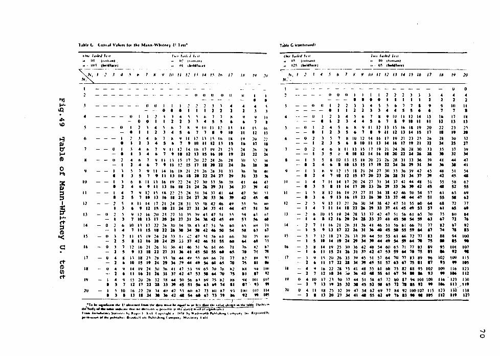
rt CD Ul rt
,,,. .. ,.,,,,.J ln1 , .... ,.,,, .. , ,,,, 01 t101tll.aHI
" • U11\ tNMlfacrl II.' 1t11u1.1111
111 1lii0Whut
~~~ --~_: __ ---~-: __ '''II /.'/I 1-1 /.~/II 17
I
y
IO
II
12
I l
14
IS
II>
IK
l'I
II
I 0
- U I I
ti 2 I
I I
11 I 0
I 2 0 I
.I 1 4 l
4 II 1 4 ~ 7 l 5
II ti 11 11 11
ti U I I I 0 I I
.1 4 1 1
I II
4 ' 11 7 M 'I Ill II I! 1145•7719
4 11 7 M 'I II 12 I.I I\ Ill l 4 5 • 7 9 10 11 11 I.I II 7 'I II I 2 14 Ill 17 l'I 21 4 • 7 9 ID 11 11 15 I• II 7 Y II II I~ 17 211 22 24 211 • 7 9 11 !! l!I 17 II 1t 11
9 11 14 11> IM 21 21 211 2M JI 7 9 11 U .. II 10 11 14 17
" • II Ill
IM 15 2.1 19
2M 14
.I.I 19
I J II M II 11 II> l'I 22 24 n JU H .Ill .IK I 1 4 • 9 II U .. II 21 14 1• 1' 11 14
I 4 7 'I 12 I~ IM 22 2' 2M .II J4 .17 41 44 I 1 5 7 ID U .. II 21 14 17 111 ll l6 1' 2 ~ M 11 14 17 21 24 2M l I I\ I• 42 41> 4'1
l • 9 11 l!I II 11 14 17 11 14 .17 41 44
U \ Y 12 II> 211 H 27 .II H W 41 47 \I '' l 7 10 I) 17 18 14 17 11 )4 111 41 4!1 49
0 I> IO II 17 22 211 Jll .14 JM 41 41 'I \II 1141 4 7 II 15 II 11 1• 10 14 111 41 46 !II !M
II 7 II I\ IY 24 2K H J • ~- 41 \I \II 111 1111 5 I 11 .. 10 14 1' .;.; 17 41 46 !II 5!1 Ml
0 7 12 II> 21 21> II )II 41 46 \I \11 111 1111 71 !I 9 U II 11 17 11 l6 41 4!1 !II !1!1 '8 •!I
U 4 K II IK 21 2M H IK 44 4'1 '' "'I 1111 11 71 1 • 10 15 " 14 19 )4 .;, 44 49 Sot ... •!I 70
- U 4 'I 14 IY 24 .Ill )II 41 47 \.I W 11\ 711 711 MJ 1 • 11 .. 11 1• 11 17 41 47 !11 !Ill '4 711 75
I 4 Y I~ 2U 211 l2 IM 44 Sii Sii 1>1 11'1 H M2 MK I l 7 11 17 11 U )) )t 45 51 ~ •l .. 74 II
I ' 10 II> JJ 2M 14 411 47 \I "'I 117 7) Mii M7 'II I l I U II J.4 111 16 41 41 !M '8 •7 71 79 ..
I~
II
4 1 ., •
14 II
l'I .. 24 11
.111 1• Jll 11
41 17
41 41
H 47 ,., !I.I
"' !Ill 711 ... 111 70
M! 7!1
KK II '14 17
llMI 91
/Y .. 'fl
I I
• • 4 .I
'I 111 7 I
I\ 111 11 ll ]O !! 17 II ~h !M 11 24
.\! 14 11 )I
IM 40 )) 1'
44 41 )IJ 41
'" " 4!1 41
"" ,... !II ~
111 ,., !o6 .. llY 11 •.I •7 74i 910
.. H
"~ ~1 7'. " "" .,, II .. t.1.a 11•1 17 ti
1111 jlll
9) " 1117 IU .. 11!1
- -··- ·-,.. -· --,-- -·---;--·- --•1" tw ... k.M ttw (I -*"••..-d Ina. the .... •a.111 tw ..-1 w U1 •u ... '''Hiatt •hoin ""the .... O••hh "'
c1w"l.od,e1 .... .,... ..,., .. , tlrll•t ~ *nuon "poo1to.k 11 , .. ,-.1 .... Cllt~IUliun ... tro. ••• ,....,.. ..... S..1111N I ti, ..... ,, I ... ,. ( or,••1h1 ii •~h ... , ···d~•onb P\.hl,.h1n1 t tt"-r•n) In· lhr11nlf'4 "'' ,...,.,..,""'el ehc pvbh•hc1. l1uul,,/( olt P\.bl,.tun1 C ...npen). Muo.lfH,. t •hi
T ~bl< C lconhnuodl
o .. , '"'''"' ,,,, ., O\ trom.,,nt u o]\ cNWr•ctt ~ ----- - -· -·-·---. N, I ! 4 6 N.',
I•·•• l4'1lrJ ln1 IO 1rom.n1 u' ( .. .,, .... ,
11 !I /fl II /] I.I 14 /j /6 IN /9 ]I)
-~----------------------------
9
Ill
II
12
ll
14
I~
I&
17
I~
20
0
u
0
I I
II
II
I 0
2 I
J 1
II
I I
J 1 4 l s 4
II
I 0
2 I
u
s l
II
4 l
" 5
I I I 0 I 0 ) ) 4 1 1 l
~ " 7 4 4 5
M 9 II • 7 I
I l D I S,1>771 l 4 4 5 5 ' H 9 IO II 12 14 • 7 I 9 II II
12 ll IS II> IK 19 9 II 11 U 14 15
' l 7 5
I IU 12 14 II> • I 10 II U
17 l'I 21 2J 2S 14 .. 17 " 11
19 21 24 21> 2K lO .. 111111114:16
II 5
II
• ll IS 17 II 11 14
K IU IJ I~ IM 2U • I II U 15 17
21
" 21> 2M )I Jl 11> 11 14 U 1' 11
)
1 9
' 15 II 20 17
21> 11
H 21 )9 )4
4 1 9 7
'" 11 22 II n 14
B )I
41 )6
I l 11 <I 12 I~ 18 21 24 21 )O H )I> )Y 42 4S 4H I 1 4 7 18 11 15 17 111 11 1• 21 11 14 17 lt 41
I 4 1 11 14 17 20 24 27 JI )4 )7 41 44 41 SI SS I l 5 I 11 14 17 111 11 1• 1' )) l6 lt 41 45 41
I ' • 12 II> IY 1.1 27 ll )4 ll 42 46 SU S4 S7 1>1 I l • 9 U .. 19 11 1• 10 )) 17 ... 44 47 51 55
2 ' Y ll 17 2! 21> lU .14 J8 42 47 SI SS llCl M l>8 I 4 7 II 14 II 11 U 1• )) 17 41 45 .. 51 57 ..
I> IU IS 19 24 2B H .17 41 47 " SI> 1>1 l>S 7U B 4 I 11 .. 18 14 JI )) 17 41 45 51 Sot 5' '1 •7
7 II Ill 21 111 ll ll> 41 41> SI '" 1>1 !>!> 71 77 11 5 9 11 17 11 1• 11 l6 40 45 51 55 59 64 67 74
1 11 IR 21 2h H 19 44 ~U 55 1>1 !>!> 72 77 ll II 5 10 14 " 14 1' )4 )t 44 49 Sot " '4 '71 75 • • 14 1'1 2S )II JI> 42 4H S4 Ml !>\ 71 77 U 19 9S • 11 15 11 1• 11 17 41 47 !11 5• '4 71 75 II 16
'I I\ JU 21> H W 4S " S7 M 70 77 ll K9 91> 102 • II 17 11 21 14 lt 4!1 51 57 •l •7 75 II 11 91
4 'I II> 22 21 lS 41 41 \~ 1>1 llK n 12 18 9S 102 109 1 7 11 II 14 _;;, l6 41 41 55 " •7 74 • 16 U "
U 4 IU 17 21 IU .17 44 SI SH I>~ 12 •O 17 94 IOI 109 Ill> 1 7 I) " 15 )1 )I 45 51 !II •5 71 .,. 15 91 " '"
U 4 11 IK J) l1 )'I 47 ~4 1>2 M 77 U 92 IOO 107 llS 121 1 I U 18 17 14 41 41 5' •1 .. 7• I) .. ti 115 111
u 0
4 4 1 1
IO II 7 I
17 IK I) I)
21 2\
" 18 )U .12 15 17
)7 )9 11 )4
44 47 )I 41
SI S4 4!1 41 ,. 1>2 51 55
"~ 1>9 !II •1 72 77 •5 .. Hll 14 71 7• 17 92 71 I)
94 100 15 ..
101 107 91 ..
109 II S
" 115 Ill> 121 116 111
' 121 1)0 Ill /119 Ila 118 llt 117

The Mann-Whitney Test Ho: Rank of MB • Rank of Non-MB Ha: Rank of MB - Rank Of Non-MB
(MB: Mouthbreathing) = 0.05
Subject Facial patterns Subject Ranks
Mouth- Non-Mouth Mouth Non-Mouth breather breather breather breather
N1=19 N2.,.19 H1•19 N2=19
-0.10 +0.05 26.5 29 -0.20 +0.60 21.5 36 -1.00 +0.06 8 30 -0.60 -0.24 15 20 +0.60 -1.08 36 6 +0.50 -0.84 34 11 -1.80 -0.98 1 9 -1.60 -0.56 2 17 +0.60 -0.12 36 24.5 -1.50 +o 02 3 28 +0.10 -0.96 32 10 -0.80 -1.06 12. 5 7 +0.10 -0.10 32 26.5 -0.80 -0.12 12.5 24.5 -0.30 -0.58 19 16 +1.60 -1.16 38 4 -1.10 -0.14 5 23 -0.20 -0.70 21.5 14 -0.50 +0.10 18 32
Total=373.5 367.5
T= 373.5 - 19(19+1) I 2 = 183.5 N, - 19 I N2 "" 19, a s 0.05
From Table W ~2 (=0.025) = 113
w 1 • •/2 = 19 x 19 - 113 - 248
w 1 • ~2 > T > w
So that the Ho can not be rejected.
It means that there is no difference between the two samples in terms of Facial Pattern
Fig.SO Statistical analysis for facial patterns
71
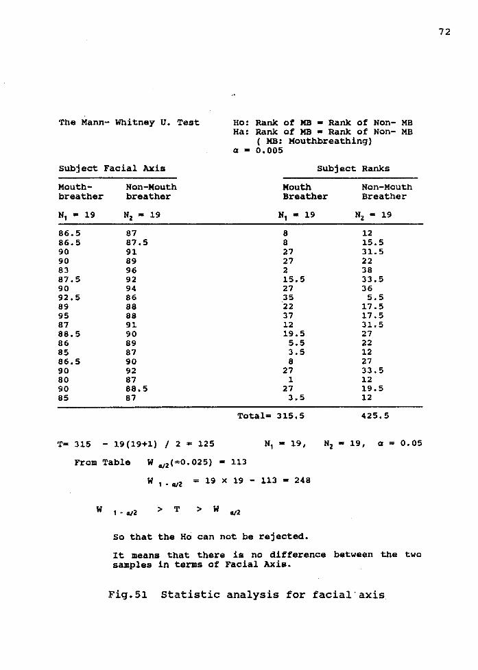
The Mann- Whitney U. Test Ho: Rank of MB • Rank of Non- MB Ha: Rank of MB • Rank of Non- MB
( MB: Mouthbreathinq) a "" o.005
subject Facial Axis Subject Ranks
Mouthbreather
N1 • 19
86.5 86.5 90 90 83 87.5 90 92.5 89 95 87 88.5 86 85 86.5 90 80 90 85
Non-Mouth breather
N2 • 19
87 87.5 91 89 96 92 94 86 88 88 91 90 89 87 90 92 87 88.5 87
Mouth Breather
8 8 27 27 2 15.5 27 35 22 37 12 19.5 5.5 3.5 8
27 l
27 3.5
Total• 315.5
Non-Mouth Breather
N2 • 19
12 15.5 31.5 22 38 33.5 36 5.5
17.5 17.5 31.5 27 22 12 27 33.5 12 19.5 12
425.5
Tm 315 - 19(19+1) / 2 "" 125 N1 • 19, N2 • 19, a • o. os
From Table w Cl/2(=0.025) - 113
w
w 1 • Cl/2 - 19 x 19 - 113 - 248
1 • Cl/2 > T > w Cl/2
so that the Ho can not be rejected.
It means that there is no difference between the two samples in terms of Facial Axis.
Fig.51 Statistic analysis for facial· axis
72

The Mann- Whitney u. Test Ho: Rank of MB .. Rank of Non- MB Ha: Rank of MB • Rank of Non- MB
( MB: Mouthbreathing) Q - 0.005
Subject Facial Depth Subject Ranks
Mouth- Non-Mouth Mouth Non-Mouth breather breather Breather Breather
N1 =19 N2 =19 N1 •19 N2 =19
84 83 20 15 82 83 9 15 88 80 34.5 2 83.5 80 18 2 82 82.5 9 11.5 81 86 5 27 88 83 34.5 15 86 87 27 30.5 81 84 5 20 91 80 37.5 2 84.5 83 22.5 15 87 85 . ·30. 5 24.5 84.5 84 22.5 20 85 81 24.5 5 87 82 30.5 9 82.5 87 11.5 30.5 91 81.5 37.5 7 88 86 34.5 27 88 83 34.5 15
Total• 448 293
Ta 448 - 19(19+1) / 2 • 258 N1 • 19, N2 c 19, a = 0.05
From Table w ~2 (=0.025) • 113
w 1 • ~2 - 19 x 19 - 113 - 248
T > W ~2
T > W 1. ~2
So that the Ho can be rejected.
It means that there i• difference between the two samples in terms of ~aoial Depth.
Fig.52 Statistics analysis for facial depth
73

The Mann- Whitney U. Test Ho: Rank of MB • Rank of Non- MB Ha: Rank of MB • Rank of Non- MB
( MB: Houthbreathing) a • 0.005
Subject Mand. Plane Angle Subject Ranks
Mouthbreather
N1 = 19
31 27 25 27 31.5 30 19 17 33 19 28 27 29 21 24.5 32 23 26 25
T= 419.5
Non-Mouth breather
N2 • 19
30 31 30 22.5 22 23 21.5 22 29.5 25 22 21.5 25 25 21 18 25 21 26
- 19(19+1) / 2 • 229.5
Mouth Breather
N1 • 19
34.5 26 19.5 26 36 32 3.5 1 38 3.5 28 26 29 6 16 37 14 .5 23.5 19.5
Total• 419;5
N, - 19 I Nz •
Non-Mouth Breather
N2 ""' 19
32 34.5 32 13 11 14. 5 8.5 11 30 19.5 11 8.5 19.5 19.5 6 2 19.5 6 23.5
321.5
19, a• 0.05
From Table W uz(•0.025) • 113
w
w 1 • uz • 19 x 19 - 113 • 248
t • uz > T > w u2
So that the Ho can not be rejected.
It means that there is no difference between the two samples in terms of Mandibular Plane Angle.
Fig.53 Statistical analysis for mandibular plane angle
74

The Mann- Whitney u. Test Ho: Ranlc ot MB • Rank of Hon- MB Ha: Rank ot MB • Rank ot Hon- MB
( MB: Mouthbreathing) Cl - 0.005
Subject Mandibular Arc Subject Ranks
Mouth- Hon-Mouth breather breather
H1 • 19 Hz• 19
30.5 30 30 20 31 25 30.5 27 27 31 25 30 37 32 37 31 28 30 25 32 27.5 34 35 31 31.5 ll 34 31.5 29 35 25 30 30 34 23.5 33 35 25
Mouth Breather
19.5 15.5 23 19.5 8.5 5 37.5 37.5 11 5 10 35 26.5 32 12 5 15.5 2 35
Total• 355
Hon-Mouth Breather
Hz • 19
15.5 1 5 8.5 23 15.5 28.5 23 15.5 28.5 32 23 23 26.5 35 15.5 32 30 5
386
T- 355 - 19(19+1) / 2 • 165 N2 • 19, a • 0.05
From Table W uz(•0.025) • 113
w t • tl/Z - 19 x 19 - 113 - 248
w 1. tl/Z > T > W tl/Z
So that the Ho can not be rejected.
It means that there is no difference between the two saaples in terms of Mandibular Arc.
Fig.54 statistical analysis for mandibular arc
75

The Mann- Whitney u. Test Ho: Rank of MB - Rank Of Non- MB Ha: Rank of MB • Rank of Non- MB
( MB: Mouthbreathinq) Q - 0.005
Subject Lower Facial Ht. Subject Ranks
Mouth- Non-Mouth breather breather
N1 • 19 Nz • 19
45 47 41 47 42 43 41.5 43 47 41.5 48 44 41 45 40 44 50 42 38 46 46 39 42 36 46 48 42 42 45 45 48 40 41 45 46.5 45 44 42
Mouth Breather
-24.5 7 13.5 9.5 33 36 7 4.5 38 2 29 13.5 29 13.5 24.5 36 7 31 20
Total• 378.5
Non-Mouth Breather
N2 • 19
33 33 17.5 17.5 9.5 20 24.5 20 13.5 29 3 1 36 13.5 24.5 4.5 24.5 24.5 13.5
362.5
T- 378.5 - 19(19+1) I 2 • 188.5
Fro• Table W _,2(•0.025) • 113
Nz • 19, a • 0. 05
w
W 1 • c/Z • 19 >< 19 - 113 Al: 2.48
> T > w «12
so that the Ho can not be rejected.
It means that there is no· difference between the two samples in terms of Lower Facial Heiqht.
Fig.SS Statistical analysis for lower facial height
76

77
1. From the result of the Mann-Whitney u.test, the null
hypothesis which stated that there was no difference between
the two samples could not be rejected at the p< 0.05 level as
far as facial pattern, facial axis, mandibular plane angle,
mandibular arc and lower facial height are concernd. It could
be concluded that both groups were from the same population
distribution. That meant there were no statistical difference
between the experimental and the control groups as far as
facial pattern, facial axis, mandibular plane angle,
mandibular arc and lower facial height were concerned.
2. The only measurement which had significant difference
was the facial depth.
3. The result showed that there was no significant effect
of the mouthbreathing on the craniof acial pattern for the
children from three to seven years of age.
4. All the means of the facial patterns of the
experimental group, the control group, and the Bolton standard
templates were within one standard deviation, on the
Brachyfacial side, i.e. though they were all within mesofacial
pattern but on the brachyfacial side of the Rickett•s norm.
5. The mean of facial pattern of the experimental is 0.2
on the brachyfacial side to the Rickett's norm. The mean of
the facial pattern of the Bolton standard from age three to
seven is 0.5 on the brachyfacial side. And the mean of the
facial pattern of the control group which is taken from the
78
Bolton study is 0.4 on the Brachfacial side, which is pretty
much close the the mean of facial pattern of its population
i,e. the Bolton standard.

CHAPTER V
DISCUSSION
1. The experimental sample size was limited by the
difficulty of getting a larger sample referred from an ENT
off ice which is a little far away from the investigator• s
department. The small size of sample made not only the
parametric analysis inadequate but also the original attempt
to compare the facial patterns between each age group
impossible. Future researches with a larger sample size might
be proper to more completely evaluate the effect of
mouthbreathing on the craniofacial development at each age
group.
2. Because the depth.of nasopharynx which was related to
air flow was established in the first or second year of life, 7
1920 and only minor changes in size of adenoid were observed
between the ages of 2 to 15 years26, it can be assumed that
the air flow capacity was established as early as age two and
kept constant until puberty when the adenoids started to
recede. We suppose that mouthbreathing developed in those
subjects who had adenoid obstruction at their age of
examination may exist for many years until puberty if
treatments were not given.
Based on the mentioned fact stated above, there is no
evidence showing that mouthbreathing has effect. on the
79

80
craniofacial deformity for those young children in this study.
Early adenoidectomy to correct the mouthbreathing to nose
breathing was recommended by Linder-Aronson57 as helpful to
prevent the facial deformity.
3. Because the control group is from the Broadbent-Brush
Growth study Center, there is no way to be absolutely sure
that they were nasal-breather. However, they were within
normal value cephalometrically. For future study, samples with
history of non-mouthbreathing from normal population will be
proper for the control group.
4 • Most studies demonstrated a positive relationship
between airway obstruction and a dolicofacial pattern. 10111213
14 15 16 17 18 The reasons that there was no significant
relationship found in this research may be due to the
following factors:
A. The period of time of mouthbreathing for the subjects
in this research had been so short that changes on the
development of facial deformity were not yet significant.
Because the size of airway was established in the early
childhood by the stable size of adenoid24 25 and the stable
depth and width of nasopharyngeal space19 20 38 , the subjects
might continue the mouthbreathing until treatment or after
puberty when adenoids gradually recede. 25 In Drs.• Vargervik
and Harvold • s animal study21 it was concluded that the changes
in mandibular morphology will only occur when lowering of the
mandible was sufficiently persistent.

81
Most parents and children didn't remember the age of
onset of mouthbreathing nor the exact period of time of the
existence of the mouthbreathing problem. So we would not be
able to know the period of time that mouthbreathing had its
effect on the craniofacial development for each subject. A
longitudinal research for mouthbreathers could be indicated
to evaluate the effect of mouthbreathing on craniofacial
development.
B. The subjects in this research were very young. Most
of them didn't have first permanent molar eruption. So the
effect of mouthbreathing could not have significant changes
on the development of facial deformity. In the study by Drs•
Vargervik and Harvold21 , they concluded that the lower face
height was increased significantly when excessive molar
extrusion occurred. Increased molar extrusion which may cause
increased lower facial height would be expected to occur most
rapidly during eruption of the first and second molars.
5. A small sample size of this study may also have an
impact on the result. The underlying distribution of the
experimental group in not known to be of a normal population.
The data of this study can't be used in parametric statistical
analysis to other studies such as Michigan, Burlington, or
Ricketts' which had a large sample size as a normal
population.
A further related research with large sample size,
matched race, age, and sex would be indicated to either

82
confirm this research or may have different result.
6. Different sources of materials for the experimental
and control groups may have another impact to this study. The
lateral head plates of the Broadbent-Brush Growth Center are
more than forty years old and might not be clear enough to
identify exactly the needed anatomical points. The anatomical
porions, which are very important in the Ricketts' facial
pattern analysis, were converted from machine porions showing
on those lateral head plates by a set of different templets.
The different templates were used according to different types
of ear roads and head fixer poles used in different years when
those lateral head plates were taken during the whole period
of time of Broadbent-Brush Growth Study. There might be errors
between the real and the converted anatomical porions and
which may influence the facial pattern analysis.
For further study, the control samples not only to be
matched to the experimental sample in race, sex and age but
also taken not from the other old study materials but by the
exact the same method as the experimental samples would be
suggested.
7. From the result that the mean of the facial pattern
of the experimental group, which is 0.2 on the brachyfacial
side, is a little bit more on the dolicofacial pattern side
when it is compared to the means of facial patterns of the
control group and the Bolton standard which are 0.4 and 0.5
on the brachyf acial pattern side seperately. · So if from

83
inspection of comparison between the means of facial patterns
of the expremental and the control group, it seems that there
is possibility of· tendency that the effect of mouthbreathing
to the growth of facial pattern may have existed even to the
young study group. Further study would be very necessary and
interesting to confirm this assumption.

84
CHAPTER VI
SUMMARY
Many studies had shown the effect of nasopharyngeal
obstruction to the development of craniof acial pattern on
children aged above six. No study had ever been done to
identify the effect on younger children. This study was trying
to investigate such effect on children from three to seven
years of age.
Nineteen children with history of chronic nasopharyngeal
obstruction were ref erred from an Ear, Nose, and Throat
specialist to us to take a lateral head X-Ray film.
Cephalometric analysis according to Ricketts' facial pattern
analysis had been calculated for each subjects. For the
control group, nineteen tracings of lateral head X-Ray films
of those who didn't have airway obstruction history were
chosen to match the experimental group by race, sex and age
from Broadbent-Brush Growth Center. They were also calculated
as the same way as the experimental group for their facial
patterns.
The result of the statistical analysis showed there is
no difference between the two groups as far as the facial
patterns were concerned. From this study the effect of
mouthbreathing on the craniofacial development for children
from three to seven years of age could not be found.

Further studies with large sample size would be
indicated.
85

BIBLIOGRAPHY
1. Brash JC, ~ckeag HTA,Scott JH The aetiology of irregularity and malocclusion of the teeth . Dent. Broad u. Kingdom,1929.
2. Bresolin Dante, Shapiro GG, Shapiro PA, Dassel sw, Furudase CT, Pierson WE, Chapko M, Bierman WC: Facial characteristics off children who breathe through the mouth. Pediatrics 73(5):622-625,1984.
3. Ricketts RM : Form on the tonsil and adenoid problem in Orthodontics, Respiartory obstruction syndrome. Am, J. Orthod.,54:495 1986.
4. Ranly OM : Theories of craniofacial growth, In: Ranly OM, ed. A synopsis of craniofacial growth. New York: AppletonCentury Croft, 1980.
5. Gwynne-Evens D, Ballard CF : Discussion on the mouthbreather. Proc.R. Soc. Med. 51:279-285, 1959.
6. Humphreys HF,Leighton BC : A survey of anteroposterior abnormalities of the jaws in children between the ages of two and five and a half years of age. Br. Dent. J. 88:3-15,1950.
7. Kingsley WS : A treatise on oral deformities as a branch of mechanical surgery, New York, 1889, D Appleton Company.
8. Leech HL : A clinical analysis of orofacial morphology and behavior of 500 patients attending an upper respiratory research clinic. Dent. Practit. Dent. Rec., 9:57,1958.
9 • o 'Ryan FS, Gallagher OM, Labanc JP, Epker BN The relation between nasorespiratory function and dentof acial morphology:A review. Am. J. Orthod.: 82(5):403-444.
10. Harvold EP, Tomer BS, Chierici G : Primate experiments ojn oral respiration. Am. J. Orhtod. 79(4): 359-372,1981.
11. Harvold EP, Vargervik K,Chierici G : Primate experiments on oral sensations and dental malocclusions. AM. J. Orthod. 63:494-508, 1973.
12. Linder-Aronson s, Backstrom A: A comparison between mouth and nose breathers with respect t occlusin and facial dimensions. Odol Revy,11:343-376,1960.
13. Linda-Aronson s Adenoid. Their effects on mode of breathing and nasal airflow and their relationship to characteristics of facial skeleton and the dention. Acta
86
87
Otolaryng. Suppl. 265:1, 1970.
14. Linder-Aronson S. : Effects of adenoidectomy on dent ion and nasopharynx. Am.J.Orthod.65(1):1-15.1974.
15. Martin R, Vig PS, Warren DW Nasal resistance and vertical dentofacial features, IADR ABSTRACT,1981.
16. Vig PS, Sarver DM, Hall DJ, Warren DW: Quantitative evaluation of nasal airflow of nasal airflow in relation to dental morphology. AM.J. Orhtod.79:263-272,1981.
17. Watson RM, Warren DW, Fischer ND : Nasal resistance, skeletal classification and mouth breathing in orthodontic patients. AM. J. Orthod. 54:367-379,1968.
18. Woodside DE, Linder-Aronson s :The channelization of upper and lower anterior face heights compared to population standard in males between ages 6 to 2 o years. Eur. J. Orthod.1:25-40,1979.
19. Brodie AG : On the growth pattern of the human head from the third month to the eighth year of life, Am. J. Anat.,68:209, 1941
20. Handelman cs, Osborne G : Growth of the nasopharynx and adenoid development from one to eighteen years.? 46(3):243-259,1976.
21. Ricketts RM : The cranial base and soft structure in cleft palate speech and breathing. Plast. Reconstr. surg.,14:47,1954.
22. Bergland o : The bony nasopharynx. Acta Odont. Scand., Supply. 35:1, 1963.
23. Subtelny JD : The significanceof adenoid tissue in orthodontia. Angle Orthodontics, 24(2) 59-69, 1954 April.
24. Scammon RE : The measrement of the body in childhood. In Harris JA, editor: The measurement of Man. Minneapolis,1930, University of Minnesota.
25. Subtelny JD, Baker HK : The significance of adenoid tissue in velopharyngeal function. Plastic and Rsconstructie surgery, 17(3):235-250, 1956.
26. Johannessen s.: Roentgenoloqic investigation of the nasopharyngeal tonsil in children of different ages.? :299-304,1967 March.
27. Miller MJ, Martin RJ, Carlo WA, Fouke JM, Strohl KP,
88
Fanaroff AA : Oral breathinq in newborn infants. J. Pediatrics, 1985,Sep:465-469.
28. Rodenstein DO, Stanescu DC : Soft palate and oronasal breathing in humans, Am Physiological Society 1984: 651-657. 29. Trask GM, Shapiro GG. Shapiro PA : The effctts of perennial allerqic rhinitis on dental and skeletal development: A comparison of siblinq pairs. Am. J. orthod 92:286-293,1987.
30. Weimert TA : JCO/Interview : On Airway obstruction in Orthodontic Paractice. J. of Orthod. 1986 Feb.:96-105.
31. Reed GF : Nasal obstruction: Cause and treatment. Postgrad.med.,34:464,1963.
32. Emslie RD, Hassler M, Zwemer JD : Mouth breathinq I: Etiology and effects. J. Am. Dent. Assoc.,44:506,1952.
3 3 • Goldmen JL, Bachman AL : Soft tissue roetgenoqraphy of the nasopharynx for adenoid. Am. Laryng. Rhin. Otol. soc. P-692, 1958.
34. Hibbert J, Whitehouse GH : The assessment of adenoidal size by radioloqical means. Clinical otolaryngology. 1978,3,43-47.
35. Khoo FY, Nalpon J : Contrast examination of nasopharynx with cinefouorograph and roentqenograph: new technic. Am. J. Roentgenol., 99:238,1967.
36. Johnson AP, Murry AM, Maran AGO : Errors in the assessment of nasopharyngeal airway by radioqraphy. Clinic Otolaryngology:1983,Nov:l017-1026.
37. Holmberq Hans, Linder-Aronson s: Cephalometric radioqraphs as a means to evaluatinq the capacity of the nasal and nasopharynqeal airway. Am. J. Orthod.,76(5): 479-490,1979.
38. Weimert TA : Evaluation of the upper airway in children. Ear, Nose and Throat J. 66:196-200,1987.
39. Quinn GW : Airway Interference Syndrome, Clinical identification and evaluation of nose breathing capabilities. Anqle Orthod. 53(4):311-319, 1983.
40. Hibbert J, Tweedie CK : The value of sign and symptoms in the diaqnosis of enlarqed adenoids. Clinical Otolarynqology,
41. crepeau J, Patriquin HB, Poliquin JF, Tetreault L, Radiographic evaluation of the symptom-producing adenoid. Otolarynqol Head Neck Surg 1982;90:548-554 (Sep-Oct).

89
42. Hannudsela A: The effect of moderate and severe atopy on the facial skeletan. Eur. J. orthod. 1981;3:187-193.
43. Shapiro GC, · Shapiro PA: Nasal airway obstruction and facial development.Clin Rev Allergy 1984;2:22S-23S.
44. Petit H: Upper airway problems and pre-Orthodontic Orthopedics.Ear,Nose and Throat J. 228-236,1987.
4S. Vergervik K, Miller AJ. Chierici G, Harvold E, Tomer BS: Morphologic response to changes in neuromuscular patterns wxpserimentally induced by altered modes of respiration. Am. J. Orhtod. 8S:11S-124 1984.
46. Meredith GM: The airway and dentofacial development.Ear,Nose and Throat J., 66:190-19S,1987.
4 7 • Linder-Aronson s: Respiratory function in relation to facial morphology and the dention~ British J. Ortho.,6:S9-71,1979.
48. Bosma, :
49. Bibby RE: The Hyoid bone position in mouth breathers and tongue-thrusters. Am. J. Orthod. 8S(S):431-433,1984.
so. Linder-Aronson S,Henrikson co : Radiociphalometric analysis of anteroposterior nasopharyngeal dimensions in 6-to 12-year old mouth breathers compared with nose breathers. ORL 3S:19-29,1973.
Sl. Cohen D.,Konak s.: The evaluatiion of radiographs of the nasopharynx. Clin. Otolaryngol 198S,10,73-78.
S2. Hibbert J, Stell PM : A radiological study of the adenoid in normal children. Clinical Otolarygology, 1979,4,321-327.
S3. Fujioka M, Young LW, Girdancy BR: Radiographic evaluation of adenoidal size in children: Adenoidal-Nasopharyngeal ratio. Am. J. Roen., 133:401-404, 1979.
S4. Richter HJ: Obstructiion of the pediatric upper airway. Ear,Nose and Throat J. 66:209-?, 1987.
ss. Linder-Aronson s, Woodside DC : Lundstrom A: Mandibular growth direction following adenoidedctomy. Am. J. Orthod.,89(4):273-284, 1986.
S6. Linder-Aronson s.: Respiratory function in relation to facial morphology and the dention. British J. ortho., 6:S9-71,1979.

90
57. Linder-Aronson S: Effects of Adenoidectomy on mode of breathinq, size of adenoids and nasal airflow. otolarynqotoloqy 35:283-302,1973.
58. Linder-Aronson S: The skeletal and dental effects of rapid maxillary expansion. British J. orthod. 66:25-29,1979.
59. Guenthner TA, Sather AH, Kern EB: The effect of Le Fort I maxillary impaction on nasal airway resistance. Am. J. orthod,:85(4):308-315,1984.
60. Varqervik K,Harold EP : Experiments on the interaction between orofacial function and morpholoqy. Ear, Nose, and Throat J. 66:201-208,1987.

APPROVAL SHEET
The thesis submitted by Tai Yang Hsi, B.D.S., has been read and approved by the following committee:
Lewis Klapper, D.M.D., M.Sc., D.Sc. Professor and Chairman Department of Orthodontics, Loyola
Richard Port, D.D.S., M.S. Assistant Professor Department of Orthodontics, Loyola
Michael Kiely, Ph.D. Professor Department of Anatomy, Loyola
The final copies have been examined by the director of the thesis and the signature which appears below verifies the fact that any necessary changes have been incorporated and that the thesis is now given final approval by the Committee with reference to content and form.
The thesis is therefore accepted in partial fulfillment of the requirements for the degree of Master of Science.
Nove/l'ber r11 1 t8f Date