Embed Size (px)
Citation preview
Degradation of the cyclin-dependent kinase inhibitor KRP1is regulated by two different ubiquitin E3 ligases
Hong Ren1, Aaron Santner1, Juan Carlos del Pozo2, James A. H. Murray3 and Mark Estelle1,*
1Department of Biology, Indiana University, Bloomington, IN 47405, USA,2Dpt. Biotecnologıa, Instituto Nacional de Investigacion y Tecnologıa Agraria y Alimentaria (INIA), 28040 Madrid, Spain, and3Institute of Biotechnology, University of Cambridge, Cambridge CB2 1QT, UK
Received 25 May 2007; revised 21 September 2007; accepted 17 October 2007.*For correspondence (fax 1 812 855 6082; e-mail [email protected]).
Summary
In animals and fungi, a group of proteins called the cyclin-dependent kinase inhibitors play a key role in cell
cycle regulation. However, comparatively little is known about the role of these proteins in plant cell cycle
regulation. To gain insight into the mechanisms by which the plant cell cycle is regulated, we studied the
cyclin-dependent kinase inhibitor KRP1 in Arabidopsis. KRP1 interacts with the CDKA;1/CYCD2;1 complex
in planta and functions in the G1–S transition of the cell cycle. Furthermore, we show that KRP1 is a likely
target of the ubiquitin/proteasome pathway. Two different ubiquitin protein ligases, SCFSKP2 and the RING
protein RKP, contribute to its degradation. These results suggest that SCFSKP2b and RPK play an important role
in the cell cycle through regulating KRP1 protein turnover.
Keywords: Arabidopsis, cell cycle, KRP ubiquitin.
Introduction
The cell cycle consists of a series of events that ultimately
lead to the formation of two daughter cells. In eukaryotes,
the fundamental mechanisms of cell cycle regulation are
highly conserved. The cycle is divided into four phases, G1,
S, G2 and M, and has two major checkpoints to control cell
cycle progression: the G1–S transition and the G2–M tran-
sition. Cell cycle progression is controlled by the activities of
cyclin-dependent kinase (CDK)/cyclin complexes. Different
combinations of CDKs and cyclins regulate passage from
one phase of the cycle to the next (De Veylder et al., 2003;
Dewitte and Murray, 2003). The activities of CDK/cyclin
complexes can be regulated by CDK inhibitors (CKIs), which
function as negative regulators of CDK activity (Sherr and
Roberts, 1999). CKIs have been identified in yeast, mammals
and plants (De Clercq and Inze, 2006). In mammals, there are
seven CKIs, which are classified into two families: the INK4
family and the Cip/Kip family (Nakayama and Nakayama,
1998; Vidal and Koff, 2000). Plants do not appear to have
INK4-type CKIs, but proteins related to the Cip/Kip family
have been identified in Arabidopsis, tobacco, maize and
tomato (Bisbis et al., 2006; Coelho et al., 2005; De Veylder
et al., 2001; Jasinski et al., 2002; Wang et al., 1997). The
Arabidopsis genome encodes seven proteins related to the
mammalian CKI p27Kip1, known as Kip-related proteins
(KRPs) or interactors/inhibitors of Cdc2 kinase (ICKs; De
Veylder et al., 2001; Vandepoele et al., 2002; Zhou et al.,
2002a,b). The only sequence similarity between KRPs and
p27Kip1 is in the conserved CDK-binding/inhibitory domain
(De Veylder et al., 2001; Wang et al., 1997). Recently, an
additional CKI was identified in Arabidopsis called SIAMESE
(SIM; Churchman et al., 2006). The SIM protein includes a
cyclin binding domain and a domain also found in the KRP
proteins. Genetic studies indicate that SIM has a role in the
control of endoreduplication.
Recent studies have begun to shed light on the function of
plant CKIs in cell cycle regulation and growth. Ectopic
expression of KRP1, KRP2, KRP4 and KRP6 confirm that
these proteins function as inhibitors of the cell cycle,
resulting in dwarfed plants with reduced cell number and
organ size (Bemis and Torii, 2007; De Veylder et al., 2001;
Wang et al., 2000; Zhou et al., 2003). Studies of transcrip-
tional regulation have shown that KRP1 transcription is
increased by low temperature and abscisic acid (ABA),
whereas KRP2 transcription is downregulated by auxin
during lateral root initiation (Himanen et al., 2002; Wang
et al., 2000). The expression patterns of KRPs during the cell
ª 2008 The Authors 705Journal compilation ª 2008 Blackwell Publishing Ltd
The Plant Journal (2008) 53, 705–716 doi: 10.1111/j.1365-313X.2007.03370.x
cycle were characterized using synchronized Arabidopsis
cultured cells (Menges and Murray, 2002; Menges et al.,
2005). These data show that there are three main patterns of
transcriptional regulation of KRP genes. KRP1 is highly
expressed in non-dividing cells and is strongly downregu-
lated during the G1 phase in cell cycle re-entry. KRP1 shows
a further clear peak of expression at the G2–M transition,
although this is threefold lower than the expression in non-
dividing cells. KRP2 is highly expressed in non-dividing
cells, and is unique in showing a peak of expression only
during the G1 phase as cells re-enter the cell cycle. In
contrast, KRP3, KRP4, KRP5, KRP6 and KRP7 are not highly
expressed in non-dividing cells, but are upregulated or peak
during the S and early G2 phases. These results implicate
KRP1 and KRP2 as primary candidates for controlling
activation of division by non-dividing cells. Among the plant
CKIs, KRP1 and KRP2 are known to be degraded by the 26S
proteasome (Jakoby et al., 2006; Verkest et al., 2005a).
Interestingly, degradation of KRP2 requires its phosphory-
lation by the CDKB1;1 complex (Verkest et al., 2005b).
However, the detailed mechanisms of KRP1 and KRP2
degradation remain to be elucidated.
In eukaryotes, ubiquitin-mediated protein degradation
plays a critical role in the cell cycle by destroying many
important cell cycle regulators (Hershko, 2005). Conjugation
of ubiquitin to a substrate requires the sequential action of
three enzymes: ubiquitin-activating enzyme (E1), ubiquitin-
conjugating enzyme (E2) and ubiquitin-protein ligase (E3).
The E3 enzymes are responsible for the specificity of the
pathway, and several classes of E3s have been implicated in
cell cycle regulation including the SCF (SKP1-cullin-F-box
protein) and RING domain ubiquitin ligases. An SCF com-
plex is composed of four subunits: RBX1, CUL1, SKP1 and an
F-box protein. The CUL1 subunit functions as a scaffold to
bind RBX1 and the SKP1-F-box protein subcomplex. The
F-box protein subunit provides specificity for SCFs and
binds substrates (Moon et al., 2004; Petroski and Deshaies,
2005). In yeast and mammals, the role of SCFs in cell cycle
regulation has been extensively investigated. SCFs are
responsible for the degradation of cyclins, CKIs, transcrip-
tion factor E2F-1 and many other cell cycle regulators
(Hershko, 2005). A well-known example is the regulation of
p27Kip1 degradation by SCFSKP2 in mammals. SCFSKP2
targets p27Kip1 for degradation to trigger the G1–S transition
of the cell cycle (Sutterluty et al., 1999; Tsvetkov et al., 1999).
The Arabidopsis genome encodes two F-box proteins
(called SKP2a and SKP2b) that are related to mammalian
SKP2 (del Pozo et al., 2002). SKP2a appears to recruit the
phosphorylated form of the transcription factor E2Fc for
degradation. Whether SKP2b also regulates E2Fc degrada-
tion is unknown. In addition to E2Fc, another cell cycle
regulator that may be an SCF substrate is CYCD3;1. This
cyclin is unstable and its degradation depends on the 26S
proteasome (Planchais et al., 2004). In transgenic plants with
reduced levels of RBX1, CYCD3;1 accumulates indicating
that SCF is involved in its degradation. However, the F-box
protein component of this SCF has not been identified
(Lechner et al., 2002; Liu et al., 2004).
To investigate the post-translational regulation of plant
CKIs, we focused on KRP1. Our data demonstrate that KRP1
interacts with the CDKA;1/CYCD2;1 complex in planta. Fur-
thermore, we show that KRP1 degradation is dependent on
SCFSKP2b and the RING protein RKP. These results provide
new insight into the mechanisms by which the plant cell
cycle is regulated by protein degradation.
Results
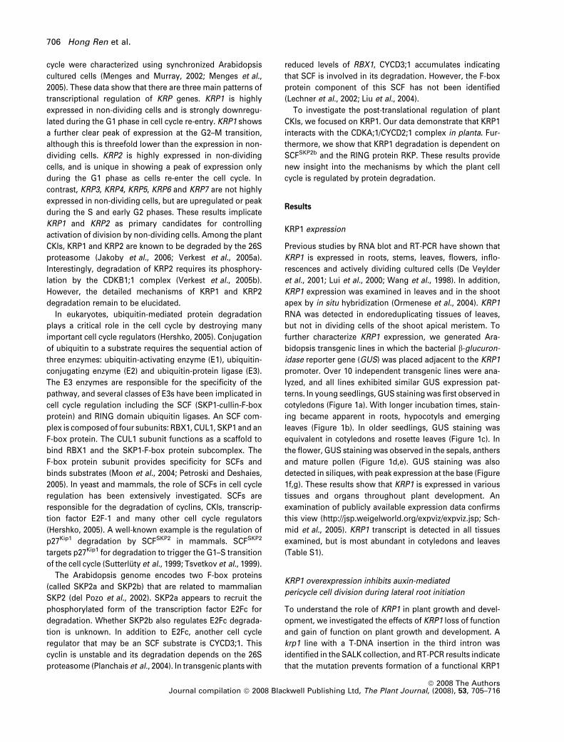
KRP1 expression
Previous studies by RNA blot and RT-PCR have shown that
KRP1 is expressed in roots, stems, leaves, flowers, inflo-
rescences and actively dividing cultured cells (De Veylder
et al., 2001; Lui et al., 2000; Wang et al., 1998). In addition,
KRP1 expression was examined in leaves and in the shoot
apex by in situ hybridization (Ormenese et al., 2004). KRP1
RNA was detected in endoreduplicating tissues of leaves,
but not in dividing cells of the shoot apical meristem. To
further characterize KRP1 expression, we generated Ara-
bidopsis transgenic lines in which the bacterial b-glucuron-
idase reporter gene (GUS) was placed adjacent to the KRP1
promoter. Over 10 independent transgenic lines were ana-
lyzed, and all lines exhibited similar GUS expression pat-
terns. In young seedlings, GUS staining was first observed in
cotyledons (Figure 1a). With longer incubation times, stain-
ing became apparent in roots, hypocotyls and emerging
leaves (Figure 1b). In older seedlings, GUS staining was
equivalent in cotyledons and rosette leaves (Figure 1c). In
the flower, GUS staining was observed in the sepals, anthers
and mature pollen (Figure 1d,e). GUS staining was also
detected in siliques, with peak expression at the base (Figure
1f,g). These results show that KRP1 is expressed in various
tissues and organs throughout plant development. An
examination of publicly available expression data confirms
this view (http://jsp.weigelworld.org/expviz/expviz.jsp; Sch-
mid et al., 2005). KRP1 transcript is detected in all tissues
examined, but is most abundant in cotyledons and leaves
(Table S1).
KRP1 overexpression inhibits auxin-mediated
pericycle cell division during lateral root initiation
To understand the role of KRP1 in plant growth and devel-
opment, we investigated the effects of KRP1 loss of function
and gain of function on plant growth and development. A
krp1 line with a T-DNA insertion in the third intron was
identified in the SALK collection, and RT-PCR results indicate
that the mutation prevents formation of a functional KRP1
706 Hong Ren et al.
ª 2008 The AuthorsJournal compilation ª 2008 Blackwell Publishing Ltd, The Plant Journal, (2008), 53, 705–716
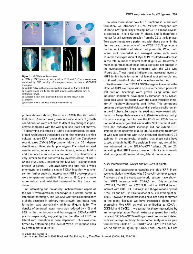
protein (data not shown; Alonso et al., 2003). Despite the fact
that the krp1 mutant was grown in a wide variety of growth
conditions, we were not able to detect any changes in phe-
notype compared with the wild-type line (data not shown).
To determine the effects of KRP1 overexpression, we gen-
erated Arabidopsis transgenic plants that express a c-Myc
epitope tagged KRP1 under the control of the Cauliflower
mosaic virus (CaMV) 35S promoter. More than 30 indepen-
dent lines exhibited similar phenotypes. Plants had serrated
rosette leaves, reduced apical dominance, reduced fertility
and a reduced numbers of lateral roots. This phenotype is
very similar to that conferred by overexpression of KRP1
(Wang et al., 2000), indicating that Myc-KRP1 is a functional
protein in planta. A 35S:Myc-KRP1 line that has a weak
phenotype and carries a single T-DNA insertion was cho-
sen for further analysis. Interestingly, KRP1 overexpressors
were temperature sensitive. If grown at 18�C, plants were
more robust and exhibited increased fertility (data not
shown).
An interesting and previously uncharacterized aspect of
the KRP1-overexpression phenotype is a severe defect in
lateral root formation. The 35S:Myc-KRP1 line exhibited only
a slight decrease in primary root growth, but lateral root
formation was dramatically inhibited (Figure 2a,b). The
density of emerged lateral roots was reduced by 41% and
96% in the hemizygous and homozygous 35S:Myc-KRP1
plants, respectively, suggesting that the effect of KRP1 on
lateral root formation is dose dependent. This was con-
firmed by determining the level of Myc-KRP1 in these lines
by protein blot (Figure 2e).
To learn more about how KRP1 functions in lateral root
formation, we introduced a CYCB1;1:GUS transgene into
35S:Myc-KRP1 plants by crossing. CYCB1;1, a mitotic cyclin,
is expressed in late G2 and M phase, and is therefore a
marker for cell cycle progression from the G2 to the M phase.
Two experiments were performed with these plants. In the
first we used the activity of the CYCB1:1:GUS gene as a
marker for initiation of lateral root primordia. When both
lateral root primordial and emerged lateral roots were
counted, overexpression of Myc-KRP1 resulted in a decrease
in the total number of lateral roots (Figure 2c). However, a
much larger fraction of these lateral roots did not emerge in
the overexpression lines compared with the wild type
(Figure 2d). These results indicate that increased levels of
KRP1 inhibit both formation of lateral root primordia and
continued growth of primordia once they are formed.
We then used the CYCB1:1:GUS transgene to examine the
effect of KRP1 overexpression on auxin-mediated pericycle
cell division. Seedlings were grown using lateral root
induction conditions developed by Himanen et al. (2002).
Seedlings were first treated with the auxin transport inhib-
itor N-1-naphthylphthalamic acid (NPA). This compound
prevents pericycle cell division, and all pericycle cells remain
in the G1 phase. Subsequently, seedlings were treated with
the auxin 1-naphthaleneacetic acid (NAA) to activate pericy-
cle cells, causing them to pass the G1–S and G2–M transi-
tions and to undergo cell division. After NPA treatment, wild-
type and 35S:Myc-KRP1 seedlings did not exhibit GUS
staining in the pericycle (Figure 2f). As expected, treatment
of wild-type seedlings with NAA produced significant GUS
staining in the pericycle, showing that these cells have
passed through the G2–M transition. In contrast, no staining
was observed in the 35S:Myc-KRP1 plants (Figure 2f),
indicating that KRP1 overexpression inhibits auxin-medi-
ated pericycle cell division during lateral root initiation.
KRP1 interacts with CDKA;1 and CYCD2;1 in planta
A crucial step towards understanding the role of KRP1 in cell
cycle regulation is to identify its CDK/cyclin complex targets.
Analyses using the yeast two-hybrid system have shown
that KRP1 interacts with CDKA;1 and D-type cyclins
(CYCD1;1, CYCD2;1 and CYCD3;1), but that KRP1 does not
interact with CDKB1;1, CYCA2;2 and B-type mitotic cyclins
(CYCB1;1 and CYCB2;1; De Veylder et al., 2001; Wang et al.,
1998). However, these interactions have not been confirmed
in the plant. Because we have transgenic plants over-
expressing Myc-KRP1 as well as antibodies to CDKA;1,
CDKB1;1 and CYCD2;1, we tested for these interactions by
immunoprecipitation. Protein extracts prepared from wild-
type and 35S:Myc-KRP1 seedlings were immunoprecipitated
with an a-c-myc antibody. Immunoblot analyses were per-
formed with a-CDKA;1, a-CDKB1;1 and a-CYCD2;1 antibod-
ies. As shown in Figure 2g, CDKA;1 and CYCD2;1, but not
(a) (b) (c)
(d) (e) (f) (g)
Figure 1. KRP1 is broadly expressed.
A 2062-bp KRP1 promoter was fused to GUS, and GUS expression was
examined by GUS staining of transgenic plants carrying a KRP1:GUS
transgene.
(a) and (b) 7-day-old light-grown seedling stained for 4 (a) or 24 h (b).
(c) Rosette leaves of a 10-day-old light-grown seedling stained for 4 h.
(d) Mature flower.
(e) A closer look at the anthers and mature pollens shown in (d).
(f) Siliques.
(g) A closer look at the base of siliques shown in (f).
KRP1 degradation by E3 ligases 707
ª 2008 The AuthorsJournal compilation ª 2008 Blackwell Publishing Ltd, The Plant Journal, (2008), 53, 705–716
CDKB1;1, co-immunoprecipitates with Myc-KRP1. These
results indicate that KRP1 interacts with CDKA;1 and
CYCD2;1 in planta.
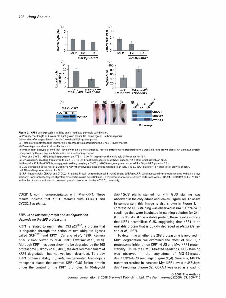
KRP1 is an unstable protein and its degradation
depends on the 26S proteasome
KRP1 is related to mammalian CKI p27Kip1, a protein that
is degraded through the action of two ubiquitin ligases
called SCFSKP2 and KPC1 (Carrano et al., 1999; Kamura
et al., 2004b; Sutterluty et al., 1999; Tsvetkov et al., 1999).
Although KRP1 has been shown to be degraded by the 26S
proteasome (Jakoby et al., 2006), the detailed mechanism of
KRP1 degradation has not yet been described. To study
KRP1 protein stability in planta, we generated Arabidopsis
transgenic plants that express KRP1–GUS fusion protein
under the control of the KRP1 promoter. In 10-day-old
KRP1:GUS plants stained for 4 h, GUS staining was
observed in the cotyledons and leaves (Figure 1c). To assist
in comparison, this image is also shown in Figure 3. In
contrast, no GUS staining was observed in KRP1:KRP1–GUS
seedlings that were incubated in staining solution for 24 h
(Figure 3b). As GUS is a stable protein, these results indicate
that KRP1 destabilizes GUS, suggesting that KRP1 is an
unstable protein that is quickly degraded in planta (Jeffer-
son et al., 1987).
To determine whether the 26S proteasome is involved in
KRP1 degradation, we examined the effect of MG132, a
proteasome inhibitor, on KRP1–GUS and Myc-KRP1 protein
stability. Unlike the DMSO-treated seedlings, GUS staining
was observed in the cotyledons of MG132-treated
KRP1:KRP1–GUS seedlings (Figure 3c,d). Similarly, MG132
treatment resulted in increased Myc-KRP1 levels in 35S:Myc-
KRP1 seedlings (Figure 3e). CDKA;1 was used as a loading
(a) (b)
(c)
(e) (f) (g) (h) (i) (j)
(d)
Figure 2. KRP1 overexpression inhibits auxin-mediated pericycle cell division.
(a) Primary root length of 2-week-old light-grown plants. He, hemizygous; Ho, homozygous.
(b) Number of emerged lateral roots in 2-week-old light-grown plants.
(c) Total lateral roots/seedling (primordia + emerged) visualized using the CYCB1;1:GUS marker.
(d) Percentage lateral root primordia from (c).
(e) Immunoblot analysis of Myc-KRP1 levels with an a-c-myc antibody. Protein extracts were prepared from 2-week-old light-grown plants. An unknown protein
recognized by the a-c-myc antibody was used as a loading control.
(f) Root of a CYCB1;1:GUS seedling grown on an ATS + 10 lM N-1-naphthylphthalamic acid (NPA) plate for 72 h.
(g) CYCB1;1:GUS seedling transferred to an ATS + 10 lM 1-naphthaleneacetic acid (NAA) plate for 12 h after initial growth on NPA.
(h) Root of a 35S:Myc-KRP1 (homozygous) seedling carrying a CYCB1;1:GUS transgene grown on an ATS + 10 lM NPA plate for 72 h.
(i) GUS expression in the root of a 35S:Myc-KRP1 (homozygous) seedling transferred to an ATS + 10 lM NAA plate for 12 h after initial growth on NPA.
(f–i) All seedlings were stained for GUS.
(j) KRP1 interacts with CDKA;1 and CYCD2;1 in planta. Protein extracts from wild-type (Col) and 35S:Myc-KRP1 seedlings were immunoprecipitated with an a-c-myc
antibody. Immunoblot analyses of protein extracts from wild-type (Col) and a-c-myc immunoprecipitates were performed with a-CDKA;1, a-CDKB1;1 and a-CYCD2;1
antibodies. Asterisk indicates an unknown protein recognized by the a-CYCD2;1 antibody.
708 Hong Ren et al.
ª 2008 The AuthorsJournal compilation ª 2008 Blackwell Publishing Ltd, The Plant Journal, (2008), 53, 705–716
control. MG132 did not affect CDKA;1 protein levels
(because this protein is stable). The stabilization of both
KRP1–GUS and Myc-KRP1 by MG132 indicates that KRP1
degradation depends on the 26S proteasome.
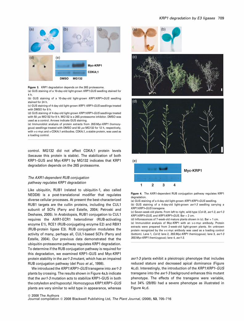
The AXR1-dependent RUB conjugation
pathway regulates KRP1 degradation
Like ubiquitin, RUB1 (related to ubiquitin-1, also called
NEDD8) is a post-translational modifier that regulates
diverse cellular processes. At present the best-characterized
RUB1 targets are the cullin proteins, including the CUL1
subunit of SCFs (Parry and Estelle, 2004; Petroski and
Deshaies, 2005). In Arabidopsis, RUB1 conjugation to CUL1
requires the AXR1-ECR1 heterodimer (RUB-activating
enzyme E1), RCE1 (RUB-conjugating enzyme E2) and RBX1
(RUB-protein ligase E3). RUB conjugation modulates the
activity of many, perhaps all, CUL1-based SCFs (Parry and
Estelle, 2004). Our previous data demonstrated that the
ubiquitin-proteasome pathway regulates KRP1 degradation.
To determine if the RUB conjugation pathway is required for
this degradation, we examined KRP1–GUS and Myc-KRP1
protein stability in the axr1-3 mutant, which has an impaired
RUB conjugation pathway (del Pozo et al., 1998).
We introduced the KRP1:KRP1–GUS transgene into axr1-3
plants by crossing. The results shown in Figure 4a,b indicate
that the axr1-3 mutation acts to stabilize KRP1–GUS in both
the cotyledon and hypocotyl. Homozygous KRP1:KRP1–GUS
plants are very similar to wild type in appearance, whereas
axr1-3 plants exhibit a pleiotropic phenotype that includes
reduced stature and decreased apical dominance (Figure
4c,d). Interestingly, the introduction of the KRP1:KRP1–GUS
transgene into the axr1-3 background enhances this mutant
phenotype. The effects of the transgene were variable,
but 34% (29/85) had a severe phenotype as illustrated in
Figure 4c,d.
Myc-KRP1
CDKA;1
DMSO MG132
(a) (b) (c)
(d) (e)
Figure 3. KRP1 degradation depends on the 26S proteasome.
(a) GUS staining of a 10-day-old light-grown KRP1:GUS seedling stained for
4 h.
(b) GUS staining of a 10-day-old light-grown KRP1:KRP1–GUS seedling
stained for 24 h.
(c) GUS staining of 4-day-old light-grown KRP1: KRP1–GUS seedlings treated
with DMSO for 8 h.
(d) GUS staining of 4-day-old light-grown KRP1:KRP1–GUS seedlings treated
with 50 lM MG132 for 8 h. MG132 is a 26S proteasome inhibitor. DMSO was
used as a control. Arrows indicate GUS staining.
(e) Immunoblot analysis of protein extracts from 35S:Myc-KRP1 (homozy-
gous) seedlings treated with DMSO and 50 lM MG132 for 12 h, respectively,
with a-c-myc and a-CDKA;1 antibodies. CDKA;1, a stable protein, was used as
a loading control.
1
(a)
(e)
(b)
2 3 4
Myc-KRP1
(c)
(d)
Figure 4. The AXR1-dependent RUB conjugation pathway regulates KRP1
degradation.
(a) GUS staining of a 4-day-old light-grown KRP1:KRP1–GUS seedling.
(b) GUS staining of a 4-day-old light-grown axr1-3 seedling carrying a
KRP1:KRP1–GUS transgene.
(c) Seven-week-old plants. From left to right, wild-type (Col-0), axr1-3, axr1-3
KRP1:KRP1–GUS, and KRP1:KRP1–GUS. Bar = 2 cm.
(d) Inflorescences of 7-week-old mature plants shown in (c). Bar = 1 cm.
(e) Immunoblot analysis of Myc-KRP1 with an a-c-myc antibody. Protein
extracts were prepared from 2-week-old light-grown plants. An unknown
protein recognized by the a-c-myc antibody was used as a loading control
(bottom). Lane 1, Col-0; lane 2, 35S:Myc-KRP1 (hemizygous); lane 3, axr1-3
35S:Myc-KRP1 (hemizygous); lane 4, axr1-3.
KRP1 degradation by E3 ligases 709
ª 2008 The AuthorsJournal compilation ª 2008 Blackwell Publishing Ltd, The Plant Journal, (2008), 53, 705–716
We also introduced the 35S:Myc-KRP1 transgene into
axr1-3 plants by crossing. As shown in Figure 4e, the axr1-3
mutation stabilized Myc-KRP1. These data indicate that the
AXR1-dependent RUB conjugation pathway regulates KRP1
degradation.
KRP1 degradation is dependent on SCFSKP2b
The RUB conjugation pathway is probably required for
the function of all cullin-based E3 ubiquitin ligases,
including SCF, CUL3-BTB and CUL4-DDB E3s (Bernhardt
et al., 2006; Chen et al., 2006; Parry and Estelle, 2004). To
determine if an SCF might be involved in KRP1 degrada-
tion, we introduced the KRP1:KRP1–GUS and 35S:Myc-
KRP1 transgenes into the axr6-3 mutant by crossing. The
axr6-3 mutant contains a recessive and temperature-sen-
sitive mutation of CUL1 that has been shown to stabilize
SCF substrates (Quint et al., 2005). As shown in Figure
5a,b,d, the axr6-3 mutant stabilized both KRP1–GUS and
Myc-KRP1, exhibiting GUS staining in the cotyledon and
an increased Myc-KRP1 protein level. Interestingly, over-
expression of either KRP1–GUS or Myc-KRP1 enhances
the axr6-3 growth defects. Plants exhibited a dramatically
decreased height and were sterile (Figures 5c and 6e).
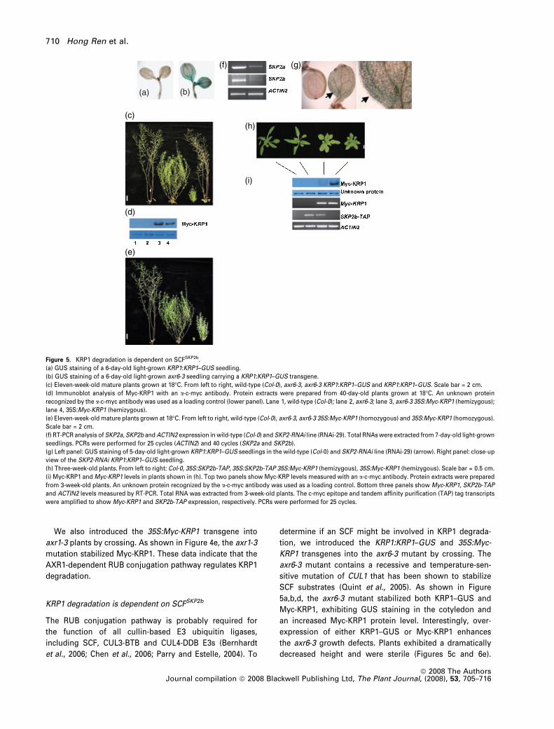
(a) (b)
(f) (g)
(h)
(i)
(c)
(d)
(e)
Figure 5. KRP1 degradation is dependent on SCFSKP2b.
(a) GUS staining of a 6-day-old light-grown KRP1:KRP1–GUS seedling.
(b) GUS staining of a 6-day-old light-grown axr6-3 seedling carrying a KRP1:KRP1–GUS transgene.
(c) Eleven-week-old mature plants grown at 18�C. From left to right, wild-type (Col-0), axr6-3, axr6-3 KRP1:KRP1–GUS and KRP1:KRP1–GUS. Scale bar = 2 cm.
(d) Immunoblot analysis of Myc-KRP1 with an a-c-myc antibody. Protein extracts were prepared from 40-day-old plants grown at 18�C. An unknown protein
recognized by the a-c-myc antibody was used as a loading control (lower panel). Lane 1, wild-type (Col-0); lane 2, axr6-3; lane 3, axr6-3 35S:Myc-KRP1 (hemizygous);
lane 4, 35S:Myc-KRP1 (hemizygous).
(e) Eleven-week-old mature plants grown at 18�C. From left to right, wild-type (Col-0), axr6-3, axr6-3 35S:Myc-KRP1 (homozygous) and 35S:Myc-KRP1 (homozygous).
Scale bar = 2 cm.
(f) RT-PCR analysis of SKP2a, SKP2b and ACTIN2 expression in wild-type (Col-0) and SKP2-RNAi line (RNAi-29). Total RNAs were extracted from 7-day-old light-grown
seedlings. PCRs were performed for 25 cycles (ACTIN2) and 40 cycles (SKP2a and SKP2b).
(g) Left panel: GUS staining of 5-day-old light-grown KRP1:KRP1–GUS seedlings in the wild-type (Col-0) and SKP2-RNAi line (RNAi-29) (arrow). Right panel: close-up
view of the SKP2-RNAi KRP1:KRP1–GUS seedling.
(h) Three-week-old plants. From left to right: Col-0, 35S:SKP2b-TAP, 35S:SKP2b-TAP 35S:Myc-KRP1 (hemizygous), 35S:Myc-KRP1 (hemizygous). Scale bar = 0.5 cm.
(i) Myc-KRP1 and Myc-KRP1 levels in plants shown in (h). Top two panels show Myc-KRP levels measured with an a-c-myc antibody. Protein extracts were prepared
from 3-week-old plants. An unknown protein recognized by the a-c-myc antibody was used as a loading control. Bottom three panels show Myc-KRP1, SKP2b-TAP
and ACTIN2 levels measured by RT-PCR. Total RNA was extracted from 3-week-old plants. The c-myc epitope and tandem affinity purification (TAP) tag transcripts
were amplified to show Myc-KRP1 and SKP2b-TAP expression, respectively. PCRs were performed for 25 cycles.
710 Hong Ren et al.
ª 2008 The AuthorsJournal compilation ª 2008 Blackwell Publishing Ltd, The Plant Journal, (2008), 53, 705–716
These results indicate that CUL1 is required for KRP1
degradation.
The involvement of CUL1 in KRP1 degradation reveals
that an SCF mediates KRP1 protein turnover. Among the SCF
subunits, the F-box protein recognizes and binds substrates.
In mammals, the F-box protein SKP2 binds CKI p27Kip1
(Sutterluty et al., 1999; Tsvetkov et al., 1999) and the
transcription factor E2F-1 (Marti et al., 1999), as well as
other cell cycle regulators (Nakayama and Nakayama, 2005),
targeting them for degradation. Arabidopsis has two SKP2-
related F-box proteins SKP2a and SKP2b that are 83%
identical at the amino acid sequence level. It has been
shown that SKP2a binds the transcription factor E2Fc and
appears to mediate its degradation (del Pozo et al., 2002,
2006). Because of the sequence and functional relationship
between KRP1 and p27Kip1, we decided to investigate the
possibility that Arabidopsis SKP2 is involved in KRP1
degradation.
To test this possibility, we examined KRP1–GUS protein
stability in the SKP2a and SKP2b T-DNA insertion mutants.
We identified SKP2a and SKP2b T-DNA insertion mutants in
the GABI-KAT and SALK collection (Alonso et al., 2003;
Rosso et al., 2003; see Figure S1a). Neither skp2a-1 and
skp2b-1 nor a skp2a-1 skp2b-1 double mutant exhibited an
obvious phenotype. In addition, none of these mutants
stabilized KRP1–GUS (data not shown). To understand the
molecular nature of the skp2a-1 and skp2b-1 mutants, we
examined SKP2a and SKP2b expression by RT-PCR in the
skp2a-1 skp2b-1 double mutant. The full-length transcripts
of SKP2a and SKP2b could not be detected. However,
truncated transcripts were detected at a high level for SKP2a
and at a low level for SKP2b (see Figure S1c). Therefore,
truncated SKP2a and SKP2b proteins could be produced. If
these truncated proteins exist, SKP2a and SKP2b will have
the F-box domain, as well as four and two leucine-
rich repeats (LRRs), respectively (see Figure S1b). Thus,
it is possible that truncated SKP2a and SKP2b could
form a functional SCF complex that targets substrates for
degradation.
As an alternative, we examined KRP1–GUS protein sta-
bility in SKP2 RNA interference (RNAi) transgenic plants with
reduced levels of both SKP2a and SKP2b. We worked with
two independent lines and obtained similar results. SKP2-
RNAi transgenic lines did not exhibit any obvious defects in
growth and development (data not shown). Here, we show
results for the line RNAi-29. Compared with wild type, the
expression of both SKP2a and SKP2b was strongly de-
creased in this line (Figure 5f). The KRP1:KRP1–GUS trans-
gene was introduced into SKP2-RNAi transgenic plants by
crossing, and F1 plants were examined for GUS expression.
GUS staining was observed in the cotyledon of RNAi-29
seedlings (Figure 5g). Therefore, SKP2-RNAi transgenic
plants stabilize KRP1–GUS, indicating that SKP2a and/or
SKP2b are involved in KRP1 degradation.
To gain further evidence for a role for Arabidopsis SKP2 in
KRP1 degradation, we examined the effect of SKP2 overex-
pression on KRP1 degradation in planta. We generated
Arabidopsis transgenic plants that express a TAP (tandem
affinity purification) tagged SKP2a or SKP2b under the
control of the CaMV 35S promoter. We introduced the
35S:SKP2a-TAP and 35S:SKP2b-TAP transgenes into
35S:Myc-KRP1 plants by crossing. A 35S:SKP2a-TAP line
did not appear to alter the effects of Myc-KRP1 overexpres-
sion (data not shown). Interestingly, two independent
35S:SKP2b-TAP lines suppressed the effects of Myc-KRP1
overexpression. Here, we show results for line 5. An obvious
phenotype of KRP1 overexpressers is serrated rosette
leaves. Plants that overexpress both SKP2b-TAP and Myc-
KRP1 did not exhibit serrated rosette leaves (Figure 5h). The
loss of serrated leaf phenotype was associated with a
decreased Myc-KRP1 protein level. Three-week-old
35S:SKP2b-TAP 35S:Myc-KRP1 plants had much less Myc-
KRP1 than 35S:Myc-KRP1 plants (Figure 5i). We also exam-
ined Myc-KRP1 transcript levels in both lines and found
them to be similar (Figure 5i), confirming that decreased
Myc-KRP1 protein levels are caused by increased degrada-
tion. Taken together, our data indicate that KRP1 degrada-
tion is dependent on an SCF complex that consists of CUL1
and SKP2b.
The RING protein RKP also contributes to KRP1 degradation
Recent studies in mammalian cells indicate that at least two
different E3s contribute to p27Kip1 degradation: SCFSkp2 and
a RING-type E3 called KPC1 (Kamura et al., 2004b; Kotoshiba
et al., 2005). Because KRP1 is related to p27Kip1, a similar
KPC-dependent protein degradation mechanism might exist
in Arabidopsis to regulate KRP1 protein turnover. Interest-
ingly, there is a KPC1-related RING finger protein called
At2g22010 in Arabidopsis. Like mammalian KPC1,
At2g22010 has a RING finger domain in the C-terminus and a
SPRY domain with an unknown function near the N-termi-
nus (Stone et al., 2005; Figure 6a). The two proteins are
approximately 25% identical, and this conservation extends
along the entire length of the proteins. Based on this rela-
tionship we have named this protein RKP (related to KPC1).
Microarray experiments indicate that RKP RNA is present
throughout plant development, with particularly high levels
of expression in the embryo and senescing leaves (Schmid
et al., 2005).
To determine if RKP functions in KRP1 degradation, we
first examined the effect of loss of RKP on KRP1–GUS levels.
A RKP T-DNA insertion mutant was identified in the SAIL
collection (Sessions et al., 2002). The rkp-1 mutant contains
a T-DNA insertion in exon 2 of RKP that prevents the
formation of full-length RKP RNA. As any protein encoded
by the truncated transcript would not include the RING
domain, rkp-1 is likely to be a null mutant. When the
KRP1 degradation by E3 ligases 711
ª 2008 The AuthorsJournal compilation ª 2008 Blackwell Publishing Ltd, The Plant Journal, (2008), 53, 705–716
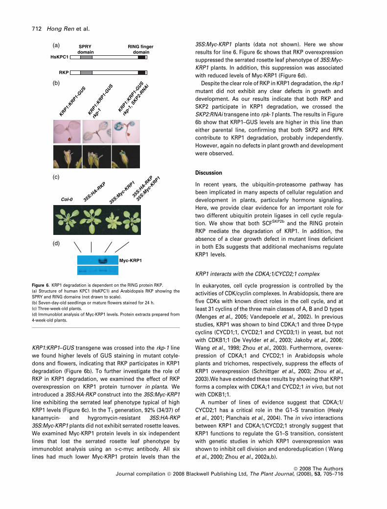
KRP1:KRP1–GUS transgene was crossed into the rkp-1 line
we found higher levels of GUS staining in mutant cotyle-
dons and flowers, indicating that RKP participates in KRP1
degradation (Figure 6b). To further investigate the role of
RKP in KRP1 degradation, we examined the effect of RKP
overexpression on KRP1 protein turnover in planta. We
introduced a 35S:HA-RKP construct into the 35S:Myc-KRP1
line exhibiting the serrated leaf phenotype typical of high
KRP1 levels (Figure 6c). In the T1 generation, 92% (34/37) of
kanamycin- and hygromycin-resistant 35S:HA-RKP
35S:Myc-KRP1 plants did not exhibit serrated rosette leaves.
We examined Myc-KRP1 protein levels in six independent
lines that lost the serrated rosette leaf phenotype by
immunoblot analysis using an a-c-myc antibody. All six
lines had much lower Myc-KRP1 protein levels than the
35S:Myc-KRP1 plants (data not shown). Here we show
results for line 6. Figure 6c shows that RKP overexpression
suppressed the serrated rosette leaf phenotype of 35S:Myc-
KRP1 plants. In addition, this suppression was associated
with reduced levels of Myc-KRP1 (Figure 6d).
Despite the clear role of RKP in KRP1 degradation, the rkp1
mutant did not exhibit any clear defects in growth and
development. As our results indicate that both RKP and
SKP2 participate in KRP1 degradation, we crossed the
SKP2:RNAi transgene into rpk-1 plants. The results in Figure
6b show that KRP1–GUS levels are higher in this line than
either parental line, confirming that both SKP2 and RPK
contribute to KRP1 degradation, probably independently.
However, again no defects in plant growth and development
were observed.
Discussion
In recent years, the ubiquitin-proteasome pathway has
been implicated in many aspects of cellular regulation and
development in plants, particularly hormone signaling.
Here, we provide clear evidence for an important role for
two different ubiquitin protein ligases in cell cycle regula-
tion. We show that both SCFSKP2b and the RING protein
RKP mediate the degradation of KRP1. In addition, the
absence of a clear growth defect in mutant lines deficient
in both E3s suggests that additional mechanisms regulate
KRP1 levels.
KRP1 interacts with the CDKA;1/CYCD2;1 complex
In eukaryotes, cell cycle progression is controlled by the
activities of CDK/cyclin complexes. In Arabidopsis, there are
five CDKs with known direct roles in the cell cycle, and at
least 31 cyclins of the three main classes of A, B and D types
(Menges et al., 2005; Vandepoele et al., 2002). In previous
studies, KRP1 was shown to bind CDKA;1 and three D-type
cyclins (CYCD1;1, CYCD2;1 and CYCD3;1) in yeast, but not
with CDKB1;1 (De Veylder et al., 2003; Jakoby et al., 2006;
Wang et al., 1998; Zhou et al., 2003). Furthermore, overex-
pression of CDKA;1 and CYCD2;1 in Arabidopsis whole
plants and trichomes, respectively, suppress the effects of
KRP1 overexpression (Schnittger et al., 2003; Zhou et al.,
2003).We have extended these results by showing that KRP1
forms a complex with CDKA;1 and CYCD2;1 in vivo, but not
with CDKB1;1.
A number of lines of evidence suggest that CDKA;1/
CYCD2;1 has a critical role in the G1–S transition (Healy
et al., 2001; Planchais et al., 2004). The in vivo interactions
between KRP1 and CDKA;1/CYCD2;1 strongly suggest that
KRP1 functions to regulate the G1–S transition, consistent
with genetic studies in which KRP1 overexpression was
shown to inhibit cell division and endoreduplication ( Wang
et al., 2000; Zhou et al., 2002a,b).
HsKPC1
(a)
(b)
(c)
(d)
RKP
SPRYdomain
RING fingerdomain
Myc-KRP1
KRP1:KRP1-
GUSKRP1
:KRP1
-GUS
rkp-
1 KRP1:KRP1-
GUS
rkp-
1,SKP2-
RNAi
Col-0 35S:HA-R
KP
35S:Myc-K
RP1
35S:H
A-RKP
35S:M
yc-K
RP1
Figure 6. KRP1 degradation is dependent on the RING protein RKP.
(a) Structure of human KPC1 (HsKPC1) and Arabidopsis RKP showing the
SPRY and RING domains (not drawn to scale).
(b) Seven-day-old seedlings or mature flowers stained for 24 h.
(c) Three-week-old plants.
(d) Immunoblot analysis of Myc-KRP1 levels. Protein extracts prepared from
4-week-old plants.
712 Hong Ren et al.
ª 2008 The AuthorsJournal compilation ª 2008 Blackwell Publishing Ltd, The Plant Journal, (2008), 53, 705–716
In addition to the CDKA;1/CYCD2;1 complex, KRP1 may
have other targets. A recent study reported that besides its
role in the G1–S transition, KRP1 also functions in the G2–M
transition to regulate mitosis entry (Weinl et al., 2005).
However, the CDK/cyclin complex target of KRP1 at the
G2–M transition is unknown. It will be important to identify
other CDK/cyclin complex targets of KRP1 in the future to
further define the role of KRP1 in cell cycle regulation.
Ectopic expression of KRP1 inhibits pericycle
activation during lateral root initiation
Lateral root formation is an example of post-embryonic
de novo organogenesis. In Arabidopsis, lateral roots are
derived from pericycle cells adjacent to the xylem poles
(Casimiro et al., 2003; Himanen et al., 2002). Pericycle acti-
vation appears to involve changes in the level of a protein
related to KRP1, called KRP2 (Himanen et al., 2002). The
KRP2 gene is expressed in pericycle cells and downregu-
lated in conditions that induce lateral root formation. Based
on these results it was proposed that KRP2 controls the G1–S
transition of pericycle cells during pericycle activation.
The KRP1 gene is also expressed in roots and is down-
regulated during lateral root formation (Himanen et al.,
2002). We demonstrate that KRP1 overexpression inhibits
auxin-mediated pericycle cell division, resulting in a dra-
matic decrease in the number of lateral roots. These results
suggest that KRP1 may also have a role in regulating
pericycle cell activation. However, the lack of a clear effect
of the krp1 mutation on lateral root development precludes a
definitive conclusion. In any case, it is clear that KRP1 is not
the only factor regulating this process.
SCFSKP2b mediates KRP1 degradation
Previous studies demonstrated that KRP1 is an unstable
protein that is degraded by the 26S proteasome (Jakoby
et al., 2006; Zhou et al., 2003). We have extended this work
using a KRP1–GUS fusion protein. Our results demonstrate
that the AXR1-RUB conjugation pathway is required for
KRP1 degradation. Because the only known substrates for
RUB conjugation are the cullin proteins, these results imply
that KRP1 degradation requires a cullin-based E3, such as an
SCF. Indeed we show that CUL1 is required for KRP1 deg-
radation, indicating that an SCF regulates KRP1 degradation.
In mammals p27Kip1 is degraded by SCFSkp2. In Arabid-
opsis, there are two proteins related to Skp2: SKP2a and
SKP2b. Despite the fact that KRP1 is only related to p27Kip1
through the cyclin- and CDK-binding/inhibitory domain, our
results indicate that SKP2b is involved in KRP1 degradation.
SKP2-RNAi lines with strongly decreased levels of both
SKP2a and SKP2b stabilize KRP1–GUS. Furthermore, we
have shown that SKP2b overexpression promotes KRP1
degradation in planta. Surprisingly, overexpression of the
closely related SKP2a protein did not suppress the effects of
Myc-KRP1 overexpression.Whether this is caused by a
difference in the function of SKP2a and SKP2b, or by an
artifact related to the specific 35S:SKP2a line, is unknown.
Thus, our results suggest that SKP2b targets KRP1 for
degradation, but do not exclude the possibility that SKP2a
has a similar function. It will be important to examine the
biochemical interactions between KRP1 and SKP2b in the
future. Taken together, our data suggest that an SCF
complex that is composed of CUL1 and SKP2b mediates
KRP1 degradation.
KRP1 degradation is also regulated by the RING protein RKP
Previous studies have identified two domains on KRP1 that
contribute to its instability, suggesting that there may be two
independent mechanisms of KRP degradation (Jakoby et al.,
2006). In mammalian cells, p27Kip1 degradation is mediated
by both SCFSkp2 and a RING protein called KPC1 (Kamura
et al., 2004b). Our genetic studies suggest that RKP, an
Arabidopsis protein related to KPC1, also contributes to
KRP1 degradation. The rkp-1 mutant stabilizes KRP1–GUS.
In addition, RKP overexpression promotes KRP1 degrada-
tion in planta. The observation that KRP1–GUS is signifi-
cantly more stable in rkp 35S:SKP2-RNAi plants than
either the rkp or 35S:SKP2-RNAi lines suggest that the
two E3s function independently. Furthermore, both path-
ways appear to be active in the same tissue. In mammals,
SCFSkp2-dependent degradation of p27Kip1 occurs in the
nucleus, whereas KPC functions in the cytoplasm (Kamura
et al., 2004a). Jakoby et al. (2006) have shown that KRP1
localizes primarily to the nucleus, but is found in different
subnuclear domains. It will be interesting to determine if
RKP and SCFSKP2b mediate KRP1 degradation in distinct
cellular compartments.
Despite the clear involvement of SCFSKP2 and RKP in KRP1
degradation, plants deficient in either or both of these E3s do
not display any growth defects. This is surprising, as
relatively modest changes in KRP1 levels exert an effect on
development. For example, the KRP1:KRP1–GUS transgene
acts to enhance the phenotype of the axr1-3 and axr6-3
mutants. Our failure to detect a phenotype in SKP2-RNAi and
rkp lines may be caused by residual SCFSKP2 activity in the
RNAi lines and redundancy between SCFSKP2 and RPK.
Alternatively, it is possible that Arabidopsis has an addi-
tional mechanism of KRP1 degradation, perhaps via another
E3 enzyme.
Experimental procedures
Plant materials and growth conditions
Arabidopsis thaliana plants were grown under 24-h light conditionsat 22�C, or 18�C when necessary. All mutants and transgenic lines
KRP1 degradation by E3 ligases 713
ª 2008 The AuthorsJournal compilation ª 2008 Blackwell Publishing Ltd, The Plant Journal, (2008), 53, 705–716
were in the Columbia ecotype. The skp2a-1 mutant (GABI-Kat293D12), a T-DNA insertion in At1g21410, was acquired fromGABI-KAT; Rosso et al., 2003). The skp2b-1 mutant (SALK_028396),a T-DNA insertion in At1g77000, was acquired from the ArabidopsisBiological Research Center (ABRC, http://www.arabidopsis.org;Alonso et al., 2003). The rkp mutant (SAIL_3_E3), a T-DNA insertionin At2g22010, was acquired from the ABRC (Sessions et al., 2002).All T-DNA mutants were confirmed by PCR and sequencing. Seedswere surface sterilized in a 30% bleach and 0.04% Triton X-100solution, and were cold treated for 2–3 days at 4�C to synchronizegermination. Seeds were grown on ATS medium [1% sucrose,5 mM KNO3, 2.5 mM KH2PO4 (pH 5.6), 2 mM MgSO4, 2 mM Ca(NO3)2,50 lm CuSO4, 1 lM ZnSO4, 0.2 lm NaMoO4, 10 lm NaCl and0.01 lM CoCl2) with or without 0.8% agar. N-1-Naphthylphthalamicacid (NPA), 1-naphthaleneacetic acid (NAA), herbicide basta(AgroEvo) and antibiotics were added to autoclaved ATS medium,when necessary.
Transgenic lines
A 2062-bp KRP1 (At2g23430) promoter was amplified from genomicDNA with primers KRP1-PF (5¢-GTTCAAGCGAGTGACACATCTC-3¢)and KRP1-PR (5¢-CTTCGATTTAGGTTACGTGTGCG-3¢). TheKRP1:GUS plasmid was constructed by cloning the KRP1 promoterto pCB308 containing the Escherichia coli GUS (Xiang et al., 1999). A602-bp KRP1 full-length cDNA was amplified from a cDNA librarywith primers KRP1-F (5¢-ACGCACACGTCACCTAAATC-3¢) and KRP1-R (5¢-CTTCACTCTAACTTTACCCATTCG-3¢). The KRP1:KRP1–GUSplasmid was constructed by cloning both the KRP1 promoter and theKRP1 full-length cDNA without the stop codon TGA to pCB308. Tomake a 35S:Myc-KRP1 construct, the KRP1 full-length cDNA withoutthe start codon ATG was fused to the C-terminus of six c-mycepitopes in pGEM7Z. The Myc-KRP1 insert was then cloned topROK2.
To make a 35S:SKP2-RNAi construct, a 433-bp cDNA fromnucleotides 256–688 of SKP2a (At1g21410) was cloned in anopposite orientation to pHANNIBAL (Wesley et al., 2001). TheSKP2-RNAi insert was then cloned to pBIN19. To make a35S:SKP2b-TAP construct, an SKP2b (At1g77000) full-length cDNAwithout the stop codon TGA was fused to the N-terminus of a TAP(tandem affinity purification) tag (Rigaut et al., 1999). The SKP2b-TAP insert was then cloned to pPILY (Ferrando et al., 2000).
A 3861-bp RKP full-length cDNA without the start codon ATG wasamplified with primers RKP-F (5¢-CACCTTGGCTGAAGACAGCCT-ACGG-3¢) and RKP-R (5¢-GCAACTAACCCGAGCTTCATGTGC-3¢), andwas cloned to pENTR/D-TOPO using the pENTR directional TOPOcloning kit (Invitrogen, http://www.invitrogen.com). The RKP insertin pENTR/D-TOPO was then cloned to pGWB15 (http://bio2.ipc.shimane-u.ac.jp/pgwbs/index.htm) containing the CaMV 35S pro-moter and three hemagglutinin epitopes to make a 35S::HA-RKPconstruct.
All the above constructs in the binary vectors were introducedinto Agrobacterium tumefaciens strain GV3101. Plants were trans-formed by the vacuum infiltration method (Bechtold and Pelletier,1998). Transgenic plants were selected on ATS plates supplementedwith the necessary antibiotics or herbicide. The KRP1:GUS andKRP1:KRP1–GUS transgenic plants are basta resistant. The35S:Myc-KRP1 and 35S::HA-RKP transgenic plants are kanamycin-and hygromycin-resistant, respectively.
GUS assays
To examine GUS expression, seedlings were incubated in a GUSstaining solution at 37�C as described by Oono et al. (1998). GUS-
stained seedlings were incubated in 70% ethanol to remove chlo-rophyll. GUS staining patterns were examined under a NikonSMZ1500 dissecting microscope (http://www.nikonusa.com).
RT-PCR analysis
Total RNAs were extracted using the TRI reagent (Sigma, http://www.sigmaaldrich.com). The first-strand cDNAs were synthesizedfrom 5 lg total RNAs using oligo(dT)20 primer and SuperScript IIRNase H) reverse transcriptase (Invitrogen). PCRs were performedwith the following gene-specific primers: SKP2a-F, 5¢-CCGCTTC-ATTTTAGTCATTAAAC-3¢; SKP2a-R1, 5¢-GGCCGTTTATATATACAA-CATAAC-3¢; SKP2a-R2, 5¢-TGATTGCAGTTATTCCCAATAG-3¢;SKP2b-F, 5¢-CATATTTACTTTTGATCTCGTGG-3¢; SKP2b-R1,5¢-CATACTAGAGAGTAGTAGACC-3¢; SKP2a-R2, 5¢-CGAGTTTAGT-CAGGTTAGTA-3¢; Myc-F, 5¢-GACTCTAGAGGATCCCCAAAGC-3¢;Myc-R, 5¢-AGCCGAATTCGATGGGGTACCG-3¢; TAP-F, 5¢-TAG-CCGTCTCAGCAGCCAACC-3¢; TAP-R, 5¢-CTTCCCCGCGGAATTCGC-GTC-3¢; ACTIN2-F, 5¢-GGCTGAGGCTGATGATATTC-3¢; ACTIN2-R,5¢-TCTGTGAACGATTCCTGGAC-3¢.
Immunoblot analysis and immunoprecipitation
Protein extracts were prepared as described by Gray et al. (1999).For immunoblot analysis, 50 lg protein extracts were mixed withSDS-PAGE sample buffer and were boiled for 5 min. Denaturedproteins were separated on a 10% acrylamide SDS gel and weretransferred to a nitrocellulose membrane. The membrane wasimmersed in Tris-buffered saline (pH 7.6) containing 5% non-fat drymilk and 0.1% Tween 20 to block non-specific binding sites. Thea-c-myc 9E 10 antibody (Covance Research Products, http://www.crpinc.com) was used at a 1:1000 dilution. The horseradishperoxidase-conjugated goat a-mouse secondary antibody (Sigma)was used at a 1:3000 dilution. Proteins were detected with theenhanced chemiluminescence (ECL) kit (Amersham PharmaciaBiotech, http://www.gelifesciences.com).
For immunoprecipitation, 5 ll a-c-myc 9E 10 antibody was addedto 3 mg protein extracts and was incubated for 1–3 h at 4�C. To collectimmune complexes, 30 ll of protein A agarose beads (Roche, http://www.roche.com) were added and were incubated for from 3 h toovernight. Immune complexes were washed three times in 1 ml ofprotein extraction buffer. Finally, agarose beads were resuspendedin SDS-PAGE sample buffer. Immunoblot analysis was carried out asdescribed above. The a-CDKA;1 antibody was used at a 1:5000dilution. The a-CDKB1;1 and a-CYCD2;1 antibodies (Healy et al.,2001) were used at a 1:3000 dilution. The horseradish peroxidase-conjugated goat a-rabbit secondary antibody (Chemicon Interna-tional, http://www.millipore.com) was used at a 1:2500 dilution.
Acknowledgements
We thank Dirk Inze for the a-CDKA;1 antibody and CYCB1;1:GUSline, William M. Gray for the axr6-3 mutant, the SALK InstituteGenomic Analysis Laboratory, ABRC and GABI-KAT for T-DNAinsertion mutants. This work was supported by grants from theNational Science Foundation (NSF 2010 MCB-0115870) to ME andthe Spanish MEC (BIO2004-01749) to JCdP.
Supplementary material
The following supplementary material is available for this articleonline:
714 Hong Ren et al.
ª 2008 The AuthorsJournal compilation ª 2008 Blackwell Publishing Ltd, The Plant Journal, (2008), 53, 705–716
Figure S1. RT-PCR analysis of SKP2a and SKP2b expression in theSKP2a and SKP2b T-DNA insertion mutants.Table S1. Developmental expression of KRP1.This material is available as part of the online article from http://www.blackwell-synergy.comPlease note: Blackwell Publishing are not responsible for thecontent or functionality of any supplementary materials suppliedby the authors. Any queries (other than missing material) should bedirected to the corresponding author for the article.
References
Alonso, J.M., Stepanova, A.N., Leisse, T.J., Kim, C.J., Chen, H.,
Shinn, P., Stevenson, D.K., Zimmerman, J., Barajas, P. and
Cheuk, R. (2003) Genome-wide insertional mutagenesis ofArabidopsis thaliana. Science, 301, 653–657.
Bechtold, N. and Pelletier, G. (1998) In planta Agrobacterium-mediated transformation of adult Arabidopsis thaliana plants byvacuum infiltration. Methods Mol. Biol. 82, 259–266.
Bemis, S.M. and Torii, K.U. (2007) Autonomy of cell proliferationand developmental programs during Arabidopsis abovegroundorgan morphogenesis. Dev. Biol. 304, 367–381.
Bernhardt, A., Lechner, E., Hano, P., Schade, V., Dieterle, M.,
Anders, M., Dubin, M.J., Benvenuto, G., Bowler, C. and Genschik,
P. (2006) CUL4 associates with DDB1 and DET1 and its downre-gulation affects diverse aspects of development in Arabidopsisthaliana. Plant J. 47, 591–603.
Bisbis, B., Delmas, F., Joubes, J., Sicard, A., Hernould, M., Inze, D.,
Mouras, A. and Chevalier, C. (2006) Cyclin-dependent kinase(CDK) inhibitors regulate the CDK-cyclin complex activities inendoreduplicating cells of developing tomato fruit. J. Biol. Chem.281, 7374–7383.
Carrano, A.C., Eytan, E., Hershko, A. and Pagano, M. (1999) SKP2 isrequired for ubiquitin-mediated degradation of the CDK inhibitorp27. Nat. Cell Biol. 1, 193–199.
Casimiro, I., Beeckman, T., Graham, N., Bhalerao, R., Zhang, H.,
Casero, P., Sandberg, G. and Bennett, M.J. (2003) DissectingArabidopsis lateral root development. Trends Plant Sci. 8, 165–171.
Chen, H., Shen, Y., Tang, X., Yu, L., Wang, J., Guo, L., Zhang, Y.,
Zhang, H., Feng, S. and Strickland, E. (2006) Arabidopsis CULLIN4forms an E3 ubiquitin ligase with RBX1 and the CDD complexin mediating light control of development. Plant Cell, 18, 1991–2004.
Churchman, M.L., Brown, M.L., Kato, N., Kirik, V., Hulskamp, M.,
Inze, D., De Veylder, L., Walker, J.D., Zheng, Z. and Oppenheimer,
D.G. (2006) SIAMESE, a plant-specific cell cycle regulator, con-trols endoreplication onset in Arabidopsis thaliana. Plant Cell, 18,3145–3157.
Coelho, C.M., Dante, R.A., Sabelli, P.A., Sun, Y., Dilkes, B.P.,
Gordon-Kamm, W.J. and Larkins, B.A. (2005) Cyclin-dependentkinase inhibitors in maize endosperm and their potential role inendoreduplication. Plant Physiol. 138, 2323–2336.
De Clercq, A. and Inze, D. (2006) Cyclin-dependent kinase inhibitorsin yeast, animals, and plants: a functional comparison. Crit. Rev.Biochem. Mol. Biol. 41, 293–313.
De Veylder, L., Beeckman, T., Beemster, G.T., Krols, L., Terras, F.,
Landrieu, I., van der Schueren, E., Maes, S., Naudts, M. and Inze,
D. (2001) Functional analysis of cyclin-dependent kinase inhibi-tors of Arabidopsis. Plant Cell, 13, 1653–1668.
De Veylder, L., Joubes, J. and Inze, D. (2003) Plant cell cycletransitions. Curr. Opin. Plant Biol. 6, 536–543.
Dewitte, W. and Murray, J.A. (2003) The plant cell cycle. Annu. Rev.Plant Biol. 54, 235–264.
Ferrando, A., Farras, R., Jasik, J., Schell, J. and Koncz, C. (2000)Intron-tagged epitope: a tool for facile detection and purificationof proteins expressed in Agrobacterium-transformed plant cells.Plant J. 22, 553–560.
Gray, W.M., del Pozo, J.C., Walker, L., Hobbie, L., Risseeuw, E.,
Banks, T., Crosby, W.L., Yang, M., Ma, H. and Estelle, M. (1999)Identification of an SCF ubiquitin-ligase complex required forauxin response in Arabidopsis thaliana. Genes Dev. 13, 1678–1691.
Healy, J.M.S., Menges, M., Doonan, J.H. and Murray, J.A.H. (2001)The Arabidopsis D-type cyclins CycD2 and CycD3 both interact invivo with the PSTAIRE cyclin-dependent kinase Cdc2a but aredifferentially controlled. J. Biol. Chem. 276, 7041–7047.
Hershko, A. (2005) The ubiquitin system for protein degradation andsome of its roles in the control of the cell division cycle. Cell DeathDiffer. 12, 1191–1197.
Himanen, K., Boucheron, E., Vanneste, S., De Almeida Engler, J.,
Inze, D. and Beeckman, T. (2002) Auxin-mediated cell cycle acti-vation during early lateral root initiation. Plant Cell, 14, 2339–2351.
Jakoby, M.J., Weinl, C., Pusch, S., Kuijt, S.J., Merkle, T., Dissmeyer,
N. and Schnittger, A. (2006) Analysis of the subcellular local-ization, function, and proteolytic control of the Arabidopsiscyclin-dependent kinase inhibitor ICK1/KRP1. Plant Physiol. 141,1293–1305.
Jasinski, S., Riou-Khamlichi, C., Roche, O., Perennes, C.,
Bergounioux, C. and Glab, N. (2002) The CDK inhibitor NtKIS1a isinvolved in plant development, endoreduplication and restoresnormal development of cyclin D3; 1-overexpressing plants. J. CellSci. 115, 973–982.
Jefferson, R.A., Kavanagh, T.A. and Bevan, M.W. (1987) GUSfusions:beta-glucouronidase as a sensitive and versatile genefusion marker in higher plants. EMBO J. 6, 3901–3907.
Kamura, T., Hara, T., Matsumoto, M., Ishida, N., Okumura, F.,
Hatakeyama, S., Yoshida, M., Nakayama, K. and Nakayama, K.I.
(2004a) Cytoplasmic ubiquitin ligase KPC regulates proteolysis ofp27(Kip1) at G1 phase. Nat. Cell Biol. 6, 1229–1235.
Kamura, T., Hara, T., Matsumoto, M., Ishida, N., Okumura, F.,
Hatakeyama, S., Yoshida, M., Nakayama, K. and Nakayama, K.I.
(2004b) Cytoplasmic ubiquitin ligase KPC regulates proteolysis ofp27Kip1 at G1 phase. Nat. Cell Biol. 6, 1229–1235.
Kotoshiba, S., Kamura, T., Hara, T., Ishida, N. and Nakayama, K.I.
(2005) Molecular dissection of the interaction between p27 andKip1 ubiquitylation-promoting complex, the ubiquitin ligase thatregulates proteolysis of p27 in G1 phase. J. Biol. Chem. 280,17694–17700.
Lechner, E., Xie, D., Grava, S. et al. (2002) The AtRbx1 protein is partof plant SCF complexes, and its down-regulation causes severegrowth and developmental defects. J. Biol. Chem. 277, 50069–50080.
Liu, F., Ni, W., Griffith, M.E., Huang, Z., Chang, C., Peng, W., Ma, H.
and Xie, D. (2004) The ASK1 and ASK2 genes are essential forArabidopsis early development. Plant Cell, 16, 5–20.
Lui, H., Wang, H., Delong, C., Fowke, L.C., Crosby, W.L. and Fobert,
P.R. (2000) The Arabidopsis Cdc2a-interacting protein ICK2 isstructurally related to ICK1 and is a potent inhibitor of cyclin-dependent kinase activity in vitro. Plant J. 21, 379–385.
Marti, A., Wirbelauer, C., Scheffner, M. and Krek, W. (1999) Inter-action between ubiquitin-protein ligase SCFSKP2 and E2F-1underlies the regulation of E2F-1 degradation. Nat. Cell Biol. 1,14–19.
Menges, M. and Murray, J.A. (2002) Synchronous Arabidopsissuspension cultures for analysis of cell-cycle gene activity. PlantJ. 30, 203–212.
KRP1 degradation by E3 ligases 715
ª 2008 The AuthorsJournal compilation ª 2008 Blackwell Publishing Ltd, The Plant Journal, (2008), 53, 705–716

Menges, M., de Jager, S.M., Gruissem, W. and Murray, J.A.H. (2005)Global analysis of the core cell cycle regulators of Arabidopsisidentifies novel genes, reveals multiple and highly specific pro-files of expression and provides a coherent model for plant cellcycle control. Plant J. 41, 546–566.
Moon, J., Parry, G. and Estelle, M. (2004) The ubiquitin-proteasomepathway and plant development. Plant Cell, 16, 3181–3195.
Nakayama, K. and Nakayama, K. (1998) Cip/Kip cyclin-dependentkinase inhibitors: brakes of the cell cycle engine during develop-ment. Bioessays, 20, 1020–1029.
Nakayama, K.I. and Nakayama, K. (2005) Regulation of the cellcycle by SCF-type ubiquitin ligases. Semin. Cell Dev. Biol. 16, 323–333.
Oono, Y., Chen, Q.G., Overvoorde, P.J., Kohler, C. and Theologis, A.
(1998) age mutants of Arabidopsis exhibit altered auxin-regulatedgene expression. Plant Cell, 10, 1649–1662.
Ormenese, S., de Almeida Engler, J., De Groodt, R., De Veylder, L.,
Inze, D. and Jacqmard, A. (2004) Analysis of the spatial expres-sion pattern of seven Kip related proteins (KRPs) in the shoot apexof Arabidopsis thaliana. Ann. Bot. 93, 575–580.
Parry, G. and Estelle, M. (2004) Regulation of cullin-based ubiquitinligases by the Nedd8/RUB ubiquitin-like proteins. Semin. CellDev. Biol. 15, 221–229.
Petroski, M.D. and Deshaies, R.J. (2005) Function and regulation ofcullin-RING ubiquitin ligases. Nat. Rev. Mol. Cell Biol. 6, 9–20.
Planchais, S., Samland, A.K. and Murray, J.A.H. (2004) Differentialstability of Arabidopsis D-type cyclins: CYCD3;1 is a highlyunstable protein degraded by a proteasome-dependent mecha-nism. Plant J. 38, 616–625.
del Pozo, J.C., Timpte, C., Tan, S., Callis, J. and Estelle, M. (1998)The ubiquitin-related protein RUB1 and auxin response inArabidopsis. Science, 280, 1760–1763.
del Pozo, J.C., Boniotti, M.B. and Gutierrez, C. (2002) ArabidopsisE2Fc functions in cell division and is degraded by the ubiquitin-SCF(AtSKP2) pathway in response to light. Plant Cell, 14, 3057–3071.
del Pozo, J.C., Diaz-Trivino, S., Cisneros, N. and Gutierrez, C.
(2006) The balance between cell division and endoreplicationdepends on E2FC-DPB, transcription factors regulated by theubiquitin-SCFSKP2A pathway in Arabidopsis. Plant Cell, 18,2224–2235.
Quint, M., Ito, H., Zhang, W. and Gray, W.M. (2005) Characterizationof a novel temperature-sensitive allele of the CUL1/AXR6 subunitof SCF ubiquitin-ligases. Plant J. 43, 371–383.
Rigaut, G., Shevchenko, A., Rutz, B., Wilm, M., Mann, M. and
Seraphin, B. (1999) A generic protein purification method forprotein complex characterization and proteome exploration. Nat.Biotechnol. 17, 1030–1032.
Rosso, M.G., Li, Y., Strizhov, N., Reiss, B., Dekker, K. and Weisshaar,
B. (2003) An Arabidopsis thaliana T-DNA mutagenized population(GABI-Kat) for flanking sequence tag-based reverse genetics.Plant Mol. Biol. 53, 247–259.
Schmid, M., Davison, T.S., Henz, S.R., Pape, U.J., Demar, M.,
Vingron, M., Scholkopf, B., Weigel, D. and Lohmann, J.U. (2005)A gene expression map of Arabidopsis thaliana development.Nat. Genet. 37, 501–506.
Schnittger, A., Weinl, C., Bouyer, D., Schobinger, U. and Hulskamp,
M. (2003) Misexpression of the cyclin-dependent kinase inhibitorICK1/KRP1 in single-celled Arabidopsis trichomes reducesendoreduplication and cell size and induces cell death. Plant Cell,15, 303–315.
Sessions, A., Burke, E., Presting, G. et al. (2002) A high-through-put Arabidopsis reverse genetics system. Plant Cell, 14, 2985–2994.
Sherr, C.J. and Roberts, J.M. (1999) CDK inhibitors: positive andnegative regulators of G1-phase progression. Genes Dev. 13,1501–1512.
Stone, S.L., Hauksdottir, H., Troy, A., Herschleb, J., Kraft, E.
and Callis, J. (2005) Functional analysis of the RING-typeubiquitin ligase family of Arabidopsis. Plant Physiol. 137, 13–30.
Sutterluty, H., Chatelain, E., Marti, A., Wirbelauer, C., Senften, M.,
Muller, U. and Krek, W. (1999) p45SKP2 promotes p27Kip1 deg-radation and induces S phase in quiescent cells. Nat. Cell Biol. 1,207–214.
Tsvetkov, L.M., Yeh, K.H., Lee, S.J., Sun, H. and Zhang, H. (1999)p27(Kip1) ubiquitination and degradation is regulated by theSCF(Skp2) complex through phosphorylated Thr187 in p27. Curr.Biol. 9, 661–664.
Vandepoele, K., Raes, J., De Veylder, L., Rouze, P., Rombauts, S.
and Inze, D. (2002) Genome-wide analysis of core cell cycle genesin Arabidopsis. Plant Cell, 14, 903–916.
Verkest, A., Manes, C.L., Vercruysse, S., Maes, S., Van Der
Schueren, E., Beeckman, T., Genschik, P., Kuiper, M., Inze, D.
and De Veylder, L. (2005a) The cyclin-dependent kinaseinhibitor KRP2 controls the onset of the endoreduplica-tion cycle during Arabidopsis leaf development throughinhibition of mitotic CDKA;1 kinase complexes. Plant Cell, 17,1723–1736.
Verkest, A., Weinl, C., Inze, D., De Veylder, L. and Schnittger, A.
(2005b) Switching the cell cycle. Kip-related proteins in plant cellcycle control. Plant Physiol. 139, 1099–1106.
Vidal, A. and Koff, A. (2000) Cell-cycle inhibitors: three familiesunited by a common cause. Gene, 247, 1–15.
Wang, H., Fowke, L.C. and Crosby, W.L. (1997) A plant cyclin-dependent kinase inhibitor gene. Nature, 386, 451–452.
Wang, H., Qi, Q., Schorr, P., Cutler, A.J., Crosby, W.L. and Fowke,
L.C. (1998) ICK1, a cyclin-dependent protein kinase inhibitorfrom Arabidopsis thaliana interacts with both Cdc2a and CycD3,and its expression is induced by abscisic acid. Plant J. 15, 501–510.
Wang, H., Zhou, Y., Gilmer, S., Whitwill, S. and Fowke, L.C. (2000)Expression of the plant cyclin-dependent kinase inhibitor ICK1affects cell division, plant growth and morphology. Plant J. 24,613–623.
Weinl, C., Marquardt, S., Kuijt, S.J., Nowack, M.K., Jakoby, M.J.,
Hulskamp, M. and Schnittger, A. (2005) Novel functions of plantcyclin-dependent kinase inhibitors, ICK1/KRP1, can act non-cell-autonomously and inhibit entry into mitosis. Plant Cell, 17, 1704–1722.
Wesley, S.V., Helliwell, C.A., Smith, N.A. et al. (2001) Constructdesign for efficient, effective and high-throughput gene silencingin plants. Plant J. 27, 581–590.
Xiang, C., Han, P., Lutziger, I., Wang, K. and Oliver, D.J. (1999) Amini binary vector series for plant transformation. Plant Mol. Biol.40, 711–717.
Zhou, Y., Fowke, L.C. and Wang, H. (2002a) Plant CDK inhibitors:Studies of interactions with cell cycle regulators in the yeast two-hybrid system and functional comparison in transgenic Arabid-opsis plants. Plant Cell Rep. 20, 967–975.
Zhou, Y., Wang, H., Gilmer, S., Whitwill, S., Keller, W. and Fowke,
L.C. (2002b) Control of petal and pollen development by the plantcyclin-dependent kinase inhibitor ICK1 in transgenic Brassicaplants. Planta, 215, 248–257.
Zhou, Y., Wang, H., Gilmer, S., Whitwill, S. and Fowke, L.C. (2003)Effects of co-expressing the plant CDK inhibitor ICK1 and D-typecyclin genes on plant growth, cell size and ploidy in Arabidopsisthaliana. Planta, 216, 604–613.
716 Hong Ren et al.
ª 2008 The AuthorsJournal compilation ª 2008 Blackwell Publishing Ltd, The Plant Journal, (2008), 53, 705–716