Embed Size (px)
Citation preview

The Pennsylvania State University
The Graduate School
Department of Bioengineering
DEFINED AND XENO-FREE CULTURE FOR HUMAN
PLURIPOTENT STEM CELLS
A Thesis in
Bioengineering
by
Tzu Ting Lin
© 2017 Tzu Ting Lin
Submitted in Partial Fulfillment
of the Requirements
for the Degree of
Master of Science
May 2017

ii The thesis of Tzu Ting Lin was reviewed and approved* by the following:
Xiaojun Lian
Assistant Professor of Biomedical Engineering
Thesis Advisor
Justin L. Brown
Associate Professor of Biomedical Engineering
William O. Hancock
Professor of Biomedical Engineering
Head of Graduate Program
*Signatures are on file in the Graduate School

iii
ABSTRACT
Owing to their ability to differentiate into all cell types in body, and therefore
greatly impact the landscape of regenerative medicine and tissue engineering, the
human pluripotent stem cells (hPSCs) including human induced pluripotent stem
cells (hiPSCs) and human embryonic stem cells (hESCs) are of great interest to
researchers in recent decades. One of the combating issues is the development of
a robust stem cell culture environment to maintain stem cells without differentiation
for long-term culture, in other words, to support their self-renewal and pluripotency.
Moreover, because hiPSCs and hESCs have practical use in in-vivo study, for such
a clinical application, it is necessary to establish a chemically defined, feeder-free
culture system to maintain large scale undifferentiated stem cells in-vitro. Various
methods have been proposed for maintaining large scale stem cell culture Some
major considerations are: culture media, extracellular matrix, and environment cues.
This study will be focused on developing and analyzing a new xeno-free stem cell
culture medium, LaSR, for supporting long-term proliferation and pluripotency for
both hiPSCs and hESCs to facilitate the basis of stem cell research.

iv
TABLE OF CONTENTS List of Figures…………………………………………………………………………...vi
List of Tables…………………………………………………………………….……..viii
Acknowledgements……………………………………………………….…………….ix
Chapter 1 Background………………………………………………………………….1
1.1 Human stem cells in culture……………………………………………….1 1.2 Important factors involved in human hPSC culture……………………..3 1.3 Culture medium……………………………………………………………..4
1.3.1 From feeder layer to defined culture system…………………6 1.3.2 Critical signaling pathways……………………………………..7 1.3.3 Growth medium………………………………………………….7
1.4 Extracellular matrix………………………………...………………............8 1.4.1 Matrigel…………………………………………………………...8 1.4.2 Alternative ECM proteins……………………………………….9 1.4.3 Laminin isoforms………………………………………………...9
1.5 Environmental cues……………………………………………………..…10 1.6 Goal of this study…………………………………………………………..11
Chapter 2 Methods……………………………………………………………………..12
2.1 Maintenance of human iPSCs and human ESCs……………………....12 2.2 Cell counting……………………………………………………………......12 2.3 Flow cytometry analysis…………………………………………………...13 2.4 Immunostaining……………………………………….…………...............13 2.5 Statistical analysis………………....…………..........………....………….13
Chapter 3 Results………………...………………………………………...................14
3.1 Comparison study between LaSR and Nutristem………………………14 3.2 Optimization of LaSR by removal of Glutamax………………………....17 3.3 Development of Xeno-free LaSR medium……………………………....20 3.4 Chemically defined lipids concentrate as a medium supplement….....24

v
Chapter 4 Discussion, Conclusion, and Future Work………………………………30
4.1 Discussion…………………………………………………………………..30 4.2 Conclusion………………………………………………...………………..32 4.3 Future Work………………………………………………………..............33
Appendix…………………………………………………………………………….......35
Bibliography……………………………………………………………………………..36

vi
LIST OF FIGURES
Figure 1.1 Stem cell culture environment. Culture medium, substrate, and
environmental cues such as oxygen gradient are contributing to stem cell fate. Figure adapted from Sanden et al. (31). ………….………………………………………………………….........4
Figure 1.2 Evolution of stem cell culture condition. The in-vitro culture of stem hiPSCs has evolved from relying on feeder cells to defined, xeno-free medium components. Figure adapted from Villa-Diaz et al. (21).………………………………………………………….……….........6
Figure 3.1 Micrographs of iPSC colonies in LaSR medium (Upper panel), and Nutristem medium (Lower panel) 2, 4, 5 days after seeding at passage 3. Scale bars are 100 µm……………………………………………….15
Figure 3.2 Growth curves of iPSC cultured in LaSR and Nutristem medium at passage 2 and 3. Cultures are seeded at 1.5 x 10⁵ cells/well. Error bars represent standard deviation of the mean for each day. n=3….16
Figure 3.3 Micrographs of iPSC colonies in LaSR medium (Upper panel), and LaSR without Glutamax medium (Lower panel) 2, 4, 5 days after seeding at passage 6. Scale bars are 100 µm………………….........17
Figure 3.4 Growth curves of iPSC cultured in LaSR and Nutristem medium from passage 2 to 6. Cultures are seeded at 1.5 x 10⁵ cells/well. Error bars represent standard deviation of the mean for each day. n=2………..18
Figure 3.5 Cell counting number on day 5 of iPSC cultured in LaSR and LaSR without Glutamax medium. n=2…………………………………..……18
Figure 3.6 Immunostaining of pluripotency marker Oct4 (green) in iPSC cultured for 6 passages in LaSR (Upper panel), and LaSR without Glutamax medium (Lower panel). Individual cell nuclei were visualized using DAPI (blue). Scale bars are 100 µm…………………………………..19
Figure 3.7 Cell counting number on day 5 of iPSC cultured in LaSR -BSA medium....................................................................................…….20
Figure 3.8 Micrographs of iPSC colonies in LaSR -BSA medium supplemented with 0.25 mg/mL HRA, 3, 4, 5 days after seeding at passage 3. Scale bars are 100 µm………………………………………………………….21
Figure 3.9 Micrographs of iPSC colonies in LaSR -BSA medium supplemented with 0.4 mg/mL HSA 3, 4, 5 days after seeding at passage 3. Scale bars are 100 µm………………………………………………………….22
Figure 3.10 Cell number of iPSC cultured in LaSR -BSA medium supplemented with 0.4 mg/mL HSA on day 0, 2, 4, 5, from passage 2 to 5. Cultures are seeded at 1.5 x 10⁵ cells/well. n=2…………………………………22
Figure 3.11 Immunostaining of pluripotency marker Oct4 (green) and Nanog (red) in iPSC cultured in LaSR +HSA medium at passage 6 (Left panel) and passage 14 (Right panel). Individual cell nuclei were visualized using DAPI (blue). Scale bars are 100 µm…………………………….........23

vii
Figure 3.12 Flow cytometry analysis of Oct4 (y-axis) and Nanog (x-axis) expression
in iPSC cultured in LaSR +HSA medium at passage 6 (Left panel) and passage 14 (Right panel)…..............………………………………….24
Figure 3.13 Micrographs of iPSC colonies in LaSR -BSA medium supplemented with 1:100, 1:250, 1:500, 1:750, 1:1000 dilution of lipid concentrate, and control (LaSR -BSA) 2 days after seeding at passage 4………..25
Figure 3.14 Immunostaining of pluripotency marker Oct4 (green) in iPSC cultured in LaSR -BSA medium and LaSR -BSA +1:500 dilution lipids medium at passage 8. Individual cell nuclei were visualized using DAPI (blue). Scale bars are 100 µm…………………………………………………..26
Figure 3.15 Micrographs of iPSC colonies in LaSR -BSA medium (Upper panel), and LaSR -BSA supplemented with 1:250 dilution of lipid concentrate (Lower panel) 2, 3, 4 days after seeding at passage 4. Scale bars are 100 µm……………………………………………………………………27
Figure 3.16 Cell counting number on day 5 (passage 1, 2), and day 4 (passage 3, 4) of iPSC cultured in LaSR -BSA medium versus LaSR + 1:250 dilution of lipid concentrate. Error bars represent standard deviation of the mean for each passage. *P < 0.05, LaSR -BSA versus LaSR -BSA+lipids; Student’s t test. n=3……………………………………….28
Figure 3.17 Immunostaining of pluripotency marker Oct4 (green) and Nanog (red) in iPSC cultured in LaSR -BSA medium and LaSR -BSA + 1:250 dilution of lipid concentrate. Individual cell nuclei were visualized using DAPI (blue). Scale bars are 100 µm…………………………………..29

viii
LISTS OF TABLES
Table 1. Average cell viability values (LaSR vs. LaSR-Glutamax)
Table 2. Formulation of Chemically Defined Lipid Concentrate (Invitrogen)

ix
ACKNOWLEDGEMENTS
First, I would like to thank Dr. Xiaojun Lian for allowing me to conduct
research in his lab, for teaching me lab techniques, for his help in the completion
in my thesis, and for his guidance and counseling during my master’s degree.
I would like to thank the committee members, Dr. Justin L. Brown and Dr.
William O. Hancock, for agreeing to be a part of my thesis committee and their
guidance in the completion of my thesis.
I would like to thank Lauren N. Randolph for her help with conducting the
experiments, analyzing data, and teaching me the techniques necessary for
success in the lab. Finally, thank you to my family in Taiwan for their support and
comfort.

1
Chapter 1
Background
1.1 Human stem cells in culture
Since the first human embryonic stem cell (hESCs) line was described1,
several hundreds of stem cells lines including hESCs and human induced
pluripotent stem cell (hiPSCs)2 have been derived and investigated for their key
signaling pathways and transcription factors which are related to self-renewal and
lineage differentiation. Nowadays, three types of stem cells are commonly cultured
in laboratory settings: ESCs, iPSCs, and adult stem cells such as mesenchymal
stem cells. Stem cells hold promising hope for cell therapies and regenerative
medicine with the ability to differentiate into all types of somatic cells.
Embryonic stem cells are stem cells derived from inner cell mass of an embryo.
They are able to self-renew and be passaged for long term culture under
appropriate culture conditions. The use of hESCs is hindered since the generation
of embryonic stem cells requires destruction of blastocysts, which is controversial
and facing ethical issues. Induced pluripotent stem cell are somatic cells
reprogrammed back to pluripotent state by delivering certain sets of transcription
factors such as Klf4, Oct4, Sox2, and c-Myc2. HiPSCs mimic hESCs with the ability
to give rise to every cell type in the body. They also represent promising cell source
2
for therapeutic applications in the fact that patient-specific stem cells can be derived
and applied3. Since hiPSCs can be derived from somatic cells, they can be made
in a patient-specific manner, and bypass the need for embryo, which means each
patient can have their own stem cell line for clinical application. Moreover, hiPSCs
that have specific disease phenotype can contribute to develop personalized drug
and understanding patient-specific basis of disease3.
Adult stem cells are multipotent somatic stem cells found in differentiated
tissues in human body, like blood or bone marrow. They are responsible for
replenishing damaged or dying cells and regenerate damaged tissues from where
they originate4. Unlike hESCs or hiPSCs, those cells only give rise to certain distinct
cell types, which is termed multipotency4. In practice, these adult stem cells have
limited self-renewal ability compared to hESCs and hiPSCs, and are very hard to
be isolated from body.
Despite the different origins of the three types of stem cells, the current
challenges with regards to stem cell culture conditions are roughly the same.
Culture conditions dictate stem cell fate to some extent. In-vivo, hESCs are in a
specialized dynamic microenvironment called stem cell niche5. In-vitro culture
settings are supposed to mimic in-vivo stem cell niche to provide appropriate cues
that govern stem cell fate. Cultured stem cells are subjected to an environment that
main components are: medium, extracellular matrix, atmosphere, and cell-cell
interaction. Each of these components combines to form a complicated network of
signaling pathways that determine stem cell fate. The increasing interest in
3
optimizing stem cell culture methods has led to the evolution of culture conditions
to meet the need of regenerative medicine and disease modeling.
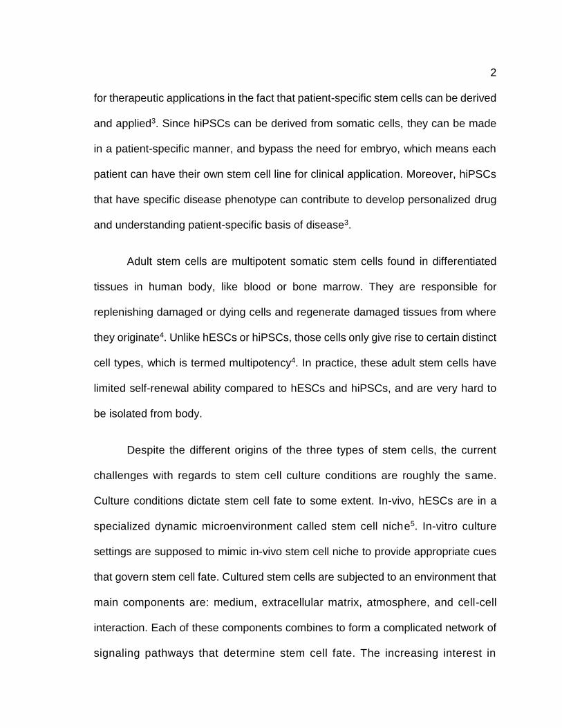
1.2 Important factors involved in hPSCs culture
The proliferation of any type of mammalian cells in vitro relies on three key
factors: cell culture media, extracellular matrix (ECM), and environmental cues5
(Figure 1.1). HPSCs require different culture conditions from murine ESCs. Mouse
embryonic stem cells (mESCs) rely on BMP4 and Stat3 signaling and leukemia
inhibitory factors (LIF) to maintain self-renewal6. However, hPSCs depend on the
interaction between different signaling pathways such as fibroblast growth factor
(FGF-2), Noggin, Activin/Nodal, and TGF-β pathways in the absence of
LIF5,7,8,9,10,11. Moreover, mESCs can be passaged and grow as single cell, but
hPSCs lose viability after dissociation as single cell. Therefore, hPSCs are usually
plated and grown as colonies. The intrinsic difference between mESCs and hPSCs
leads to different consideration of culture conditions to support hPSCs proliferation
in vitro12. To successfully culture hPSCs and maintain their pluripotency in vitro, it
is critical to consider the combination of culture medium, extra cellular matrix, and
environment cues. Ideally, a new stem cell culture protocol can be made based on
one or in combination of the three contributing factors.

4
1.3 Culture medium
There is growing interest in optimizing stem cell culture, not only because
cell culture is the basis of basic stem cell research in laboratory, but also due to the
possible clinical applications of pluripotent stem cells in human body. Growth
medium is one of the most critical components for stem cell culture, and it also has
experienced dramatic evolution since it was first developed for human embryonic
Figure 1.1 Stem cell culture environment. Culture medium, substrate, and environmental cues such as oxygen gradient are contributing to stem cell fate31. Figure adapted from Sanden et al. (31).

5
stem cell culture1. In the beginning of stem cell culture, one of the biggest challenges
was to determine the necessary components in a culture medium to support
undifferentiated hPSCs. Researchers have been spending a lot of efforts trying to
understand the key signaling pathways involved in the process for developing a
robust culture medium.
It is very clear that culture medium dramatically influences cell proliferation
and cell fate. Stem cell fate is always balanced between proliferation and
differentiation. Therefore, the component a of stem cell culture medium is usually
determined by its ability to support self-renewal and pluripotency. In other words,
the medium is required to maintain a large-scale undifferentiated hPSCs as well as
to promote their self-renewal. Stem cells from different sources are diverse.
Different stem cell type might require distinct culture conditions owing to different
signaling pathways. Thus, it is impossible to develop an universal stem cell culture
medium.
Furthermore, some other general considerations are also required. Stem cell
culture conditions must allow propagation of normal phenotype and karyotype cells.
Culture conditions need to be well established and avoid any undefined matrix and
medium component such as serum, for therapeutic applications. The earliest
version of culture medium for human embryonic stem cell was composed of fetal
bovine serum (FBS)5, which is an undefined component secreted by mouse
embryonic fibroblasts.
6
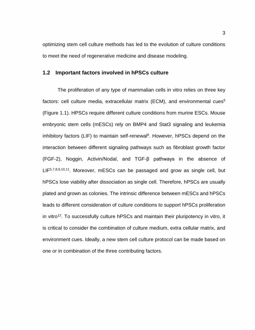
1.3.1 From feeder layer to defined culture system
When several hESC lines were first brought to culture, cells were maintained
on feeder layers such as mouse embryonic fibroblasts (MEFs) in medium containing
serum. Feeder cells secret growth factors and extracellular matrix to support stem
cell growth. However, it is extremely difficult to determine which components are
present in the medium due to the undefined secretion of these factors by different
feeder cells. The preparation of feeder cells takes lots of time and efforts. Moreover,
feeder cells and their secretion products might be a source of pathogens for stem
cells which raises concerns when it comes to therapeutic applications. For example,
it has been reported that immunogenic sialic acid (Neu5Gc) is present in the co-
culture of hESCs and MEFs with serum replacements13. Thus, the undefined culture
condition created by feeder cells impairs the potential use in clinical application, and
must be replaced by more defined components.
Serum is also not fully defined since there are too many variabilities between
different batches of serum products. Nowadays, more and more efforts have been
focusing on the development of fully-defined medium by providing key growth
factors or cytokines
without the need of
feeder cells (Figure
1.2).
Figure 1.2 Evolution of stem cell culture condition. The in-vitro culture of stem hiPSCs has evolved from relying on feeder cells to defined, xeno-free medium components. Figure adapted from Villa-Diaz et al. (21).

7
1.3.2 Critical signaling pathways
Nowadays, serum-free medium supplemented with several growth
factors and cytokines is used in most stem cell culture. One of the critical factors
added to culture medium is bFGF, which facilitates to culture undifferentiated
hESCs and hiPSCs14. Different cell lines may require different growth factors. For
example, LIF is important for the proliferation of mouse ESCs, but has no effects for
human stem cells12. Other soluble factors involved in stem cell culture include BMP
family proteins, which can contribute to hESCs differentiation15. Nodal, activin A,
and TGF-β are used to promote self-renewal of hESCs by the inhibition of BMP
signaling16. Small molecules such as retinoic acid, hormones, and CHIR99021 are
used in differentiation medium to promote stem cell differentiation towards certain
pathway5, likewise, there are many signaling molecules that can support
undifferentiated stem cell population remaining undiscovered. Inhibition of GSK3
signaling has been shown to maintain pluripotency state of mouse and human
embryonic stem cells17.
1.3.3 Growth medium
Considering that cell culture is dynamic and changing due to the secretion
and release of metabolites by cells, it takes plenty of time to achieve fully defined
culture conditions. In recent years, researchers have been using more chemically
defined medium to replace xenogeneic components. The first culture medium
termed TeSR1 containing FGF-2, lithium chloride (LiCl), γ-aminobutyric acid

8
(GABA), TGF-β ,and pipeolic acid that supports feeder-free stem cell culture was
developed by Thomson and colleagues18. More recently, Thomson and colleagues
developed a chemically defined medium termed E8 medium, which is derived from
TeSR containing 8 components and lacks serum albumin and β-mercaptoethanol.
This E8 medium is reported to successfully support undifferentiated human stem
cells including hESCs and hiPSCs during daily passages19,20.
1.4 Extracellular matrix
Cell-matrix interaction plays an important role in regulating stem cell fate
including self-renewal, differentiation, and pluripotency. Extracellular components
include organic matrix derived from animal cells, hydrogel, matrix proteins, synthetic
chemicals, and some are commercial available products5.
1.4.1 Matrigel
Matrigel is now the most widely used coating on tissue culture plate for
feeder-free stem cell culture. It is a basement membrane matrix composed of
laminin, collagen, heparan sulfate proteoglycan, and undefined growth factors.
However, Matrigel is produced from mouse Engelbreth-Holm-Swarm (EHS)
sarcoma and ECM proteins derived from mouse embryonic fibroblasts21, and
therefore limits the clinical application of hPSCs. Thus, efforts have been focused
on the study of ECM proteins to replace Matrigel for stem cell culture. It should be

9
noted that Matrigel cannot be used to culture clinical-grade hPSCs. Many different
ECM have been reported to successfully culture hPSCs in vitro.
1.4.2 Alternative ECM proteins
Both natural proteins and synthetic biomaterials have been proposed
recently trying to generate a defined hPSC culture environment. ECM proteins such
as laminin, collagen and fibronectin have been proved to maintain hPSC self-
renewal21. However, not all of them are suitable for hPSC cultivation. Stem cell
culture typically involves two issues: first, the ability to promote proliferation, and,
secondly, the ability to maintain pluripotency during cell culture. People have
studied different types of ECM proteins such as collagen, fibronectin, and many
other synthetic biomaterials. They are shown to be able to promote stem cell
proliferation, however, they fail to support undifferentiated stem cell population, thus
not an ideal candidate.
1.4.3 Laminin isoforms
Among different types of ECM, Laminin draws most attention because they
are endogenously produced by both induced human pluriotent stem cells (hiPSCs)
and human embryonic stem cells (hESCs) to support self-renewal and pluripotency
of hPSCs. Laminin contains α, β, and γ chains. There are five α chains, three β
chains, and three γ chains, and laminin is named by its various combination of each
chain (α1-α5, β1-β3, γ1-γ3). The different isoforms are synthesized in different cell
types and have distinct functions. It is shown that α5 laminins are endogenously
10
synthesized by hPSCs and significantly promote hPSCs proliferation and survival22.
Laminin-521 therefore has been used for stem cell culture coating recently23.
A recently study has shown that α-5 laminin isoforms synthesized by
undifferentiated hPSCs play a key role in regulating hPSCs survival22, which is
consistent with previous findings including laminin-511 is an important regulator
during embryo development24, and laminin-521 can support ideal stem cell culture.
Knocking down of LAMA5 gene by shRNA and Cas9-mediated disruption results in
reduced hPSCs survival and significantly increased apoptosis. Additionally, the self-
renewal and survival were rescued by culturing the LAMA5 deficient hPSCs on
exogenously laminin coating. Based on those studies, laminin α-5 isoforms present
good potential to support undifferentiated hPSCs in vitro.
1.5 Environmental cues
There are several environmental cues including cues from both physical and
physiological environments which affect stem cells growth and differentiation. For
example, temperature, humidity, cell density, rigidity of culture plate, osmosity,
oxygen diffusion velocity, and multicellular associations5. Among these
environmental cues, oxygen is the one of the most important factors that influences
stem cell growth.
Cell are usually passaged and having medium change in a laminar flow
hood, and being maintained in incubators under normoxic oxygen condition.

11
The physiological environment of mammalian embryo is hypoxic. Stem cell culture
condition is supposed to mimic the in vivo environment in order to reconstruct stem
cell niche. However, traditional stem cell culture has been implemented under
normoxia (~21% O2). Low O2 tension can prevent spontaneous hESCs
differentiation25. Moreover, physiological O2 also facilitates hESCs recovery and
reduces chances of abnormal chromosome. Therefore, optimal O2 would be
required to maintain healthy stem cells as well as helping the expansion of stem cell
population without unwanted spontaneous differentiation.
1.6 Goal of this study
Owing to their ability to impact the landscape of regenerative medicine and
tissue engineering, the human pluripotent stem cells including induced pluripotent
stem cells and human embryonic stem cells are of great interest to researchers.
One of the combating issues is the development of a good stem cell medium for
prolonged culture and maintenance of the stem cells without further differentiation
into any cell type. Because hiPSCs and hESCs have practical use in in-vivo study,
it is important to be able to culture these cells in animal-contamination free medium
to avoid the transmittance of animal-pathogens, and induce immunogenic
responses. Therefore, this study considers the development of a xeno-free culture
medium for hiPSCs and hESCs for long term culture and we analyze this new
medium against existing xeno-free media, Nutristem. Glutamax modulation was
also conducted for medium optimization.

12
Chapter 2
Methods
2.1 Maintenance of human iPSCs and human ESCs
Human embryonic stem cells (H9, H7)1, and transgene and vector free
human iPSCs (19-9-7)2 were maintained at 37°C/5% CO2 on Matrigel (Corning)
coated tissue culture plates in LaSR medium. Matrigel plate was coated with a 1:100
dilution using DMEM/F12 (Life Technologies) and incubated at 37°C for 30 minutes
prior to use. Cells were routinely passaged with 0.5mM EDTA in PBS. In brief, cells
were washed twice with EDTA solution, then incubated with EDTA at 37°C for 10
minutes. Then EDTA was removed, and cell suspension was collected.
2.2 Cell counting
All experiments were done on 6-well tissue culture plates. Before starting cell
counting, cells were adapted to the new medium for 1 passage. Cell were
dissociated from the plate with 0.5mM EDTA on the day of counting, then 10 μl of
cell suspension was subjected to hemocytometer for counting. Cell viability assay
was performed by adding 10 μl of Trypanblue to the 10 μl cell suspension, then 10
μl of the mixed solution was subjected to hemocytometer for counting.

13
2.3 Flow cytometry analysis
To analyze Oct4 and Nanog expression, cells were dissociated into single
cell suspension using 0.5mM EDTA in DPBS for 10 min at 37ºC and then fixed with
1% paraformaldehyde for 20 min. at room temperature. Cells were stained with
primary and secondary antibodies in DPBS with 0.1% Triton X-100 and 0.5% BSA.
FACS gating was based on the corresponding isotype antibody control.
2.4 Immunostaining
Cells were fixed with 4% paraformaldehyde for 15 min. at room temperature
and then immunostained using primary and secondary antibodies in DPBS with
0.4% Triton X-100 and 5% non-fat dry milk. Nuclei were stained with Hoechst
33342. A Nikon TI Eclipse epifluorescence microscope was used for imaging
analysis.
2.5 Statistical analysis
All results are represented as means ± SE. Students’ T-test is used for
comparisons between two groups.

14
Chapter 3
Results
In this chapter, we first compared LaSR medium to a commercially available
stem cell culture medium, Nutristem, to evaluate LaSR medium in terms of the
ability to support stem cell self-renewal. Next, we removed one of the components,
Glutaxmax from LaSR, trying to examine the effect of Glutamax in the medium for
supporting stem cell culture, and minimize the formula of LaSR. Last, we developed
a xeno-free version of LaSR by replacing bovine serum albumin in the medium by
human recombinant albumin, human serum albumin, and chemically lipid
concentrate step by step. Cells cultured in different medium were analyzed for
pluripotency markers by immunostaining and flow cytometry.
3.1 Comparison study between LaSR and Nutristem
LaSR basal medium was first developed by previous work to be used for
endothelial differentiation26. Here, we added two growth factors together with
Glutamax and vitamin C to the LaSR basal medium to form a new stem cell culture
medium, which is termed LaSR. To evaluate its ability to support stem cell
proliferation as well as maintaining stem cell pluripotency, we compare the
proliferation rate of hiPSCs cultured in LaSR medium with the hiPSCs cultured in
Nutristem (Biological Industries), which is designed to support long-term culture of
hiPSCs and hESCs. HiPSCs were seeded onto a 6-well tissue culture plate

15
with the same seeding density on day 0 for each condition, and they were cultured
in each condition for one passage to be adapted to the new medium before counting.
Cells cultured in both conditions exhibited normal cell morphology, which includes
uniform colonies of tightly packed cells and distinct colony edges (Figure 3.1).
Then cell counting was done on day 2, 4, 5 after seeding. The cell counting data
have shown that hiPSCs’ growth rates in LaSR were similar to those seen when
cells were cultured in Nutristem (Figure 3.2). They reached the similar cell number
at the end of each passage, and the growth curves fit what we expect to see for the
hiPSCs. This suggests that LaSR has the same ability as Nutristem to support stem
cells proliferation, and can be used for long-term cell culture.
Day 2 Day 4 Day 5
Figure 3.1 Micrographs of iPSC colonies in LaSR medium (Upper panel), and Nutristem medium (Lower panel) 2, 4, 5 days after seeding at passage 3. Scale bars are 100 µm.

16
Figure 3.2 Growth curves of iPSC cultured in LaSR and Nutristem medium at passage 2 and 3.
Cultures are seeded at 1.5 x 10⁵ cells/well. Error bars represent standard error of the mean for
each day. n=2.

17
3.2 Optimization of LaSR by removal of Glutamax
Glutamax is an improved direct substitute for L-glutamine, which is an
important nutrient in cell culture. It is more stable than L-glutamine and does not
degrade in aqueous solutions. To understand its effect in the regulation of stem cell
proliferation, and try to simplify the formula of LaSR, we removed Glutamax from
the LaSR medium, and compared the proliferation growth rate of cells cultured in
LaSR medium with the cells cultured in LaSR without Glutamax medium. Again, the
cells were cultured in each condition for one passage to be adapted to the new
environment. The cell morphology was not affected by the removal of Glutamax
(Figure 3.3).
Day 2 Day 5 Day 4
Figure 3.3 Micrographs of iPSC colonies in LaSR medium (Upper panel), and LaSR without Glutamax medium (Lower panel) 2, 4, 5 days after seeding at passage 6. Scale bars are 100 µm.
18
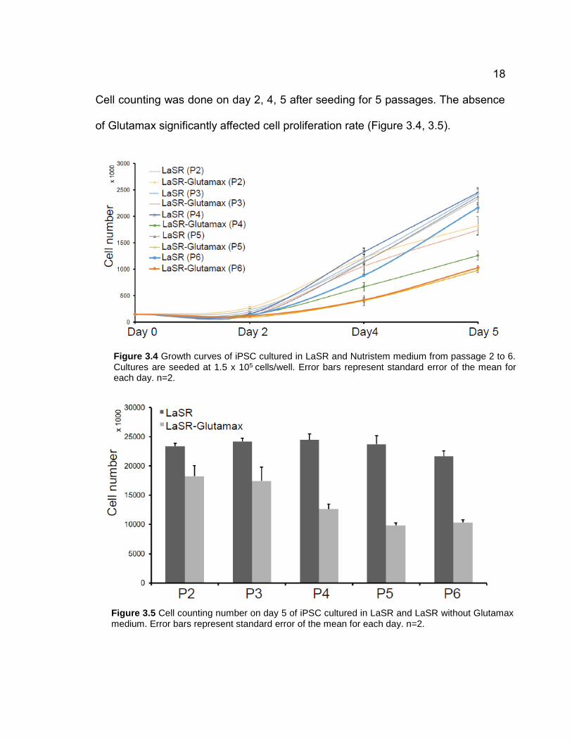
Cell counting was done on day 2, 4, 5 after seeding for 5 passages. The absence
of Glutamax significantly affected cell proliferation rate (Figure 3.4, 3.5).
Figure 3.4 Growth curves of iPSC cultured in LaSR and Nutristem medium from passage 2 to 6. Cultures are seeded at 1.5 x 10⁵ cells/well. Error bars represent standard error of the mean for each day. n=2.
Figure 3.5 Cell counting number on day 5 of iPSC cultured in LaSR and LaSR without Glutamax medium. Error bars represent standard error of the mean for each day. n=2.

19
The cell numbers at the end of each passage for the cells cultured in LaSR without
Glutamax medium were only about half of those cultured in LaSR. However, the
removal of Glutamax did not affect cell survival at all (Table 1). This result shows
that Glutamax is critical for the stem cell proliferation. It might have some beneficial
effects to the cells, or it might cancel some toxic effects of other LaSR medium
components. We then examined the pluripotency of cells by immunostaining against
Oct4 for each condition. Data have demonstrated that both LaSR medium and LaSR
without Glutamax medium supported undifferentiated human iPSCs culture (Figure
3.6).
Figure 3.6 Immunostaining of pluripotency marker Oct4 (green) in iPSC cultured for 6 passages in LaSR (Upper panel), and LaSR without Glutamax medium (Lower panel). Individual cell nuclei were visualized using DAPI (blue). Scale bars are 100 µm.
20
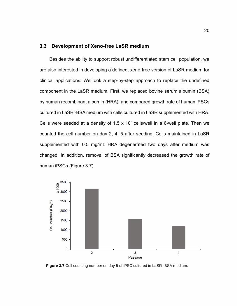
3.3 Development of Xeno-free LaSR medium
Besides the ability to support robust undifferentiated stem cell population, we
are also interested in developing a defined, xeno-free version of LaSR medium for
clinical applications. We took a step-by-step approach to replace the undefined
component in the LaSR medium. First, we replaced bovine serum albumin (BSA)
by human recombinant albumin (HRA), and compared growth rate of human iPSCs
cultured in LaSR -BSA medium with cells cultured in LaSR supplemented with HRA.
Cells were seeded at a density of 1.5 x 10⁵ cells/well in a 6-well plate. Then we
counted the cell number on day 2, 4, 5 after seeding. Cells maintained in LaSR
supplemented with 0.5 mg/mL HRA degenerated two days after medium was
changed. In addition, removal of BSA significantly decreased the growth rate of
human iPSCs (Figure 3.7).
Figure 3.7 Cell counting number on day 5 of iPSC cultured in LaSR -BSA medium.
21
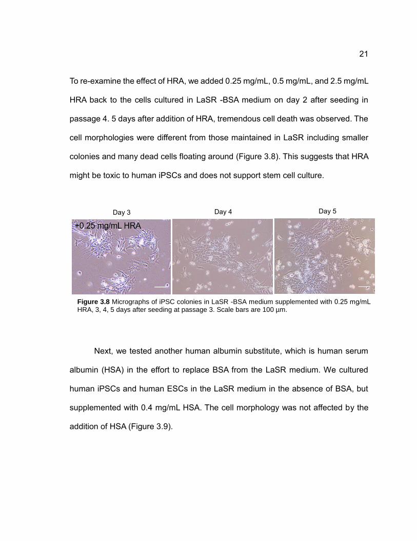
To re-examine the effect of HRA, we added 0.25 mg/mL, 0.5 mg/mL, and 2.5 mg/mL
HRA back to the cells cultured in LaSR -BSA medium on day 2 after seeding in
passage 4. 5 days after addition of HRA, tremendous cell death was observed. The
cell morphologies were different from those maintained in LaSR including smaller
colonies and many dead cells floating around (Figure 3.8). This suggests that HRA
might be toxic to human iPSCs and does not support stem cell culture.
Next, we tested another human albumin substitute, which is human serum
albumin (HSA) in the effort to replace BSA from the LaSR medium. We cultured
human iPSCs and human ESCs in the LaSR medium in the absence of BSA, but
supplemented with 0.4 mg/mL HSA. The cell morphology was not affected by the
addition of HSA (Figure 3.9).
Day 4 Day 5 Day 3
Figure 3.8 Micrographs of iPSC colonies in LaSR -BSA medium supplemented with 0.25 mg/mL HRA, 3, 4, 5 days after seeding at passage 3. Scale bars are 100 µm.

22
Cell counting was done on day 2, 4, 5 after seeding for 5 passages. Cells exhibited
robust proliferation comparable to cells cultured in normal LaSR (Figure 3.10).
Day 3 Day 4 Day 5
Figure 3.9 Micrographs of iPSC colonies in LaSR -BSA medium supplemented with 0.4 mg/mL HSA 3, 4, 5 days after seeding at passage 3. Scale bars are 100 µm.
Figure 3.10 Cell number of iPSC cultured in LaSR -BSA medium supplemented with 0.4 mg/mL HSA on day 0, 2, 4, 5, from passage 2 to 5. Cultures are seeded at 1.5 x 10⁵ cells/well. n=2.

23
The cells were further cultured to passage 14 to test whether LaSR HSA medium
supports long-term hiPSCs cultivation. Immunostaining against Oct4 and Nanog in
passage 6 and 14 confirmed the cells cultured in this medium were sill pluripotent
(Figure 3.11).
Flow cytometry was also performed to quantify the percentage of double-positive
cells against Oct4 and Nanog. 79.1%, 79.3% of the cells were double-positive in
passage 6 and 14, respectively (Figure 3.12). Overall, the data show that LaSR
HSA medium can maintain long-term stable undifferentiated stem cell culture.
Figure 3.11 Immunostaining of pluripotency marker Oct4 (green) and Nanog (red) in iPSC cultured
in LaSR +HSA medium at passage 6 (Left panel) and passage 14 (Right panel). Individual cell
nuclei were visualized using DAPI (blue). Scale bars are 100 µm.

24
3.4 Chemically defined lipids concentrate as a medium supplement
However, serum albumin varies from batch to batch, and what kinds of
molecules present in the serum are unknown. Although we identified a human
source albumin that can be supplemented in LaSR medium for long-term stem cell
culture, the undefined components in the serum still hamper the possible clinical
application of stem cells cultured in this medium. We sought to replace BSA from
the medium by chemically defined lipid concentrate (Invitrogen). It is a combination
of saturated and unsaturated fatty acids designed for a wide variety of applications
(Table 2). We first cultured the human iPSCs in LaSR -BSA medium in the presence
of different dilutions of lipids to test which dilution is most suitable for supporting
stem cell culture. Each well had the same seeding density (150K cells/well) on day
0, and we compared the number of cells at the end of each passage.
Figure 3.12 Flow cytometry analysis of Oct4 (y-axis) and Nanog (x-axis) expression in iPSC
cultured in LaSR +HSA medium at passage 6 (Left panel) and passage 14 (Right panel).
25
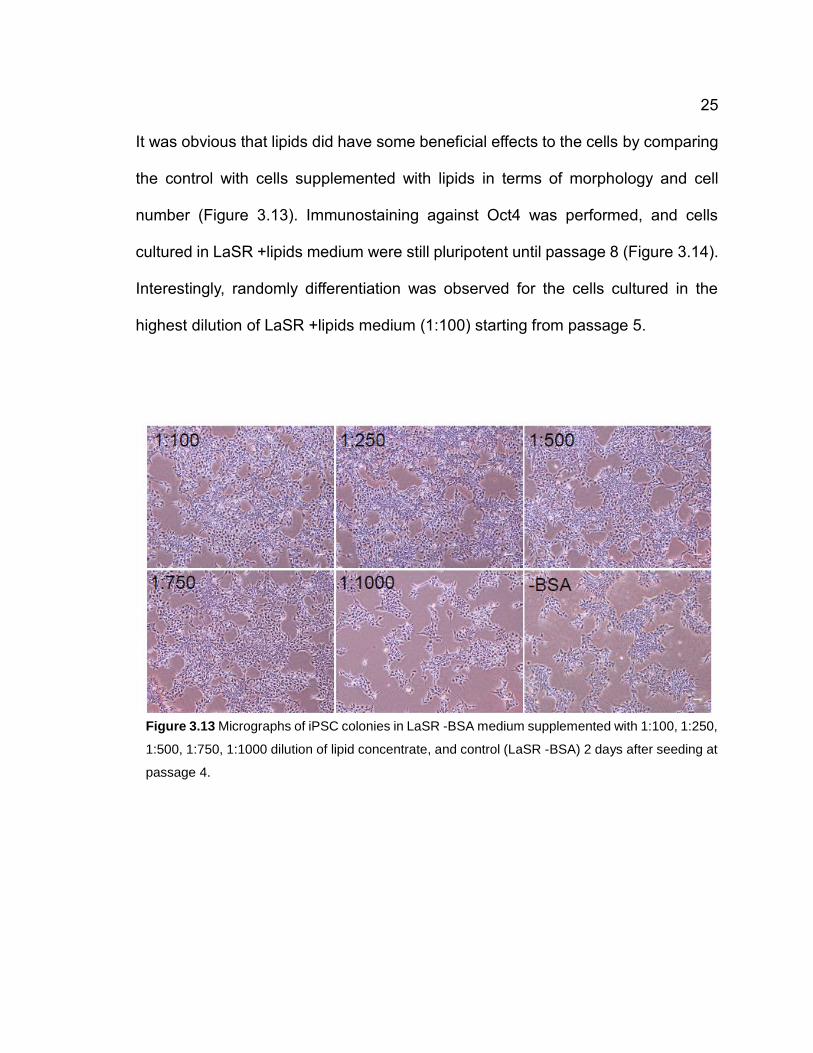
It was obvious that lipids did have some beneficial effects to the cells by comparing
the control with cells supplemented with lipids in terms of morphology and cell
number (Figure 3.13). Immunostaining against Oct4 was performed, and cells
cultured in LaSR +lipids medium were still pluripotent until passage 8 (Figure 3.14).
Interestingly, randomly differentiation was observed for the cells cultured in the
highest dilution of LaSR +lipids medium (1:100) starting from passage 5.
Figure 3.13 Micrographs of iPSC colonies in LaSR -BSA medium supplemented with 1:100, 1:250,
1:500, 1:750, 1:1000 dilution of lipid concentrate, and control (LaSR -BSA) 2 days after seeding at
passage 4.

26
Figure 3.14 Immunostaining of pluripotency marker Oct4 (green) in iPSC cultured in LaSR -BSA
medium and LaSR -BSA +1:500 dilution lipids medium at passage 8. Individual cell nuclei were
visualized using DAPI (blue). Scale bars are 100 µm.
27
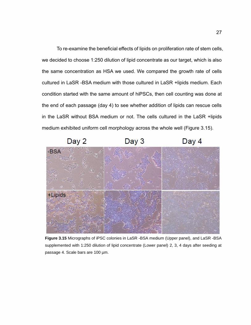
To re-examine the beneficial effects of lipids on proliferation rate of stem cells,
we decided to choose 1:250 dilution of lipid concentrate as our target, which is also
the same concentration as HSA we used. We compared the growth rate of cells
cultured in LaSR -BSA medium with those cultured in LaSR +lipids medium. Each
condition started with the same amount of hiPSCs, then cell counting was done at
the end of each passage (day 4) to see whether addition of lipids can rescue cells
in the LaSR without BSA medium or not. The cells cultured in the LaSR +lipids
medium exhibited uniform cell morphology across the whole well (Figure 3.15).
Figure 3.15 Micrographs of iPSC colonies in LaSR -BSA medium (Upper panel), and LaSR -BSA
supplemented with 1:250 dilution of lipid concentrate (Lower panel) 2, 3, 4 days after seeding at
passage 4. Scale bars are 100 µm.

28
Moreover, we found that the cell number significantly increased in the presence of
lipids over 4 passages, showing lipids did have some beneficial effects on cell
proliferation (Figure 3.16).
Immunostaining against Oct4 and Nanog for the cells in LaSR-BSA and LaSR
+lipids was performed, and both conditions were double-positive for the
pluripotency markers (Figure 3.17).
Figure 3.16 Cell counting number on day 5 (passage 1, 2), and day 4 (passage 3, 4) of iPSC cultured
in LaSR -BSA medium versus LaSR + 1:250 dilution of lipid concentrate. Error bars represent
standard error of the mean for each passage. *P < 0.05, LaSR -BSA versus LaSR -BSA+lipids;
Student’s t test. n=3.

29
Figure 3.17 Immunostaining of pluripotency marker Oct4 (green) and Nanog (red) in iPSC cultured
in LaSR -BSA medium and LaSR -BSA + 1:250 dilution of lipid concentrate. Individual cell nuclei
were visualized using DAPI (blue). Scale bars are 100 µm.

30
Chapter 4
Discussion, Conclusion, and Future Work
4.1 Discussion
Previous study has claimed Glutamax can decrease blastocyst apoptosis
and prolong the shelf life of culture medium to at least 1 year27. By removal of
Glutamax from LaSR, we have shown Glutamax is an essential component in our
medium, cells without Glutamax gradually lose their ability to proliferate as normal.
A research group has proposed that glutamine is essential for survival of human
iPSCs, because their energy production relies mainly on glutamine oxidation28. Our
initial hypothesis was that the deprivation of Glutamax will make human stem cells
enter dormant state, where they stop proliferation but still maintain their life, and can
be awakened simply by the addition of Glutamax into culture medium. This concept
of dormant state could facilitate the knowledge of maintaining stem-cell-derived
progenitor cells. More work needs to be done to investigate the initiation of dormant
state solely by small molecules for the human stem cells.

31
We found that the addition of HRA cannot support stem cell culture, whereas
HSA can successfully support long-term stem cell culture. The major difference
between the two human sourced of albumin is that HRA is a purely recombinant
protein that only contains albumin itself. However, HSA is derived from human
serum, and undefined lipids or other unknown molecules may be collected as well.
It has been suggested that albumin-associated lipids promote human ESCs self-
renewal, not the albumin itself29. The result of this study is consistent to what we
observed when we tested both HRA and HSA in the role of regulating stem cell
proliferation, and explains the reason why HRA fails to maintain stem cell self-
renewal.
Random differentiation was observed when cells were cultured in the LaSR
supplemented with 1:100 dilution of lipid concentrate, which is the highest
concentration we have tested. This may suggest that the high concentration of lipid
concentrate could activate some signaling pathways related to stem cell
differentiation. The concentration of culture medium supplements must be tested
carefully to avoid unwanted side effects such as random differentiation and
karyotype abnormality.

32
4.2 Conclusion
We have successfully demonstrated that both human iPSCs and human
ESCs cultured in LaSR medium exhibited comparable growth with cells cultured in
Nutristem, which is a widely used serum-free medium for stem cell culture. In
addition, cells maintained pluripotency and normal morphology through long-term
cell culture. The cost of stem cell culture medium could be reduced by our LaSR
medium, which has less growth factor and cytokine components. This advantage
will facilitate the research of stem cell-based therapies for a variety of diseases.
The xeno-free LaSR medium was developed by replacing BSA by lipid
concentrate. Our data show the XF-LaSR has the ability to maintain hiPSC self-
renewal and pluripotency through long-term passage, which are the two most
important characteristics of stem cells. This medium can be used to culture stem
cells that are meant to be used for clinical applications. Due to the low cost and
robust ability to support stem cell proliferation, we have been using regular LaSR
medium for all routine culture in the lab. In other words, we have developed two

33
versions of LaSR: regular LaSR and XF-LaSR. They can serve for different
purposes. If xeno-free and chemically defined culture environment is not required,
regular LaSR can be used for routine cell culture for a lower cost of medium,
whereas the XF-LaSR is a defined medium that can be used to generate clinical-
grade human stem cells.
4.3 Future Work
As we have shown, HRA does not support stem cell proliferation, and causes
rapid cell death no matter what concentration we use. This is surprising to us since
HRA is not a toxic molecule to stem cells. Even though HRA itself is not able to
promote stem cell proliferation, it should not kill the cells. More work needs to be
done to investigate the signaling pathways triggered by the treatment of HRA in
order to fully understand the mechanisms involved. This knowledge may allow us
to get better understanding of HRA for developing a new medium.
Extracellular matrix is also a critical factor impacting stem cell fate. For the
generation of clinical-grade human stem cells, it is required to consider both the
medium component and the ECM we used to culture the cells. Matrigel is a most

34
widely-used ECM protein for routine stem cell culture in-vitro. However, it contains
undefined molecules from animal source. More work needs to be done to find a
more appropriate ECM protein which can allow long-term propagation of human
stem cells. It has been reported that Laminin isoforms can be used to culture stem
cells in-vitro. However, the cost of Laminin is considerable. The cost for maintaining
daily cell culture would be much higher if Laminin is used as a substrate.
Chemically-defined synthetic material such as hydrogel may be a good alternative30
since they are stable and made from standard procedures. Most importantly,
synthetic substrates are fully defined and exhibit very little variation from batch to
batch. Further considerations of the existing methods, medium, ECM components
are required to generate clinical-grade human stem cells and their derivative tissues
for the purpose of regenerative medicine and potential drug screening models.

35
APPENDIX
Table 1. Average cell viability values (LaSR vs. LaSR-Glutamax)
Table 2. Formulation of Chemically Defined Lipid Concentrate (Invitrogen)
Passage 2 Passage 3 Passage 4 Passage 5 Passage 6
LaSR 88% 92% 87% 86% 85%
LaSR-Glu. 96% 91% 85.5% 87% 86%
Components Concentration (mg/L)
Arachidonic Acid 2
Cholesterol 220
DL-alpha-Tocopherol Acetate 70
Ethyl Alcohol 100% N/A
Linoleic Acid 10
Linolenic Acid 10
Myristic Acid 10
Oleic Acid 10
Palmitic Acid 10
Palmitoleic Acid 10
Pluronic F-68 90000
Stearic Acid 10
Tween 80® 2200

36
BIBLIOGRAPHY
1. Thomson, J. A. et al. Embryonic stem cell lines derived from human
blastocysts. Science 282, 1145–7 (1998).
2. Takahashi, K. et al. Induction of Pluripotent Stem Cells from Adult Human
Fibroblasts by Defined Factors. Cell 131, 861–872 (2007).
3. Qin, Y. & Gao, W. Q. Concise Review: Patient-Derived Stem Cell Research
for Monogenic Disorders. Stem Cells 34, 44–54 (2016).
4. Eyler, C. E. et al. Adult Stem Cells. Cell (2011). doi:10.1007/978-1-61779-
002-7
5. Chen, K. G., Mallon, B. S., Mckay, R. D. G. & Robey, P. G. Cell Stem Cell
Human Pluripotent Stem Cell Culture: Considerations for Maintenance,
Expansion, and Therapeutics. Stem Cell 14, 13–26 (2014).
6. Evans, M. J. & Kaufman, M. H. Establishment in culture of pluripotential cells
from mouse embryos. Nature 292, 154–156 (1981).
7. James, D., Levine, A. J., Besser, D. & Hemmati-Brivanlou, A.
TGFbeta/activin/nodal signaling is necessary for the maintenance of
pluripotency in human embryonic stem cells. Development 132, 1273–1282
(2005).
8. Vallier, L., Alexander, M. & Pedersen, R. a. Activin/Nodal and FGF pathways
cooperate to maintain pluripotency of human embryonic stem cells. J. Cell Sci.
118, 4495–4509 (2005).
9. Wang, G. et al. Noggin and bFGF cooperate to maintain the pluripotency of
human embryonic stem cells in the absence of feeder layers. Biochem.
Biophys. Res. Commun. 330, 934–942 (2005).
10. Xiao, L., Yuan, X. & Sharkis, S. J. Activin A maintains self-renewal and
regulates fibroblast growth factor, Wnt, and bone morphogenic protein
pathways in human embryonic stem cells. Stem Cells 24, 1476–1486 (2006).
11. Xu, R.-H. et al. Basic FGF and suppression of BMP signaling sustain
undifferentiated proliferation of human ES cells. Nat. Methods 2, 185–190
(2005).
12. Ginis, I. et al. Differences between human and mouse embryonic stem cells.
Dev. Biol. 269, 360–380 (2004).

37
13. Martin, M. J., Muotri, A., Gage, F. & Varki, A. Human embryonic stem cells
express an immunogenic nonhuman sialic acid. Nat Med 11, 228–232 (2005).
14. Xu, C. et al. Feeder-free growth of undifferentiated human embryonic stem
cells. Nat. Biotechnol. 19, 971–974 (2001).
15. Xu, R.-H. et al. BMP4 initiates human embryonic stem cell differentiation to
trophoblast. Nat. Biotechnol. 20, 1261–1264 (2002).
16. Rao, B. M. & Zandstra, P. W. Culture development for human embryonic stem
cell propagation: Molecular aspects and challenges. Curr. Opin. Biotechnol.
16, 568–576 (2005).
17. Ding, S. & Schultz, P. G. A role for chemistry in stem cell biology. Nat.
Biotechnol. 22, 833–840 (2004).
18. Ludwig, T. E. et al. Derivation of human embryonic stem cells in defined
conditions. Nat. Biotechnol. 24, 185–187 (2006).
19. Chen, G. et al. Chemically defined conditions for human iPS cell derivation
and culture. Nat. Methods 8, 424–429 (2011).
20. Chen, G., Hou, Z., Gulbranson, D. R. & Thomson, J. A. Actin-myosin
contractility is responsible for the reduced viability of dissociated human
embryonic stem cells. Cell Stem Cell 7, 240–248 (2010).
21. Villa-Diaz, L. G., Ross, A. M., Lahann, J. & Krebsbach, P. H. Concise review:
The evolution of human pluripotent stem cell culture: From feeder cells to
synthetic coatings. Stem Cells 31, 1–7 (2013).
22. Laperle, A. et al. ??-5 Laminin Synthesized by Human Pluripotent Stem Cells
Promotes Self-Renewal. Stem Cell Reports 5, 195–206 (2015).
23. Lu, H. F. et al. A defined xeno-free and feeder-free culture system for the
derivation, expansion and direct differentiation of transgene-free patient-
specific induced pluripotent stem cells. Biomaterials 35, 2816–2826 (2014).
24. Domogatskaya, A., Rodin, S., Boutaud, A. & Tryggvason, K. Laminin-511 but
not -332, -111, or -411 enables mouse embryonic stem cell self-renewal in
vitro. Stem Cells 26, 2800–9 (2008).
25. Ezashi, T., Das, P. & Roberts, R. M. Low O2 tensions and the prevention of
differentiation of hES cells. Proc. Natl. Acad. Sci. U. S. A. 102, 4783–8 (2005).
26. Lian, X. et al. Efficient differentiation of human pluripotent stem cells to
endothelial progenitors via small-molecule activation of WNT signaling. Stem
Cell Reports 3, 804–816 (2014).

38
27. Zhao, M. H., Kim, N. H. & Cui, X. S. GlutaMAX prolongs the shelf life of the
culture medium for porcine parthenotes. Theriogenology 85, 368–375 (2016).
28. Tohyama, S. et al. Glutamine Oxidation Is Indispensable for Survival of
Human Pluripotent Stem Cells. Cell Metab. 23, 663–674 (2016).
29. Garcia-Gonzalo, F. R. & Belmonte, J. C. I. Albumin-associated lipids regulate
human embryonic stem cell self-renewal. PLoS One 3, 1–10 (2008).
30. Higuchi, A. et al. Long-term xeno-free culture of human pluripotent stem cells
on hydrogels with optimal elasticity. Sci. Rep. 5, 18136 (2015).
31. Van Der Sanden, B., Dhobb, M., Berger, F. & Wion, D. Optimizing stem cell
culture. J. Cell. Biochem. 111, 801–807 (2010).