Embed Size (px)
Citation preview
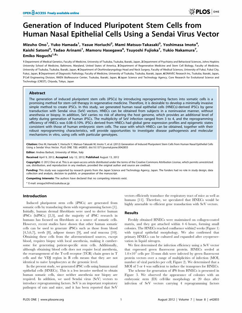
Generation of Induced Pluripotent Stem Cells fromHuman Nasal Epithelial Cells Using a Sendai Virus VectorMizuho Ono1, Yuko Hamada1, Yasue Horiuchi2, Mami Matsuo-Takasaki3, Yoshimasa Imoto4,
Kaishi Satomi5, Tadao Arinami1, Mamoru Hasegawa6, Tsuyoshi Fujioka7, Yukio Nakamura7,
Emiko Noguchi1,8*
1 Department of Medical Genetics, Faculty of Medicine, University of Tsukuba, Tsukuba, Ibaraki, Japan, 2 Department of Psychiatry and Behavioral Sciences, Johns Hopkins
University School of Medicine, Baltimore, Maryland, United States of America, 3 Department of Regenerative Medicine and Stem Cell Biology, Faculty of Medicine,
University of Tsukuba, Tsukuba, Ibaraki, Japan, 4 Department of Otorhinolaryngology Head and Neck Surgery, Faculty of Medical Sciences, University of Fukui, Fukui City,
Fukui, Japan, 5 Department of Diagnostic Pathology, Faculty of Medicine, University of Tsukuba, Tsukuba, Ibaraki, Japan, 6 DNAVEC Research Inc, Tsukuba, Ibaraki, Japan,
7 Cell Engineering Division, RIKEN BioResource Center, Tsukuba, Ibaraki, Japan, 8 Japan Science and Technology Agency, Core Research for Evolutional Science and
Technology (CREST), Chiyoda, Tokyo, Japan
Abstract
The generation of induced pluripotent stem cells (iPSCs) by introducing reprogramming factors into somatic cells is apromising method for stem cell therapy in regenerative medicine. Therefore, it is desirable to develop a minimally invasivesimple method to create iPSCs. In this study, we generated human nasal epithelial cells (HNECs)-derived iPSCs by genetransduction with Sendai virus (SeV) vectors. HNECs can be obtained from subjects in a noninvasive manner, withoutanesthesia or biopsy. In addition, SeV carries no risk of altering the host genome, which provides an additional level ofsafety during generation of human iPSCs. The multiplicity of SeV infection ranged from 3 to 4, and the reprogrammingefficiency of HNECs was 0.08–0.10%. iPSCs derived from HNECs had global gene expression profiles and epigenetic statesconsistent with those of human embryonic stem cells. The ease with which HNECs can be obtained, together with theirrobust reprogramming characteristics, will provide opportunities to investigate disease pathogenesis and molecularmechanisms in vitro, using cells with particular genotypes.
Citation: Ono M, Hamada Y, Horiuchi Y, Matsuo-Takasaki M, Imoto Y, et al. (2012) Generation of Induced Pluripotent Stem Cells from Human Nasal Epithelial CellsUsing a Sendai Virus Vector. PLoS ONE 7(8): e42855. doi:10.1371/journal.pone.0042855
Editor: Andrea Barbuti, University of Milan, Italy
Received April 9, 2012; Accepted July 12, 2012; Published August 13, 2012
Copyright: � 2012 Ono et al. This is an open-access article distributed under the terms of the Creative Commons Attribution License, which permits unrestricteduse, distribution, and reproduction in any medium, provided the original author and source are credited.
Funding: This study was supported by research grants from the Japan Science and Technology Agency, Japan. The funders had no role in study design, datacollection and analysis, decision to publish, or preparation of the manuscript.
Competing Interests: The authors have declared that no competing interests exist.
* E-mail: [email protected]
Introduction
Induced pluripotent stem cells (iPSCs) are generated from
somatic cells by transducing them with reprogramming factors [1].
Initially, human dermal fibroblasts were used to derive human
iPSCs (hiPSCs) [2,3], and the majority of iPSC research in
humans has focused on fibroblasts as a source of somatic cells.
However, recent studies have shown that other human somatic
cells can be used to generate iPSCs such as those from blood
[4,5,6,7], teeth [8], adipose tissues [9], and oral mucosa [10].
Obtaining these cells from the aforementioned sources, except
blood, requires biopsy with local anesthesia, making it cumber-
some for generating patient-specific stem cells. Additionally,
although obtaining blood cells does not require local anesthesia,
the rearrangement of the T-cell receptor (TCR) chain genes in T
cells and the VDJ region in B cells means that they are not
identical to naı̈ve lymphocytes at the genomic level.
In the present study, we generated iPSC cells using human nasal
epithelial cells (HNECs). This is a less invasive method to obtain
human somatic cells, since neither anesthesia nor biopsy are
required. In addition, we used Sendai virus (SeV) vectors to
introduce reprogramming factors. SeV is an important respiratory
pathogen of rats and mice, and it has been reported that SeV
vectors efficiently transduce the respiratory tract of mice as well as
humans [11]. Therefore, we speculated that HNECs would be
highly amenable to efficient gene transduction with SeV vectors.
Results
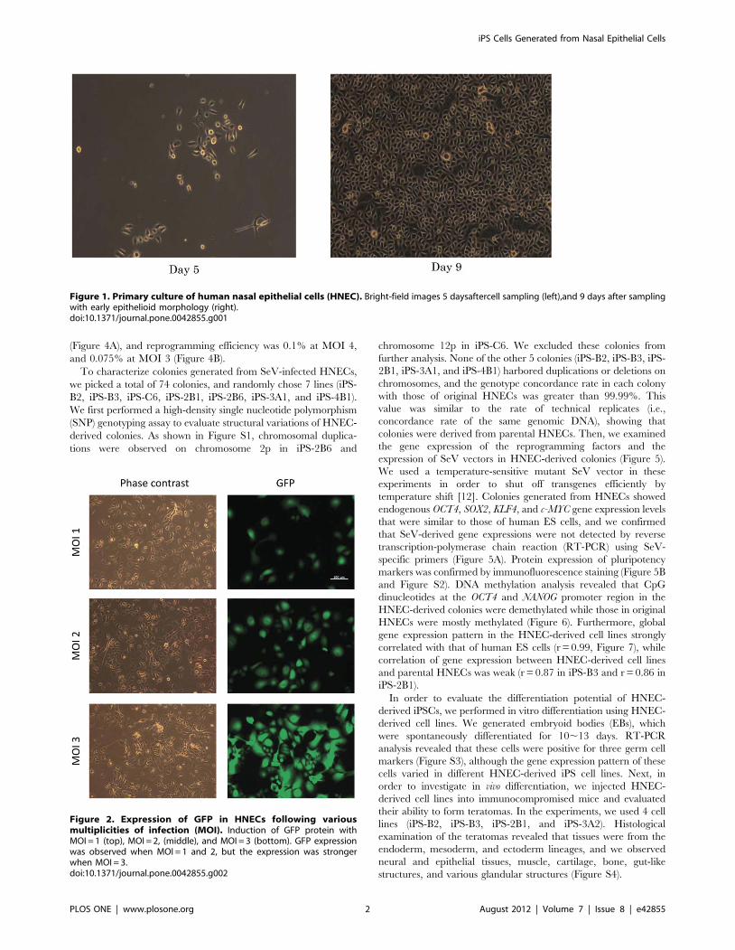
Freshly obtained HNECs were maintained on collagen-coated
matrix, and they got attached within 4–6 hours, forming small
colonies. The HNECs reached confluence within2 weeks (Figure 1)
with typical epithelial morphology. We also confirmed that
primary HNECs can be cultured and expanded after cryopreser-
vation in liquid nitrogen.
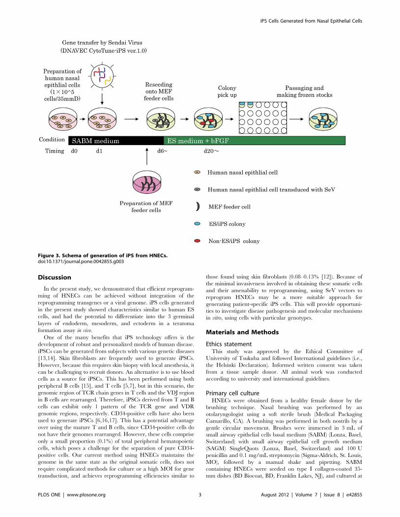
We first determined the infection efficiency using a SeV vector
that expressed green fluorescent protein. HNECs seeded at
1.06105 cells per 35-mm dish were infected by green fluorescent
protein vectors over a range of multiplicities of infection (MOI,
number of viral particles per cell; Figure 2). We determined that a
MOI of 3 or 4 was sufficient to induce the transgenes for HNECs.
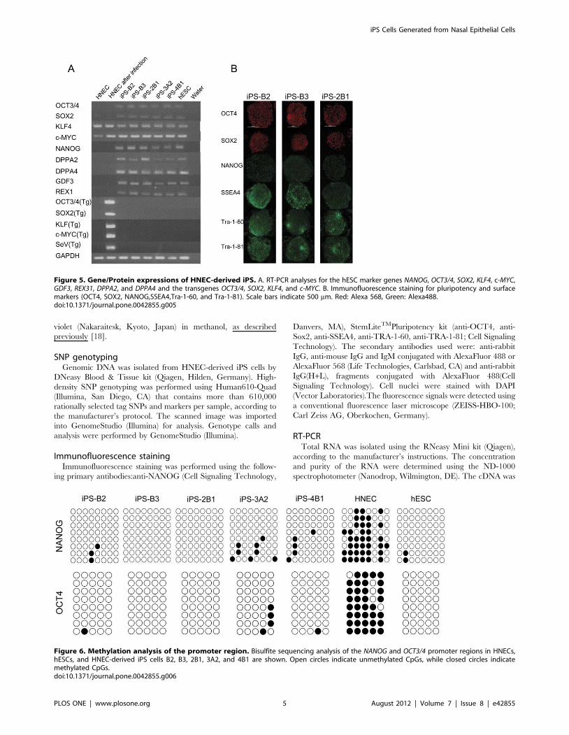
The scheme for generation of iPS from HNECs is presented in
Figure 3. We observed the appearance of colonies with an
embryonic stem (ES) cell-like morphology at 20 days after
infection of SeV vectors carrying 4 reprogramming factors
PLOS ONE | www.plosone.org 1 August 2012 | Volume 7 | Issue 8 | e42855
(Figure 4A), and reprogramming efficiency was 0.1% at MOI 4,
and 0.075% at MOI 3 (Figure 4B).
To characterize colonies generated from SeV-infected HNECs,
we picked a total of 74 colonies, and randomly chose 7 lines (iPS-
B2, iPS-B3, iPS-C6, iPS-2B1, iPS-2B6, iPS-3A1, and iPS-4B1).
We first performed a high-density single nucleotide polymorphism
(SNP) genotyping assay to evaluate structural variations of HNEC-
derived colonies. As shown in Figure S1, chromosomal duplica-
tions were observed on chromosome 2p in iPS-2B6 and
chromosome 12p in iPS-C6. We excluded these colonies from
further analysis. None of the other 5 colonies (iPS-B2, iPS-B3, iPS-
2B1, iPS-3A1, and iPS-4B1) harbored duplications or deletions on
chromosomes, and the genotype concordance rate in each colony
with those of original HNECs was greater than 99.99%. This
value was similar to the rate of technical replicates (i.e.,
concordance rate of the same genomic DNA), showing that
colonies were derived from parental HNECs. Then, we examined
the gene expression of the reprogramming factors and the
expression of SeV vectors in HNEC-derived colonies (Figure 5).
We used a temperature-sensitive mutant SeV vector in these
experiments in order to shut off transgenes efficiently by
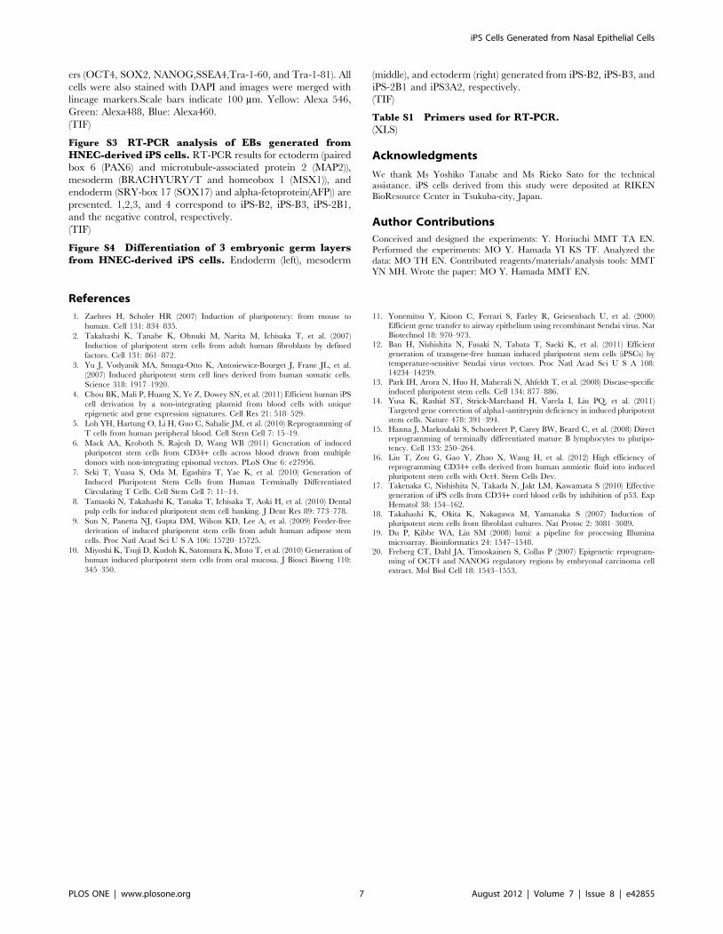
temperature shift [12]. Colonies generated from HNECs showed
endogenous OCT4, SOX2, KLF4, and c-MYC gene expression levels
that were similar to those of human ES cells, and we confirmed
that SeV-derived gene expressions were not detected by reverse
transcription-polymerase chain reaction (RT-PCR) using SeV-
specific primers (Figure 5A). Protein expression of pluripotency
markers was confirmed by immunofluorescence staining (Figure 5B
and Figure S2). DNA methylation analysis revealed that CpG
dinucleotides at the OCT4 and NANOG promoter region in the
HNEC-derived colonies were demethylated while those in original
HNECs were mostly methylated (Figure 6). Furthermore, global
gene expression pattern in the HNEC-derived cell lines strongly
correlated with that of human ES cells (r = 0.99, Figure 7), while
correlation of gene expression between HNEC-derived cell lines
and parental HNECs was weak (r = 0.87 in iPS-B3 and r = 0.86 in
iPS-2B1).
In order to evaluate the differentiation potential of HNEC-
derived iPSCs, we performed in vitro differentiation using HNEC-
derived cell lines. We generated embryoid bodies (EBs), which
were spontaneously differentiated for 10,13 days. RT-PCR
analysis revealed that these cells were positive for three germ cell
markers (Figure S3), although the gene expression pattern of these
cells varied in different HNEC-derived iPS cell lines. Next, in
order to investigate in vivo differentiation, we injected HNEC-
derived cell lines into immunocompromised mice and evaluated
their ability to form teratomas. In the experiments, we used 4 cell
lines (iPS-B2, iPS-B3, iPS-2B1, and iPS-3A2). Histological
examination of the teratomas revealed that tissues were from the
endoderm, mesoderm, and ectoderm lineages, and we observed
neural and epithelial tissues, muscle, cartilage, bone, gut-like
structures, and various glandular structures (Figure S4).
Figure 1. Primary culture of human nasal epithelial cells (HNEC). Bright-field images 5 daysaftercell sampling (left),and 9 days after samplingwith early epithelioid morphology (right).doi:10.1371/journal.pone.0042855.g001
Figure 2. Expression of GFP in HNECs following variousmultiplicities of infection (MOI). Induction of GFP protein withMOI = 1 (top), MOI = 2, (middle), and MOI = 3 (bottom). GFP expressionwas observed when MOI = 1 and 2, but the expression was strongerwhen MOI = 3.doi:10.1371/journal.pone.0042855.g002
iPS Cells Generated from Nasal Epithelial Cells
PLOS ONE | www.plosone.org 2 August 2012 | Volume 7 | Issue 8 | e42855
Discussion
In the present study, we demonstrated that efficient reprogram-
ming of HNECs can be achieved without integration of the
reprogramming transgenes or a viral genome. iPS cells generated
in the present study showed characteristics similar to human ES
cells, and had the potential to differentiate into the 3 germinal
layers of endoderm, mesoderm, and ectoderm in a teratoma
formation assay in vivo.
One of the many benefits that iPS technology offers is the
development of robust and personalized models of human disease.
iPSCs can be generated from subjects with various genetic diseases
[13,14]. Skin fibroblasts are frequently used to generate iPSCs.
However, because this requires skin biopsy with local anesthesia, it
can be challenging to recruit donors. An alternative is to use blood
cells as a source for iPSCs. This has been performed using both
peripheral B cells [15], and T cells [5,7], but in this scenario, the
genomic region of TCR chain genes in T cells and the VDJ region
in B cells are rearranged. Therefore, iPSCs derived from T and B
cells can exhibit only 1 pattern of the TCR gene and VDR
genomic regions, respectively. CD34-positive cells have also been
used to generate iPSCs [6,16,17]. This has a potential advantage
over using the mature T and B cells, since CD34-positive cells do
not have their genomes rearranged. However, these cells comprise
only a small proportion (0.1%) of total peripheral hematopoietic
cells, which poses a challenge for the separation of pure CD34-
positive cells. Our current method using HNECs maintains the
genome in the same state as the original somatic cells, does not
require complicated methods for culture or a high MOI for gene
transduction, and achieves reprogramming efficiencies similar to
those found using skin fibroblasts (0.08–0.13% [12]). Because of
the minimal invasiveness involved in obtaining these somatic cells
and their amenability to reprogramming, using SeV vectors to
reprogram HNECs may be a more suitable approach for
generating patient-specific iPS cells. This will provide opportuni-
ties to investigate disease pathogenesis and molecular mechanisms
in vitro, using cells with particular genotypes.
Materials and Methods
Ethics statementThis study was approved by the Ethical Committee of
University of Tsukuba and followed International guidelines (i.e.,
the Helsinki Declaration). Informed written consent was taken
from a tissue sample donor. All animal work was conducted
according to university and international guidelines.
Primary cell cultureHNECs were obtained from a healthy female donor by the
brushing technique. Nasal brushing was performed by an
otolaryngologist using a soft sterile brush (Medical Packaging
Camarillo, CA). A brushing was performed in both nostrils by a
gentle circular movement. Brushes were immersed in 3 mL of
small airway epithelial cells basal medium (SABM) (Lonza, Basel,
Switzerland) with small airway epithelial cell growth medium
(SAGM) SingleQuots (Lonza, Basel, Switzerland) and 100 U
penicillin and 0.1 mg/mL streptomycin (Sigma-Aldrich, St. Louis,
MO), followed by a manual shake and pipetting. SABM
containing HNECs were seeded on type I collagen-coated 35-
mm dishes (BD Biocoat, BD, Franklin Lakes, NJ), and cultured at
Figure 3. Schema of generation of iPS from HNECs.doi:10.1371/journal.pone.0042855.g003
iPS Cells Generated from Nasal Epithelial Cells
PLOS ONE | www.plosone.org 3 August 2012 | Volume 7 | Issue 8 | e42855

37uC in an atmosphere of 5% CO2. After 24 and 48 h of culture,
non-adherent HNECs were collected and transferred to a new
type I collagen-coated dish. SABM was changed every 2 days, and
cells were cultured with SABM with SAGM SingleQuots and
Reagent Pack (Lonza, Basel, Switzerland).
Induction of pluripotent stem cellsAfter 2 weeks of culture, HNECs were collected and 1.06105
cells were transferred to a new 35-mm dish coated with type Icollagen (BDBiocoat, BD, Franklin Lakes, NJ), and cultured at
37uC in an atmosphere of 5% CO2 overnight. HNECs were
infected with SeV vectors (DNAVEC, Tsukuba, Japan) that
individually carried each of OCT3/4, SOX2, KLF4, and c-MYC at
an MOI of 3 or 4. Infected cells were cultured in SABM
supplemented with SAGM SingleQuots. After 24 h of infection,
the medium was changed to fresh medium. At 6–7 days after
infection, 1.0–2.06105 cells were collected and transferred to a
10 cm-dish containing mitomycin C-inactivated mouse embryonic
fibroblast (MEF) feeder cells (1.56106 cells per dish). After an
additional 24 h of incubation, the medium was changed to hiPSC
medium, which consisted of Dulbecco’s modified Eagle medium/
F12 medium (Life Technologies, Carlsbad, CA) supplemented
with 20% Knock-out Serum Replacement (KSR; Life Technol-
ogies, Carlsbad, CA), 2 mML-glutamine (Life Technologies),
1 mM non-essential amino acids (Sigma-Aldrich, St. Louis,
MO), 0.1 mM b-mercaptoethanol (Sigma-Aldrich, St. Louis,
MO), and 4 ng/mL basic fibroblast growth factor (WAKO,
Osaka, Japan). The hiPSC medium was changed every day. The
generated iPSCs were maintained on mitomycin C-inactivated
MEF feeder cells in hiPSC 3 medium, and the cells were passaged
with CTK solution consisted of 0.25% trypsin, 1 mg/mL
collagenase IV, 20% KSR (all from Life Technologies, Carlsbad,
CA), 20 mM CaCl2 (Wako) in PBS(-) (Life Technologies,
Carlsbad, CA).
Reprogramming efficiencyReprogramming efficiency was calculated as the number of iPS
colonies formed per number of infected cells seeded. iPS colonies
were identified based on ES cell-like morphology and crystal violet
staining. Crystal violet staining was performed with 0.1% crystal
Figure 4. Morphologies of HNEC-derived iPS. A. Colonies generated from HNECs show a round shape with large nucleoli and scant cytoplasm,similar to the morphologyof human ES cells. B. Crystal violet staining of growing cells. Efficiency of iPS cell generation is 0.75% at MOI 3 (left), and0.1% at MOI 4 (right).doi:10.1371/journal.pone.0042855.g004
iPS Cells Generated from Nasal Epithelial Cells
PLOS ONE | www.plosone.org 4 August 2012 | Volume 7 | Issue 8 | e42855
violet (Nakaraitesk, Kyoto, Japan) in methanol, as described
previously [18].
SNP genotypingGenomic DNA was isolated from HNEC-derived iPS cells by
DNeasy Blood & Tissue kit (Qiagen, Hilden, Germany). High-
density SNP genotyping was performed using Human610-Quad
(Illumina, San Diego, CA) that contains more than 610,000
rationally selected tag SNPs and markers per sample, according to
the manufacturer’s protocol. The scanned image was imported
into GenomeStudio (Illumina) for analysis. Genotype calls and
analysis were performed by GenomeStudio (Illumina).
Immunofluorescence stainingImmunofluorescence staining was performed using the follow-
ing primary antibodies:anti-NANOG (Cell Signaling Technology,
Danvers, MA), StemLiteTMPluripotency kit (anti-OCT4, anti-
Sox2, anti-SSEA4, anti-TRA-1-60, anti-TRA-1-81; Cell Signaling
Technology). The secondary antibodies used were: anti-rabbit
IgG, anti-mouse IgG and IgM conjugated with AlexaFluor 488 or
AlexaFluor 568 (Life Technologies, Carlsbad, CA) and anti-rabbit
IgG(H+L), fragments conjugated with AlexaFluor 488(Cell
Signaling Technology). Cell nuclei were stained with DAPI
(Vector Laboratories).The fluorescence signals were detected using
a conventional fluorescence laser microscope (ZEISS-HBO-100;
Carl Zeiss AG, Oberkochen, Germany).
RT-PCRTotal RNA was isolated using the RNeasy Mini kit (Qiagen),
according to the manufacturer’s instructions. The concentration
and purity of the RNA were determined using the ND-1000
spectrophotometer (Nanodrop, Wilmington, DE). The cDNA was
Figure 5. Gene/Protein expressions of HNEC-derived iPS. A. RT-PCR analyses for the hESC marker genes NANOG, OCT3/4, SOX2, KLF4, c-MYC,GDF3, REX31, DPPA2, and DPPA4 and the transgenes OCT3/4, SOX2, KLF4, and c-MYC. B. Immunofluorescence staining for pluripotency and surfacemarkers (OCT4, SOX2, NANOG,SSEA4,Tra-1-60, and Tra-1-81). Scale bars indicate 500 mm. Red: Alexa 568, Green: Alexa488.doi:10.1371/journal.pone.0042855.g005
Figure 6. Methylation analysis of the promoter region. Bisulfite sequencing analysis of the NANOG and OCT3/4 promoter regions in HNECs,hESCs, and HNEC-derived iPS cells B2, B3, 2B1, 3A2, and 4B1 are shown. Open circles indicate unmethylated CpGs, while closed circles indicatemethylated CpGs.doi:10.1371/journal.pone.0042855.g006
iPS Cells Generated from Nasal Epithelial Cells
PLOS ONE | www.plosone.org 5 August 2012 | Volume 7 | Issue 8 | e42855

synthesized using the T7 Oligo (dT) Primer (Ambion, Austin, TX),
1000 U/mL ReverTra Ace (TOYOBO, Osaka, Japan), 5000 U
RNaseoutTM recombinant ribonuclease inhibitor (Life Technolo-
gies). RT-PCR was performed with AmpliTaq Gold 360 Master
Mix (Life Technologies). The primers used for RT-PCR are listed
in Table S1.
Global gene expression analysisTotal RNA was isolated from HNEC-derived iPS cells using the
RNeasy Mini Kit. We used Illumina Bead Array with single-color
array (Illumina) as a microarray platform. For the Illumina
BeadArray assay, cRNA was synthesized with an Illumina RNA
Amplification kit (Life Technologies), according to the manufac-
turer’s instructions. In brief, 500 ng of total RNA were reverse
transcribed to synthesize first- and second-strand cDNA, purified
with spin columns, and then in vitro transcribed to synthesize
biotin-labeled cRNA. A total of 750 ng biotin-labeled cRNA was
hybridized to each Illumina Human-Ref8 v3.0 BeadChip array
(Illumina) at 55uC for 18 h. The hybridized BeadChip was washed
and labeled with streptavidin-Cy3 (GE Healthcare, Buckingham-
shire, UK) and then scanned with the Illumina BeadStation 500
System (Illumina). The scanned image was imported into
GenomeStudio software (Illumina) for analysis. Twenty-two
thousand transcripts representing 8 whole-genome samples can
be analyzed on a single BeadChip. GenomeStudio output of the
microarray data was processed with lumi package for the R
language [19] on R version 2.10.0 (http://www.R-project.org/).
Bisulfite sequencingGenomic DNA was isolated from HNECs, human ES cells, and
iPS cells derived from HNECs using DNeasy Blood & Tissue kit
(Qiagen), and was treated with sodium bisulfite using EpiTect
Bisulfite kit (Qiagen), according to the manufacturer’s instructions.
Converted DNA was used as the template for PCR using primer
sets previously described to amplify the promoter regions of OCT4
[20] and NANOG [2]. The purified PCR products were TA-cloned
into pCR4-TOPO vector using TOPO TA Cloning Kit for
Sequencing (Life Technologies). The insert sequences of randomly
picked clones were analyzed using the ABI 3130 DNA analyzer
(Life Technologies).
In vitro differentiationiPS cells were cultured on Petri dishes for one days in hiPSC
medium, then were cultured free floating to induce EBs for 10–13
days in EB medium, consisting of DMEM/Ham’s F12 contain-
ing5% KSR, 2 mML-glutamin, 161024 M nonessential amino
acids, and 161024 M 2-mercaptoethanol.
Teratoma formationAll mouse procedures were conducted in compliance with
institutional animal use guidelines. hiPSCs grown on MEF feeder
layers were collected by CTK solution into tubes, and centrifuged,
and the pellets were suspended in 10 mM Y-27632 (Wako) in cold
Hanks’ Balanced Salt Solution (Life Technologies). The cells from
a confluent 60-mm dish were injected (56106 cells in 200 mL per
injection) into 1 testis of SCID mice (Japan Charles River,
Yokohama, Japan) using a 1-mL syringe. At 8–10 weeks post
injection, teratomas were dissected, fixed in 4% paraformalde-
hyde, and embedded in paraffin. The sections were stained with
hematoxylin and eosin.
Supporting Information
Figure S1 Genome-wide SNP genotyping analysis. A.
Log R ratio and B allele frequency plots of chromosome 12 in iPS-
C6. B. Log R ratio and B allele frequency of chromosome 2 in iPS-
2B6. Red circles indicate duplicated chromosomal regions.
(TIF)
Figure S2 Protein expressions of HNEC-derived iPS.Immunofluorescence staining for pluripotency and surface mark-
Figure 7. Global gene expression analysis. Gene expression patterns were compared between HNEC-derived iPS cells (iPS-B3) and HNECs (left,top), between HNEC-derived iPS cells (iPS-2B1) and HNECs (left, bottom), between HNEC-derived iPS cells (iPS-B3) and human ES cells (hESC, right,top), and between HNEC-derived iPS cells (iPS-2B1) and hESCs (right, bottom). Blue dotted lines indicate 2-fold changes. r represents the correlationcoefficient.doi:10.1371/journal.pone.0042855.g007
iPS Cells Generated from Nasal Epithelial Cells
PLOS ONE | www.plosone.org 6 August 2012 | Volume 7 | Issue 8 | e42855
ers (OCT4, SOX2, NANOG,SSEA4,Tra-1-60, and Tra-1-81). All
cells were also stained with DAPI and images were merged with
lineage markers.Scale bars indicate 100 mm. Yellow: Alexa 546,
Green: Alexa488, Blue: Alexa460.
(TIF)
Figure S3 RT-PCR analysis of EBs generated fromHNEC-derived iPS cells. RT-PCR results for ectoderm (paired
box 6 (PAX6) and microtubule-associated protein 2 (MAP2)),
mesoderm (BRACHYURY/T and homeobox 1 (MSX1)), and
endoderm (SRY-box 17 (SOX17) and alpha-fetoprotein(AFP)) are
presented. 1,2,3, and 4 correspond to iPS-B2, iPS-B3, iPS-2B1,
and the negative control, respectively.
(TIF)
Figure S4 Differentiation of 3 embryonic germ layersfrom HNEC-derived iPS cells. Endoderm (left), mesoderm
(middle), and ectoderm (right) generated from iPS-B2, iPS-B3, and
iPS-2B1 and iPS3A2, respectively.
(TIF)
Table S1 Primers used for RT-PCR.(XLS)
Acknowledgments
We thank Ms Yoshiko Tanabe and Ms Rieko Sato for the technical
assistance. iPS cells derived from this study were deposited at RIKEN
BioResource Center in Tsukuba-city, Japan.
Author Contributions
Conceived and designed the experiments: Y. Horiuchi MMT TA EN.
Performed the experiments: MO Y. Hamada YI KS TF. Analyzed the
data: MO TH EN. Contributed reagents/materials/analysis tools: MMT
YN MH. Wrote the paper: MO Y. Hamada MMT EN.
References
1. Zaehres H, Scholer HR (2007) Induction of pluripotency: from mouse to
human. Cell 131: 834–835.
2. Takahashi K, Tanabe K, Ohnuki M, Narita M, Ichisaka T, et al. (2007)
Induction of pluripotent stem cells from adult human fibroblasts by defined
factors. Cell 131: 861–872.
3. Yu J, Vodyanik MA, Smuga-Otto K, Antosiewicz-Bourget J, Frane JL, et al.
(2007) Induced pluripotent stem cell lines derived from human somatic cells.
Science 318: 1917–1920.
4. Chou BK, Mali P, Huang X, Ye Z, Dowey SN, et al. (2011) Efficient human iPS
cell derivation by a non-integrating plasmid from blood cells with unique
epigenetic and gene expression signatures. Cell Res 21: 518–529.
5. Loh YH, Hartung O, Li H, Guo C, Sahalie JM, et al. (2010) Reprogramming of
T cells from human peripheral blood. Cell Stem Cell 7: 15–19.
6. Mack AA, Kroboth S, Rajesh D, Wang WB (2011) Generation of induced
pluripotent stem cells from CD34+ cells across blood drawn from multiple
donors with non-integrating episomal vectors. PLoS One 6: e27956.
7. Seki T, Yuasa S, Oda M, Egashira T, Yae K, et al. (2010) Generation of
Induced Pluripotent Stem Cells from Human Terminally Differentiated
Circulating T Cells. Cell Stem Cell 7: 11–14.
8. Tamaoki N, Takahashi K, Tanaka T, Ichisaka T, Aoki H, et al. (2010) Dental
pulp cells for induced pluripotent stem cell banking. J Dent Res 89: 773–778.
9. Sun N, Panetta NJ, Gupta DM, Wilson KD, Lee A, et al. (2009) Feeder-free
derivation of induced pluripotent stem cells from adult human adipose stem
cells. Proc Natl Acad Sci U S A 106: 15720–15725.
10. Miyoshi K, Tsuji D, Kudoh K, Satomura K, Muto T, et al. (2010) Generation of
human induced pluripotent stem cells from oral mucosa. J Biosci Bioeng 110:
345–350.
11. Yonemitsu Y, Kitson C, Ferrari S, Farley R, Griesenbach U, et al. (2000)Efficient gene transfer to airway epithelium using recombinant Sendai virus. Nat
Biotechnol 18: 970–973.
12. Ban H, Nishishita N, Fusaki N, Tabata T, Saeki K, et al. (2011) Efficientgeneration of transgene-free human induced pluripotent stem cells (iPSCs) by
temperature-sensitive Sendai virus vectors. Proc Natl Acad Sci U S A 108:14234–14239.
13. Park IH, Arora N, Huo H, Maherali N, Ahfeldt T, et al. (2008) Disease-specific
induced pluripotent stem cells. Cell 134: 877–886.14. Yusa K, Rashid ST, Strick-Marchand H, Varela I, Liu PQ, et al. (2011)
Targeted gene correction of alpha1-antitrypsin deficiency in induced pluripotentstem cells. Nature 478: 391–394.
15. Hanna J, Markoulaki S, Schorderet P, Carey BW, Beard C, et al. (2008) Directreprogramming of terminally differentiated mature B lymphocytes to pluripo-
tency. Cell 133: 250–264.
16. Liu T, Zou G, Gao Y, Zhao X, Wang H, et al. (2012) High efficiency ofreprogramming CD34+ cells derived from human amniotic fluid into induced
pluripotent stem cells with Oct4. Stem Cells Dev.17. Takenaka C, Nishishita N, Takada N, Jakt LM, Kawamata S (2010) Effective
generation of iPS cells from CD34+ cord blood cells by inhibition of p53. Exp
Hematol 38: 154–162.18. Takahashi K, Okita K, Nakagawa M, Yamanaka S (2007) Induction of
pluripotent stem cells from fibroblast cultures. Nat Protoc 2: 3081–3089.19. Du P, Kibbe WA, Lin SM (2008) lumi: a pipeline for processing Illumina
microarray. Bioinformatics 24: 1547–1548.20. Freberg CT, Dahl JA, Timoskainen S, Collas P (2007) Epigenetic reprogram-
ming of OCT4 and NANOG regulatory regions by embryonal carcinoma cell
extract. Mol Biol Cell 18: 1543–1553.
iPS Cells Generated from Nasal Epithelial Cells
PLOS ONE | www.plosone.org 7 August 2012 | Volume 7 | Issue 8 | e42855