Embed Size (px)
Citation preview

ORIGINAL ARTICLE
Defective response inhibition and collicular noradrenalineenrichment in mice with duplicated retinotopic mapin the superior colliculus
Chantal Mathis • Elise Savier • Jean-Bastien Bott • Daniel Clesse • Nicholas Bevins • Dominique Sage-Ciocca •
Karin Geiger • Anaıs Gillet • Alexis Laux-Biehlmann • Yannick Goumon • Adrien Lacaud •
Vincent Lelievre • Christian Kelche • Jean-Christophe Cassel • Frank W. Pfrieger • Michael Reber
Received: 5 February 2014 / Accepted: 28 February 2014 / Published online: 20 March 2014
� The Author(s) 2014. This article is published with open access at Springerlink.com
Abstract The superior colliculus is a hub for multisen-
sory integration necessary for visuo-spatial orientation,
control of gaze movements and attention. The multiple
functions of the superior colliculus have prompted
hypotheses about its involvement in neuropsychiatric
conditions, but to date, this topic has not been addressed
experimentally. We describe experiments on genetically
modified mice, the Isl2-EphA3 knock-in line, that show a
well-characterized duplication of the retino-collicular and
cortico-collicular axonal projections leading to hyperstim-
ulation of the superior colliculus. To explore the functional
impact of collicular hyperstimulation, we compared the
performance of homozygous knock-in, heterozygous
knock-in and wild-type mice in several behavioral tasks
requiring collicular activity. The light/dark box test and
Go/No-Go conditioning task revealed that homozygous
mutant mice exhibit defective response inhibition, a form
of impulsivity. This defect was specific to attention as other
tests showed no differences in visually driven behavior,
motivation, visuo-spatial learning and sensorimotor abili-
ties among the different groups of mice. Monoamine
quantification and gene expression profiling demonstrated a
specific enrichment of noradrenaline only in the superficial
layers of the superior colliculus of Isl2-EphA3 knock-in
mice, where the retinotopy is duplicated, whereas transcript
levels of receptors, transporters and metabolic enzymes of
the monoaminergic pathway were not affected. We dem-
onstrate that the defect in response inhibition is a conse-
quence of noradrenaline imbalance in the superficial layers
of the superior colliculus caused by retinotopic map
duplication. Our results suggest that structural abnormali-
ties in the superior colliculus can cause defective response
inhibition, a key feature of attention-deficit disorders.
Keywords Retinotopy � Visual system � EphA signaling �Superior colliculus � Noradrenaline � Response inhibition �Attention-deficit disorders
Introduction
The superior colliculus (SC) is a midbrain structure that
integrates sensory inputs from multiple modalities (Wal-
lace et al. 1993; Holmes and Spence 2005; May 2006) and
plays a central role in visuo-spatial orientation, attention
and sensorimotor processing (Stein 1984; May 2006;
Gandhi and Katnani 2011). Defects in SC function have
Electronic supplementary material The online version of thisarticle (doi:10.1007/s00429-014-0745-5) contains supplementarymaterial, which is available to authorized users.
C. Mathis � J.-B. Bott � K. Geiger � A. Gillet � C. Kelche �J.-C. Cassel
Laboratory of Adaptative and Cognitive Neurosciences, CNRS,
University of Strasbourg UMR 7364, 67000 Strasbourg, France
e-mail: [email protected]
E. Savier � D. Clesse � A. Laux-Biehlmann � Y. Goumon �A. Lacaud � V. Lelievre � F. W. Pfrieger � M. Reber (&)
Institute of Cellular and Integrative Neurosciences, CNRS UPR
3212, University of Strasbourg, 5, rue blaise Pascal,
67084 Strasbourg, France
e-mail: [email protected]
N. Bevins
Molecular Neurobiology Laboratory, The Salk Institute,
La Jolla, San Diego, CA 92037, USA
N. Bevins
Department of Neurosciences, University of California, La Jolla,
San Diego, CA 92039, USA
D. Sage-Ciocca
Chronobiotron, UMS 3415, CNRS, 67084 Strasbourg, France
123
Brain Struct Funct (2015) 220:1573–1584
DOI 10.1007/s00429-014-0745-5

been associated with a number of neuropathological and
neuropsychiatric disorders including epilepsy (Ross and
Coleman 2000), schizophrenia (Fuentes 2001) and autism
spectrum disorder (ASD) (Kleinhans et al. 2011). Recently,
collicular hyperstimulation has been proposed to underlie
attention-deficit/hyperactivity disorder (ADHD) symptoms,
especially the impulsivity and distractibility associated
with the disorder (Overton 2008; Miller 2009; Dommett
et al. 2009). However, direct experimental evidence for
such a link remains elusive.
The SC presents a particular feature, namely the topo-
graphic organization of its sensory inputs (Sperry 1963;
Lemke and Reber 2005; May 2006). Axons of retinal
ganglion cells (RGCs) project to the superficial layers of
the SC along spatial axes reflecting their position along
corresponding axes in the retina (the retino-collicular map).
Layer V neurons of the V1 cortex also project in a topo-
graphic manner to the superficial layers of the SC, the
cortico-collicular map, which is in register with the retino-
collicular map (May 2006; Triplett et al. 2012). This cre-
ates a topographic representation of the visual field in the
superficial layers of the SC, also called retinotopy. Audi-
tory and somatosensory afferents projecting to deep layers
of the SC are also aligned with the visual maps (Meredith
and Stein 1985; King et al. 1998; May 2006) enhancing
perception of salient stimuli and influencing decision and
overt behavior (Stein et al. 2009).
We took advantage of a specific disruption of the reti-
notopy in the superficial layers of the SC that has been
observed in the Isl2-EphA3 knock-in mice (Fig. 1; Brown
et al. 2000). In this mouse model, the EphA3 tyrosine
kinase receptor, which acts as a guidance molecule during
map formation, is over-expressed by a subset of RGCs.
This leads to a well-characterized duplication of the retino-
collicular and cortico-collicular maps along the anterior–
posterior axis of the SC. Over-expression of the EphA3
receptor neither affects retinal organization and integrity,
nor the topography of collicular somatosensory inputs
Fig. 1 Topographic retino-
collicular projections in WT and
Isl2-EphA3KI animals.
Micrographs illustrate nasal
1,10-dioctadecyl-3,3,3030-tetramethylindocarbocyanine
perchlorate (Dil) injections in
P8 retinas and the
corresponding termination
zone(s) in the SC. Top an
injection in nasal WT retina
leads to a single caudal
termination zone in the SC.
Middle an injection in a nasal
EphA3KI/? retina leads to two
caudal termination zones in the
SC. Bottom an injection in a
nasal EphA3KI/KI retina leads
to two distant termination zones
in the SC. Scale bars 1 mm
1574 Brain Struct Funct (2015) 220:1573–1584
123

(Brown et al. 2000; Reber et al. 2004; Triplett et al. 2009;
Bevins et al. 2011; Triplett et al. 2012). The duplicated
visual maps are functional as single visual stimuli trigger
the activation of two distinct areas in the SC (Triplett et al.
2009). Unlike other mouse models that target Eph/ephrin
signaling (Dottori et al. 1998; Feldheim et al. 2000; Feld-
heim 2004), the genetic modification in the Isl2-EphA3
knock-in mice affects only a subset of RGCs and does not
affect other structures in the brain (Brown et al. 2000;
Reber et al. 2004, Thaler et al. 2004).
To determine if hyperstimulation of the SC, due to
duplication of the retinotopic projections, influences col-
licular-related behavior, wild-type (WT), heterozygous
(EphA3KI/?) and homozygous (EphA3KI/KI) Isl2-EphA3KI
mice were subjected to a series of well-established
behavioral tests. As a first approach, we tested general
visual ability (cliff test, optokinetic reflex, Morris water
maze with visible platform) as the effects of disrupted
EphAs gradients in the RGCs and duplicated retinotopy in
the SC on visual perception have never been described
before. We then focused on general sensorimotor (loco-
motor activity, circadian rhythmicity, light/dark box test)
and integrative features (beam walking test) and on col-
licular-related behavior, especially visuo-spatial orientation
and memory (Morris water maze with hidden platform) and
response inhibition (Go/No-Go task). Our results show that
EphA3KI/KI mutant mice exhibit defective response inhi-
bition when compared to WT or EphA3KI/? littermates.
Visual acuity, sensorimotor activity, visuo-spatial learning,
motivation and memory were similar in the different
genotypes. Molecular characterization demonstrated ele-
vated noradrenaline levels in the superficial layers of the
SC in EphA3KI/KI animals where the retinotopy is dupli-
cated. Expression levels of receptors, transporters and
enzymes of the monoaminergic signaling pathway were
similar to WT littermates. Interestingly, these changes
resemble specific symptoms of the adult and predominantly
inattentive-type of ADHD patients (Diamond 2005; Bie-
derman and Faraone 2005).
Materials and methods
Animals
Mice were bred and housed in our mouse facility (Chro-
nobiotron, UMS 3415, CNRS, Strasbourg) and tested
during the light phase (ZT2–ZT10) of their light/dark cycle
except for indicated experiments. All procedures were in
accordance with national (council directive 87/848, Octo-
ber 1987) and European community (2010/63/EU) guide-
lines. Official agreement numbers for animal
experimentation were 67-292 for CM, 67-215 for J-CC and
67-358 for KG, AG was under their responsibility. Mice
were genotyped by PCR of genomic DNA from tail biop-
sies as described previously (Reber et al. 2004). Four- to
seven-month-old male littermates of each genotype
(EphA3KI/KI, EphA3KI/? and WT) on a mixed genetic
background (C57/Bl6 9 129Sv/J) were subjected to
behavioral tests and molecular analyses. Standard labora-
tory rodent food and water were available ad libitum
throughout all experiments, except for the Go/No-Go task,
for which all mice were kept at 85 % of their free-feeding
weight.
Behavioral tests
Three distinct cohorts of 4- to 7-month-old WT, EphA3KI/?
and EphA3KI/KI males littermates were characterized using
fixed sequences of test ranging as much as possible from
the least to the most invasive test. Inter-test intervals (ITI)
varied along the sequences to limit order effect. The first
cohort of 4- to 7-month-old males littermates (n = 6–9 per
group) was first tested in the light/dark box test (Boeuf
et al. 2009) (ITI 5 days) and then only in the Go/No-Go
task (Meziane et al. 1993). The second cohort of 4- to
7-month-old males littermates (n = 7 per group) was
dedicated to sensorimotor evaluations. They were first
tested for circadian wheel running activity (Mendoza et al.
2008) and general locomotor activity (Yassine et al. 2013)
(ITI 15 days) followed by the Morris water maze paradigm
(Moreau et al. 2008) (ITI 15 days), the beam walking test
(Moreau et al. 2008) (ITI 3 days) and the visual cliff test
(Gibson and Walk 1960) (ITI 21 days). The optokinetic
reflex (Douglas et al. 2005) was studied on a third cohort of
4-month-old (n = 7–10) male littermates. Detailed
descriptions can be found in Online resource 1.
Molecular analysis
Transcript levels were analyzed by semi-quantitative PCR
and monoamine levels were measured by high-pressure
liquid chromatography as described in the Online resource
1.
Statistical analysis
Unless otherwise indicated, data were analyzed by analysis
of variance with repeated measure factors to study inter-
actions between genotype and side, trial, day, 15-min
block, quadrant, runway (rANOVA). All statistical out-
comes were confirmed by a Kruskal–Wallis test applied on
the light–dark single factors or within each repeated mea-
sure, as group sizes in behavioral studies were relatively
small. When required, post hoc analyses were performed
with the Newman–Keuls (NK) multi-comparison test
Brain Struct Funct (2015) 220:1573–1584 1575
123
(Statistica 8.0; Statsoft, Inc., Tulsa, OK). The time spent in
the goal quadrant of the water maze was compared to the
15-s chance value by means of a t test. The 15-s chance
value corresponds to the time spent for random search in
four quadrants during the 60 s probe test. All behavioral
data are expressed as mean ± standard error of the mean
(SEM). HPLC and qPCR data were analyzed using the
non-parametric Kruskal–Wallis (KW) test. All expression
data are represented using boxplots (min, q1, median, q3,
max).
Results
The functional contribution of the SC in specific behavior
has been investigated in a variety of experiments, including
electrophysiological recording, inactivation and lesion
approaches (Binns 1999; Huberman and Niell 2011) but
little has been done at a more integrated level in animal
models with congenital defects.
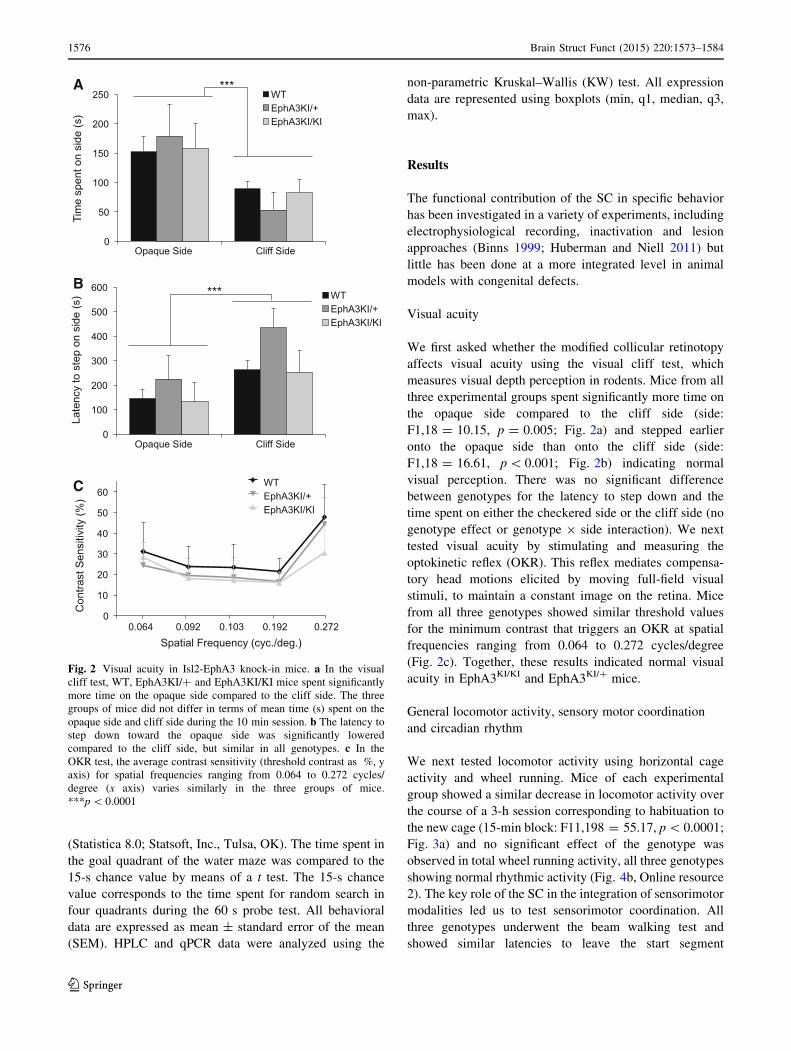
Visual acuity
We first asked whether the modified collicular retinotopy
affects visual acuity using the visual cliff test, which
measures visual depth perception in rodents. Mice from all
three experimental groups spent significantly more time on
the opaque side compared to the cliff side (side:
F1,18 = 10.15, p = 0.005; Fig. 2a) and stepped earlier
onto the opaque side than onto the cliff side (side:
F1,18 = 16.61, p\ 0.001; Fig. 2b) indicating normal
visual perception. There was no significant difference
between genotypes for the latency to step down and the
time spent on either the checkered side or the cliff side (no
genotype effect or genotype 9 side interaction). We next
tested visual acuity by stimulating and measuring the
optokinetic reflex (OKR). This reflex mediates compensa-
tory head motions elicited by moving full-field visual
stimuli, to maintain a constant image on the retina. Mice
from all three genotypes showed similar threshold values
for the minimum contrast that triggers an OKR at spatial
frequencies ranging from 0.064 to 0.272 cycles/degree
(Fig. 2c). Together, these results indicated normal visual
acuity in EphA3KI/KI and EphA3KI/? mice.
General locomotor activity, sensory motor coordination
and circadian rhythm
We next tested locomotor activity using horizontal cage
activity and wheel running. Mice of each experimental
group showed a similar decrease in locomotor activity over
the course of a 3-h session corresponding to habituation to
the new cage (15-min block: F11,198 = 55.17, p\ 0.0001;
Fig. 3a) and no significant effect of the genotype was
observed in total wheel running activity, all three genotypes
showing normal rhythmic activity (Fig. 4b, Online resource
2). The key role of the SC in the integration of sensorimotor
modalities led us to test sensorimotor coordination. All
three genotypes underwent the beam walking test and
showed similar latencies to leave the start segment
A
B
C
Fig. 2 Visual acuity in Isl2-EphA3 knock-in mice. a In the visual
cliff test, WT, EphA3KI/? and EphA3KI/KI mice spent significantly
more time on the opaque side compared to the cliff side. The three
groups of mice did not differ in terms of mean time (s) spent on the
opaque side and cliff side during the 10 min session. b The latency to
step down toward the opaque side was significantly lowered
compared to the cliff side, but similar in all genotypes. c In the
OKR test, the average contrast sensitivity (threshold contrast as %, y
axis) for spatial frequencies ranging from 0.064 to 0.272 cycles/
degree (x axis) varies similarly in the three groups of mice.
***p\ 0.0001
1576 Brain Struct Funct (2015) 220:1573–1584
123

(genotype x trial: F6,54 = 0.65, p[ 0.10, not shown) and
to reach the platform, which decreased significantly during
subsequent trials (trial: F3,54 = 16.48, p\ 0.0001;
Fig. 3b). Sensorimotor coordination and latency to leave the
start segment were similar among genotypes. Moreover, we
tested whether the running activity of knock-in mice fol-
lows light-entrained and endogenous circadian patterns. All
three genotypes showed similar running activity in 12 h
light–dark and dark–dark cycles with similar endogenous
period (WT: 23.57 ± 0.26 h, EphA3KI/?: 23.76 ± 0.35 h
and EphA3KI/KI: 23.69 ± 0.26 h; Fig. 4). Together these
results indicate normal locomotor activity, sensory motor
processing and circadian activity in EphA3KI/KI and
EphA3KI/? animals.
Visuo-spatial orientation and memory
We then tested vision and motor skills using the Morris
water maze visible platform test, where mice must locate a
cue at close range, and swim toward it. After 2 days of
habituation, mice were tested for their performance in
reaching a visible platform. Swim speed and distance were
measured in four trials. Swim speed remained stable and
similar for all groups. Swimming distance was similarly
reduced among all groups over the four consecutive trials
(trial: F3,54 = 16.07, p\ 0.0001). No significant differ-
ence was observed among genotypes or genotype 9 trial
interactions (Fig. 5a). Next we used a variant of the Morris
water maze test where the platform is hidden to evaluate
visuo-spatial learning and memory. Here, mice must find
the hidden platform based on distant visual cues outside the
pool. Over the course of the four training days, mice of all
three genotypes showed similar swim speeds and learned
the position of the hidden platform equally well (day:
F3,54 = 20.67, p\ 0.0001; Fig. 5b). No difference was
observed between genotypes, suggesting that EphA3KI/?
and EphA3KI/KI animals are able to learn a task requiring
visuo-spatial orientation abilities. In a probe test performed
24 h later, all mice showed a clear bias toward the target
quadrant where they spent significantly more time than the
15-s chance level (WT: t6 = 6.68, p = 0.0005, EphA3KI/?:
t6 = 4.62, p = 0.004; EphA3KI/KI: t6 = 6.01, p = 0.001;
Fig. 5c). Taken together, these results indicated normal
visuo-spatial orientation, preserved motivation to reach a
visible and hidden platform and intact spatial learning and
memory in EphA3KI/KI and EphA3KI/? mice.
Anxiety, response inhibition
As the behavioral output in several tasks (e.g., visual cliff,
Go/No-Go and Morris water maze) can be modulated by
levels of anxiety, they were determined in the Isl2-EphA3
knock-in mice using the light/dark box test (Crawley
2007). This conflict test evaluates anxiety based on the
tendency of a mouse to explore a novel environment
against the aversive effect of a brightly lit open field (the
light box). We measured both the time spent in the light
box (aversive environment) and the number of attempts to
enter this box (defined as an incomplete body entrance).
Animals from the three genotypes spent a similar amount
of time in the aversive environment (the light box) indi-
cating comparable levels of anxiety (Fig. 6a). In support of
that, habituation times in a novel activity cage and latency
to leave the start segment in the beam walking test, pre-
sented above, did not differ between the three genotypes
further suggesting that the Isl2-EphA3KI animals exhibit
normal levels of anxiety. Surprisingly, EphA3KI/KI and
EphA3KI/? mice made significantly fewer attempts to enter
A
B
Fig. 3 Locomotor activity and sensorimotor coordination in Isl2-
EphA3 knock-in mice. a During the 3-h habituation phase, EphA3KI/
KI and EphA3KI/? mice did not differ from their WT littermates in
terms of exploration of a new environment (expressed as mean
horizontal activity per 15-min block). b EphA3KI/KI mice did not
differ from their WT littermates in terms of mean time per trial to
reach the platform over 4 trials of the beam test. In all three
genotypes, this parameter decreased significantly over consecutive
trials
Brain Struct Funct (2015) 220:1573–1584 1577
123

the light box (incomplete body entrances) compared to
their WT littermates (attempts: F2,21 = 4.24, p\ 0.05,
NK post hoc: p\ 0.05; Fig. 6b). In other words,
EphA3KI/KI and EphA3KI/? mice were less hesitant and
entered the light box more readily suggesting that they fail
to refrain from exploring an aversive environment. In
addition, EphA3KI/KI and EphA3KI/? mice showed a
decreased latency for complete body entrance into the light
box compared to WT littermates (latency: F2,21 = 3.24,
p = 0.06; Fig. 6c). This provides further evidence that they
did not hesitate to enter an aversive environment. However,
EphA3KI/KI mice showed no increase in time spent in the
light box and no impairment in the visual cliff test, opto-
kinetic reflex and both versions of the water maze in which
performance depends on intact visual abilities (Yassine
et al. 2013). Alternatively, reduced hesitation to enter the
light box could be related to a diminished response inhi-
bition, a key feature of impulsivity (Chamberlain and Sa-
hakian 2007).
To confirm defects in response inhibition of knock-in
mice, we performed a Go/No-Go task. Go/No-Go para-
digms are based on a cue discrimination conditioning and
are commonly used to assess attention and response inhi-
bition, but also learning and memory functions in humans
and mice (Meziane et al. 1993; Aron and Poldrack 2005;
Gubner et al. 2010; Loos et al. 2010). This test required food
restriction, during which the mice were kept at 85 % of their
weight to ensure motivation for food reward. Mice of all
three genotypes showed similar weight loss and motivation
for food during food restriction (not shown) (Meziane et al.
1993). In our version of the task, mice were conditioned to
run successively down two runaways differing in colors,
one color runaway being always baited with food (Go trail)
and the other never baited (No-Go trial). Both EphA3KI/?
and WT littermates progressively learned to discriminate
between the reinforced (Go trials) and non-reinforced (No-
Go trials) runways as indicated by a significant decrease in
running time on Go trials and stable running times on No-
Go trials (Go trials: F2,34 = 18.9, p\ 0.0001; Fig. 7a, b)
as usually observed in this task (Meziane et al. 1993). This
suggested normal learning, motivation and response inhi-
bition in EphA3KI/? and WT mice. Running duration of
EphA3KI/KI animals decreased similarly than WT and
EphA3KI/? littermates on Go trials. Surprisingly, and in
A BFig. 4 Circadian activity in
Isl2-EphA3 knock-in mice. All
three groups of mice showed
similar endogenous periods
after a 15 days of light–dark
(LD) cycle followed by 10 days
of constant darkness (DD)
(a) and similar diurnal and
nocturnal wheel running activity
(b)
1578 Brain Struct Funct (2015) 220:1573–1584
123

contrast to WT and EphA3KI/?, EphA3KI/KI running times
also significantly decreased on No-Go trials (No-Go trails:
F4,34 = 4.03, p\ 0.01, NK p\ 0.05; Fig. 7a, b) indicat-
ing their failure to refrain themselves from running in the
non-reinforced runway on No-Go trials. Preserved perfor-
mances of the EphA3KI/KI animals on Go trials suggested
intact motivation for food and efficient learning. A
discrimination learning deficit in these mice is unlikely
since amnesic treatments are known to affect essentially Go
running times (Meziane et al. 1993, 1998). In addition, their
performance in the visible and hidden versions of the Morris
water maze as well as in the visual cliff test and optokinetic
reflex suggests that their visual acuity and visuo-spatial
memory are comparable to those of WT and EphA3KI/?
littermates. Taken together, these results further support the
hypothesis of a defective response inhibition in the
EphA3KI/KI animals.
A
B
C
Fig. 5 Visuo-spatial orientation, spatial navigation, learning and
memory in Isl2-EphA3 knock-in mice. a In the visible platform test
of the Morris water maze paradigm, all three groups of mice required
similar mean swimming distances per trial to reach the visible platform
and showed a similar decrease in the swimming distance over
consecutive trials. b During the 4-day-long training period in the
hidden platform test of the Morris water maze paradigm, Isl2-EphA3
knock-in mice and their WT littermates required similar swimming
distances to reach the platform and showed a similar decrease over
consecutive trials. c In the 60-s probe test without platform, mice spent
significantly more time in the target quadrant compared to the mean
time in other quadrants regardless of their genotype. ***p\ 0.0001
A
B
C
Fig. 6 Anxiety-related behavior in Isl2-EpA3 knock-in mice. a In thelight/dark box test, Isl2-EphA3 knock-in mice spent the same amount
of time (s) in the light box as their WT littermates. b EphA3KI/KI
animals showed a significant decrease in the number of attempts to
enter the light box compared to the WT littermates. c EphA3KI/KI
and EphA3KI/? animals showed a tendency to a decreased latency
(s) to enter the light box compared to their WT littermates. *p\ 0.05
Brain Struct Funct (2015) 220:1573–1584 1579
123

In principle, this defective behavior could be caused by
impaired attention or increased distraction (Barkley 2004).
To test this possibility, we repeated the reinforced Go task,
but added visual (flashing light) and auditory (tone) dis-
tractors. Mice of all genotypes showed significantly
increased running times by reducing their speed in trials
with tones (70 dB tone: F1,18 = 5.48, p\ 0.05; 90 dB
tone: F1,18 = 9.18, p\ 0.01; Fig. 7c) and flash lights
(F1,18 = 92.06, p\ 0.0001; Fig. 7c) compared to non-
distracted trials. Notably, all EphA3KI/KI mice increased
their running times when exposed to a flashing light, (one
mouse stopped to look toward the origin of the stimulus)
although the difference between EphA3KI/KI and WT lit-
termates did not reach statistical significance (Flash
latency: F2,18 = 1.17, p = 0.33; Fig. 7c). These data
indicate that a flashing light and loud tones are effective
distractors during the Go task.
Analysis of regional monoamine levels
The observed defective response inhibition in EphA3KI/KI
mice, corresponding to an ADHD phenotypic feature, could
be induced by abnormal catecholamine levels (van der Kooij
andGlennon2007; Sontag et al. 2010). To test this possibility,
we determined levels of monoamine neurotransmitters in
distinct areas of themousebrain, namely the superficial layers
of the superior colliculus (SC), the prefrontal cortex, the
striatum, the parietal cortex and the cerebellum, all involved
in attentional processes and motor control (Himelstein et al.
2000; Aron and Poldrack 2005; Biederman and Faraone
2005; Overton 2008). Levels of dopamine, adrenaline and
serotonin were not significantly different between genotypes
in the five structures studied (Fig. 8; Online resource 2). In
contrast, the levels of noradrenaline were significantly
increased in the superficial layers of the SC of EphA3KI/KI
compared to their EphA3KI/? and WT littermates (KW test
p\ 0.05; Figs. 8a, 9). The increase in noradrenaline in the
superficial layers of the SC prompted us to examine the
expression of receptors, transporters and enzymes that are
involved in monoaminergic metabolism and associated with
attention-deficit diseases (Himelstein et al. 2000; Biederman
and Faraone 2005). All three genotypes showed similar
expression of transporters, metabolic enzymes and down-
stream receptors of dopamine, noradrenaline, adrenaline and
A
C
BFig. 7 Go/No-Go performance
in Isl2-EpA3 knock-in mice.
a Over the three sessions, WT,
EphA3KI/? and EphA3KI/KI
mice reduced their mean
running time per trial in the
reinforced Go trials. b Over the
three sessions, WT and
EphA3KI/? mice show stable
mean running time in the non-
reinforced No-Go trials, as
opposed to EphA3KI/KI
littermates, which also reduced
their running times in No-Go
trials NK *p\ 0.05. c Auditory(70, 90 dB tone) and visual
(flash light) distractors led to
significant increases in the
running times in Go trials of all
three genotypes. Note that
EphA3KI/KI mice appeared
slightly more sensitive to a
visual distractor than their
littermates. *p\ 0.05;
**p = 0.01; ***p\ 0.0001
1580 Brain Struct Funct (2015) 220:1573–1584
123

serotonin in the superficial layers of the SC and in other brain
regions (Online resource 2).
Discussion
Our study provides first evidence for specific behavioral
and molecular changes in mice with genetically altered
retinotopy in the superior colliculus and consequently
enhanced visual inputs. In the Go/No-Go task, EphA3KI/KI
mice performed normally on Go trials by increasing their
running speed, but they were completely unable to inhibit
their running response on No-Go trials.
In the light/dark box test, EphA3KI/KI mice entered the
aversive light box more readily than control mice. Alto-
gether, our behavioral tests revealed that EphA3KI/KI mice
exhibit defective response inhibition, a form of impulsivity.
The observation that heterozygous EphA3KI/? mice behave
like WT littermates in the Go/No-Go task suggests that a
partial duplication of the retino-collicular map (Brown et al.
2000) is not sufficient to trigger defective response inhibi-
tion. The observed behavioral changes were remarkably
A
C
E
D
BFig. 8 Monoamine
concentrations in selected brain
regions of Isl2-EphA3 knock-in
mice. Radar-plot representation
of total dopamine, adrenaline,
noradrenaline and serotonin
content (median values, ng/mg
of proteins) in the a superficial
layers of the SC, b prefrontal
cortex, c cerebellum, d striatum
and e parietal cortex. The
noradrenaline content was
significantly increased in
superficial SC layers of
EphA3KI/KI compared to
EphA3KI/? and WT
littermates. *p\ 0.05 KW test.
SC superior colliculus
Brain Struct Funct (2015) 220:1573–1584 1581
123

specific, as all other paradigms tested, namely vision, visuo-
spatial orientation, sensorimotor function, motivation,
learning and memory as well as exploratory behavior and
anxiety were similar in WT, EphA3KI/? and EphA3KI/KI
mice. Defective response inhibition could be the conse-
quence of enhanced levels of noradrenaline that we detected
in the superficial layers of the SC of EphA3KI/KI mice.
Enhanced noradrenaline levels in the SC could alter the
behavior of the EphA3KI/KI mice by modulating the signal-
to-noise ratio in this structure (Mooney et al. 1990; Tan et al.
1999) and thereby changing its level of activation (Dommett
et al. 2009). In hamsters, in vivo and in vitro studies dem-
onstrated a suppression of collicular neuron response upon
noradrenaline application (Mooney et al. 1990; Tan et al.
1999). In rats, Sato and Kayama reported that iontophoreti-
cally applied noradrenaline exerts an excitatory action,
indicating an increase of the signal-to-noise ratio, in accor-
dance with our hypothesis (Sato and Kayama 1983). Whe-
ther noradrenaline increases or decreases the signal-to-noise
ratio in the superficial layers of the SC is still debated.
However, it clearly affects the processing of salient stimuli in
a context-specific manner (Sato and Kayama 1983; Mooney
et al. 1990; Tan et al. 1999).
The increase in noradrenaline was specific to the super-
ficial layers of the SC, where the retinotopy is duplicated.
Moreover, the increase only concerned noradrenaline,
whereas other monoamines including dopamine, serotonin
and adrenaline showed similar concentrations for all
genotypes and brain regions. The increase in noradrenaline
was not accompanied by changes in transcript levels of
genes involved in monoamine metabolism. Therefore, we
hypothesize that the increase of noradrenaline in the
superficial layers of the SC may be the consequence of the
duplication of the RGCs projections, which are functional,
as shown by optical intrinsic imaging (Triplett et al. 2009).
Previous studies revealed that RGC axons release nor-
adrenaline upon activation (Osborne and Patel 1985).
Alternatively, the increase may come from a duplication of
projections from the locus coeruleus (LC), the major source
of noradrenaline in the brain, to the superficial layers of the
SC (Takemoto et al. 1978; Fritschy et al. 1990). Whether
LC projections to the SC are duplicated is unknown as the
mapping of the LC to the SC is hindered by the small size
and specific sub-nuclei organization of the LC. However, it
appears possible given that cortico-collicular projections
are also duplicated in the EphA3KI/KI animals although
projecting V1 neurons do not express ectopic EphA3
(Triplett et al. 2009). RGCs project to different brains areas,
including lateral geniculate nucleus (LGN) and non-image
forming structures such as the suprachiasmatic nucleus
(SCN), the medial tegmental nucleus (MTN) or the olivary
pretectal nucleus (OPN). Triplett and colleagues show no
targeting defects in the LGN of Isl2-EphA3 animals
(Triplett et al. 2009). The same group recently demon-
strated that among 1 % of RGCs projecting to the SCN
(the intrinsically photoreceptive RGCs—ipRGCs), 3 % are
A B
C D
Fig. 9 Monoamine content in the SC of Isl2-EphA3 knock-in mice.
Boxplot representation (min, q1, median, q3, max) of total a dopa-
mine, b adrenaline, c noradrenaline and d serotonin content (in ng/mg
of proteins) in the superficial layers of the superior colliculus (SC)
showing significant increase in noradrenaline in EphA3KI/KI animals
compared to EphA3KI/? and WT littermates. *p\ 0.05 KW test
1582 Brain Struct Funct (2015) 220:1573–1584
123

Isl2-positive and that these SCN-targeting Isl2-positive
RGCs only transiently innervate the SCN during the
development (Triplett et al. 2014). MTN and OPN also
show innervation by Isl2-positive RGCs at early postnatal
stages which is pruned by P6 (Triplett et al. 2014). The
behavioral and molecular changes in EphA3KI/KI mice
including defective response inhibition and noradrenaline
enrichment in the superficial layers of the SC phenocopy
some of the symptoms observed in ADHD patients, spe-
cifically the adult and predominantly inattentive-type
(Barkley 1997; Aron and Poldrack 2005; Biederman and
Faraone 2005; Bekker et al. 2005; Fisher et al. 2011;
American Psychiatric Association 2013). These symptoms
are also main features of Autism Spectrum Disorder (ASD)
(Murray 2010). Our findings support the hypothesis that
adult ADHD patients present collicular hyperstimulation
leading to the appearance of impulsivity and attentional
impairments (Overton 2008; Miller 2009; Dommett et al.
2009). Moreover, they are in line with the idea that dys-
regulation of the central noradrenergic systems contributes
to the pathophysiology of ADHD (Biederman and Spencer
1999). Currently, progress on the etiology, diagnosis and
treatment of ADHD is hindered by the limited number of
experimental models. Most of the available rodent models
are based on impaired monoaminergic transmission (van der
Kooij and Glennon 2007; Sontag et al. 2010) and present
some of the phenotypic features of ADHD patients. Our
findings suggest that EphA3KI/KI animals may serve as a new
model to study ADHD pathology and complement the lim-
ited arsenal of ADHD/ADD-related experimental approaches
to understand and treat these neuropsychologic diseases.
Acknowledgments The authors thank Dr. Sophie Reibel-Foisset,
Nicolas Lethenet and Laurence Huck (Chronobiotron, Unite Mixte de
Service 3415, Centre National de la Recherche Scientifique, Stras-
bourg) for animal care and Pedwin Pallet for help with recordings of
the optokinetic reflex. This work was supported by Partner University
Fund (M.R.), Centre National de la Recherche Scientifique (CNRS)
and Universite de Strasbourg (UdS). Publication costs are supported
by the Neurex network (TriNeuron – Program Interreg IV Upper
Rhine) http://www.neurex.org.
Conflict of interest The authors report no biomedical financial
interests or potential conflicts of interest.
Open Access This article is distributed under the terms of the
Creative Commons Attribution License which permits any use, dis-
tribution, and reproduction in any medium, provided the original
author(s) and the source are credited.
References
American Psychiatric Association (2013) Diagnostic and statistical
manual of mental disorders, 5th edn. doi: 10.1176/appi.books.
9780890423349
Aron AR, Poldrack RA (2005) The cognitive neuroscience of
response inhibition: relevance for genetic research in attention-
deficit/hyperactivity disorder. Biol Psychiatry 57:1285–1292.
doi:10.1016/j.biopsych.2004.10.026
Barkley RA (1997) Behavioral inhibition, sustained attention, and
executive functions: constructing a unifying theory of ADHD.
Psychol Bull 121:65–94
Barkley RA (2004) Driving impairments in teens and adults with
attention-deficit/hyperactivity disorder. Psychiatr Clin North Am
27:233–260. doi:10.1016/S0193-953X(03)00091-1
Bekker EM, Overtoom CCE, Kooij JJS et al (2005) Disentangling
deficits in adults with attention-deficit/hyperactivity disorder.
Arch Gen Psychiatry 62:1129–1136. doi:10.1001/archpsyc.62.
10.1129
Bevins N, Lemke G, Reber M (2011) Genetic dissection of EphA
receptor signaling dynamics during retinotopic mapping. J Neu-
rosci 31:10302–10310. doi:10.1523/JNEUROSCI.1652-11.2011
Biederman J, Faraone SV (2005) Attention-deficit hyperactivity
disorder. Lancet 366:237–248. doi:10.1016/S0140-6736(05)
66915-2
Biederman J, Spencer T (1999) Attention-deficit/hyperactivity disor-
der (ADHD) as a noradrenergic disorder. Biol Psychiatry
46:1234–1242. doi:10.1016/S0006-3223(99)00192-4
Binns KE (1999) The synaptic pharmacology underlying sensory
processing in the superior colliculus. Prog Neurobiol 59:129–159
Boeuf J, Trigo JM, Moreau P-H et al (2009) Attenuated behavioural
responses to acute and chronic cocaine in GASP-1-deficient
mice. Eur J Neurosci 30:860–868. doi:10.1111/j.1460-9568.
2009.06865.x
Brown A, Yates PA, Burrola P et al (2000) Topographic mapping
from the retina to the midbrain is controlled by relative but not
absolute levels of EphA receptor signaling. Cell 102:77–88.
doi:10.1016/S0092-8674(00)00012-X
Chamberlain SR, Sahakian BJ (2007) The neuropsychiatry of
impulsivity. Curr Opin Psychiatry 20:255–261. doi:10.1097/
YCO.0b013e3280ba4989
Crawley JN (2007) What’s Wrong With My Mouse: Behavioral
Phenotyping of Transgenic and Knockout Mice. Wiley, USA.
doi:10.1111/j.1601-183X.2008.00424_1.x
Diamond A (2005) Attention-deficit disorder (attention-deficit/
hyperactivity disorder without hyperactivity): a neurobiologi-
cally and behaviorally distinct disorder from attention-deficit/
hyperactivity disorder (with hyperactivity). Dev Psychopathol
17:807–825. doi:10.1017/S0954579405050388
Dommett EJ, Overton PG, Greenfield SA (2009) Drug therapies for
attentional disorders alter the signal-to-noise ratio in the superior
colliculus. Neuroscience 164:1369–1376. doi:10.1016/j.neu
roscience.2009.09.007
Dottori M, Hartley L, Galea M et al (1998) EphA4 (Sek1) receptor
tyrosine kinase is required for the development of the cortico-
spinal tract. Proc Natl Acad Sci USA 95:13248–13253
Douglas RM, Alam NM, Silver BD et al (2005) Independent visual
threshold measurements in the two eyes of freely moving rats
and mice using a virtual-reality optokinetic system. Vis Neurosci
22:677–684. doi:10.1017/S0952523805225166
Feldheim DA (2004) Loss-of-function analysis of EphA receptors in
retinotectal mapping. J Neurosci 24:2542–2550. doi:10.1523/
JNEUROSCI.0239-03.2004
Feldheim DA, Kim YI, Bergemann AD et al (2000) Genetic analysis
of ephrin-A2 and ephrin-A5 shows their requirement in multiple
aspects of retinocollicular mapping. Neuron 25:563–574
Fisher T, Aharon-Peretz J, Pratt H (2011) Dis-regulation of response
inhibition in adult attention deficit hyperactivity disorder
(ADHD): an ERP study. Clin Neurophysiol 122:2390–2399.
doi:10.1016/j.clinph.2011.05.010
Brain Struct Funct (2015) 220:1573–1584 1583
123

Fritschy JM, Geffard M, Grzanna R (1990) The response of
noradrenergic axons to systemically administered DSP-4 in the
rat: an immunohistochemical study using antibodies to nor-
adrenaline and dopamine-beta-hydroxylase. J Chem Neuroanat
3:309–321
Fuentes LJ (2001) Selective attention deficit in schizophrenia. Rev
Neurol 32:387–391
Gandhi NJ, Katnani HA (2011) Motor functions of the superior
colliculus. Annu Rev Neurosci 34:205–231. doi:10.1146/
annurev-neuro-061010-113728
Gibson EJ, Walk RD (1960) The ‘‘visual cliff’’. Sci Am 202:64–71
Gubner NR, Wilhelm CJ, Phillips TJ, Mitchell SH (2010) Strain
differences in behavioral inhibition in a Go/No-go task demon-
strated using 15 inbred mouse strains. Alcohol Clin Exp Res
34:1353–1362. doi:10.1111/j.1530-0277.2010.01219.x
Himelstein J, Newcorn JH, Halperin JM (2000) The neurobiology of
attention-deficit hyperactivity disorder. Front Biosci 5:461–478
Holmes NP, Spence C (2005) Multisensory integration: space, time
and superadditivity. Curr Biol CB 15:R762–R764. doi:10.1016/j.
cub.2005.08.058
Huberman AD, Niell CM (2011) What can mice tell us about how
vision works? Trends Neurosci 34:464–473. doi:10.1016/j.tins.
2011.07.002
King AJ, Schnupp JW, Thompson ID (1998) Signals from the
superficial layers of the superior colliculus enable the develop-
ment of the auditory space map in the deeper layers. J Neurosci
Off J Soc Neurosci 18:9394–9408
Kleinhans NM, Richards T, Johnson LC et al (2011) fMRI evidence
of neural abnormalities in the subcortical face processing system
in ASD. NeuroImage 54:697–704. doi:10.1016/j.neuroimage.
2010.07.037
Lemke G, Reber M (2005) Retinotectal mapping: new insights from
molecular genetics. Annu Rev Cell Dev Biol 21:551–580.
doi:10.1146/annurev.cellbio.20.022403.093702
Loos M, Staal J, Schoffelmeer ANM et al (2010) Inhibitory control
and response latency differences between C57BL/6 J and DBA/
2 J mice in a Go/No-Go and 5-choice serial reaction time task
and strain-specific responsivity to amphetamine. Behav Brain
Res 214:216–224. doi:10.1016/j.bbr.2010.05.027
May PJ (2006) The mammalian superior colliculus: laminar structure
and connections. Prog. Brain Res 151:321–378
Mendoza J, Pevet P, Challet E (2008) High-fat feeding alters the
clock synchronization to light. J Physiol 586:5901–5910. doi:10.
1113/jphysiol.2008.159566
Meredith MA, Stein BE (1985) Descending efferents from the
superior colliculus relay integrated multisensory information.
Science 227:657–659
Meziane H, Devigne C, Tramu G, Soumireu-Mourat B (1993) Effects
of anti-CCK-8 antiserum on acquisition and retrieval by mice in
an appetitive task. Peptides 14:67–73
Meziane H, Dodart JC, Mathis C et al (1998) Memory-enhancing
effects of secreted forms of the beta-amyloid precursor protein in
normal and amnestic mice. Proc Natl Acad Sci USA
95:12683–12688
Miller L (2009) Perspectives on sensory processing disorder: a call for
translational research. Front Integr Neurosci. doi:10.3389/neuro.
07.022.2009
Mooney RD, Bennett-Clarke C, Chiaia NL et al (1990) Organization
and actions of the noradrenergic input to the hamster’s superior
colliculus. J Comp Neurol 292:214–230. doi:10.1002/cne.
902920205
Moreau P-H, Cosquer B, Jeltsch H et al (2008) Neuroanatomical and
behavioral effects of a novel version of the cholinergic
immunotoxin mu p75-saporin in mice. Hippocampus 18:610–622.
doi:10.1002/hipo.20422
Murray MJ (2010) Attention-deficit/hyperactivity disorder in the
context of autism spectrum disorders. Curr Psychiatry Rep
12:382–388. doi:10.1007/s11920-010-0145-3
Osborne NN, Patel S (1985) The presence of dopamine-?-hydroxy-
lase-like enzyme in the vertebrate retina. Neurochem Int 7:51–56
Overton PG (2008) Collicular dysfunction in attention deficit
hyperactivity disorder. Med Hypotheses 70:1121–1127. doi:10.
1016/j.mehy.2007.11.016
Reber M, Burrola P, Lemke G (2004) A relative signalling model for
the formation of a topographic neural map. Nature 431:847–853.
doi:10.1038/nature02957
Ross KC, Coleman JR (2000) Developmental and genetic audiogenic
seizure models: behavior and biological substrates. Neurosci
Biobehav Rev 24:639–653
Sato H, Kayama Y (1983) Effects of noradrenaline applied ionto-
phoretically on rat superior collicular neurons. Brain Res Bull
10:453–457
Sontag TA, Tucha O, Walitza S, Lange KW (2010) Animal models of
attention deficit/hyperactivity disorder (ADHD): a critical
review. ADHD Atten Deficit Hyperact Disord 2:1–20. doi:10.
1007/s12402-010-0019-x
Sperry RW (1963) Chemoaffinity in the orderly growth of nerve fiber
patterns and connections. Proc Natl Acad Sci USA 50:703–710
Stein BE (1984) Development of the superior colliculus. Annu Rev
Neurosci 7:95–125
Stein BE, Stanford TR, Rowland BA (2009) The neural basis of
multisensory integration in the midbrain: its organization and
maturation. Hear Res 258:4–15. doi:10.1016/j.heares.2009.03.
012
Takemoto I, Sasa M, Takaori S (1978) Role of the locus coeruleus in
transmission onto anterior colliculus neurons. Brain Res
158:269–278
Tan H, Mooney RD, Rhoades RW (1999) Effects of norepinephrine
upon superficial layer neurons in the superior colliculus of the
hamster: in vitro studies. Vis Neurosci 16:557–570
Thaler JP, Koo SJ, Kania A et al (2004) A postmitotic role for Isl-
class LIM homeodomain proteins in the assignment of visceral
spinal motor neuron identity. Neuron 41:337–350
Triplett JW, Owens MT, Yamada J et al (2009) Retinal input instructs
alignment of visual topographic maps. Cell 139:175–185. doi:10.
1016/j.cell.2009.08.028
Triplett JW, Phan A, Yamada J, Feldheim DA (2012) Alignment of
multimodal sensory input in the superior colliculus through a
gradient-matching mechanism. J Neurosci 32:5264–5271.
doi:10.1523/JNEUROSCI.0240-12.2012
Triplett JW, Wei W, Gonzalez C et al (2014) Dendritic and axonal
targeting patterns of a genetically-specified class of retinal
ganglion cells that participate in image-forming circuits. Neural
Dev 9:2. doi:10.1186/1749-8104-9-2
Van der Kooij MA, Glennon JC (2007) Animal models concerning
the role of dopamine in attention-deficit hyperactivity disorder.
Neurosci Biobehav Rev 31:597–618. doi:10.1016/j.neubiorev.
2006.12.002
Wallace MT, Meredith MA, Stein BE (1993) Converging influences
from visual, auditory, and somatosensory cortices onto output
neurons of the superior colliculus. J Neurophysiol 69:1797–1809
Yassine N, Lazaris A, Dorner-Ciossek C et al (2013) Detecting spatial
memory deficits beyond blindness in tg2576 Alzheimer mice.
Neurobiol Aging 34:716–730. doi:10.1016/j.neurobiolaging.
2012.06.016
1584 Brain Struct Funct (2015) 220:1573–1584
123