Embed Size (px)
Citation preview
ARTICLE
De novo protein structure generation from incompletechemical shift assignments
Yang Shen Æ Robert Vernon Æ David Baker ÆAd Bax
Received: 20 August 2008 / Accepted: 28 October 2008 / Published online: 26 November 2008
� US Government 2008
Abstract NMR chemical shifts provide important local
structural information for proteins. Consistent structure
generation from NMR chemical shift data has recently
become feasible for proteins with sizes of up to 130 resi-
dues, and such structures are of a quality comparable to
those obtained with the standard NMR protocol. This study
investigates the influence of the completeness of chemical
shift assignments on structures generated from chemical
shifts. The Chemical-Shift-Rosetta (CS-Rosetta) protocol
was used for de novo protein structure generation with
various degrees of completeness of the chemical shift
assignment, simulated by omission of entries in the
experimental chemical shift data previously used for the
initial demonstration of the CS-Rosetta approach. In
addition, a new CS-Rosetta protocol is described that
improves robustness of the method for proteins with
missing or erroneous NMR chemical shift input data. This
strategy, which uses traditional Rosetta for pre-filtering of
the fragment selection process, is demonstrated for two
paramagnetic proteins and also for two proteins with solid-
state NMR chemical shift assignments.
Keywords NMR chemical shift � Protein
structure prediction � Solid-state NMR structure
determination � Paramagnetic protein � CS-Rosetta
Chemical shifts are key to protein NMR spectroscopy not
only because they allow separate observation of each 1H,13C, and 15N nucleus in the molecule, but also as they carry
important information on the local conformation (Saito
1986; Spera and Bax 1991; Williamson and Asakura 1993;
Williamson et al. 1995; Asakura et al. 1997; Ando et al.
1998; Cornilescu et al. 1999; Castellani et al. 2003; Neal
et al. 2006), including secondary structure (Wishart et al.
1991), hydrogen bonding (Wagner et al. 1983; Shen and
Bax 2007) and the position and orientation of aromatic
rings (Haigh and Mallion 1979; Case 1995). Protein
structural information derived from chemical shifts, such as
the backbone u/w torsion angles predicted by the program
TALOS (Cornilescu et al. 1999), is widely used in NMR
structure determination, but almost invariably as a com-
plement to conventional NOE distance restraints or to
internuclear distance restraints obtained by solid-state
NMR. Recently, several computational approaches have
been developed to use the NMR chemical shifts alone as
input for protein structure generation (Cavalli et al. 2007;
Gong et al. 2007; Shen et al. 2008; Wishart et al. 2008).
These approaches, represented by CHESHIRE (Cavalli
et al. 2007), CS-Rosetta (Shen et al. 2008) and CS23D
(Wishart et al. 2008), match the experimental chemical
shifts of the backbone and 13Cb atoms, which are com-
monly available at the early stage of the conventional NMR
structure determination procedure, to a structural database
to identify protein fragments with similar chemical shifts.
Because the structural database of proteins for which actual
Electronic supplementary material The online version of thisarticle (doi:10.1007/s10858-008-9288-5) contains supplementarymaterial, which is available to authorized users.
Y. Shen � A. Bax (&)
Laboratory of Chemical Physics, National Institute of Diabetes
and Digestive and Kidney Diseases, National Institutes of
Health, Bethesda, MD 20892-0520, USA
e-mail: [email protected]
R. Vernon � D. Baker
Department of Biochemistry and Howard Hughes Medical
Institute, University of Washington, Seattle, WA 98195, USA
123
J Biomol NMR (2009) 43:63–78
DOI 10.1007/s10858-008-9288-5
NMR assignments are available remains relatively small,
empirical relationships (Cornilescu et al. 1999; Neal et al.
2003; Kontaxis et al. 2005; Shen and Bax 2007) are
commonly used to ‘‘assign’’ chemical shift values to nuclei
in proteins of known structure. Selected protein fragments
are then used as input for a fragment assembly procedure,
which also aims to optimize empirical energy terms related
to hydrogen bonding, hydrophobic packing, etc., to gen-
erate an all-atom protein structure. These approaches have
been evaluated for over two dozen proteins with sizes of up
to 15 kD and a wide variety of folds. For the vast majority,
convergence is obtained, which then invariably yields all-
atom protein models that compare well with experimental
structures, with root-mean-square deviations (rmsd’s) from
the conventionally determined reference structure in the
0.7–2 A range for the backbone atoms, and *1.4–3 A
when considering all atoms. Structures generated by the
CS-Rosetta procedure for nine structural genomics target
proteins, prior to completion of the conventional NMR
structure determination process (Shen et al. 2008), prove
the procedure to be a viable alternative for small to med-
ium-size proteins (Gryk and Hoch 2008).
To date, the chemical shift based structure determination
methods have been evaluated for proteins with complete or
nearly complete NMR chemical shift assignments. In prac-
tice, however, resonance assignments are often incomplete,
and also may contain a small fraction of erroneous assign-
ments. Often, a completeness of[80–90% of the backbone
sequence-specific assignments makes it possible to obtain a
sufficient number of side-chain resonance and NOE assign-
ments for deriving a dense network of distance restraints,
needed for the conventional NMR structure determination
procedure. The present study investigates the impact of
incomplete chemical shift assignments on the NMR
chemical shift based CS-Rosetta protocol by using chemi-
cal shift assignments with various degrees of completeness
or correctness, simulated by omission and/or modification
of entries in the experimental chemical shift data. For
cases where a substantial fraction of the chemical shifts is
missing or in error and the standard fragment CS-Rosetta
protocol is found to fail, a more robust hybrid fragment
selection method is described which largely resolves this
limitation.
In recent years, several viable routes to resonance
assignment and structure determination of small globular
proteins by solid-state NMR (ssNMR) have been demon-
strated (Castellani et al. 2002; Igumenova et al. 2004;
Siemer et al. 2005; Zech et al. 2005; Nadaud et al. 2007;
Loquet et al. 2008; Manolikas et al. 2008), relying mostly
on 13C–13C, 15N–13C, and/or indirectly measured 1H–1H
distance restraints. Chemical shift assignments of ssNMR
spectra typically are obtained by sophisticated two- and
three-dimensional 13C-detected analogs of the widely used
triple resonance J-connectivity experiments used in solu-
tion NMR. However, with few exceptions (Agarwal et al.
2006; Chevelkov et al. 2006), 1H resonance assignments
are usually not determined when studying a protein
structure by these methods. For a variety of technical
reasons, spectral resolution obtained for small proteins by
ssNMR is often lower than what can be obtained for such
proteins in solution (Tycko 1996), resulting in increased
signal overlap and a considerable fraction of missing
resonance assignments. For cases where protein structures
have been determined both by solution and by solid-state
NMR methods, results are generally quite similar (Man-
olikas et al. 2008), and chemical shifts observed in the
solid state generally agree well with those seen in solution
(Igumenova et al. 2004; Zech et al. 2005). On the other
hand, exceptions are often seen for residues involved in
intermolecular contacts, i.e., surface-exposed residues,
reflecting the different protein sample conditions. It is
therefore interesting to evaluate to what extent the
CS-Rosetta approach is applicable to proteins whose
chemical shifts have been determined by solid state NMR.
Indeed, as we demonstrate for two small proteins, ubiq-
uitin and GB3, CS-Rosetta yields good structural models
when using solely the ssNMR chemical shift assignments
as input.
A second challenging area, where often a considerable
fraction of chemical shift assignments are missing, con-
cerns paramagnetic metalloproteins. About 25% of all
proteins in living systems contain metal ions (Andreini
et al. 2004) and in many of these cases the metal is para-
magnetic (Fe2?/3?, Cu2?, Co2?, Ni3?), where the presence
of unpaired electrons causes very rapid transverse relaxa-
tion for nearby nuclei, interfering with use of the standard1H-detected triple resonance assignment strategy (Ikura
et al. 1990; Montelione and Wagner 1990). Although13C-detected experiments can yield relief (Bertini et al.
2005; Bermel et al. 2006), collection of 1H–1H NOE
restraints remains problematic in the vicinity of paramag-
netic centers. The degree of paramagnetic broadening
scales with the inverse sixth power of the distance to the
metal, resulting in a sphere with radius of ca. 5–15 A
around the metal where assignments are missing. In addi-
tion, if protons are observed and assigned, they may
contain paramagnetic pseudo-contact contributions to their
chemical shifts, which are not easily accounted for in the
absence of a known structure, and therefore can impact the
molecular fragment search of the CS-Rosetta protocol in a
similar manner as assignment errors. We will show, how-
ever, that the hybrid CS-Rosetta protocol is quite tolerant
to these problems, and demonstrate its application to two
small paramagnetic proteins of known structure.
64 J Biomol NMR (2009) 43:63–78
123
Methods and materials
In this work, the original complete experimental chemical
shift assignments, including d15N, d13C0, d13Ca, d13Cb,
d1Ha and d1HN, for proteins MrR16 (90 residues; PDB
code: 1YWX; 514 available chemical shifts from BMRB
#6799) and TM1442 (110 residues; PDB code: 1SBO; 647
available chemical shifts from BMRB #5921) are used. The
entries of the chemical shift assignments of each protein
are regrouped and/or modified to create new datasets that
simulate the chemical shift inputs with various degrees of
completeness and/or chemical shift errors. The CS-Rosetta
protein structure generation protocol is carried out for these
differently prepared chemical shift input data sets, but
following exactly the same computational procedures. The
impact of the incompleteness and/or incorrectness of
chemical shift assignments on the CS-Rosetta procedure
are evaluated both by monitoring the accuracy of the
selected fragments and by the quality and convergence of
the generated CS-Rosetta all-atom models.
Preparation of chemical shift datasets
Three groups of incomplete or partially erroneous chemical
shift assignments were generated using the original (nearly
complete) experimental chemical shift assignments of pro-
teins MrR16 and TM1442. Details regarding the
assignments of the two paramagnetic proteins, and the pro-
teins studied by solid-state NMR, are also provided below.
Simulated datasets with missing chemical shifts
assignments for certain types of nuclei
Depending on the strategy used for backbone resonance
assignment, chemical shift assignments for certain types of
backbone may not be available. Table 1 lists the chemical
shift datasets generated for MrR16 and TM1442 by omit-
ting the entries of the experimental chemical shift
assignments of up to four types of nuclei (represented by
datasets Ia–Ii). Except for datasets Ig (containing d15N,
d13Ca, d13Cb and d13C0 for all residues) and Ii (d13Ca and
d13Cb), these datasets all include d15N, d13Ca and d1HN,
which constitute the minimum set of protein backbone
chemical shifts required for conventional triple resonance
assignment. Dataset Ig, containing only 13C and 15N
chemical shifts, was generated to simulate a typical solid-
state NMR chemical shift dataset. Dataset Ik contains no
chemical shifts and is included to allow comparison of the
impact of chemical shifts over standard Rosetta fragment
selection.
Datasets with unassigned residues
To simulate the situation of proteins with unassigned res-
idues, for both MrR16 and TM1442 two sets of
‘incomplete’ chemical shift assignments were generated by
omitting all chemical shifts (d15N, d13C0, d13Ca, d13Cb,
d1Ha and d1HN) for *10% or *20% of the residues from
their original complete chemical shift datasets. Two dif-
ferent sets of partial chemical shift assignments were
generated in this manner. First, a favorable but perhaps
unrealistic set was generated where the unassigned residues
are evenly distributed along the protein sequence by
deleting chemical shifts of residue numbers N 9 10 (data
set IIa) or N 9 5 (data set IIb), where N = 1, 2, 3,….
Second, two more realistic sets of partial assignments were
generated where the unassigned residues are consecutive
along the protein sequence, exemplifying the situation
where residues of one or two segments in the protein are
not assigned. Considering that such unassigned stretches of
residues are often located in loop or turn regions, we
arbitrarily selected such regions with length of ca. 8–10%
of the entire sequence, and removed their chemical shift
assignments from the datasets. For MrR16, the deleted
regions comprise two loops, residues 24–32 (between the
second b-strand and the first a-helix, referred to as loop I)
and 43–50 (which connects the first a-helix and the third
b-strand, referred to as loop II); for TM1442, the two loops,
comprise residues 21–29 (between the third b-strand and
the first a-helix, loop I) and 52–59 (between the fourth
b-strand and the second a-helix, loop II). For each protein,
three chemical shift assignment datasets were generated
and named as follows: dataset IIc, for which all assign-
ments of loop I are omitted, simulating the situation that
the residues in loop I are ‘‘unassigned’’; dataset IId, for
which the residues in loop II are ‘‘unassigned’’; and dataset
IIe, for which the residues in both loops I and II, com-
prising *16-19% of the total number of residues in the
protein, are ‘‘unassigned’’.
Table 1 Chemical shift datasets with partial assignment of backbone
nuclei
Dataset name d15N d1HN d13Ca d13Cb d13C0 d1Ha
Ia d d d d 9 d
Ib d d d 9 d d
Ic d d d d d 9
Id d d d 9 9 d
Ie d d d d 9 9
If d d d 9 d 9
Ig d 9 d d d 9
Ih d d d 9 9 9
Ii 9 9 d d 9 9
Ij d d d d d d
Ik 9 9 9 9 9 9
d, Present chemical shifts; 9, absent chemical shifts
J Biomol NMR (2009) 43:63–78 65
123

Simulated datasets with artificial errors
In practice, various kinds of chemical shift assignment
errors can occur during the protein resonance assignment
process, either resulting from mistakes during automated
resonance assignment, or from human errors. In order to
evaluate the impact of such ‘‘random’’ errors on the
CS-Rosetta structure generation, several chemical shift
assignment datasets were generated by swapping the
chemical shift assignments for two dipeptides with identi-
cal amino acid types along the protein sequence: dataset
IIIa, for which the chemical shift assignments of the two
dipeptides have the same secondary structures (for MrR16,
Val42-Leu43 and Val85-Leu86, both in a-helices; for
TM1442, Ile16-Val17 and Ile47-Val48, both in b-strands)
were swapped; dataset IIIb, for which the chemical shift
assignments of two dipeptides in different secondary
structures were swapped (for MrR16, Leu39-Val40 in the
first a-helix, Leu50-Val51 in the third b-strand; for TM1442,
Ser52-Ser53 in the loop between the fourth b-strand and
second a-helix, and Ser82-Ser83 in the last b-strand).
Chemical shift referencing errors also are common, and
the resulting ‘‘artificial’’ chemical shift offsets are easily
simulated by systematically altering chemical shifts of
certain types of nuclei. Here, we evaluate two such data-
sets: IIIc, for which 1.0 ppm was added to all 13Ca and 13Cb
chemical shifts as the artificial chemical shift referencing
error; and dataset IIId, for which 1.7 ppm was added to all13Ca and 13Cb chemical shifts.
Experimental chemical shifts from solid state NMR
The 15N, 13Ca, 13Cb and 13C0 chemical shifts of GB3 and
ubiquitin, as determined by ssNMR spectroscopy, were
taken from the BMRB (accession codes 15283 (Nadaud
et al. 2007) and 7111 (Manolikas et al. 2008)). For both
proteins, the high-resolution solution NMR structures
(PDB entries 2OED (Ulmer et al. 2003) and 1D3Z
(Cornilescu et al. 1998), respectively), were used as
the experimental reference structures to evaluate the
CS-Rosetta all-atom models.
Experimental chemical shifts from paramagnetic proteins
The 15N, 13Ca, 13Cb, 13C0, 1Ha and 1HN chemical shift
assignments of two paramagnetic proteins, calbindin (75
residues; with a paramagnetic Yb3? ion in the C-terminal
metal binding site and Ca2? in the N-terminal site) and
ferredoxin (98 residues; with a [2Fe–2S] cofactor), were
taken from BMRB entries 15594 (Barnwal et al. 2008) and
5148 (Muller et al. 2002), respectively. The experimental
structure of calbindin is taken from a 1.6 A X-ray structure
(PDB entry 4ICB) of diamagnetic Ca2?-calbindin (Svensson
et al. 1992); the NMR structure (PDB entry 1JQ4) of the
[2Fe–2S] ferredoxin (Muller et al. 2002), for which the
above NMR chemical shift assignments were obtained, is
used as the experimental reference structure for this protein.
Protein fragment selection and structure generation
protocols
The newly extended Rosetta protein structural database,
comprising a total of 9,523 proteins, was supplemented
with predicted 13Ca, 13Cb, 13C0, 15N, 1Ha and 1HN chemical
shifts by the program SPARTA (Shen and Bax 2007).
Then, for each 3-residue and 9-residue fragment in the
query protein, selection of database fragment candidates
was performed in two different ways:
(1) Standard MFR fragment selection: 200 fragment
candidates with best matched backbone NMR chem-
ical shifts and amino acid sequence patterns were
selected by using a standard MFR search of the
protein structural database (Kontaxis et al. 2005;
Shen et al. 2008).
(2) Hybrid fragment selection: As indicated in Fig. 1,
an exhaustive search was first conducted throughout
the protein structural database by using the standard
Rosetta method (Rohl et al. 2004) to find the 2,000
database fragments with the best matched amino
acid sequence and sequence-derived secondary
structure patterns. A second search was then
performed on these 2,000 fragment candidates to
select the 200 fragments with the best matched
chemical shifts pattern according to a chemical shift
score of
ECS ¼X
i;j
ci �dexp
i;j � dcalci;j
rcalci;j
" #2,N ð1Þ
as defined in Eq. 3 of Shen et al. (2008), where di,j
stands for the chemical shifts of atom i (i = 13Ca, 13Cb,13C0, 15N, 1Ha and 1HN) of residue j in the fragment;
dexpi;j is the experimental chemical shift in the target
segment; dcalci;j and rcalc
i;j denote the SPARTA-derived
chemical shifts and uncertainties, respectively, for the
fragments in the protein structural database; N is the
total number of chemical shifts in the fragment; ci is
the weighting factor for each atom type (1.0 for 13Ca,13Cb, 13C0, 1Ha; 0.9 for 1HN and 15N). For all tests,
proteins with significant sequence homology, as judged
by a PSI-BLAST (Altschul et al. 1997) e-score \0.05
to the target protein were excluded from the protein
structural database before fragment searching. Note
that this removal is only needed for the tests carried out
66 J Biomol NMR (2009) 43:63–78
123

in this study; in real applications the presence of
homologous proteins will increase the quality of the
resulting structures.
The selected fragments, represented by their idealized
backbone torsion angles and the secondary structure clas-
sification for each residue, were used in the standard
Rosetta manner as inputs for a Monte Carlo assembly and
relaxation process to generate ca. 10,000 Rosetta all-atom
models for each protein. These all-atom models were fur-
ther evaluated in terms of fitness with respect to the input
chemical shift data, following the same procedure used in
the standard CS-Rosetta protocol (Shen et al. 2008), con-
tributing to the empirical energy term that is used for the
selection of final all-atom models.
All CS-Rosetta structure generations were performed
using Rosetta@home (http://boinc.bakerlab.org/rosetta/)
supported by the BOINC server or the Biowulf PC/Linux
cluster at the NIH (http://biowulf.nih.gov).
Evaluation of CS-Rosetta structure generation
To evaluate the influence of the completeness of chemical
shift assignments on the CS-Rosetta protein structure
generation process, the following parameters are monitored
and analyzed:
1. Average value for the coordinate rmsd between 200
selected fragments and the experimental coordinates of
the corresponding target segment, representing the
average accuracy or ‘‘quality’’ of the selected fragments.
2. Lowest coordinate rmsd of any of the 200 selected
fragments relative to the experimental coordinates of
the corresponding target segment, representing the
accuracy or quality of the best fragment.
3. Raw Rosetta all-atom empirical energy of the assem-
bled full-atom models.
4. Re-scored Rosetta all-atom empirical energy, which
includes the agreement with the input chemical shift
data (Shen et al. 2008).
5. Ca coordinate rmsd of Rosetta all-atom models relative
to the experimental protein structure, representing the
‘‘accuracy’’ of the generated all-atom models.
6. Ca coordinate rmsd of the ten models with the lowest re-
scored empirical Rosetta all-atom models relative to the
model with the lowest energy, representing the conver-
gence of the generated all-atom models. Clustering of
these lowest energy models within *2.0 A from the
model with the lowest energy, or within *2.0 A from
the experimental structure, is taken as the criterion for a
successful prediction (Shen et al. 2008).
Results and discussion
During the CS-Rosetta structure generation, the input
chemical shifts serve two major functions: fragment selec-
tion and re-scoring of the Rosetta models (Fig. 1). Use of
the chemical shift information during the fragment search
process significantly increases the accuracy of selected
fragments over the use of sequence information alone (Shen
et al. 2008), and dramatically improves convergence of the
structure generation process. Evaluation of the agreement
between the final Rosetta-generated models and the input
experimental chemical shifts also provides an important
selection criterion for eliminating structures whose back-
bone angles have diverged from those of the original input
fragments during the Rosetta optimization procedure. In
practice, frequently not all chemical shifts (d15N, d13C0,d13Ca, d13Cb, d1Ha and d1HN) of all residues are available,
depending on the resonance assignment strategy chosen
and/or missing connectivities in the assignment pathway,
most often resulting from conformational exchange on an
Fig. 1 Flow chart of CS-Rosetta structure generation protocol. In the
hybrid fragment selection procedure, shown in red, step 1 selects 200
fragments from an initial cohort of 2,000 fragments which has been
extracted from the structural database by standard Rosetta methods. In
the standard CS-Rosetta method, step 1 takes its fragments directly
from the 2,200,000 fragments present in the structural database
J Biomol NMR (2009) 43:63–78 67
123
intermediate time scale. The completeness of the chemical
shift assignment will impact both the fragment selection and
the re-scoring steps, and thereby the entire CS-Rosetta
structure generation procedure. The impact of missing
chemical shifts on each of these steps will be discussed
below.
Absence of assignments for certain types of nuclei
It is well recognized that secondary chemical shifts of
different nuclei in any given residue are correlated (Sup-
plementary Fig. S1), and this correlation can be used
effectively to identify potential errors in chemical shift
referencing (Wang et al. 2005). The structural information
contained in the chemical shifts of the different types of
backbone nuclei therefore may be partly redundant. The
standard CS-Rosetta protocol utilizes chemical shifts of all
backbone and 13Cb atoms to select the best matched
3-residue and 9-residue fragments. This redundancy sug-
gests that the absence of assignments for some of this set of
six nuclei (N, HN, Ca, Ha, Cb, C0) may not significantly
decrease the accuracy of the selected fragments. This issue
will be evaluated below for the chemical shift combina-
tions listed in Table 1.
Omission of a single type of chemical shift (d13C0, d13Cb
or d1Ha) is found to have very little adverse impact on the
quality of selected fragments (Fig. S2A–C), either when
using the regular MFR selection protocol or the hybrid
method. There also appears little systematic difference in
the accuracy of fragments selected with the regular MFR
protocol or the hybrid method when using these sets of
chemical shifts, although the individual sets of fragments
selected by the two methods can differ substantially. This
holds true both when considering the average backbone
rmsd relative to the reference structure, and for the rmsd of
the fragment most closely matching the reference structure
(Fig. S2). In passing, we note that the moderate differences
in the quality of the fragments are not that easy to evaluate
from Figures such as S2, but these differences propagate
during the Monte Carlo Rosetta structure generation
process, dramatically impacting the yield of converged
structures.
Although the accuracy of the fragments selected when
omitting two types of chemical shifts (either d13C0/d13Cb,
d13C0/d1Ha, d1Ha/d13Cb, or d1HN/d1Ha) decreases some-
what (Fig. S2D–G), this decrease is small compared to the
variation in accuracy seen for different fragments along the
sequence of the two proteins.
For MrR16, the quality of fragments obtained by using
the chemical shift assignments of only 1HN, 15N, and 13Ca,
or sets containing only d13Ca and d13Cb is not much lower
than for sets derived using more complete assignments
(Fig. S2). As a result, the convergence of the CS-Rosetta
structure generation process remains adequate and permits
assembly of reasonable Rosetta models, albeit with raw
Rosetta all-atom energies that are not as low as for struc-
tures obtained from using all six types of chemical shifts
for fragment searching (Fig. S3). Similar results are
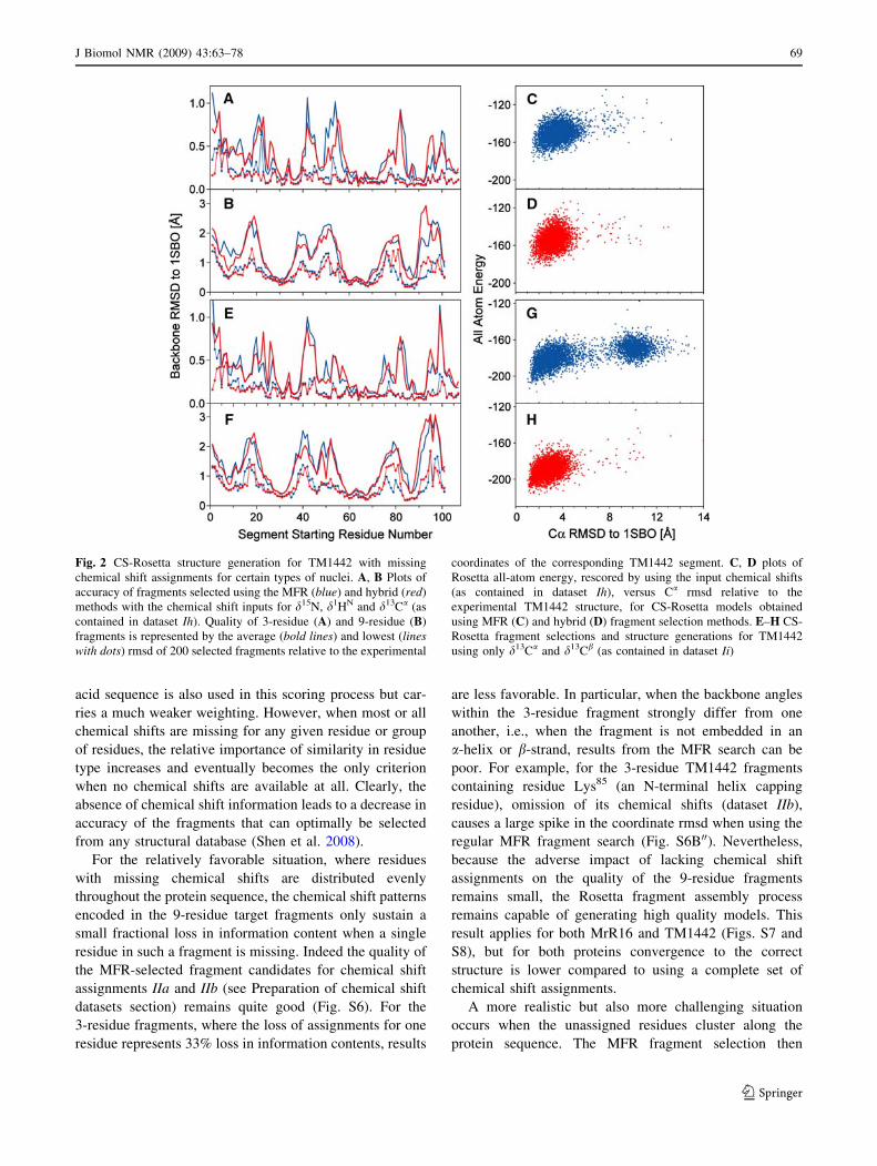
obtained for TM1442 (Fig. 2).
Remarkably, even though the accuracy of the resulting
structures decreases when just using 1HN, 15N, and 13Ca
chemical shifts, or just 13Ca and 13Cb chemical shifts,
lowest energy structures remain close to the reference
structure, in particular when the hybrid fragment selection
method is used. A survey of the energies of the Rosetta-
assembled structures and their accuracies (Fig. S3) indi-
cates that the original MFR fragment selection results in
higher yields during structure generation than the hybrid
fragment selection method when assignments are relatively
complete. However, for MrR16, the hybrid method out-
performs the regular MFR method for datasets Id, If and Ih
(Fig. S3); for TM1442 the hybrid method outperforms the
regular MFR approach for datasets Ib, If, and Ih (Fig. S4).
For the case where no chemical shifts are available, only
the standard Rosetta approach can be used. No conver-
gence is then reached for MrR16, whereas for TM1442 the
lowest energy models fall within 4 A from the reference
structure and relaxed convergence criteria are met
(Fig. S5).
The calculations discussed above, and summarized in
Fig. 2 and S2–S5 indicate that the resonance assignments
of not all six types of nuclei are required for success of the
CS-Rosetta structure generation process. The order of
importance of each type of chemical shift can be ranked as
d13Ca * d13Cb [ d1Ha * d13C0[ d15N * d1HN. For
proteins where all or the vast majority of these chemical
shifts are available, the standard MFR fragment selection
protocol tends to yield better accuracy of the selected MFR
fragments and higher convergence, as well as lower energy
when generating the all-atom Rosetta models. The calcu-
lations also suggest that the chemical shift assignment
dataset needed for the CS-Rosetta protocol at a minimum
comprises d15N, d1HN and d13Ca, which also are the cor-
nerstone nuclei during triple resonance backbone
assignment, complemented by either d13C0, d13Cb or d1Ha.
Absence of assignments for subsets of residues
The standard MFR fragment selection procedure, imple-
mented in the previously described CS-Rosetta protocol,
relies primarily on the match between the experimental13Ca, 13Cb, 13C0, 15N, 1HN and 1Ha secondary chemical
shift values of each residue in any given 3- or 9-residue
query fragment, and the SPARTA-generated secondary
shift values for the corresponding residues in any fragment
present in the structural database. The similarity in amino
68 J Biomol NMR (2009) 43:63–78
123
acid sequence is also used in this scoring process but car-
ries a much weaker weighting. However, when most or all
chemical shifts are missing for any given residue or group
of residues, the relative importance of similarity in residue
type increases and eventually becomes the only criterion
when no chemical shifts are available at all. Clearly, the
absence of chemical shift information leads to a decrease in
accuracy of the fragments that can optimally be selected
from any structural database (Shen et al. 2008).
For the relatively favorable situation, where residues
with missing chemical shifts are distributed evenly
throughout the protein sequence, the chemical shift patterns
encoded in the 9-residue target fragments only sustain a
small fractional loss in information content when a single
residue in such a fragment is missing. Indeed the quality of
the MFR-selected fragment candidates for chemical shift
assignments IIa and IIb (see Preparation of chemical shift
datasets section) remains quite good (Fig. S6). For the
3-residue fragments, where the loss of assignments for one
residue represents 33% loss in information contents, results
are less favorable. In particular, when the backbone angles
within the 3-residue fragment strongly differ from one
another, i.e., when the fragment is not embedded in an
a-helix or b-strand, results from the MFR search can be
poor. For example, for the 3-residue TM1442 fragments
containing residue Lys85 (an N-terminal helix capping
residue), omission of its chemical shifts (dataset IIb),
causes a large spike in the coordinate rmsd when using the
regular MFR fragment search (Fig. S6B00). Nevertheless,
because the adverse impact of lacking chemical shift
assignments on the quality of the 9-residue fragments
remains small, the Rosetta fragment assembly process
remains capable of generating high quality models. This
result applies for both MrR16 and TM1442 (Figs. S7 and
S8), but for both proteins convergence to the correct
structure is lower compared to using a complete set of
chemical shift assignments.
A more realistic but also more challenging situation
occurs when the unassigned residues cluster along the
protein sequence. The MFR fragment selection then
Fig. 2 CS-Rosetta structure generation for TM1442 with missing
chemical shift assignments for certain types of nuclei. A, B Plots of
accuracy of fragments selected using the MFR (blue) and hybrid (red)
methods with the chemical shift inputs for d15N, d1HN and d13Ca (as
contained in dataset Ih). Quality of 3-residue (A) and 9-residue (B)
fragments is represented by the average (bold lines) and lowest (lineswith dots) rmsd of 200 selected fragments relative to the experimental
coordinates of the corresponding TM1442 segment. C, D plots of
Rosetta all-atom energy, rescored by using the input chemical shifts
(as contained in dataset Ih), versus Ca rmsd relative to the
experimental TM1442 structure, for CS-Rosetta models obtained
using MFR (C) and hybrid (D) fragment selection methods. E–H CS-
Rosetta fragment selections and structure generations for TM1442
using only d13Ca and d13Cb (as contained in dataset Ii)
J Biomol NMR (2009) 43:63–78 69
123

becomes dominated by residue type similarity between the
query fragment and fragments present in the structural
database. The accuracy of fragments that include such
unassigned segments, selected by the standard MFR
method, is severely affected (Fig. S6C, D), in particular
when the missing assignments are located outside regions
of secondary structure (datasets IIc and IId). Interestingly,
the quality of these fragments tends to be much lower than
what is achieved with the standard Rosetta fragment
selection method (Fig. S5), highlighting that the simple
residue similarity scoring used by the MFR method per-
forms much worse than the far more elaborate Rosetta
fragment selection protocol (Rohl et al. 2004). Unsurpris-
ingly, the subsequent Rosetta structure assembly protocol,
using standard MFR fragments as input, can fail to obtain a
converged low-energy fold (Figs. S7, S8). On the other
hand, for MrR16 the CS-Rosetta structure generation for
dataset Ic, lacking assignments for residues 24–32, remains
successful and finds a converged low-energy fold, where
the backbone of the lowest energy model deviates by 1.8 A
from the experimental reference structure (Fig. S7). Even
while the quality of 9-residue fragments encompassing this
region with missing assignments is poor, the accuracy of
the best 3-residue fragments selected remains quite good
for this region, and it is the powerful combinatorial engine
of Rosetta which can exploit the presence of a relatively
small subset of accurate fragments for this single region
during the assembly process. For the case where two
regions with missing assignments are present in the protein
(dataset IIe), CS-Rosetta with standard MFR selection no
longer is able to obtain converged low energy structures
(Fig. 3, S7 and S8).
One way to improve the selection of suitable fragments,
and thereby the CS-Rosetta structure generation process, for
proteins with extended segments of missing chemical shift
assignments is to take advantage of the standard Rosetta
fragment selection procedure (Rohl et al. 2004), which
searches for matched database fragments based on a rela-
tively sophisticated procedure that simultaneously exploits
residue type similarity and predicted secondary structure.
Amino acid sequence similarity alone provides less struc-
tural information than the backbone chemical shifts, and
therefore results in a wider distribution of selected peptide
conformations. The average quality of Rosetta-selected
fragments therefore is significantly lower than for MFR
selection based on chemical shifts, but the quality of the best
fragments (out of 200 selected) remains quite good, in par-
ticular for the 3-residue fragments (Shen et al. 2008). A
preferred way to score the fragments therefore would
directly combine, with suitable weight factors, the amino
acid sequence based Rosetta fragment score with the
chemical shift component of the MFR score. For technical
reasons, however, this is not easily accomplished and we
therefore resort to a simpler protocol which equally takes
advantage of the strengths of both approaches. This hybrid
fragment selection procedure first uses standard Rosetta to
select the 2000 database fragments (out of over 2,200,000)
that are most compatible in terms of amino acid sequence,
and then uses MFR chemical shift scoring to narrow down
this set to fragments that are most compatible with the
Fig. 3 CS-Rosetta structure for TM1442 with missing chemical
shifts. A, B Plots of accuracy of fragment candidates selected using
the MFR (blue) and hybrid (red) methods using chemical shift values
d15N, d1HN, d13Ca, d13Cb, d13C0 and d1Ha for residues 1–20, 30–51
and 60–120 (as contained in the dataset IIe). For each 3-residue (A)
and 9-residue (B) segment of TM1442, 200 fragments were selected.
Average (bold lines) and lowest (lines with dots) rmsd of these
fragments relative to the experimental coordinates of the
corresponding TM1442 segment are plotted with respect to the
position of the first segment residue in the TM1442 sequence. The
regions corresponding to the ‘‘unassigned’’ residues are shaded; the
secondary structure elements are displayed at the top. C, D Plots of
Rosetta all atom energy, rescored by using the input chemical shifts
(as contained in dataset IIe), versus Ca rmsd relative to the
experimental TM1442 structure, for CS-Rosetta models obtained
using MFR (C) and hybrid (D) fragment selection methods
70 J Biomol NMR (2009) 43:63–78
123
experimental shifts. When complete chemical shifts are
available, this hybrid method performs slightly worse than
the regular MFR procedure (Figs. S2–S4). However, when
significant segments in the protein lack assignments, the
hybrid method remains successful at generating low energy,
converged results. For example, when using the ‘hybrid’
fragments selected with chemical shift datasets IIc–IIe,
lacking chemical shifts for two extended loop regions, the
Rosetta fragment assembly and relaxation protocol results in
near-convergence for TM1442, yielding lowest energy
models that are within 2.5 A Ca rmsd relative to the refer-
ence structure (Fig. 3).
Impact of chemical shift assignment errors
A potential error during conventional and/or automated
backbone resonance assignments is exemplified by the case
where chemical shift assignments of two di- or tripeptide
sequences of similar amino acid sequence, embedded
between residues with similar chemical shifts, are acci-
dentally interchanged. Below, we consider the case where
assignments for two dipeptides with identical amino acid
types are interchanged.
For the favorable situation where the two dipeptides are
located in segments with the same secondary structure, as
exemplified in dataset IIIa, the chemical shift patterns in
the 3-residue and 9-residue fragments are virtually
unchanged and there is essentially no adverse impact on the
fragment selection, neither for the standard MFR nor the
hybrid approach (Fig. S9). Clearly, generation of Rosetta
structures also remains unaffected (Figs. S10 and S11).
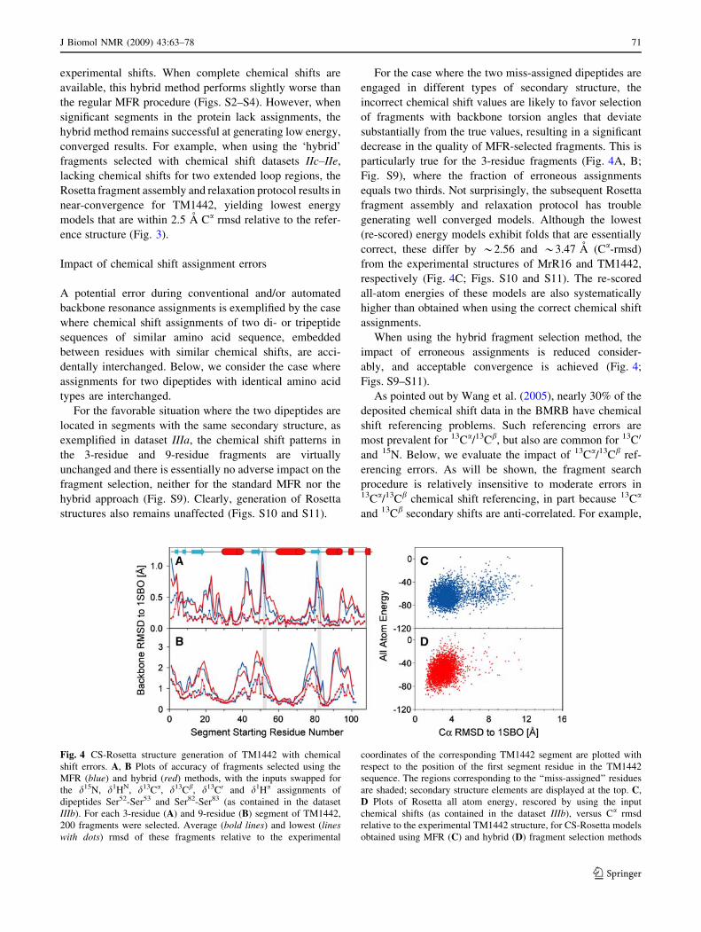
For the case where the two miss-assigned dipeptides are
engaged in different types of secondary structure, the
incorrect chemical shift values are likely to favor selection
of fragments with backbone torsion angles that deviate
substantially from the true values, resulting in a significant
decrease in the quality of MFR-selected fragments. This is
particularly true for the 3-residue fragments (Fig. 4A, B;
Fig. S9), where the fraction of erroneous assignments
equals two thirds. Not surprisingly, the subsequent Rosetta
fragment assembly and relaxation protocol has trouble
generating well converged models. Although the lowest
(re-scored) energy models exhibit folds that are essentially
correct, these differ by *2.56 and *3.47 A (Ca-rmsd)
from the experimental structures of MrR16 and TM1442,
respectively (Fig. 4C; Figs. S10 and S11). The re-scored
all-atom energies of these models are also systematically
higher than obtained when using the correct chemical shift
assignments.
When using the hybrid fragment selection method, the
impact of erroneous assignments is reduced consider-
ably, and acceptable convergence is achieved (Fig. 4;
Figs. S9–S11).
As pointed out by Wang et al. (2005), nearly 30% of the
deposited chemical shift data in the BMRB have chemical
shift referencing problems. Such referencing errors are
most prevalent for 13Ca/13Cb, but also are common for 13C0
and 15N. Below, we evaluate the impact of 13Ca/13Cb ref-
erencing errors. As will be shown, the fragment search
procedure is relatively insensitive to moderate errors in13Ca/13Cb chemical shift referencing, in part because 13Ca
and 13Cb secondary shifts are anti-correlated. For example,
Fig. 4 CS-Rosetta structure generation of TM1442 with chemical
shift errors. A, B Plots of accuracy of fragments selected using the
MFR (blue) and hybrid (red) methods, with the inputs swapped for
the d15N, d1HN, d13Ca, d13Cb, d13C0 and d1Ha assignments of
dipeptides Ser52-Ser53 and Ser82-Ser83 (as contained in the dataset
IIIb). For each 3-residue (A) and 9-residue (B) segment of TM1442,
200 fragments were selected. Average (bold lines) and lowest (lineswith dots) rmsd of these fragments relative to the experimental
coordinates of the corresponding TM1442 segment are plotted with
respect to the position of the first segment residue in the TM1442
sequence. The regions corresponding to the ‘‘miss-assigned’’ residues
are shaded; secondary structure elements are displayed at the top. C,
D Plots of Rosetta all atom energy, rescored by using the input
chemical shifts (as contained in the dataset IIIb), versus Ca rmsd
relative to the experimental TM1442 structure, for CS-Rosetta models
obtained using MFR (C) and hybrid (D) fragment selection methods
J Biomol NMR (2009) 43:63–78 71
123
a 4 ppm reference error could change a typical b-sheet
secondary 13Ca shift of -1 ppm to an a-helical 3 ppm
value. However, the ?2 ppm b-sheet secondary 13Cb shift
would become ?6 ppm, completely incompatible with a
helical conformation, preventing the residue from being
misidentified as helical. To first order, the impact of13Ca/13Cb referencing errors is small when both 13Ca and13Cb shift data are available, and manifests itself mainly as
a steeper 13Ca/13Cb chemical shift gradient when selecting
fragments, and increased total energies when rescoring the
energies of the Rosetta models.
The impact of 13Ca/13Cb chemical shift referencing
errors on CS-Rosetta structure generation was evaluated
using the chemical shift assignment datasets IIIc and IIId.
When 1.0 ppm offset was added to d13Ca/b (dataset IIIc),
comparable to the average d13Ca/b prediction errors (r in
Eq. 1) (Gong et al. 2007; Shen and Bax 2007), the
accuracy of the selected fragments slightly decreases (Fig.
S9), with a very small adverse impact on subsequent
Rosetta structure generation (Figs. S10 and S11). The
impact of chemical shift referencing errors appears to be
insensitive to the type of fragment selection method used:
For MrR16, standard MFR yields slightly better results
(Fig. S10); for TM1442, the hybrid method is slightly
favorable (Fig. S11).
When the d13Ca/b offset error is increased to 1.7 ppm
(dataset IIIc), convergence and accuracy of the resulting
structures decreases noticeably (Figs. S10 and S11), but the
folds remain essentially correct. However, when the offset
error is increased to 2.7 ppm, which corresponds to the
approximate difference between d13Ca/b values referenced
to TMS and DSS (Wishart et al. 1995; Markley et al.
1998), fragment selection results are poor and no accept-
able structures are obtained with the CS-Rosetta protocol
(data not shown).
When the chemical shift referencing error affects only a
single type of nucleus, e.g. 13Ca or 13C0, an erroneous bias
towards selection of helical or extended fragments can
occur, resulting in poorer fragment quality and decreased
performance of the CS-Rosetta protocol (results not
shown). Even in these cases, the impact of 15N or 13C
chemical shift referencing errors of up to 1 ppm have very
little adverse effect on CS-Rosetta performance.
Chemical shift referencing errors readily can be detected
by automated methods (Moseley et al. 2004; Wang et al.
2005). For this purpose, a script has been added to the
CS-Rosetta package which applies reference error correc-
tions when the referencing error exceeds the average
uncertainty in the database chemical shifts (1.0 ppm for
d13Ca/b and d13C0; 0.3 ppm for d1Ha). These referencing
corrections are based on the method described by Markley
and coworkers (Wang et al. 2005), and correlations
between (Dd13Ca- Dd13Cb) and Dd13Ca/b/Dd13C0/Dd1H
are shown in Fig. S1.
A situation similar to the chemical shift referencing
problem discussed above can arise when chemical shifts
are measured from TROSY spectra (Pervushin et al. 1998),
when the displacement between the observed resonance
frequency and the true chemical shift (1JNH/2 for d15N and
d1HN) is not taken into account. However, considering that
this error is much smaller than the standard error in the
predicted database chemical shifts, no adjustment of the
chemical shift values is required.
A larger apparent referencing error can result from
deuteration effects (Venters et al. 1996; Gardner et al.
1997) on d13Ca (with deuterium isotope shifts of -0.5 to
-0.9 ppm) and d13Cb (-0.7 to -1.3 ppm). These isotope
effects on the backbone chemical shifts are relatively
uniform and mostly smaller than the 1 ppm referencing
error, discussed above. Although it is beneficial to apply
uniform isotope shift corrections of ?0.7 and ?0.9 ppm to
d13Ca and d13Cb values, respectively, the absence of such
corrections shows little adverse impact on the performance
of CS-Rosetta (data not shown). Nevertheless, a script has
been added to the CS-Rosetta package which adjusts the
d13Ca and d13Cb chemical shifts by the residue-type-spe-
cific values reported by Cavanagh et al. (2007).
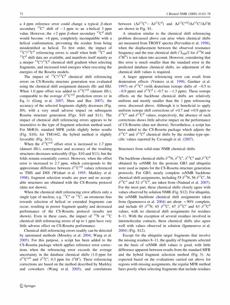
Structures from solid-state NMR chemical shifts
The backbone chemical shifts d15N, d13C0, d13Ca and d13Cb
obtained by ssNMR for the proteins GB3 and ubiquitin
were used as inputs for the CS-Rosetta structure generation
protocols. For GB3, nearly complete ssNMR backbone
chemical shift assignments, including 55 d15N, 56 d13C0, 56
d13Ca and 52 d13Cb, are taken from (Nadaud et al. 2007).
For the most part, these chemical shifts closely agree with
values observed by solution NMR (Fig. S12). For ubiquitin,
the ssNMR backbone chemical shift assignments taken
from (Igumenova et al. 2004) are about *90% complete,
and include 65 d15N, 65 d13C0, 67 d13Ca and 63 d13Cb
values, with no chemical shift assignments for residues
8–11. With the exception of several residues involved in
intermolecular contacts, these chemical shifts also agree
well with values observed in solution (Igumenova et al.
2004) (Fig. S12).
Except for the ubiquitin target fragments that involve
the missing residues 8–11, the quality of fragments selected
on the basis of ssNMR shift values is good, with little
difference apparent between results from the standard MFR
and the hybrid fragment selection method (Fig. 5). As
expected based on the evaluations carried out above for
regions with missing assignments, the regular MFR method
fares poorly when selecting fragments that include residues
72 J Biomol NMR (2009) 43:63–78
123
8–11, whereas the hybrid method shows no decrease in
structural quality for this region.
Importantly, either selection method yields fragments
from the ssNMR chemical shifts that suffice for generating
converged, high quality all-atom models for both proteins
(Fig. 5C, F). When the MFR method is used to select the
fragments, the coordinate rms deviations for GB3 between
the lowest energy model and the experimental solution
NMR structure are 0.71 A for the backbone atoms (N, Ca
and C0) and 1.28 A for all non-hydrogen atoms. For
ubiquitin these numbers are 0.69 and 1.22 A. When the
fragments are selected by the hybrid procedure, the coor-
dinate rmsd’s are slightly higher: 0.73 and 1.70 A for
backbone and all non-hydrogen GB3 atoms, respectively,
and 0.86 and 1.49 A for ubiquitin.
Considering the generally somewhat lower spectral
resolution attainable by ssNMR compared to solution
NMR, detailed structural studies of globular proteins by
ssNMR mostly have remained restricted to relatively
small systems, typically less than *80 residues. Clearly,
CS-Rosetta provides a powerful new complementary tool
for generating structural models of such proteins once
chemical shift assignments have been completed, without
requiring the extensive internuclear distance information
which sometimes can be difficult to obtain.
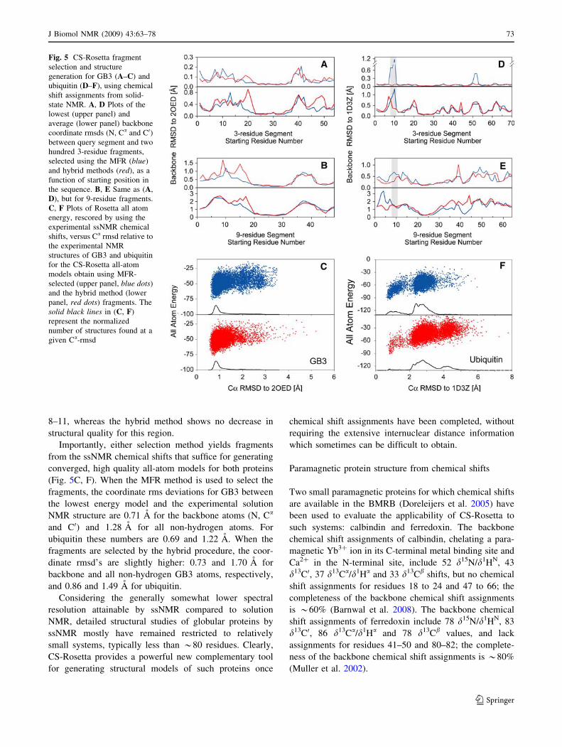
Paramagnetic protein structure from chemical shifts
Two small paramagnetic proteins for which chemical shifts
are available in the BMRB (Doreleijers et al. 2005) have
been used to evaluate the applicability of CS-Rosetta to
such systems: calbindin and ferredoxin. The backbone
chemical shift assignments of calbindin, chelating a para-
magnetic Yb3? ion in its C-terminal metal binding site and
Ca2? in the N-terminal site, include 52 d15N/d1HN, 43
d13C0, 37 d13Ca/d1Ha and 33 d13Cb shifts, but no chemical
shift assignments for residues 18 to 24 and 47 to 66; the
completeness of the backbone chemical shift assignments
is *60% (Barnwal et al. 2008). The backbone chemical
shift assignments of ferredoxin include 78 d15N/d1HN, 83
d13C0, 86 d13Ca/d1Ha and 78 d13Cb values, and lack
assignments for residues 41–50 and 80–82; the complete-
ness of the backbone chemical shift assignments is *80%
(Muller et al. 2002).
Fig. 5 CS-Rosetta fragment
selection and structure
generation for GB3 (A–C) and
ubiquitin (D–F), using chemical
shift assignments from solid-
state NMR. A, D Plots of the
lowest (upper panel) and
average (lower panel) backbone
coordinate rmsds (N, Ca and C0)between query segment and two
hundred 3-residue fragments,
selected using the MFR (blue)
and hybrid methods (red), as a
function of starting position in
the sequence. B, E Same as (A,
D), but for 9-residue fragments.
C, F Plots of Rosetta all atom
energy, rescored by using the
experimental ssNMR chemical
shifts, versus Ca rmsd relative to
the experimental NMR
structures of GB3 and ubiquitin
for the CS-Rosetta all-atom
models obtain using MFR-
selected (upper panel, blue dots)
and the hybrid method (lower
panel, red dots) fragments. The
solid black lines in (C, F)
represent the normalized
number of structures found at a
given Ca-rmsd
J Biomol NMR (2009) 43:63–78 73
123
With the absence of chemical shift assignments for long
segments in each of these two proteins, the standard
CS-Rosetta protocol, using MFR fragment selection, fails to
converge for both proteins (Fig. S13). However, the hybrid
fragment selection procedure performs much better, in
particular for those target fragments involving the unas-
signed residues (Fig. 6A, B, D, E), permitting the structure
assembly phase to be successful (Fig. 7). Interestingly, this
improved performance is not dominated by recognition of
the relatively common EF-hand and Fe–S metal-binding
sites as, for testing purposes, proteins with a PSI-BLAST
e-score \0.05 had been removed from the database. Sub-
sequent manual evaluation of the 9-residue fragments
covering the regions lacking chemical shifts showed the
presence of six 9-residue fragments for calbindin segment
54–62, which were taken from EF-hand containing proteins
that had escaped detection by the PSI-BLAST filter.
For both proteins, the Rosetta fragment assembly and
relaxation procedure generates a number of good all-atom
models, with the lowest energy models having backbone
coordinates that differ by less than 2 A from their respec-
tive reference structures when only including residues
involved in secondary structure (Fig. 6C, F). Although, the
standard convergence criterion (10 lowest energy structures
cluster with 2 A from the lowest energy structure) is not
met for either protein (Fig. S13), when relaxing this limit to
3.3 A both structures are converged.
For calbindin, the coordinate rmsd’s between the lowest
energy all-atom model and the 1.6-A X-ray structure of
calbindin D9K (Svensson et al. 1992) are 1.5 and 2.1 A for
the backbone atoms (N, Ca and C0) and for all heavy atoms
involved in secondary structure, respectively. The Ca2?
binding loops of both metal binding sites are remarkably
well formed in the CS-Rosetta structures (Fig. 7A), even
Fig. 6 CS-Rosetta structure generation for paramagnetic calbindin
(A–C) and ferredoxin (D–F). A, D Plots of the lowest (upper panel)
and average (lower panel) backbone coordinate rmsds (N, Ca and C0)between query segment and two hundred 3-residue fragment candi-
dates, selected using the MFR (blue) and hybrid methods (red), as a
function of starting position in the sequence. The regions lacking
chemical shift assignments are shaded. B, E Same as (A, D), but for
9-residue fragments. C, F Plots of Rosetta all-atom energy, rescored
by the experimental chemical shifts, versus Ca rmsd of final al-atom
models (including only residues located in elements of secondary
structure) relative to the corresponding X-ray (calbindin) and NMR
(ferredoxin) structure. Only results from CS-Rosetta all-atom models
obtained by the hybrid fragment selection procedure are shown; when
using fragments from the standard MFR method, Rosetta fails to
converge. Residues included in the backbone rmsd calculation include
3–14, 25–40, 46–53 and 63–74 for calbindin, and 4–11, 15–22, 27–
34, 54–56, 71–75 and 91–93 for ferredoxin
74 J Biomol NMR (2009) 43:63–78
123

with the second metal binding site lacking all of its
chemical shift assignments and the absence of any
restraints on metal chelation for both metal binding sites.
For the first Ca2? binding loop, a pseudo-EF-hand, the four
backbone carbonyl groups are properly positioned and
point towards the location where Ca2? is found in the
X-ray structure. Even the bidentate sidechain ligating
group of Glu27 adopts a conformation suitable for metal
chelation. For the second site, a regular EF-hand, the
backbone carbonyl of Glu60 and the sidechains of Asp54
and Glu65 are well positioned for metal binding, but the
sidechains of Asn56 and Asp58 point away from the position
where the metal ion is observed in the X-ray structure.
For the secondary structure elements of ferredoxin, the
lowest energy Rosetta model deviates from the experi-
mental NMR structure obtained for the same protein by
2.06 A for the backbone and by 3.54 A for all non-
hydrogen atoms. Two of the four Cys sidechains that ligate
the [2Fe–2S] cluster are in close proximity, even though
the loop conformations differ substantially from the
experimentally determined structure (Fig. 7B).
Concluding remarks
Although previous reports have clearly demonstrated the
potential of using chemical shifts to determine good quality
all-atom structures for small proteins (Cavalli et al. 2007;
Shen et al. 2008), these studies were based on relatively
ideal cases where complete or nearly complete backbone
assignments were available, in the absence of assignment
errors. Our present study demonstrates that the CS-Rosetta
procedure and its new variant, which uses a hybrid
fragment selection procedure, are remarkably tolerant to
such incompleteness and errors. Clearly, a study such as
the present one, which evaluates the impact of missing or
erroneous assignments, is never complete. We simply have
evaluated the impact for two proteins, and have made an
attempt to evaluate representative cases of missing
assignments. Both proteins chosen for the current study,
MrR16 and TM1442, yielded good (albeit not exceptional)
results when originally studied with complete data sets, and
these systems therefore are likely to be more robust to
incompleteness or assignment errors than proteins which
only yield borderline convergence to begin with.
The CS-Rosetta protocol uses the chemical shift infor-
mation at two stages: first for fragment selection, and then
again when evaluating the final full-atom models. There
are two primary reasons for the improved performance of
the CS-Rosetta protocol over a conceptually similar, earlier
attempt to integrate chemical shift information into Rosetta
(Bowers et al. 2000). First, the quality of fragments
selected has improved considerably by the use of SPARTA
to ‘‘assign’’ better chemical shifts to a structural database.
SPARTA uses both a more advanced algorithm to assign
these chemical shifts, but also benefits from a considerable
expansion of entries in the BMRB for which complete
chemical shift and high resolution structural information is
available (Doreleijers et al. 2005). Second, a number of
improvements in the Rosetta Monte Carlo assembly pro-
cess have been made in recent years, most notably the
incorporation of explicit all atom refinement with a phys-
ically realistic force field (Das and Baker 2008).
The adverse impact of errors and incompleteness on the
CS-Rosetta protocol results primarily from decreased
Fig. 7 Comparison of experimental (blue) and lowest energy CS-
Rosetta (red) structure for paramagnetic calbindin (A) and ferredoxin
(B). Superposition is optimized for residues in secondary structure,
defined in the caption to Fig. 6. The sidechains of residues involved in
metal binding including their metal-ligating oxygen atoms, as well as
the X-ray positions of the Ca2? ions (cyan) are shown. Metal-ligating
residues (atoms) include Ala14 (O), Glu17 (O), Asp19 (O), Gln22 (O),
Glu27 (Oe1/Oe2), Asp54 (Od1), Asn56 (O), Asp58 (Od1), Glu60 (O) and
Glu65 (Oe1/Oe2). (B) Backbone ribbon representation of the lowest-
energy CS-Rosetta structure (red) superimposed on the experimental
X-ray structure (blue) for ferredoxin, with superposition optimized for
the residues in secondary structure (See caption to Fig. 6). The
sidechain S atoms of Cys42, Cys47, Cys50 and Cys82, which coordinate
the [2Fe–2S] cluster are marked as solid spheres. Figures made using
Molmol (Koradi et al. 1996)
J Biomol NMR (2009) 43:63–78 75
123

quality of the fragment library, and has relatively little
impact on the rescoring of the final full-atom models. The
hybrid CS-Rosetta protocol first limits the selection of
fragments to a *0.1% fraction of the total structural
database on the basis of the standard Rosetta selection
mechanism. In the next step, it uses MFR to select the 200
fragments from this ensemble that agree best with experi-
mental chemical shifts. This reduces the impact of
chemical shift errors because only fragments compatible
with standard Rosetta criteria are available for selection.
Moreover, in the absence of any chemical shift informa-
tion, the Rosetta pre-selection of the top 0.1% fragments
yields better results than the less sophisticated MFR pro-
cedure, which had been designed primarily to find
fragments with similar chemical shifts and/or RDCs (Del-
aglio et al. 2000; Kontaxis et al. 2005). In the absence of
assignment errors or missing assignments, the initial
Rosetta pre-selection used in the hybrid procedure is not
beneficial and actually results in a small decrease in per-
formance. On the other hand, for cases where significant
fractions of assignments are missing or ambiguous, the
hybrid procedure is considerably more robust.
For all evaluations, including those of the two para-
magnetic proteins, homologous proteins were first
eliminated from the structural database. In practice, this is
clearly disadvantageous as Rosetta no longer can take
advantage of standard structural elements, such as Ca2?-
ligating EF-hand sequences, present in the database. Indeed
30 proteins containing a total of 64 EF-hands were
removed prior to fragment searching. Similarly, proteins
containing the relatively common Fe2S2 cluster were
removed prior to searching for fragments for ferredoxin
assembly. While for calbindin the CS-Rosetta protocol
resulted in remarkably good backbone structures for its
metal binding sites, even in the absence of chemical shift
information, loop conformations in ferredoxin were poor.
Nevertheless, using the hybrid protocol, CS-Rosetta was
able to generate the remainder of the ferredoxin structure
quite well, suggesting that even for these challenging
systems the method will be quite useful.
For the two proteins for which a structure was generated
from solid-state NMR chemical shifts, lacking 1H chemical
shifts, the standard MFR-based protocol and the hybrid
CS-Rosetta method performed comparably well. For both
proteins, the final structures obtained from these smaller
input data sets approach the quality of structures obtained
from solution NMR chemical shifts, indicating that
CS-Rosetta may be a particularly useful complement when
working with samples in the solid state.
Although CS-Rosetta considerably reduces the amount
of spectral data collection time required for structure
generation compared to conventional procedures, the
amount of computational time required typically is very
high. Although for simple systems such as GB3, generation
of less than one hundred structures may suffice to reach
convergence (Shen et al. 2008), for many other proteins as
many as 10,000 models may be required. Rosetta assembly
and minimization of each model takes 5–10 min on a
single CPU, and in practice use of a large cluster or a
central server such as BOINC is required to take advantage
of this technology.
We also note that the CS23D program (Wishart et al.
2008) performs very well for the test datasets used in our
study (Supplementary material). The major strength of
CS23D is that it takes optimal advantage of sequence
homologues present in the database during fragment selec-
tion. Such homologues were present in the structural
database for all six proteins evaluated in our work (see
Supplementary material Table S2), but were excluded from
the database for CS-Rosetta testing. On the other hand, based
on a limited number of tests, techniques such as CS-Rosetta
and Cheshire are believed to be superior for proteins that lack
significant homology to previously solved structures.
Software availability
The CS-Rosetta software package with its newly imple-
mented hybrid fragment selection module can be
downloaded from http://spin.niddk.nih.gov/bax/.
Acknowledgments This work was funded by the Intramural
Research Program of the NIDDK, NIH, and by the Intramural AIDS-
Targeted Antiviral Program of the Office of the Director, NIH; the
NIGMS, NIH, and the Howard Hughes Medical Institutes (to D. B.).
We also thank Rosetta@home participants and the BOINC project for
contributing computing power.
References
Agarwal V, Diehl A, Skrynnikov N, Reif B (2006) High resolution
H-1 detected H-1, C-13 correlation spectra in MAS solid-state
NMR using deuterated proteins with selective H-1, H-2 isotopic
labeling of methyl groups. J Am Chem Soc 128:12620–12621
Altschul SF, Madden TL, Schaffer AA, Zhang JH, Zhang Z, Miller
W, Lipman DJ (1997) Gapped BLAST and PSI-BLAST: a new
generation of protein database search programs. Nucleic Acids
Res 25:3389–3402
Ando I, Kameda T, Asakawa N, Kuroki S, Kurosu H (1998) Structure
of peptides and polypeptides in the solid state as elucidated by
NMR chemical shift. J Mol Struct 441:213–230
Andreini C, Bertini I, Rosato A (2004) A hint to search for
metalloproteins in gene banks. Bioinformatics 20:1373–1380
Asakura T, Demura M, Date T, Miyashita N, Ogawa K, Williamson
MP (1997) NMR study of silk I structure of Bombyx mori silk
fibroin with N-15- and C-13-NMR chemical shift contour plots.
Biopolymers 41:193–203
Barnwal RP, Rout AK, Chary KVR, Atreya HS (2008) Rapid
measurement of pseudocontact shifts in paramagnetic proteins
by GFT NMR spectroscopy. Open Magn Reson J 1:16–28
76 J Biomol NMR (2009) 43:63–78
123
Bermel W, Bertini I, Felli IC, Piccioli M, Pierattelli R (2006) C-13-
detected protonless NMR spectroscopy of proteins in solution.
Prog Nucl Magn Reson Spectrosc 48:25–45
Bertini I, Luchinat C, Parigi G, Pierattelli R (2005) NMR spectros-
copy of paramagnetic metalloproteins. Chembiochem 6:
1536–1549
Bowers PM, Strauss CEM, Baker D (2000) De novo protein structure
determination using sparse NMR data. J Biomol NMR 18:
311–318
Case DA (1995) Calibration of ring-current effects in proteins and
nucleic acids. J Biomol NMR 6:341–346
Castellani F, van Rossum B, Diehl A, Schubert M, Rehbein K, Oschkinat
H (2002) Structure of a protein determined by solid-state magic-
angle-spinning NMR spectroscopy. Nature 420:98–102
Castellani F, van Rossum BJ, Diehl A, Rehbein K, Oschkinat H (2003)
Determination of solid-state NMR structures of proteins by means
of three-dimensional 15N–13C–13C dipolar correlation spectroscopy
and chemical shift analysis. Biochemistry 42:11476–11483
Cavalli A, Salvatella X, Dobson CM, Vendruscolo M (2007) Protein
structure determination from NMR chemical shifts. Proc Natl
Acad Sci USA 104:9615–9620
Cavanagh J, Fairbrother WJ, Palmer AG, Rance M, Skelton NJ (2007)
Protein NMR spectroscopy: principles and practice, 2nd edn.
Academic Press, San Diego, CA
Chevelkov V, Rehbein K, Diehl A, Reif B (2006) Ultrahigh resolution
in proton solid-state NMR spectroscopy at high levels of
deuteration. Angew Chem Int Ed 45:3878–3881
Cornilescu G, Marquardt JL, Ottiger M, Bax A (1998) Validation of
protein structure from anisotropic carbonyl chemical shifts in a
dilute liquid crystalline phase. J Am Chem Soc 120:6836–6837
Cornilescu G, Delaglio F, Bax A (1999) Protein backbone angle
restraints from searching a database for chemical shift and
sequence homology. J Biomol NMR 13:289–302
Das R, Baker D (2008) Macromolecular modeling with Rosetta. Annu
Rev Biochem 77:363–382
Delaglio F, Kontaxis G, Bax A (2000) Protein structure determination
using Molecular Fragment Replacement and NMR dipolar
couplings. J Am Chem Soc 122:2142–2143
Doreleijers JF, Nederveen AJ, Vranken W, Lin JD, Bonvin A,
Kaptein R, Markley JL, Ulrich EL (2005) BioMagResBank
databases DOCR and FRED containing converted and filtered
sets of experimental NMR restraints and coordinates from over
500 protein PDB structures. J Biomol NMR 32:1–12
Gardner KH, Rosen MK, Kay LE (1997) Global folds of highly
deuterated, methyl-protonated proteins by multidimensional
NMR. Biochemistry 36:1389–1401
Gong HP, Shen Y, Rose GD (2007) Building native protein
conformation from NMR backbone chemical shifts using Monte
Carlo fragment assembly. Protein Sci 16:1515–1521
Gryk MR, Hoch JC (2008) Local knowledge helps determine protein
structures. Proc Natl Acad Sci USA 105:4533–4534
Haigh CW, Mallion RB (1979) Ring current theories in nuclear
magnetic resonance. Prog Nucl Magn Reson Spectrosc 13:
303–344
Igumenova TI, McDermott AE, Zilm KW, Martin RW, Paulson EK,
Wand AJ (2004) Assignments of carbon NMR resonances for
microcrystalline ubiquitin. J Am Chem Soc 126:6720–6727
Ikura M, Kay LE, Bax A (1990) A novel approach for sequential
assignment of 1H, 13C, and 15N spectra of larger proteins:
heteronuclear triple-resonance three-dimensional NMR spectros-
copy. Application to calmodulin. Biochemistry 29:4659–4667
Kontaxis G, Delaglio F, Bax A (2005) Molecular fragment replace-
ment approach to protein structure determination by chemical
shift and dipolar homology database mining. Meth Enzymol
394:42–78
Koradi R, Billeter M, Wuthrich K (1996) MOLMOL: a program for
display and analysis of macromolecular structures. J Mol
Graph 14:51–55
Loquet A, Bardiaux B, Gardiennet C, Blanchet C, Baldus M, Nilges
M, Malliavin T, Boeckmann A (2008) 3D structure determina-
tion of the Crh protein from highly ambiguous solid-state NMR
restraints. J Am Chem Soc 130:3579–3589
Manolikas T, Herrmann T, Meier BH (2008) Protein structure
determination from C-13 spin-diffusion solid-state NMR spec-
troscopy. J Am Chem Soc 130:3959–3966
Markley JL, Bax A, Arata Y, Hilbers CW, Kaptein R, Sykes BD,
Wright PE, Wuthrich K (1998) IUPAC-IUBMB-IUPAB inter-
union task group on the standardization of data bases of protein
and nucleic acid structures determined by NMR spectroscopy.
Pure Appl Chem 70:117–142
Montelione GT, Wagner G (1990) Conformation-independent
sequential NMR connections in isotope-enriched polypeptides
by 1H–13C–15N triple-resonance experiments. J Magn Reson
87:183–188
Moseley HNB, Sahota G, Montelione GT (2004) Assignment
validation software suite for the evaluation and presentation of
protein resonance assignment data. J Biomol NMR 28:341–355
Muller J, Lugovskoy AA, Wagner G, Lippard SJ (2002) NMR
structure of the [2Fe–2S] ferredoxin domain from soluble
methane monooxygenase reductase and interaction with its
hydroxylase. Biochemistry 41:42–51
Nadaud PS, Helmus JJ, Jaroniec CP (2007) 13C and 15N chemical
shift assignments and secondary structure of the B3 immuno-
globulin-binding domain of streptococcal protein G by magic-
angle spinning solid-state NMR spectroscopy. Biomol NMR
Assign 1:117–120
Neal S, Nip AM, Zhang HY, Wishart DS (2003) Rapid and accurate
calculation of protein H-1, C-13 and N-15 chemical shifts.
J Biomol NMR 26:215–240
Neal S, Berjanskii M, Zhang HY, Wishart DS (2006) Accurate
prediction of protein torsion angles using chemical shifts and
sequence homology. Magn Reson Chem 44:S158–S167
Pervushin K, Riek R, Wider G, Wuthrich K (1998) Transverse
relaxation-optimized spectroscopy (TROSY) for NMR studies of
aromatic spin systems in 13C-labeled proteins. J Am Chem Soc
120:6394–6400
Rohl CA, Strauss CEM, Misura KMS, Baker D (2004) Protein
structure prediction using Rosetta. Meth Enzymol 383:66–93
Saito H (1986) Conformation-dependent C13 chemical shifts—a
new means of conformational characterization as obtained by
high resolution solid state C13 NMR. Magn Reson Chem 24:
835–852
Shen Y, Bax A (2007) Protein backbone chemical shifts predicted
from searching a database for torsion angle and sequence
homology. J Biomol NMR 38:289–302
Shen Y, Lange O, Delaglio F, Rossi P, Aramini JM, Liu GH, Eletsky
A, Wu YB, Singarapu KK, Lemak A, Ignatchenko A, Arrow-
smith CH, Szyperski T, Montelione GT, Baker D, Bax A (2008)
Consistent blind protein structure generation from NMR chem-
ical shift data. Proc Natl Acad Sci USA 105:4685–4690
Siemer AB, Ritter C, Ernst M, Riek R, Meier BH (2005) High-
resolution solid-state NMR spectroscopy of the prion protein
HET-s in its amyloid conformation. Angew Chem Int Ed
44:2441–2444
Spera S, Bax A (1991) Empirical correlation between protein
backbone conformation and Ca and Cb 13C nuclear magnetic
resonance chemical shifts. J Am Chem Soc 113:5490–5492
Svensson LA, Thulin E, Forsen S (1992) Proline cis-trans isomers in
calbindin D9K observed by X-ray crystallography. J Mol Biol
223:601–606
J Biomol NMR (2009) 43:63–78 77
123
Tycko R (1996) Prospects for resonance assignments in multidimen-
sional solid-state NMR spectra of uniformly labeled proteins.
J Biomol NMR 8:239–251
Ulmer TS, Ramirez BE, Delaglio F, Bax A (2003) Evaluation of
backbone proton positions and dynamics in a small protein by
liquid crystal NMR spectroscopy. J Am Chem Soc 125:
9179–9191
Venters RA, Farmer BT, Fierke CA, Spicer LD (1996) Characterizing
the use of perdeuteration in NMR studies of large proteins C-13,
N-15 and H-1 assignments of human carbonic anhydrase II.
J Mol Biol 264:1101–1116
Wagner G, Pardi A, Wuthrich K (1983) Hydrogen-bond length and
H-1-NMR chemical-shifts in proteins. J Am Chem Soc
105:5948–5949
Wang LY, Eghbalnia HR, Bahrami A, Markley JL (2005) Linear
analysis of carbon-13 chemical shift differences and its appli-
cation to the detection and correction of errors in referencing and
spin system identifications. J Biomol NMR 32:13–22
Williamson MP, Asakura T (1993) Empirical comparisons of models
for chemical-shift calculation in proteins. J Magn Reson B
101:63–71
Williamson MP, Kikuchi J, Asakura T (1995) Application of H1
NMR chemical shifts to measure the quality of protein
structures. J Mol Biol 247:541–546
Wishart DS, Sykes BD, Richards FM (1991) Relationship between
nuclear magnetic resonance chemical shift and protein secondary
structure. J Mol Biol 222:311–333
Wishart DS, Bigam CG, Holm A, Hodges RS, Sykes BD (1995) 1H,
13C and 15N random coil NMR chemical shifts of the common
amino acids. I. Investigations of nearest-neighbor effects.
J Biomol NMR 5:67–81
Wishart DS, Arndt D, Berjanskii M, Tang P, Zhou J, Lin G (2008)
CS23D: a web server for rapid protein structure generation using
NMR chemical shifts and sequence data. Nucleic Acids Res
36:496–502
Zech SG, Wand AJ, McDermott AE (2005) Protein structure
determination by high-resolution solid-state NMR spectroscopy:
application to microcrystalline ubiquitin. J Am Chem Soc
127:8618–8626
78 J Biomol NMR (2009) 43:63–78
123