Embed Size (px)
Citation preview

* Corresponding author. E-mail address: [email protected] (S. S. Ghani) © 2021 Growing Science Ltd. All rights reserved. doi: 10.5267/j.ccl.2021.1.007
Current Chemistry Letters 10 (2021) 235–254
Contents lists available at GrowingScience
Current Chemistry Letters
homepage: www.GrowingScience.com
Dates (Phoenix Dactylifera L.) extracts derived nanoparticles and its application
Syed Sauban Ghania* and Iqbal Hussaina aDepartment of General Studies, Jubail Industrial College, Jubail Industrial City,31961. Kingdom of Saudi Arabia
C H R O N I C L E A B S T R A C T
Article history: Received October 18, 2020 Received in revised form January 28, 2020 Accepted January 29, 2021 Available online January 29, 2021
Plant-mediated green synthesis of metallic nanoparticles (NPs) has become the most deserving alternative to chemical synthesis as this process is economical and energy-efficient, and environmentally benign. For the last twenty to thirty years, different plant sources are being utilized for the fabrication of green NPs, and few of them have used the extract of Phoenix Dactylifera L. as reducing, capping, or stabilizing agents. This review provides a detailed outline of the extraction method from various parts of dates and their synthesis with different metal salts using these extracts. The applied techniques of characterization and application of these nanoparticles have also been thoroughly discussed. The phytochemicals present in the extract were responsible for reducing the metals. Except for a few, all the investigations reported the spherical NPs but have variations in their size. These NPs have high prospects in applications such as antimicrobial, anticancer, antioxidant, and catalytic activities. This work may lead the path for additional advancement in this field, and researchers may take up the future work for the large scale production of NPs and their application using date extracts.
© 2021 Growing Science Ltd. All rights reserved.
Keywords: Green synthesis Antimicrobial Characterization Phytochemicals Dates extract
1. Introduction
The term "nano" in science is labeled for one billionth part, i.e., 10−9. The sizes of nanoparticles obtained from metals range from 1 to 100 nm.1 The shapes, sizes, and compositions of metallic nanoparticles are considerably associated with their properties that have been exploited for their superior physical, chemical, mechanical, thermal, and biological properties as compared to the bulk materials.2, 3 The main aim of the synthesis of nanoparticles (NPs) with control over particle size, shape, and crystalline nature is that it can be used for potential applications, such as bio-medical, biosensor, a catalyst for bacterial biotoxin elimination and lower cost electrode.4-6 Researchers have made an enormous effort in synthesizing metal NPs by applying different physical and chemical methods. However, all these methods utilize expensive as well as toxic reducing and stabilizing agents.7-13 They are also more likely to have toxic unreacted chemicals and by-products that make most of these synthesized NPs unfit for biomedical applications. Despite the advantages of metal nanoparticles synthesized from chemical means, there exist potentially worrying toxic effects.14 Industrial scale production and use of NPs may cause unintentional human and environmental exposure to these substances. Several researchers and social activists have raised their concerns about the environmental impact and toxicity of chemically synthesized nanoparticle-based products.15 Humans can get exposed to these chemically fabricated nanoparticles right from the origin of generation, transportation to final

236
applications in different products.16 According to the Wilson Centre data, exposure to 45% of the 580 categorized NPs have been marked as potentially dangerous.17 The purposeful use of these chemically synthesized metallic NPs in many commercial products, such as vectors of drugs, sunscreens, toothpaste, cosmetics, plastic products, textiles, paints, and gasoline components, are likely source of human exposure and enters into the body through different routes, including injection, inhalation or ingestion. Consequently, an intoxicating number of organs and tissues, including the central nervous system.18-21 Ray and his coworkers22 have raised issues regarding the potential impact of emerging NPs that are chemically synthesized on the environment. Griffitt et al23 reported the effects of particle composition on the toxicity of chemically produced metallic nanomaterials in aquatic organisms and observed that nanosilver and nano copper cause toxicity in all organisms tested 48-hr median lethal concentrations as low as 40 and 60 µg/L. The toxicity of gold nanorods is due to hexadecyl cetyltrimethylammonium bromide (CTAB), which is the most widely used surfactant for synthesizing nanorods.24 The work by Karlsson et al25 focussed on several chemically synthesized metal oxide nanoparticles (CuO, TiO2, ZnO, Fe3O4, Fe2O3) and found that each of them has a varying degree of cytotoxicity and ability to cause DNA damage and oxidative stress. Many of the surfactants used to control and shape the nanoparticles' size are toxic, so there is a need to find alternatives to surfactants, templates, or other substances to stabilize and control nanoparticle shape during synthesis.22 There are encouraging results advocating the green nano synthesis can guide or design the production and application of greener nanomaterials across the range of compositions, sizes, shapes, and functionality. Therefore, the synthesis of nanoparticles seeks an eco-friendly approach and green materials for the current scenario.26 Consequently, the need for environmental friendly molecules that provide solutions to growing challenges related to environmental issues led to a novel practice of using the green source for the synthesis of nanoparticles.27 Green syntheses is generally carried out by using both eukaryotic and prokaryotic microorganisms such as yeasts, fungi, and bacteria or by utilizing templates like membranes, viruses DNA, and diatoms. The comparison of the different routes for NPs green synthesis is shown in Table 1.
Table. 1. Different routes for NPs green synthesis Plant route Microorganism route Green chemical route
Uses the plant part like leaf, stem, seed, shoot, root, petal, fruit, extract either fresh or dried; the extract of any of those plant parts in distilled water will carry the reducing and capping agent needed for
fabricating the NPs.
Microorganism, either fungi or algae, or bacteria grows on culture media where it will
release the reducing agent or the capping agent needed to
reduce the metal ion
Eco-friendly pure chemicals like chitosan, starch, sucrose, calcium alginate are used as a
capping agent to form spherical NPs.
But the most common method of green synthesis of NPs is through plants and their extracts. To synthesize nanoparticles at a larger scale, the plants have proved to be the ideal contender for reducing metal ions since the beginning of the 20th century. The phytochemicals present in the plant's extract can biologically reduce the metal ions and minimize the agglomeration and oxidation of NPs. Therefore, plants are considered as alternate reducing and capping agents in NPs synthesis. The use of plant extracts in the synthesis of NPs eliminates the complicated steps such as intracellular synthesis, multiple purification steps, maintenance of microbial cell culture, or prolonged incubation time, etc. Additionally, the plant-mediated synthesis is typically carried out at neutral pH and occurs at ambient temperatures.28 It is also well established that NPs synthesized with plant source shows the best compatibility with biomolecules.29 Green synthesis is one of the most popular methods due to the abundant availability of materials, simplicity, and safe to handle in addition to low cost and minimum toxic pollutants. The significant steps involved in this method are selecting eco-friendly reduction agents, H2O as solvents are generally preferred, and non-toxic substances as stabilizers. According to the available literature, numerous parts of the plants such as leaves, fruits, barks, peels, roots, buds,

S. S. Ghani and I. Hussain / Current Chemistry Letters 10 (2021)
237
flowers, twigs, peel, latex, and seeds are extensively used in the synthesis of metal and metal oxide (silver, gold, platinum, titanium, iron, cobalt and nickel) nanoparticles of different sizes and shapes.30-
36 Numerous research pieces have concluded that plant-mediated synthesis of metal NPs shows potential biological activity. Plant extract contains many bioactive and phytochemical compounds like flavonoids, iridoids, alkaloids, lignans, glycosides, phenolic acids, and etheric oils, benzenoids, quinoids, and steroids.37- 39 Bioactive compounds present in plant extract are responsible for reducing metal ions and acting as capping agents for metal NPs exhibiting bacteriostatic effects.40 Many biosynthesized NPs have been used to treat bacterial infections41 and are also useful in antitumor therapy.42 Plant extract–NP bioconjugates possess capable antioxidant activity and anti-inflammatory properties.43 The possible array of sizes, shapes, and compositions of green synthesized NPs converts into a wide range of nanomaterial applications. We have seen considerable advancement in synthesizing NPs of the desired morphology and size during the last decades to make them application-specific. Innumerable green NPs have been synthesized using plant species varieties with Ag, Au, Pt, or Cu. They have their applications ranging from biomedical purposes to catalytic water treatment or as environmental sensors and many other applications. The NPs from the extracts of Phoenix dactylifera L. has also acquired adequate applications in antimicrobial, antioxidant, anticancer, and catalytic activities. However, much less research has been carried out for synthesizing metal NPs with the extracts of the different parts of the plant species date palm (Phoenix dactylifera L.), as evident from Fig. 1. Considering the scarcity of detailed work, this review has been motivated to disseminate up-to-date information on the methods of extraction from different parts of the date palm, synthesis of Ag, Au, Pt, Zn, Cu, Pd nanoparticles prepared using date palm extracts, the characterization techniques involved, and some prospects on the potential applications in future.
Fig. 1. Number of Publications for the synthesis of NPs from dates
2. Date Palm Tree Date palm tree (Phoenix dactylifera L.) is considered one of the oldest and main staple ancient crops in the Arabian Peninsula. Date palm belongs to the Arecaceae family (Angiosperms, monocotyledon), consisting of about 200 genera and more than 2,500 species. The date palm fruit is an important staple food resource in the Middle East and Asia because of its sweet taste. It has had a considerable effect on people's daily lives of the Arabian Peninsula for a long. Due to the fast-growing demand, and economical production of the dates has increased over the years. Worldwide dates are cultivated on about 2.9 million acres of land in around 35 countries.44 Phoenix dactylifera L. is one of the genera which are native to the tropical or subtropical regions. The name of the species dactylifera means "finger-bearing," which refers to the fruit clusters produced by this plant.45 The uniqueness of
0
1
2
3
4
5
6
7
8
9
2020 2019 2018 2017 2016 2015 2014 2013 2012 2011
Num
ber o
f Pub
licat
ion
Year
NPs from Dates

238
the date palm tree led them to adapt to adverse circumstances. Date palm trees are generally cultivated in the sand and grown in soil where soil water is near the surface. The quality of soil and water influences the date palm tree's growth as its optimum growth is found where soil water is close to the surface, whereas its growth is not found in saline conditions. Phoenix dactylifera L. has an average height of around 30 m and is considered the tallest of all the date palm species. The size of the fruit ranges to 100 mm × 40 mm.46 The fruits of the date palm contain a single seed enclosed by a fleshy pericarp. They are usually oblong; however, some varieties may have nearly spherical shapes consisting of several nutrients like carbohydrates, proteins, fat, minerals, and vitamins.47 Fruits of Phoenix dactylifera L. possesses phytochemicals as shown in Fig. 2, such as carotenoids, polyphenols, phenolic acids, isoflavones, lignans, flavonoids, tannins, and sterols48 that may provide antioxidant, antimutagenic, free-radical scavenging, antimicrobial, and immunomodulatory medicinal properties.49, 50 The leaves of date palm trees are elongated and are divided into leaflets and rachis. Every year, the date palm tree gives rise to nearly 10-20 new leaves to adapt to hot and dry conditions.51 The cellulose content in date palm leaves is more than that of lignin, making it appropriate for automotive applications.52 The date palm leaves are also used as a traditional medicine for the treatment diarrhea, intestinal hemorrhage, jaundice, and diabetes.53, 54 Date palm seeds are one of the bulk residues produced by date palm trees annually. The seeds are also utilizable, but a few studies investigated their use in some applications. The date seeds constitute only 8-15 % of mass to that of the date palm fruit.55 Substantial amounts of dietary components like oleic acid, polyphenols, and dietary fibers are present in date seed in addition to many minerals. Date seeds are commonly used in traditional medicine for the treatment of diabetes, hypertension, gastrointestinal, and cardiovascular disorders as well as to boost the immune system.56
Fig. 2. Phytochemicals present in extracts from different parts of Phoenix dactylifera L.
3. Methods of Extraction from Phoenix Dactylifera L The extract's preparation depends on which part of the date palm plant has been employed in the experiment because the concentration of the phytochemicals present in the extract may differ. The extraction methods may vary even if the different parts of the same date plant are being used. In this

S. S. Ghani and I. Hussain / Current Chemistry Letters 10 (2021)
239
part, we summarized the standard techniques applied for extraction from the fruits, seeds, leaves, or pollens of Phoenix dactylifera L. as discussed in the literature for synthesizing different metal NPs. 3.1 Preparation of fruit extract of Phoenix Dactylifera L The preparation of Phoenix dactylifera L. fruit extract usually has been reported by two main methods. Some workers reported the first method for the extraction Zafar et al,57 Al-Radadi,58 Abusahid and Kandiah,59 Zaheer,60 in this method they have taken the fruits of Phoenix dactylifera L. which were repeatedly washed with running tap water and then with deionized water to remove dust and soil particles and to sterilize some even use Tween 20. The dates are dried to remove the traces of water at room temperature or in the oven at 50 °C, followed by weighing and then chopping into fine pieces. The chopped date fruits were soaked in about 250 mL distilled water, and the solution is stirred and heated at 60°C for around 30-40 min and filtered through Whatman filter paper No. 1 twice. The resultant extract used as reducing and stabilizing agents was stored in amber bottles and kept in the refrigerator at 4 °C for further use. The other method reported by Mohamed61, after washing the date fruits, was homogenized thoroughly in an electric blender and one liter of deionized water and then centrifuged at 4000 rpm for about 10 min. The supernatant was used without further purification, and the pellets were thrown away. Both these methods yielded the light yellow extract. 3.2 Preparation of leaf extract of Phoenix Dactylifera L The leaves of the Phoenix Dactylifera L. were also used for preparing the extract as reported by Aitenneite et al,62 Zayed and Eisa,63 Ismail et al.64 The leaves after washing with deionized water were soaked in 70% ethanol for 5 minutes. The leaves were dried at 80 °C in the oven to remove moisture content. The dried leaves were crushed in a grinder to powder and even more fine by sieving. The powders were then boiled by taking 2g /100 mL of distilled water. After cooling at room temperature, it was filtered two folds, first by the muslin cloth and then by the Whatman filter paper. Rashid et al65 have reported passing the extract through a 0.25 µm syringe filter to obtain super filtered leaf extract. The reddish-brown Date leaves extract were then stored in sterile bottles at 4 °C. 3.3 Preparation of seed extract of Phoenix Dactylifera L The seed extract's preparation was initiated by thoroughly washing the seeds to remove any adhering flesh, dust, and contamination, and then the seeds are air-dried for a day. After drying, two different approaches are found in the literature. Salama et al66 and Bouhlali et al67 directly grounded the date seeds into a coarse powder using a hammer mill and were further grounded into fine powder by using a heavy-duty grinder. To produce a particle size of 1 mm-2 mm or less, the powders were passed through screens of about 1 mm to about 2 mm. The date seeds were then kept in an airtight container. Whereas the procedures adopted by Khatami and Pourseyedi68 and El-Naggar et al69 were a bit different. They took around 20g of the simply grind date seeds with 100 mL of deionized water, boiled for few hours under constant stirring. The mixture was then cooled at room temperature and filtered through Whatman filter paper. To be used further as reducing and stabilizing agents, it was kept at 4 °C. 4. Synthesis of Metal Nanoparticles Using Date Parts The plant-based method for synthesizing metal NPs is dominantly dependent on the phytochemicals in the plant extract. Plant extracts' capability due to their biomolecules and phytochemicals is recognized in the assembly of various well-ordered nanostructures having diversity in sizes and morphologies. In the synthesis of metallic NPs, the biomolecules in the extract have definite roles both as reducing as well as capping/stabilizing agents.70, 71 The source of the plant extract influences the typical characteristics of metallic NPs as they may have various concentrations with different

240
combinations of organic reducing/stabilizing agents. The synthesis of metal nanoparticles using different parts of date palm extract is quite similar to synthesizing other plant extract mediated synthesis. The extracts from the different parts of Phoenix Dactylifera L. are mixed with a metal salt solution at room temperature. The reaction is simple and fast as the color change is observed within minutes, indicating the formation of the product. Numerous nanoparticles of the metals such as Ag, Au, Pt, and Cu have been synthesized using the similar procedures44, 61, 72-77 with slight variations. It has been established that the amount of synthesized NPs as well their characteristics depends on the metal salt concentration, the pH of the reaction medium, temperature and, the concentration of the extract and its nature.78 4.1 Nanoparticles with date fruit extract The synthesis of silver nanoparticles (AgNPs) using the Phoenix Dactylifera L. fruit extract was reported by Farhadi et al.44 The author has synthesized spherical silver nanoparticles (AgNPs) with sizes of about 25-60 nm. Silver nitrate treated with date fruit extract showed yellowish-brown color because of the excitation of surface plasmon resonance (SPR) vibrations in AgNPs. The maximum intensity of SPR was observed after 10 min, implying that the reduction of Ag+ ions to AgNPs is completed. The accountability of this reduction is on the hydroxyl and carboxyl groups in the fruit extract. It was also found that if the fruit extract amount increases, there is a gradual increase of the AgNPs size. The zeta potential with a high negative value of -35mV confirms its expected stability. Further, Zoya Zaheer60 too reported the formation of AgNPs by reduction through date fruit extract. The crystalline and spherical shaped AgNPs were having their SRP at 425 nm. They also found that the concentration of the date fruit extract directly influences the shape and position of the SRP spectra as the peaks moved to higher wavelengths with the increase in the concentration of the extract. The produced AgNPs were effective against the series of gram-positive bacteria and exhibited appreciable catalytic activity towards the degradation of NPs. Shaikh et al73 reported a study of the synthesis of AgNPs having the surface plasmon resonance at 445 nm, and the sizes were between 12-140 nm. The findings again concluded that the phytochemicals, tannins, flavonoids, and proteins are responsible for converting Ag+ to Ag0. The reported AgNPs possessed considerable antioxidant and antimicrobial potentials. Similar results were also obtained by Zafar et al57 and Mohammed et al74 for the synthesized AgNPs through date fruit extract. Najlaa Al-Radadi58 has reported the synthesis of Platinum nanoparticles (PtNPs) by mixing different ratios of the date fruit extract and hexachloroplatinic acid (H2PtCl6.6(H2O) used as a metal salt. The color change to yellowish-brown was monitored to confirm the formation of PtNPs by reducing platinum from Pt (IV) to Pt (0). As the concentration of platinum salt increased, the SRP peak increased and was found in the range of 321-329 nm. It was also observed that at higher pH, small size NPs were formed and were more efficient in capping the NPs. The XRD pattern further confirmed the crystalline nature and face-centered cubic (fcc) structure of the PtNPs. The synthesized PtNPs were also effective against colon carcinoma cells and breast cells and showed considerable antibacterial potential against gram-positive and gram-negative bacteria. Elwy A. Mohamed reported the Copper nanoparticles (CuNPs) production using the seedless date fruit,61 by the same reduction method with some modification as cetyltrimethylammonium bromide was added in the stirring Date extract followed by the addition of copper sulfate pentahydrate. The synthesized CuNPs have been confirmed through XRD with crystal faces, and the peaks showed the same pattern as Mustafa Biçer and Ilkay Sisman79 for the CuNPs. The FTIR spectrum further affirmed that Cu2+ is reduced to Cu0 because of the carbonyl and hydroxyl groups present in the extract. The CuNPs were spherical, and the SRP emerged at around 560-570 nm. The stability of the synthesized CuNPs was supported with the zeta potential values of +41 mV, and it was due to the capping effect of the phenolic compounds in the date fruit extract.

S. S. Ghani and I. Hussain / Current Chemistry Letters 10 (2021)
241
4.2 Nanoparticles with date seed extract The study of green synthesis of nanoparticles from the seed extracts of Phoenix Dactylifera L. without using the metal was patented by Awad et al,80 by mixing the seed powder with 38% hydrochloric acid solution at 1000 rpm speed. The NPs were first isolated through filtration and then by centrifugation. The synthesized date seed NPs were having a size varying between 1-200 nm. These Date NPs were very effective against a variety of bacterial strains responsible for food poisoning. Khatami et al68 also reported the date palm seed aqueous extract mediated synthesis of AgNPs by adding varying concentrations of silver nitrate solution to the seed extract resulting in the reduction of Ag+ to Ag0. The spherical shape AgNPs nanocrystals with fcc structure confirmed by XRD were produced with the size ranging between 1-40 nm and having a uniform distribution according to the Dynamic Light Scattering (DLS) analysis. The phytochemicals attributed to reducing silver ions to silver in AgNPs are the hydroxyl, carboxyl, and amine groups in the date seed extract. These AgNPs also tend to exhibit antifungal and antibacterial activities. Following the similar synthesis method of AgNPs via date seed extracts were performed by Ansari and Alzohairy,81 wherein the AgNPs absorption spectra have the SPR peak at 430 nm. The spherical shaped AgNPs produced were having a size between 14-30 nm, supported by DLS measurement of an average diameter of 32.1 nm. These AgNPs were effective against S. aureus. Alajmi et al82 reported the synthesis of AgNPs through the mixed Phoenix dactylifera seeds and Ziziphus spina-christi extracts. The synthesized AgNPs were of mixed shape, and the maximum was irregularly shaped, whereas few were spherical with the size ranging between 22.8 -13.2 nm. These AgNPs were further studied as a replacement for the sulfadiazine drug. Mohammadi et al83 synthesized AgNPs through the ethanolic extract of Phoenix dactylifera seeds and aqueous silver nitrate (AgNO3). The formed AgNPs were separated from the mixture via centrifugation at 12000 rpm. FTIR spectrum of the synthesized AgNPs revealed that carbonyl groups and free amine groups in the seed extract bind and stabilize the AgNPs. The absorption spectra band at around 438 nm further confirms the formation of AgNPs. It was also observed that an increase in the seed extract concentration affected the size and the size distribution of the NPs. The XRD analysis shows that the crystallized AgNPs have an fcc lattice structure. The XRD further discloses that some metabolite components have crystallized on the AgNPs surface, a strong proof of seed extract components' participation in the synthesized AgNPs. In the spherical-shaped AgNPs, most of them were having a particle size below 19 nm. The authors have also concluded that the AgNPs synthesized by Phoenix dactylifera seed extract exhibit strong antioxidant activity because of the capped phenolic groups. Similar methods of synthesis of AgNPs via date seed extract have also been reported by Qidwai et al.84 The date seed extract has been used as capping agent for the synthesis of Zinc oxide nanoparticles (ZnO-NPs) was reported by Naggar et al,69 wherein, it was shown that the incorporation of Phoenix dactylifera seed extract effectively reduces and stabilizes the ZnO-NPs. To shield the in situ produced ZnO-NPs from aggregation, it was synthesized on the cotton fabrics. The role of the date seed extract comes into effect as the phytochemicals in it mainly have hydroxyl groups (OH-), which has a negative charge on the O atom, and it would easily connect with the Zn+2 through the ionic-dipolar interaction among the H atom of the phenolic group and O atom of the zinc oxide particles. The spherical shape ZnO-NPs capped with seed extract having a small size reflects its good quality. The DLS analysis exhibited the hydrodynamic size of 97 nm of the ZnO-NPs with a narrow size distribution. The XRD pattern confirms the crystalline nature of the ZnO-NPs. Further, the antibacterial assay evaluation established that ZnO-NPs capped with date seed extract is far superior inhibition than the uncapped ZnO-NPs on the cotton fabrics. 4.3 Nanoparticles with date leaf extract Most of the nanoparticles derived from the leaf extract of Phoenix dactylifera are from silver (Ag) metal. The silver nanoparticles (AgNPs) synthesized from Date leaves extract in a single step have been
242
reported by Rashid et al.65 The reduction of Ag+ to Ag0 was observed, varying the concentration of the leaf extract. The absorbance intensity peak appeared within 420-450 nm, and the rise in absorbance intensity was proportional to the leaf extract concentration and led to the decrease in particle size. The FTIR spectrum further corroborated that the phytochemicals in the leaf extract such as proteins, sucrose, flavonoids, and amines were involved as the reducing and capping agents in synthesizing AgNPs as there was an apparent shifting of bands for the phenolic hydroxyl groups, carboxyl groups, and the amines. The XRD studies gave the size of 26.3 nm for the spherical crystallized AgNPs, which is further supported by Scanning Electron Microscopy (SEM), and the size ranges between 30-85 nm. The synthesized AgNPs tested to be an effective antibacterial activity against E. coli and K. pneumoniae with a potency of 100%. Later on, Aitenneite et al62 proposed the synthesis of AgNPs from leaf extract of Phoenix dactylifera but adopting a slightly different methodology. The unique synthetic procedure involves mixing leaf extract and the aqueous silver nitrate solutions and then placing the mixture under microwave exposure for a few minutes. The SPR peaks for the synthesized AgNPs were obtained at 420 nm. The extract's components were responsible for the reduction of Ag+ and can be justified as the amount of AgNPs produced increases with the increment of extract concentration. The XRD and TEM investigations further revealed the fcc structure, and the size of the spherical-shaped AgNPs is about 40 nm. The authors further suggested that the synthesized AgNPs are an effective catalyst for reducing nitro group to amino group for the substituted phenols.
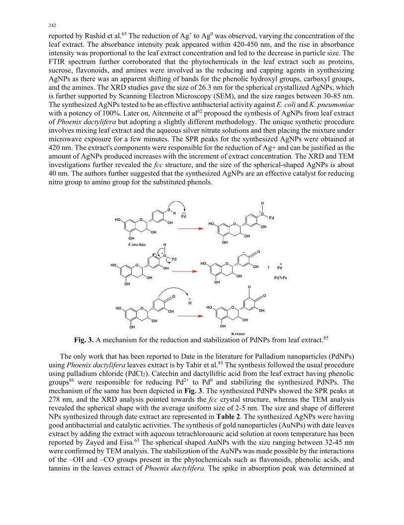
Fig. 3. A mechanism for the reduction and stabilization of PdNPs from leaf extract.85
The only work that has been reported to Date in the literature for Palladium nanoparticles (PdNPs) using Phoenix dactylifera leaves extract is by Tahir et al.85 The synthesis followed the usual procedure using palladium chloride (PdCl2). Catechin and dactyllifric acid from the leaf extract having phenolic groups86 were responsible for reducing Pd2+ to Pd0 and stabilizing the synthesized PdNPs. The mechanism of the same has been depicted in Fig. 3. The synthesized PdNPs showed the SPR peaks at 278 nm, and the XRD analysis pointed towards the fcc crystal structure, whereas the TEM analysis revealed the spherical shape with the average uniform size of 2-5 nm. The size and shape of different NPs synthesized through date extract are represented in Table 2. The synthesized AgNPs were having good antibacterial and catalytic activities. The synthesis of gold nanoparticles (AuNPs) with date leaves extract by adding the extract with aqueous tetrachloroauric acid solution at room temperature has been reported by Zayed and Eisa.63 The spherical shaped AuNPs with the size ranging between 32-45 nm were confirmed by TEM analysis. The stabilization of the AuNPs was made possible by the interactions of the –OH and –CO groups present in the phytochemicals such as flavonoids, phenolic acids, and tannins in the leaves extract of Phoenix dactylifera. The spike in absorption peak was determined at

S. S. Ghani and I. Hussain / Current Chemistry Letters 10 (2021)
243
552 nm as more AuNPs were formed due to the reduction of Au3+ ions. TEM analysis further clarified that the spherical shaped AuNPs are sized between 32-45 nm. Further studies on the catalytic behavior of AuNPs demonstrated that they are size-dependent towards the degradation of nitrophenol to aminophenol.
5. Characterization of Nanoparticles of Phoenix Dactylifera L. Usually, the synthesized NPs are analyzed for their morphological features as the properties of the NPs are influenced by their morphology and the structural features that signify the nature and composition of the NPs. The standard characterization techniques that are generally used for analyzing both these properties are UV-Visible spectroscopy, Fourier transform infrared (FTIR) spectroscopy, Dynamic light scattering (DLS), Scanning electron microscopy (SEM), Transmission electron microscopy (TEM), and X-ray diffraction (XRD). 5.1 UV-visible spectroscopy The characterization by UV-Visible spectroscopy is based on the basic light principles known as the beer-lambert law. The synthesis of NPs from their respective salts of the Ag, Au, Pt, Pd, Cu, or Zn is monitored through UV-Vis spectroscopy as they give their characteristic peaks at different absorptions. The absorbance of the nanoparticle samples at light wavelengths ranging from 300-800 nm and their sizes in between 1-100 nm is frequently used to confirm nanoparticles' synthesis.87 The strong absorption band known as surface plasmon resonance (SPR) is shown only in the spectrum of metal NPs, not in the spectra of metal solutions.88 The AgNPs possess absorption peaks in the range of 400-450 nm, whereas the AuNPs have it at around 500-550 nm. The UV-vis intensity and absorptions are influenced by the NPs shape and size. It is the primary technique employed by many authors89, 90 for the stability and formation of metallic NPs. The absorption peaks in the UV-Vis spectra for PdNPs are usually below 300 nm.91 PtNPs usually have their maximum absorption between 300-350 nm. At higher concentrations, it may present a small redshift. An increase in the concentration of the date extract, or the reaction time, a gradual increase in the absorption peaks indicates the formation of NPs. The green synthesized metallic NPs frequently exhibit surface plasmon resonance (SPR) that helps in the absorption in the UV-Visible region. Researchers have determined that ZnO nanoparticles with date extracts displayed broad peaks in the visible region and an intense peak in the UV region employing UV-Visible spectroscopy. 5.2 Fourier transform infrared (FTIR) spectroscopy Fourier transform infrared spectroscopy (FTIR) is a powerful surface chemical analytical tool for characterization. The nature of the organic functional groups such as carbonyls, hydroxyl, or any other required group is attached to the NPs surface can be evaluated through FTIR.92 FTIR analysis is carried out to detect the surface capping and purity of the formed NPs and is measured from 250-4000 cm-1. The common approach is comparing the FTIR spectrum of the date extract before synthesis with that of the NPs synthesized with it is made that gives vital data of the functional groups involved in the reduction and stabilization. The prominent functional groups show phenolic O–H, amide NH, C=O, C–OH stretching bands. They may be present in carbohydrates, flavonoids, terpenoids, proteins, and phenolic compounds and are responsible for reducing metal ions, paving the way for the synthesis of NPs. In work reported by Al-Radadi,58 the combining of phytochemicals with PtNPs was shown by shifting of -NH frequency of the amino acid group from 2900 cm-1 to 2790 cm-1.

244
Fig. 4. FT-IR spectra of Date fruit extract and AgNPs from Date fruit extract.57
5.3 Dynamic light scattering (DLS) Dynamic light scattering (DLS) is one of the techniques to estimate the size of the NPs and is also used to characterize the surface charge and the size distribution of the particles.93 In DLS, particles' hydrodynamic size is measured by analyzing the scattered light intensity modulation as a function of time.94 The DLS technique's advantages is that it is a table top instrument that is easy to operate, fast, sensitive, and selective to size and concentration changes of the NPs. Additionally, the NPs with high molecular mass are also analyzed and do not require calibration. However, the disadvantage with the DLS technique is that it can measure the size of the monodispersed NPs even at a very low level. Still, it is not accurate in measuring polydisperse nanoparticles with large size.95 DLS also measures the zeta potential, which is the charge acquired by particles in the solution, and it indicates the stability of the nanoparticles. The average particle size of the NPs obtained from DLS is typically larger than those obtained from microscopic techniques. It measures all the substances adsorbed on NPs surface and not only the metallic core. A study by Tahir et al85 has observed that the particle size distribution of the PdNPs synthesized from date leaf extract ranged from 5-18 nm. Another survey by Ansari and Alzohairy81 indicated the average diameter of 32.1 nm, whereas Khatami and Pourseyedi68 have reported the average size of 27 nm for the AgNPs synthesized from date seed extract. The hydrodynamic diameter of the synthesized CuNPs reported by Mohamed61 was approximately 78 nm, as shown in Fig. 5. All the size analyzed for the Phoenix dactylifera mediated synthesis of NPs falls within the stipulated range for establishing the nanomaterials. 5.4 Scanning electron microscopy (SEM) Scanning electron microscopy (SEM) is a versatile, high-resolution imaging technique that delivers insight into the morphology, topology, and surface characteristic of the fabricated NPs. Based on the electron scanning principle, SEM determines the NPs shapes, sizes, distribution, and the dispersion of nanoparticles in bulk at the nanoscale level. SEM's main advantage is that it determines the morphology of NPs smaller than 10 nm but is not useful in determining its internal structure.96 From the SEM study, Tahir et al85 have confirmed that most of the PdNPs synthesized by Date leaves extract are spherical and have a uniform distribution, and the PdNPs are well dispersed. A similar analysis using SEM was reported by Qidwai et al84 for AgNPs from date seed extract revealing mostly the spherical shape without any agglomeration. In another study of NPs with Phoenix dactylifera fruit extract, Ezhilarasi

S. S. Ghani and I. Hussain / Current Chemistry Letters 10 (2021)
245
et al97 reported the rod-shaped surface of well-dispersed NPs having little agglomeration. The interesting feature from the literature report58, 63, 65, 98, 99 reveals that the morphology of almost 99 % of the nanoparticles derived using extracts from different parts of Phoenix dactylifera is spherical. 5.5 Transmission electron microscopy (TEM)
Transmission electron microscopy (TEM) is the general analytical tool that is crucial for determining the size of nanoparticles and the size distribution and morphology. TEM can deliver data even from low to high magnifications and can scan particles in a wide size range. Because of the continuous demand to visualize the lattice arrangements in the crystalline NPs, it has to generate high atomic resolution images. This considerable enhancement of TEM power is possible by integrating many sophisticated techniques, and one such advancement is high-resolution TEM. The resolution of TEM is a thousand times higher and has enhanced effectiveness in terms of three-dimensional resolution in comparison to SEM.100 But the limitation with the TEM technique is that it gives low magnification if the particle size is less than 100 nm. Farhadi et al44 noticed the date extract synthesized AgNPs are spherical with a size ranging from 25-60 nm, whereas a similar study by Zafar and Zafar57 reported the size between 20-100 nm through TEM. Using the TEM images, Al-Radadi58 observed the variation in size of PtNPs at different pH conditions.
Table 2. Synthesis of NPs from date extract Type of
NPs Parts extracted Size
(nm) Shape Characterization Application Ref.
Pt Fruit 1.3-2.6 Spherical UV-vis, FTIR, XRD, TEM, EDX, TGA
Anticancer, Antibacterial 58
Ni Fruit 32±5 Rod-shaped
UV-vis, FTIR, XRD, TEM, Photoluminescence spectroscopy
Antibacterial, Photocatalytic degradation
97
Ag Fruit 25-60 Spherical UV-vis, FTIR, XRD, SEM, TEM, EDX Antibacterial, Catalytic
44
Ag Fruit -- -- UV-vis Antioxidant, Antibacterial
59
Ag Fruit 12.2-140.2
Spherical UV-vis, SEM, EDX Antioxidant, Antimicrobial
73
Ag Fruit 20-100 Spherical UV-vis, FTIR, TEM Antimicrobial, Cytotoxicity
57
Ag Fruit 20-60 Spherical UV-vis, FTIR, SEM, TEM, DLS Antibacterial, Anticancer 74 Cu Fruit 78 Spherical UV-vis, FTIR, TEM, DLS, XRD -- 61 Ag Fruit 3-30 Spherical UV-vis, SEM, TEM, DLS, EDX Catalytic, Antimicrobial,
Optical 60
Ag Seed 14-30 Spherical UV-vis, SEM, TEM, DLS Bactericidal 71 Ag Seed 17-19 Spherical UV-vis, FTIR, SEM, TEM, XRD Antioxidant,
Anticancer 83
Ag Seed 1-40 Spherical UV-vis, XRD TEM, DLS Antibacterial, Antifungal
68
Ag Seed 13.2-22.8 Irregular UV-vis, SEM TEM, DLS Anti-toxoplasma activity 82 ZnO Seed 97nm Spherical TEM, SEM, XRD, EDX Antibacterial 69 Ag Leaf 30-85 Spherical UV-vis, FTIR, SEM, XRD, Bactericidal 65 Pd Leaf 5-21 Spherical UV-vis, FTIR, SEM, TEM, XRD, DLS,
EDX Antioxidant, Antibacterial,
Hemolytic, Catalytic
85
Ag Leaf 20-60 Spherical UV-vis, TEM, XRD Catalytic 62 Au Leaf 32-45 Spherical UV-vis, FTIR, TEM Catalytic 63 Ag Root Hair 15-41 Spherical UV-vis, FTIR, TEM, SEM, XRD, EDX Anticancer, Antibacterial 98 Au Pollen 95 Spherical UV-vis, SEM, EDX Anticancer 99 Ag Pollen 27 Spherical UV-vis, SEM, EDX Anticancer 99
5.6 X-ray diffraction (XRD) The X-ray diffraction (XRD) is a crucial technology to determine the structural information phase identification about the crystalline form of metallic NPs. Strong X-rays can penetrate materials deeply and gives data on the bulk structure.101 The widening of the peaks in the XRD endorses the fabrication of nano-sized particles. The crystal structure explains the atoms' arrangement, position, and power of diffraction peaks. The XRD technique can identify the nanoparticles in single as well as in multiple

246
phases. The XRD is a simple, very delicate, easy to use, and non-destructive technology. The main reason for using this technique for determining the structure of NPs is because the X-rays measure the wavelength on an atomic scale. The operation of XRD is based on Bragg's law.102 The Debye-Scherrer equation is applied to determine the crystalline size of the NPs from the XRD data. The Bragg reflection is calculated through the formula: 𝑑 = 𝐾𝜆𝛽𝐶𝑜𝑠𝜃
(where d is the crystalline size of the NPs in nm, K is the Scherrer constant, λ is the wavelength of X-ray radiation, β is the angular full width at half maximum, and θ is the diffraction angle).103 However, for small nanoparticles that are less than hundreds of atoms in size, it isn't easy to obtain the structural and additional parameters with their precise dimensions. Al-Radadi58 has used the XRD spectra to ascertain the crystallinity of PtNPs synthesized from Phoenix dactylifera fruit extract, and the pattern shows an index plane for dissimilar values of 2θ. The Braggs reflection identified at 2θ values of 39. 38, 45.88, and 67.33° correspond to the index plane of 1 1 1, 2 0 0 and 2 2 0, respectively, which is the characteristic feature of fcc structure as shown in Figure 5. The synthesis of CuNPs was well established by Mohamed61 that exhibited peaks at angle 43.27, 50.41, and 74.17° corresponding to 1 1 1, 2 0 0, and 2 2 0 Bragg's plane inferring that the copper crystallites have fcc lattices.
Fig. 5. (a) SEM of AgNPs from seed extract83 (b) DLS of CuNPs from fruit showing Particle Size
Distribution61 (c) XRD of AgNPs from leaves65 (d) TEM of AgNPs using fruit extract.57
6. Application of Nanoparticles and Future Perspectives The Phoenix dactylifera L. mediated nanoparticles' synthesis is gradually gaining popularity among scientists because of appreciable properties such as economical, non-hazardous, significantly soluble in water, biocompatible, and lack toxic stabilizers. Therefore, the green synthesis route provides ease and extra benefits towards biomedical application. These nanoparticles find their use in different biomedical applications due to their higher surface to volume ratio and some explicit properties. As the green synthesized NPs are expanding in biomedicine, special attention is given towards their cytotoxicity to determine appropriate strategies for the assessment, communication, and management of NPs to safeguard human health besides the environment. The influence of cytotoxicity studied concerning the fabricated NiO nanoparticles towards A549 cell lines exhibited excellent outcome.97

S. S. Ghani and I. Hussain / Current Chemistry Letters 10 (2021)
247
Reactive oxidizing species oxidizes the protein and lipids resulting in toxic electrophiles toxicity to wrong pathways of cellular signal transduction. The NiO NPs synthesized through Phoenix dactylifera L. extract induces cytotoxicity. It possesses the ability to release metal ions and additional features such as impeccable morphology and definite surface area, enabling them to participate in cellular activities. The suitability of AuNPs and AgNPs for application in the medical field is due to their biocompatible nature and great affinities towards biological molecules like lipids, proteins, antibodies, or even microorganisms such as bacteria and viruses.104 When these AgNPs/AuNPs are functionalized with biomolecules facilitates them as biomarkers for drug delivery and finding of disease.105 Phoenix dactylifera L. seeds have a high capacity in forming AgNPs from AgNO3 solution. Therefore, they are utilized as a bioreactor for manufacturing AgNPs at the industrial level, with remarkable properties like highly stable, enhanced antibacterial and antifungal activity, low cost, and environmental compatibility. These properties empower the AgNPs with higher effectiveness as compared with conventional drugs like colistin and polymyxin.68 In general, the AgNPs/AuNPs have exhibited broad-spectrum antibiotic action against microbes. The charismatic feature of the AuNPs from leaf extract of Phoenix dactylifera L. is that it is feasible to tune its size within the nanoscale, which shows size-dependent catalytic activity. Hence, these AuNPs are frequently used for the degradation of 4-nitrophenol to 4-aminophenol.63 The synthesized AgNPs from date fruit extract can be potentially used for antioxidant activity. This high antioxidant potential is attributed to the high amount of polyphenolic compounds in date extract. Various researches have also emphasized that the AgNPs from date fruits extract show convincing antimicrobial results against gram-positive and gram-negative bacteria such as E. coli, S. aureus, K. pneumoniae, and C. albicans,65, 73 and there is no report to Date on its resistance. PtNPs from Date extracts has shown encouraging outcomes as an anticancer agent. They were successful to a considerable extent in obstructing the growth of human cancer cells such as hepatocellular carcinoma cells (HepG-2), colon carcinoma cells (HCT-116), and breast cancer cells (MCF-7). Henceforth, PtNPs find their application as an economical substitute for the treatment of cancer.58 In addition to antimicrobial and catalytic activity, PdNPs formed from the date leaf extract actively scavenged DPPH free radicals, making the water purification easier and cheaper.85 Investigations have shown that the date extracts have the effective antioxidant and mutagenic activity at a slightly higher level than the extract of other plants.106, 107 The antioxidant activity is credited to the comprehensive range of a phenolic compound in dates including p-coumaric, ferulic, and sinapic acids, flavonoids and procyanidins as well as the presence of vitamin C.108 The concentration and composition of phenolic acids, isoflavones, lignans, and flavonoids, tannins, and sterols varies depending on conditions such as the variety of dates, the geographical origin, and soil type, but are comparable with any other plant extracts that are generally used in green synthesis of NPs.48,109 For example, the concentration of carotenoid in fruit extract of dates (0.97 mg/100 g) is higher than that of the figs (0.032 mg/100 g).48 Many researchers have reported50,110,111 that the number of phenolic acids in Date extracts are significantly higher than the other plants. However, we could not find any comprehensive report or review comparing the advantages that date extracts have over other plant extracts to synthesize NPs. In the last decade, green synthesized nanoparticles from Phoenix dactylifera L. have played a considerable role in developing the pharmaceutical field. It unfolds a new perception owing to its anticancer and antimicrobial properties. In the future, these NPs might be used for tumor therapy, depending on their adjustable size, toxicity, and biocompatibility. The antioxidant feature can give them a significant place in the nutraceutical and food industries. Unexpectedly, less work has been reported in the literature either of the synthesis or application of NPs from dates. Regardless of the potential advantages of green NPs in biosensors, their application is marginal and so far has not made its impact compared with other applications of NPs from Date. Nevertheless, these NPs have therapeutic and diagnostic applications in the medical field for humans, but no research has been carried out probing their application in veterinary medicine. At the pace at which the applications of NPs from dates are investigated, it is obvious that it may influence the global consumption of NPs products derived from date palms in the near future. Therefore, avenues are open for the researchers to

248
design new strategies or routes for synthesizing NPs from dates that may find application in the veterinary field. 7. Conclusion In the last two decades, the green synthesis of metallic NPs has gathered considerable scientific community response. The present work emphasizes nanoparticles' synthesis using extracts from various parts of the Phoenix dactylifera L. plant. The extraction methodologies, the synthetic strategies, the characterization techniques involving UV-Vis, FTIR, DLS, SEM, TEM, and XRD and their applications have been discussed in detail. Date seeds, leaves, or fruits used as a green source played a dual role as reducing and stabilizing agent with immense potential to produce numerous NPs with metal or metal oxides like Ag, Au, Pt, Pd, Cu, CuO, and ZnO. The different synthetic routes discussed in this review were useful for fabricating particles in the nano range and were dependent on the conditions such as pH, temperature, and concentration of the extract. Additionally, all the synthesis was a single step process and found to be more eco-friendly, economical, non-toxic, less time consuming, and benign. The date extracts containing phytochemicals such as flavonoids, alkaloids, carotenoids, polyphenols, etc., were responsible for the NPs synthesis. A little discrepancy in Phoenix dactylifera L. extract's chemical composition was observed in sample collection from different regions. Still, it does not induce any striking differencing in their reducing, capping, or stabilizing effect. It can be inferred from the studies that the application of these NPs are impacted by their size, shape, or surface area, and they have prospective applications in catalytic and biomedical fields, but the researchers are more concentrated on their antibacterial and anticancer activities. Continued research needs to be carried out to make the date extract mediated NPs the leading NPs of the future. References 1. Khan A., Rashid R., Murtaza G., and Zahra A. (2014) A gold nanoparticles: synthesis and
applications in drug delivery. Trop. J. Pharm. Res., 13 (7) 1169-1177. 2. Lee S. H., Rho W. Y., Park S. J., Kim J., Kwon O. S., and Jun B. H. (2018) Multifunctional self-
assembled monolayers via microcontact printing and degas-driven flow guided patterning. Sci. Rep., 8 (1) 16763.
3. Lee S. H., and Jun B. H. (2019) Silver nanoparticles: synthesis and application for nanomedicine. Int. J. Mol. Sci., 20 (4) 865.
4. Antonyraj C. A., Jeong J., Kim B., Shin S., Kim S., Lee K. Y., and Cho J. K. (2013) Selective oxidation of HMF to DFF using Ru/calumina catalyst in moderate boiling solvents toward industrial production. J. Ind. Eng. Chem., 19 (3) 1056-1059.
5. Frances N., Nikolay A. P., Michael J. F. B., Tim G., and Paul A. M. (2009) Novel one-pot synthesis and characterization of bioactive thiol-silicate nanoparticles for biocatalytic and biosensor applications. Nanotechnology, 20 (5) 055612.
6. Staniland S. S. (2007) Magnetosomes: Bacterial Biosynthesis of Magnetic Nanoparticles and Potential Biomedical Applications. In: Nanotechnologies for the Life Sciences. Wiley-VCH Verlag GmbH & Co. KGaA.
7. Green S. J., Pietron J. J., Stokes J. J., Hostetler M. J., Vu H., Wuelfing W. P., and Murray R. W. (1998) Three-dimensional monolayers: voltammetry of alkanethiolate stabilized gold cluster molecules. Langmuir, 14 (19) 5612-5619.
8. Brust M., Walker, M., Bethell D., Schiffrin D. J., and Whyman R. (1994) Synthesis of thiol-derivatised gold nanoparticles in a two-phase liquid-liquid system. J. Chem. Soc. Chem. Commun., 7 801-802.

S. S. Ghani and I. Hussain / Current Chemistry Letters 10 (2021)
249
9. Sahoo G. P., Bar H., Bhui D. K., Sarkar P., Samanta S., Pyne S., Ash S., and Misra A. (2011) Synthesis and photo physical properties of star shaped gold nanoparticles. Colloid. Surf. A, 375 (1-3) 30-34.
10. Hu J., Wang Z., and Li J. (2007) Gold nanoparticles with special shapes: controlled synthesis, surface-enhanced Raman scattering, and the application in biodetection. Sensors, 7 (12) 3299-3311.
11. Xu F., Zhang Q., and Gao Z. (2013) Simple one-step synthesis of gold nanoparticles with controlled size using cationic Gemini surfactants as ligands: Effect of the variations in concentrations and tail lengths. Colloid. Surf. A, 417 201-210.
12. Biswal J., Ramnani S. P., Shirolikar S., and Sabharwal S. (2011) Synthesis of rectangular plate like gold nanoparticles by in situ generation of seeds by combining both radiation and chemical methods. Radiat. Phys. Chem., 80 (1) 44-49.
13. Ackerson C. J., Jadzinsky P. D., Sexton J. Z., Bushnell D. A., and Kornberg R. D. (2010) Synthesis and bioconjugation of 2 and 3 nm-diameter gold nanoparticles. Bioconjugate Chem., 21 (2) 214-218.
14. Zoroddu M. A., Medici S., Ledda A., Nurchi V. M., Lachowicz J. I., and Peana M. (2014) Toxicity of Nanoparticles. Curr. Med. Chem., 21 (33) 3837-3853.
15. Zhang L., Bai R., Liu Y., Meng L., Li B., Wang L., Xu L., Le Guyader L., and Chen C. (2012) The dose-dependent toxicological effects and potential perturbation on the neurotransmitter secretion in brain following intranasal instillation of copper nanoparticles. Nanotoxicology, 6 (5) 562-575.
16. Sajid M., Ilyas M., Basheer C., Tariq M., Daud M., Baig N., and Shehzad F. (2015) Impact of nanoparticles on human and environment: review of toxicity factors, exposures, control strategies, and future prospects. Environ. Sci. Pollut. Res. Int., 22 (6) 4122-4143.
17. Sawicki K., Czajka M., Kucharek M. M., Fal B., Drop B., Wielgosz S. M., Sikorska K., Kruszewski M., and Skrzypczak L. K. (2019) Toxicity of metallic nanoparticles in the central nervous system. Nanotechnol. Rev., 8 (1) 175-200.
18. Han D., Tian Y., Zhang T., Ren G., and Yang Z. (2011) Nano-zinc oxide damages spatial cognition capability via over-enhanced long-term potentiation in hippocampus of Wistar rats. Int. J. Nanomedicine, 6 1453-1461.
19. Oszlánczi G., Vezér T., Sárközi L., Horváth E., Kónya Z., and Papp A. (2010) Functional neurotoxicity of Mn-containing nanoparticles in rats. Ecotoxicol. Environ. Saf., 73 (8) 2004- 2009.
20. Singh S. P., Rahman M. F., Murty U. S., Mahboob M., and Grover P. (2013) Comparative study of genotoxicity and tissue distribution of nano and micron sized iron oxide in rats after acute oral treatment. Toxicol. Appl. Pharmacol., 266 (1) 56-66.
21. Papp A., Oszlánczi G., Horváth E., Paulik E., Kozma G., Sápi A., Kónya Z., and Szabó A. (2012) Consequences of subacute intratracheal exposure of rats to cadmiumoxide nanoparticles: Electrophysiological and toxicological effects. Toxicol. Ind. Health, 28 (10) 933- 941.
22. Ray P. C., Yu H., and Fu P. P. (2009) Toxicity and environmental risks of nanomaterials: challenges and future needs. J. Environ. Sci. Health C Environ. Carcinog. Ecotoxicol. Rev., 27 (1) 1-35.
23. Griffitt R. J., Luo J., Gao J., Bonzongo J. C., and Barber D. S. (2008) Effects of particle composition and species on toxicity of metallic nanomaterials in aquatic organisms. Environ. Toxicol. Chem., 27 (9) 1972-1978.
24. Pernodet N., Fang X., Sun Y., Bakhtina A., Ramakrishnan A., Sokolov J., Ulman A., and Rafailovich M. (2006) Adverse effects of citrate/gold nanoparticles on human dermal fibroblasts. Small, 2 (6) 766-773.
25. Karlsson H. L., Cronholm P., Gustafsson J., and Moller L. (2008) Copper oxide nanoparticles are highly toxic: A comparison between metal oxide nanoparticles and carbon nanotubes. Chem. Res. Toxicol., 21 (9) 726-1732.

250
26. Rajakumar G., Gomathi T., Thiruvengadam M., Rajeswari V. D., Kalpana V. N., and Chung I. M. (2017) Evaluation of anti-cholinesterase, antibacterial and cytotoxic activities of green synthesized silver nanoparticles using from Millettia pinnata flower extract. Microb. Pathog., 103 123-128.
27. Ahmed S., Ahmad M., Swami B. L., and Ikram S. (2016) A review on plants extract mediated synthesis of silver nanoparticles for antimicrobial applications: a green expertise. J. Adv. Res., 7 (1) 17-28.
28. Velusamy P., Kumar G. V., Jeyanthi V., Das J., and Pachaiappan R. (2016) Bio-inspired green nanoparticles: synthesis, mechanism, and antibacterial application. Toxicol. Res., 32 (2) 95-102.
29. Raghunandan D., Bedre M. D., Basavaraja S., Sawle B., Manjunath S. Y., and Venkataraman A. (2010) Rapid biosynthesis of irregular shaped gold nanoparticles from macerated aqueous extracellular dried clove buds (Syzygium aromaticum) solution. Colloids Surf. B Biointerfaces, 79 (1) 235-240.
30. Benakashani F., Allafchian A. R., and Jalali S. A. H. (2016) Biosynthesis of silver nanoparticles using Capparis spinosa L. leaf extract and their antibacterial activity. Karbala International Journal of Modern Science, 2 (4) 251-258.
31. Phongtongpasuk S., Poadang S., and Yongvanich N. (2016) Environmental-friendly method for synthesis of silver nanoparticles from dragon fruit peel extract and their antibacterial activities. Energy Procedia, 89 239-247.
32. Satishkumar M., Sneha K., Won S. W., Cho C. W., Kim S., and Yun Y. S. (2009) Cinnamon zeylanicum bark extract and powder mediated green synthesis of nano-crystalline silver particles and its antibacterial activity. Colloids Surf. B Biointerfaces, 73 (2) 332-338.
33. Annu A. S., Kaur G., Sharma P., Singh S., and Ikram S. (2018) Fruit waste (peel) as bio-reductant to synthesize silver nanoparticles with antimicrobial, antioxidant and cytotoxic activities. J. Appl. Biomed., 16 (3) 221-231.
34. Ahmad N., Sharma S., Alam M. K., Singh V. N., Shamsi S. F., Mehta B. R., and Fatma A. (2010) Rapid synthesis of silver nanoparticles using dried medicinal plant of basil. Colloids Surf. B Biointerfaces, 81 (1) 81-86.
35. Dakshayani S. S., Marulasiddeshwara M. B., Kumar M. N. S., Ramesh G., Kumar P. R., Devarajac S., and Hosamani R. (2019) Antimicrobial, anticoagulant and antiplatelet activities of green synthesized silver nanoparticles using Selaginella (Sanjeevini) plant extract. Int. J. Biol. Macromol., 131 787-797.
36. Skiba M. I., Vorobyova M. I., Pivovarov A., and Makarshenko N. P. (2020) Green synthesis of silver nanoparticles in the presence of polysaccharide: optimization and characterization. J. Nanomater., 2020 3051308.
37. Ahmad, D. S. (2011) Review: natural products from Genus Selaginella (Selaginellaceae). Nus. Biosci., 3 (1) 44-58.
38. Blazics B., Alberti A., and Kery A. (2009) Antioxidant activity of different phenolic fractions separated from Euphrasia rostkoviana hayne. Acta. Pharm. Hung., 79 (1) 11-16.
39. Petrichenko V. M., Sukhinina T. V., Babiyan L. K., and Shramm N. I. (2006) Chemical composition and antioxidant properties of biologically active compounds from Euphrasia brevipila. Pharm. Chem. J., 40 312-316.
40. Ahmed M. J., Murtaza G., Mehmood A., and Bhatti T. M. (2015) Green synthesis of silver nanoparticles using leaves extract of Skimmia laureola: characterization and antibacterial activity. Mater. Lett., 153 10-13.
41. Sankar R., Karthik A., Prabu A., Karthik S., Shivashangari K. S., and Ravikumar V. (2013) Origanum vulgare mediated biosynthesis of silver nanoparticles for its antibacterial and anticancer activity. Colloids Surf. B Biointerfaces, 108 80-84.
42. Qu D., Sun W., Chen Y., Zhou J., and Liu C. (2014) Synthesis and in vitro antineoplastic evaluation of silver nanoparticles mediated by Agrimoniae herba extract. Int. J. Nanomed., 9 (1) 1871-1882.
43. Ahmad N., Bhatnagar S., Ali S. S., and Dutta R. (2015) Phytofabrication of bioinduced silver nanoparticles for biomedical applications. Int. J. Nanomed., 10 (1) 7019-7030.

S. S. Ghani and I. Hussain / Current Chemistry Letters 10 (2021)
251
44. Farhadi S., Ajerloo B., and Mohammadi A. (2017) Green biosynthesis of spherical silver nanoparticles by using date palm (phoenix dactylifera) fruit extract and study of their antibacterial and catalytic activities. Acta Chim. Slov., 64 (1) 129-143.
45. Ashraf Z., and Hamidi E. Z. (2011) Date and date processing: a review. Food Rev. Int., 27 (2) 101-133.
46. Jaradat A. A., and Zaid A. (2004) Quality traits of date palm fruits in a center of origin and center of diversity. J. Food Agric. Environ., 2 (1) 208-217.
47. Barghini P., Gioia D. D., Fava F., and Ruzzi M. (2007) Vanillin production using metabolically engineered Escherichia coli under non-growing conditions. Microb. Cell Fact, 6 (1) 13.
48. Martín-Sánchez A. M., Cherif S., Ben-Abda J., Barber-Vallés X., Pérez-Álvarez J. Á., and Sayas-Barberá E. (2014) Phytochemicals in date co-products and their antioxidant activity. Food Chem., 158 513-520.
49. Sosnowska J., and Balslev H. (2009) American palm ethnomedicine: A meta-analysis, J. Ethnobiol. Ethnomed., 5 43-53.
50. Benmeddour Z., Mehinagic E., Meurlay D. L., and Louaileche H. (2013) Phenolic composition and antioxidant capacities of ten Algerian Date (Phoenix dactylifera L.) cultivars: A comparative study. J. Funct. Foods, 5 (1) 346-354.
51. Salem A. O. M., Rhouma S., Zehdi S., Marrakchi M., and Trifi M. (2008) Morphological variability of Mauritanian date-palm (Phoenix dactylifera L.) cultivars as revealed by vegetative traits. Acta Bot. Croat., 67 (1) 81-90.
52. Al-Oqla F. M., and Sapuan S. M. (2014) Natural fiber reinforced polymer composites in industrial applications: feasibility of date palm fibers for sustainable automotive industry. J. Cleaner Prod., 66 347-354.
53. Karar M. G. E., and Kuhnert N. (2017) Herbal drugs from Sudan: traditional uses and phytoconstituents. Pharmacogn. Rev., 11 (22) 83-103.
54. Aryangat A. V., and Gerich J. E. (2010) Type 2 diabetes: postprandial hyperglycemia and increased cardiovascular risk. Vasc. Health Risk Manag., 6 145-155.
55. Besbes S., Ghorbel R., Salah R. B., Masmoudi M., Jedidi F., Attia H., and Blecker C. (2010) Date fiber concentrate: chemical compositions, functional properties and effect on quality characteristics of beef burgers. J. Food Drug Anal., 18 (1) 8-14.
56. Hasan M., and Mohieldein A. (2016) In vivo evaluation of anti diabetic, hypolipidemic, antioxidative activities of Saudi date seed extract on streptozotocin induced diabetic rats. J. Clin. Diagn. Res., 10 (3) 06-12.
57. Zafar S., and Zafar A. (2019) Biosynthesis and characterization of silver nanoparticles using phoenix dactylifera fruits extract and their in vitro antimicrobial and cytotoxic effects. Open Biotechnol. J., 13 37-46.
58. Al-Radadi N.S. (2019) Green synthesis of platinum nanoparticles using Saudi's Dates extract and their usage on the cancer cell treatment. Arabian J. Chem., 12 (3) 330-349.
59. Abusahid Z., and Kandiah M. (2019) In Vitro Green synthesis of phoenix dactylifera silver nanoparticles: assessing their antioxidant and antimicrobial properties. Int. J. Nanosci., 18 (5) 1850031.
60. Zaheer Z. (2017) Biogenic synthesis, optical, catalytic, and in vitro antimicrobial potential of Ag nanoparticles prepared using Palm date fruit extract. J. Photochem. Photobiol. B Biol., 178 584-592.
61. Mohamed E. A. (2020) Green synthesis of copper & copper oxide nanoparticles using the extract of seedless dates. Heliyon, 6 (1) e03123.
62. Aitenneite H., Abboud Y., Tanane O., Solhy A., Sebti S., and El-Bouari A. (2016) A Rapid and green microwave-assisted synthesis of silver nanoparticles using aqueous Phoenix Dactylifera L. (date palm) leaf extract and their catalytic activity for 4-Nitrophenol reduction. J. Mater. Environ. Sci., 7 (7) 2335-2339.
63. Zayed M. F., and Eisa W. H. (2014) Phoenix dactylifera L. leaf extract phytosynthesized gold nanoparticles, controlled synthesis and catalytic activity. Spectrochim. Acta Part A, 121 238-244.

252
64. Ismail M. S., Abuzaid O. I., and El-Ashmawy I. M. (2017) Effect of aqueous extract of tops of date palm leaves on blood glucose of diabetic rats. Pak. J. Pharm. Sci., 30 (5) 20317-22037.
65. Rashid M. I., Mujawar L. H., Rehan Z. A., Qari H., Zeb J., Almeelbi T., and Ismail I. M. I. (2016) One-step synthesis of silver nanoparticles using Phoenix dactylifera leaves extract and their enhanced bactericidal activity. J. Mol. Liq., 223 1114-1122.
66. Salama A. A., Ismael N. M., and Megeed M. M. (2019) Using date seed powder nanoparticles and infusion as a sustainable source of nutraceuticals. J. Food Nutr. Sci., 7 (3) 39-48.
67. Bouhlali E. D. T., Alem C., Ennassir J., Benlyas M., Mbark A. N., and Zegzouti Y. F. (2017) Phytochemical compositions and antioxidant capacity of three Date (Phoenix dactylifera L.) seeds varieties grown in the South East Morocco. J. Saudi Soc. Agric. Sci., 16 (4) 350-357.
68. Khatami M., and Pourseyedi S. (2015) Phoenix dactylifera (date palm) pit aqueous extract mediated novel route for synthesis high stable silver nanoparticles with high antifungal and antibacterial activity. IET Nanobiotechnol., 9 (4) 184-190.
69. El-Naggar M. E., Shaarawy S., and Hebeish A. A. (2017) Multifunctional properties of cotton fabrics coated with in situ synthesis of Zinc oxide nanoparticles capped with date seed extract. Carbohydr. Polym., 181 307-316.
70. Sathishkumar M., Sneha K., Kwak I. S., Mao J., Tripathy S. J., and Yun Y. S. (2009) Phyto-crystallization of Palladium Through Reduction Process Using Cinnamon Zeylanicum Bark Extract. J. Hazard Mater., 171 (1-3) 400-404.
71. Yang X., Li Q., Wang H., Huang J., Lin L., Wang W., Sun D., Su Y., Opiyo J. B., Hong L., Wang Y., He N., and Jia L. (2010) Green synthesis of palladium nanoparticles using broth of Cinnamomum camphora leaf. J. Nanopart. Res., 12 1589-1598.
72. Charti I., Eddahbi A., Abboud Y., and El Bouari A. (2017) Rapid and green microwave-assisted synthesis of silver nanoparticles using aqueous phoenix dactylifera l. (date palm) wood extract and evaluation of catalytic and antibacterial activities. Micro Nanosyst., 9 (2) 134-139.
73. Shaikh A. E., Satardekar K. V., Khan R. R., Tarte N. A., and Barve S. S. (2018) Silver nanoparticles: green synthesis using Phoenix dactylifera fruit extract, characterization, and antioxidant and antimicrobial activities. Appl. Nanosci., 8 407-415.
74. Mohammed A. E., Al-Qahtani A., Al-Mutairi A., Al-Shamri B., and Aabed K. (2018) Antibacterial and cytotoxic potential of biosynthesized silver nanoparticles by some plant extracts. Nanomaterials, 8 (6) 382.
75. Das P., and Karankar V. S. (2019) New avenues of controlling microbial infections through antimicrobial and anti-biofilm potentials of green mono-and multi-metallic nanoparticles: A review. J. Microbiol. Methods, 167 105766.
76. Nanaei M., Nasseri M. A., Allahresani A., and Kazemnejadi M. (2019) Phoenix dactylifera L. extract: antioxidant activity and its application for green biosynthesis of Ag nanoparticles as a recyclable nanocatalyst for 4-nitrophenol reduction. SN Appl. Sci., 1 853.
77. Athinarayanan J., Periasamy V. S., and Alshatwi A. A. (2019) Phoenix dactylifera lignocellulosic biomass as precursor for nanostructure fabrication using integrated process. Int. J. Biol. Macromol., 134 1179-1186.
78. Dwivedi A. D., and Gopal K. (2011) Plant-mediated biosynthesis of silver and gold nanoparticles. J. Biomed. Nanotechnol., 7 (1) 163-164.
79. Biçer M., and Sisman I. (2010) Controlled synthesis of copper nano/microstructures using ascorbic acid in aqueous CTAB solution. Powder Technol., 198 (2) 279-284.
80. Awad M. A. G., Al-Olayan E. M., Yehia H. M., Ortashi K. M. O., Ali H. S. M., and Elkhadragy M. F. (2017) Method of preparing date palm seed nanoparticles. US Patent 9623067B1.
81. Ansari M. A., and Alzohairy M. A. (2018) One-pot facile green synthesis of silver nanoparticles using seed extract of phoenix dactylifera and their bactericidal potential against MRSA. Evid. Based Complement. Alternat. Med., 2018 1860280.
82. Alajmi R. A., Al-Megrin W. A., Metwally D., Al-Subaie H., Altamrah N., Barakat A. M., Al-Moneim A. E., Al-Otaibi T. T., and El-Khadragy M. (2019) Anti-Toxoplasma activity of silver nanoparticles green synthesized with Phoenix dactylifera and Ziziphus spina-christi extracts which

S. S. Ghani and I. Hussain / Current Chemistry Letters 10 (2021)
253
inhibits inflammation through liver regulation of cytokines in Balb/c mice. Biosci. Rep., 39 (5) BSR20190379.
83. Mohammadi G., Zangeneh M. M., Zangeneh A., and Haghighi Z. M. S. (2020) Chemical characterization and anti‐breast cancer effects of silver nanoparticles using Phoenix dactylifera seed ethanolic extract on 7,12‐Dimethylbenz[anthracene]‐induced mammary gland carcinogenesis in sprague dawley male rats. Appl. Organometal. Chem., 34 (1) e5136.
84. Qidwai A., Kumar R., and Dikshit A. (2018) Green synthesis of silver nanoparticles by seed of Phoenix sylvestris L. and their role in the management of cosmetics embarrassment. Green Chem. Lett. Rev., 11 (2) 176-188.
85. Tahir K., Nazir S., Ahmad A., Li B., Khan A. U., Shah S. A. A., Khan G. M., Khan Z. U. H., Khan Q., and Khan F. U. (2016) Biodirected synthesis of palladium nanoparticles using Phoenix dactylifera leaves extract and their size dependent biomedical and catalytic applications. RSC Adv., 6 (89) 85903-85916.
86. Ziouti A., Modafar C. E., Fleuriet A., Boustani S. E., and Macheix J. J. (1996) Phenolic compounds in date palm cultivars sensitive and resistant to Fusarium oxysporum. Biol. Plant., 38 (3) 451-457.
87. Feldheim D. L., and Foss C. A. (2002) Metal nanoparticles: synthesis, characterization, and applications. CRC Press: Boca Raton, FL, USA.
88. Tomaszewska E., Soliwoda K., Kadziola K., Celichowski G., Cichomski M., Szmaja W., and Grobelny J. (2013) Detection limits of DLS and UV-Vis spectroscopy in characterization of polydisperse nanoparticles colloids. J. Nanomater., 2013 313081.
89. Noruzi M. (2015) Biosynthesis of gold nanoparticles using plant extracts. Bioprocess. Biosyst. Eng., 38 (1) 1-14.
90. Sharma V. K., Yngard R. A., and Lin Y. (2009) Silver nanoparticles: Green synthesis and their antimicrobial activities. Adv. Colloid Interface Sci., 145 (1-2) 83-96
91. Luo C., Zhang Y., and Wang Y. (2005) Palladium nanoparticles in poly(ethyleneglycol): the efficient and recyclable catalyst for Heck reaction. J. Mol. Catal. A Chem., 229 (1-2) 7-12.
92. Chithrani B. D., Ghazani A. A., and Chan W. C. W. (2006) Determining the size and shape dependence of gold nanoparticle uptake into mammalian cells. Nano Lett., 6 (4) 662-668.
93. Jiang J., Oberdörster G., Biswas P. (2009) Characterization of size, surface charge, and agglomeration state of nanoparticle dispersions for toxicological studies. J. Nanopart. Res., 11 77-89.
94. Berne B. J., and Pecora R. (2000) Dynamic Light Scattering: With Applications to Chemistry, Biology, and Physics, Dover, New York, NY, USA.
95. Jans H., Liu X., Austin L., Maes G., and Huo Q. (2009) Dynamic light scattering as a powerful tool for gold nanoparticle bioconjugation and biomolecular binding studies. Anal. Chem., 81 (22) 9425-9432.
96. Tran Q. H., Nguyen V. Q., and Le A. (2013) Silver nanoparticles: Synthesis, properties, toxicology, applications and perspectives. Adv. Nat. Sci. Nanosci. Nanotechnol., 4 (3) 033001.
97. Ezhilarasi A. A., Vijaya J. J., Kennedy L. J., and Kaviyarasu K. (2020) Green mediated NiO nano-rods using Phoenix dactylifera (Dates) extract for biomedical and environmental applications. Mater. Chem. Phys., 241 122419.
98. Oves M., Aslam M., Rauf M. A., Qayyum S., Qari H. A., Khan M. S., Alam M. Z., Tabrez S., Pugazhendhi A., and Ismail I. M. I. (2018) Antimicrobial and anticancer activities of silver nanoparticles synthesized from the root hair extract of Phoenix dactylifera. Mat. Sci. Eng. C, 89 429-443.
99. Banu H., Renuka N., Faheem S. M, Ismail R, Singh V., Saadatmand Z., Khan S. S., Narayanan K., Raheem A., Premkumar K., and Vasanthakumar G. (2018) Gold and silver nanoparticles biomimetically synthesized using date palm pollen extract-induce apoptosis and regulate p53 and bcl-2 expression in human breast adenocarcinoma cells. Biol. Trace Elem. Res., 186 (1) 122-134.
100. Eppler A. S., Rupprechter G., Anderson E. A., and Somorjai G. A. (2000) Thermal and chemical stability and adhesion strength of Pt nanoparticle arrays supported on silica studied by

254
transmission electron microscopy and atomic force microscopy. J. Phys. Chem. B, 104 (31) 7286- 7292.
101. Huang J., Li Q., Sun D., Lu Y., Su Y., and Yang X. (2007) Biosynthesis of silver and gold nanoparticles by novel sundried Cinnamomum camphora leaf. Nanotechnology, 18 105104- 105114.
102. Waseda Y., Matsubara E., and Shinoda K. (2011) X-Ray Diffraction Crystallography. Springer Berlin.
103. Prathna T. C., Chandrasekaran N., Raichur A. M., and Mukherjee A. (2011) Biomimetic synthesis of silver nanoparticles by Citrus limon (lemon) aqueous extract and theoretical prediction of particle size. Colloids Surf. B Biointerfaces, 82 (1) 152-159.
104. Victor S. U., and Roberto V. B. J. (2015) Gold and silver nanotechnology on medicine. J. Chem. Biochem., 3 (1) 21-33.
105. Lin M., Pei H., Yang F., Fan C., and Zuo X. (2013) Applications of gold nanoparticles in the detection and identification of infectious diseases and biothreats. Adv. Mater., 25 (25) 3490-3496.
106. Mohamed D. A. and Al-Okbi S. (2005) In vitro evaluation of antioxidant activity of different extracts of Phoenix dactylifera L. fruits as functional foods. Dtsch. Lebensm. Rundsch., 101 305- 308.
107. Allaith A. A. A. (2008) Antioxidant activity of Bahraini date palm (Phoenix dactylifera L.) fruit of various cultivars. Int. J. Food Sci. Technol., 43 (6) 1033-1040.
108. Mansouri A., Embarek G., Kokkalou E., and Kefalas P. (2005) Phenolic profile and antioxidant activity of the Algerian ripe date palm fruit (Phoenix dactylifera). Food Chem., 89 (3) 411-420.
109. Al-Turki S., Shahba M. A., and Stushnoff C. J. (2010) Diversity of antioxidant properties and phenolic content of date palm (Phoenix dactylifera L.) fruits as affected by cultivar and location. J. Food Agri. Environ., 8 (1) 253-260.
110. Lemine F. M. M., Ahmed M. V. O. M., Maoulainine L. B. M., Bouna Z. E. A. O., Samb A., and Boukhary A. O. M. S. (2014) Antioxidant activity of various Mauritanian date palm (Phoenix dactylifera L.) fruits at two edible ripening stages. Food Sci. Nutr., 2 (6) 700-705.
111. Chaira N., Smaali M. I., Martinez-Tomé M., Mrabet A., Murcia M. A., Ferchichi A. (2009) Simple phenolic composition, flavonoid contents and antioxidant capacities in water- methanol extracts of Tunisian common date cultivars (Phoenix dactylifera L.). Int. J. Food Sci. Nutr., 60 (7) 316-329.
© 2021 by the authors; licensee Growing Science, Canada. This is an open access article distributed under the terms and conditions of the Creative Commons Attribution (CC-BY) license (http://creativecommons.org/licenses/by/4.0/).