Embed Size (px)
Citation preview

RESEARCH ARTICLE Open Access
Anti-cancer effects of Ajwa dates (Phoenixdactylifera L.) in diethylnitrosamine inducedhepatocellular carcinoma in Wistar ratsFazal Khan1,2, Tariq Jamal Khan1, Gauthaman Kalamegam2* , Peter Natesan Pushparaj2, Adeel Chaudhary3,Adel Abuzenadah3, Taha Kumosani1,4, Elie Barbour1,5 and Mohammed Al-Qahtani2
Abstract
Background: Hepatocellular carcinoma (HCC) accounts for major cancer-related deaths despite current advancedtherapies. Treatment and prognosis of HCC is better in patients with preserved liver function. Many natural productsincluding ajwa dates (Phoenix dactylifera L.), are claimed to have hepatoprotective and HCC inhibitory effects, butmost lack scientific validation. To prove our hypothesis, we attempted to evaluate the HCC inhibitory effects, andother beneficial properties of the aqueous extract of ajwa dates (ADE) in a rat model of diethylnitrosamine (DEN)induced liver cancer.
Methods: Thirty-two male rats were divided into four groups of eight each as follows, Group A: untreated control;Group B: DEN control (180 mg/kg bw), Group C: DEN + ADE 0.5 g/kg bw; and Group D: DEN +1.0 g/kg bw. Ratsfrom all groups were assessed for liver cancer progression or inhibition by evaluating histological, biochemical,antioxidant enzyme status, cytokines and gene expression profiles.
Results: DEN treatment Groups (B, C, D) showed histological features of HCC and in rats treated with ADE (Groups C,D) partial to complete reversal of normal liver architecture was observed. Antioxidant enzymes such as superoxidedismutase (SOD), glutathione reductase (GR), glutatione peroxidase (GPx) and catalase (CAT) were increased, while theliver enzymes alanine aminotransferase (ALT), aspartate aminotransferase (AST) and alkaline phosphatase (ALP) levelsand lipid peroxidation were significantly decreased in Group C and Group D compared to Group B. Pro-inflammatorycytokines such as interleukin (IL)-1α, IL-1β,, GM-CSF) were increased in the serum of rats in Group B while the anti-tumor cytokines (IL-2, IL-12) were increased in ADE treated Groups (C, D). In addition, Alpha-Feto Protein (AFP) and IL-6gene expression levels were upregulated in Group B, while they were significantly downregulated in ADE treatedGroups (C, D).
Conclusions: ADE helped in the reversal of DEN damaged liver towards normal. Restoration of anti-oxidant enzymes,liver enzymes, cytokines balance and gene expression to normal levels following ADE treatment indicates that ADEimproves liver function and inhibits HCC. ADE can, therefore, be used together with conventional therapeutics for HCC.
Keywords: Ajwa date extract, Hepatocellular carcinoma, In vivo, Anti-oxidants, Cytokines, Quantitative real-time PCR
* Correspondence: [email protected] of Excellence in Genomic Medicine Research (CEGMR), KingAbdulaziz University (KAU), PO BOX 80216, Jeddah 21589, Kingdom of SaudiArabiaFull list of author information is available at the end of the article
© The Author(s). 2017 Open Access This article is distributed under the terms of the Creative Commons Attribution 4.0International License (http://creativecommons.org/licenses/by/4.0/), which permits unrestricted use, distribution, andreproduction in any medium, provided you give appropriate credit to the original author(s) and the source, provide a link tothe Creative Commons license, and indicate if changes were made. The Creative Commons Public Domain Dedication waiver(http://creativecommons.org/publicdomain/zero/1.0/) applies to the data made available in this article, unless otherwise stated.
Khan et al. BMC Complementary and Alternative Medicine (2017) 17:418 DOI 10.1186/s12906-017-1926-6

BackgroundHepatocellular carcinoma (HCC) is one of the leadingcause of cancer-associated deaths, and globally it is threetimes more common in men than women [1]. HCC isone of the leading cause of cancer related deaths in theKingdom of Saudi Arabia (KSA). HCC is the 4th mostcommon cancer in males and 8thcommon cancer infemales accounting for 4.3% of the reported cancer casesin the year 2013 [2]. A global estimation of 40,710 newcases and 28,920 deaths due to liver and intrahepatic bileduct cancers in 2017 highlights the need for improve-ment in current management strategies [1]. Commonrisk factors for liver cancer include heavy alcoholconsumption, hepatitis B virus (HBV), hepatitis C virus,obesity, tobacco smoking, diabetes and genetic factors.Partial hepatectomy, embolization and sorafenib (Nexavar®)[3] offer partial to complete success depending upon thestage of the disease.The incidence of high mortality and associated side
effects following chemotherapy and/or radiotherapy in-crease the demand for alternative medicine for thecancer treatment. Not surprisingly, many potent antican-cer compounds were isolated from the plants viz. doxo-rubicin, taxol, etoposide, cisplatin, vinblastine, vindesine,vincristine and topotecan [4]. Given the current interestsin alternative medicine, it is imperative that much-concerted effects are undertaken to identify novelcompounds that will aid therapy and also help to avoidits unjustifiable use.Date fruit (Phoenix dactylifera L.) is a native fruit of
arid region, largely cultivated as an economical and foodcrop in the Middle East, Southern Europe, North Africa,South America, India, and Pakistan [5]. Of the manyvarieties of date fruits, ajwa dates are unique for itsmedicinal properties and these are specifically grownin the city of Al-Madina Al-Munawwarah, KSA [5].Ajwa dates are a rich source of energy, and readilyprovide sugars, proteins, vitamins, high dietary fibers,minerals and fats [6]. They contain various phytochemi-cals like sterols, polyphenols, flavonoids and glycosides[6]. Ajwa dates have hypolipidemic, antioxidant, anti-in-flammatory, cardioprotective, nephroprotective andhepatoprotective effects [7]. In addition, the concen-trated polyphenols and ex vivo digested extracts fromajwa dates inhibited growth and proliferation of coloncancer Caco-2 cell line [8]. Also, (1 → 3)-β-D-glucan,which is an essential component of dates had aninhibitory effect on sarcoma-180 in vivo in mice [9].Recently, we identified that methanolic extract of ajwadates inhibited human breast adenocarcinoma (MCF-7) by causing cell cycle inhibition and induction ofapoptosis [10]. Interestingly, so far no studies havebeen done to identify the effects of ajwa or otherdates on hepatic cancer.
Many in vitro and in vivo models of hepatic cancerhave been utilized in the past to evaluate various drugs.In vivo hepatic cancer model development is done usingvarious chemical methods, of which diethylnitrosamine(DEN) is a chemical carcinogen, commonly utilized toinduce liver carcinogenesis by increasing oxidative stressand liver damage [11]. Hence in the present study, weused the DEN-induced hepatic cancer model in Wistarrats and evaluated the cancer inhibition effects of theaqueous extract of ajwa dates (ADE). Histological,biochemical, antioxidant enzyme status, as well as the re-lated cytokines and gene expression profiles was studied.
MethodsAnimalsThe animals used in this study was approved by theInstitutional Ethics Committee, King Abdulaziz Univer-sity (KAU), Saudi Arabia vide approval number HA-02-J-008. Thirty-two albino male rats (Wistar strain,5-6 weeks old) that weighed between 100 g - 120 g werepurchased through the animal holding unit (AHU), KingFahd Medical Research Center (KFMRC, KAU). All ratswere housed in the AHU under standard animal housingconditions of 25 °C ± 3 °C, 55 ± 2% humidity, 12 hrsday/night cycle, and had access to standard rat pelletand water ad libitum.
Preparation of aqueous extract of ajwa date (ADE)Ajwa dates used in the present study were sourced fromAl Madina Dates Co. (Tomoor), Madinah Al Munawarah,Kingdom of Saudi Arabia. Ajwa dates sample was depos-ited with the Biology Department, Faculty of Science,KAU. Dr. Dhafer Ahmed Al-Zahrani, Plant Taxonomistconfirmed the genus, species and origin of date fruit(Specimen voucher number: P. dactylifera L. #PD17569).Ajwa dates flesh was mixed with ultrapure distilled waterat a ratio of 1:3. The date suspension was continuouslyagitated and incubated at 25 °C on a shaker (at 60 rpm)placed within the incubator (25 °C) for 48 hrs. The date-water extract was then filtered through muslin cloth andWhatman no. 1 filter paper. The filtrate was then centri-fuged at 4500 rpm for 10 min. The supernatant was againfiltered and the aqueous extract of ajwa dates (ADE) thusobtained was stored at −80 °C until use.
Experimental designWistar male rats (n = 32) were acclimatized for 1 weekprior to start of experiments. Rats were randomlydivided into four experimental groups (n = 8). HCC in-duction was done using diethylnitrosamine (DEN).Briefly, DEN was dissolved in corn oil and two doses(180 mg/kg body weight (bw); at 15 days interval) wereadministered orally to all rats in Group B, C and D usingintragastric gavage. Untreated control (Group A) was
Khan et al. BMC Complementary and Alternative Medicine (2017) 17:418 Page 2 of 10

administered vehicle (corn oil) alone. Treatment withADE was started on the next day following DEN treat-ment. All rats in Group C and Group D were treated withADE 0.5 g/kg bw and 1.0 g/kg bw respectively, daily for10 weeks; while the rats in Group A (untreated control)and Group B (DEN control) were administered vehiclealone. Following completion of treatment, the animalswere sacrificed using an injection of chloral hydrate(400 mg/kg bw; i.p). Blood and liver tissues were collectedfrom all the animals in different experimental groups.Serumwas separated by centrifugation (2500 rpm× 10min)and stored at −80 °C until further analyzed. Part of theliver tissue was fixed in 10% neutral buffered formalin for24 h or in RNA later for subsequent use in histologicaland molecular analysis respectively.
HistologyFormalin fixed liver tissues were dehydrated using gradi-ent concentrations of ethanol; then washed in xyleneand embedded in paraffin wax. Tissue blocks were sec-tioned at 5-6 μM thickness, deparaffinized and stainedwith hematoxylin and eosin and analyzed under micro-scope (Olympus Instruments, Tokyo, Japan).
Anti-oxidant enzymesThe following anti-oxidant enzymes, namely superoxidedismutase (SOD, Sigma 19,160), glutathione reductase(GR, Sigma GRSA), glutathione peroxidase (GPx, SigmaCGP1) and catalase (CAT, Sigma CAT100) wereanalyzed using respective kits and manufacturer’s proto-cols. SOD and CAT were end point assays and theabsorbance was obtained at 440 nm and 520 nm respect-ively. GR and GPx were kinetic assays and the absorb-ance at 412 nm and 340 nm was obtained every 10 srespectively.
Lipid peroxidation (LPO) assayLPO assay was carried out using the Sigma kit(MAK085) according to the manufacturer’s instructions.Following end point assay, the absorbance at 532 nmwas determined spectrophotometrically.
Liver enzymesThe activity of the following liver enzymes namely, ala-nine aminotransferase (ALT, Abcam 105,134), aspartateaminotransferase (AST, Abcam 105,135) and alkalinephosphatase (ALP, Abcam 83,369) were measured usingrespective kits according to the manufacturer’s instruc-tions. Following incubation for 5 min, the absorbance at570 nm (ALT, AST) and 405 nm were determinedspectrophotometrically.All spectrophotometric analysis were done using the mi-
croplate reader (SpectraMax® i3x, Molecular Devices,Sunnyvale, CA).
Multiplex cytokine assaySerum samples from ADE treated and control rats were an-alyzed for both pro-inflammatory and anti-inflammatorycytokines using the Rat Cytokine Magnetic 10-Plex Panel(Novex®) according to the manufacturer’s instructions.Briefly, the magnetic beads in solution were added andwashed twice with 1X wash buffer. Standards (1:3 serialdilution) and samples (1:2 dilution) were prepared andadded to the washed beads and incubated on an orbitalshaker at 500 rpm for 2 hrs. The plate was then incubatedwith detection antibodies for 1 hr and streptavidin-RPEantibodies for 30 min incubation. Between incubations withdifferent antibodies, the plate was washed twice with washbuffer. Following antibody incubation, the plate was finallywashed thrice and resuspended in wash buffer and analyzedusing on MAGPIX® instrument (Luminex, USA). Dataobtained was analyzed using the Luminex® xPONENT®
multiplex assay analysis software.
Quantitative real time-PCR (qRT-PCR)Total ribonucleic acid (RNA) was extracted from theliver tissue of both control and treated rats using PureLink® RNA Mini Kit (Ambion™, Thermo Fischer Scien-tific) according to the manufacturer’s protocol. Oncolumn, DNase-I treatment was included in the proto-col. First-strand cDNA synthesis was carried out usingrandom hexamers (High Capacity cDNA ReverseTranscription Kit, Applied Biosystems). Primers weredesigned using qPrimerDepot (National Institute ofHeath, USA) and PrimerBank (The MassachusettsGeneral Hospital, USA), and the primer sequences aregiven in Table 1. The qRT-PCR analysis was performedusing the ABI StepOnePlus™ Real-Time PCR System(Applied Biosystems) using SYBR green dye master mixand relative quantification was performed using thecomparative 2 –ΔΔCt method which analyzes the relativechange in gene expression based on Ct values (fluores-cence denoting cDNA copy numbers) of housekeepinggene and gene of interest).
Statistical analysisStatistical analyses were performed using Prism GraphPadversion 6.0. One way ANOVA (analysis of variance) andstudent unpaired t-test for comparisons between groups
Table 1 Primer sequences
Genes Primer type Sequence
AFP Forward 5′-CTGTATGCTCCCACCATTCTTT-3′
Reverse 5′-TTGATGCTCTCTTTGTCTGGAA-3′
IL6 Forward 5′-CCGGAGAGGAGACTTCACAG-3′
Reverse 5′-CAGAATTGCCATTGCACAAC-3′
GAPDH Forward 5’GACTCTACCCACGGCAAGTT-3′
Reverse 5’GGTGATGGGTTTCCCGTTGA-3′
Khan et al. BMC Complementary and Alternative Medicine (2017) 17:418 Page 3 of 10

and within groups respectively. All the values wereexpressed as mean ± SEM (standard error of the mean).Asterisk (*) and hash (#) indicates statistical significance,with the level significance for comparisons set at P < 0.05.
ResultsHistologyLiver sections from the control group showed normallobular pattern with central vein and peripheral portalveins (Fig. 1a). In contrast, the animals treated withDEN showed evidence of inflammatory cell infiltrationand fibrosis. The normal lobular pattern was replacedwith clusters of atypical cells; hepatocytes with increasednuclear to cytoplasmic ratio and dense connective tissue(Fig. 1b). However, treatment with ADE (0.5 g/kg bwand 1.0 g/kg bw) promoted partial to complete reversalof liver architecture (Fig. 1c and d).
Antioxidant enzymesIn general, the activity of the anti-oxidant enzymes SOD,GR, GPx and CAT were decreased in treated Groups (B,C, D) compared to normal untreated control (Group A).Compared to DEN control, the ADE treated Groups (C,D) demonstrated increases in activity for SOD, GR andGPx and these increases in values were statisticallysignificant (p < 0.05) (Fig. 2a–c). However, CAT demon-strated an increase only in Group D and this was notstatistically significant (Fig. 2d).
Lipid peroxidation (LPO) assayMalondialdehyde (MDA) levels as determined by LPOassay, showed mild increases in Groups (B, C), and a de-crease in Group D compared to untreated normal con-trol (Group A). Compared to Group B (DEN control),LPO demonstrated an increase in Group C and a de-crease in Group D these values in the ADE treatedgroups were statistically significant (p < 0.05) (Fig. 2e).
Liver enzymesIn general, the activity of the liver enzymes ALT, ASTand ALP were increased in treated Groups (B, C, D)compared to normal untreated control (Group A). Com-pared to DEN control, the ADE treated Groups (C, D)demonstrated decreases in activity for ALT, AST andALP and these decreases in values statistically significant(p < 0.05) (Fig. 3a–c).Additional results from anti-oxidant enzymes (SOD,
GR, GPx, CAT), LPO and liver enzymes (ALT, AST,ALP) are summarized in Table 2.
CytokinesIn general, the cytokines analyzed demonstrated a de-crease in the proinflammatory cytokines (IL-1β, IL-1α,and GM-CSF) and an increase in the anti-tumor cytokinesIL-2 and IL-12 compared to Group B (DEN control).The concentrations of IL-1α were 1867.3 4 ± 84.78,
2754.82 ± 69.12, 1899.23 ± 17.07 and 1349.25 ± 192.88for the Groups A, B, C and D respectively. Only the
Fig. 1 Haematoxylin and eosin (H&E) staining: Representative images of the liver sections (a, b, c, d) from rats treated with aqueous date extract(ADE) following DEN induced HCC are shown above. a - untreated control (Group A) shows normal hepatocytes, intact cell membrane and centralvain; b - DEN control (Group B) liver shows altered hepatocyte morphology, with loss of cell membrane, increased nuclear size and connective tissueinfiltration; c, d - the ajwa treated Groups (C: 0.5 g/kg bw; D: 1.0 g/kg bw) shows normal liver pattern with radially arranged hepatocytes similar to thatof the control and this was more pronounced in Group D than Group C. Magnification (40X). Inset (e) is the magnified image of the boxed area in B(DEN treated), where changes in cell morphology, nuclear pattern and connective tissue infiltration are clearly demonstrated. Inset (f) is the magnifiedimage of the boxed area in D (Ajwa 1 g/kg bw), which shows normal histological pattern of the liver. Thin black arrows indicate normal hepatocytemorphology. Thick black arrows indicate connective tissue infiltration. Black arrow heads indicate abnormal nuclear morphology
Khan et al. BMC Complementary and Alternative Medicine (2017) 17:418 Page 4 of 10

Fig. 2 Analysis of antioxidant enzymes and lipid peroxidation levels from the serum samples of rats treated with aqueous date extract (ADE)following DEN induced HCC. Antioxidant enzymes namely, (a) - superoxide dismutase (SOD); (b) - glutathione reductase (GR); (c) - glutathioneperoxidase (GPx); (d) - catalase (CAT); and (e) lipid peroxidase levels were analyzed. One-way ANOVA and student t test were used to computestatistics between groups and within groups respectively. All values were expressed as mean ± SEM. Comparisons were made between untreatedcontrol (Group A) and DEN control (Group B) and their significance were represented using asterisk (*p < 0.05 and **p < 0.01, ***p < 0.001) andhash (# p < 0.05, ## p < 0.01 and ### p < 0.001) respectively
Fig. 3 Analysis of liver enzymes from the serum samples of rats treated with aqueous date extract (ADE) following DEN induced HCC. Liverenzymes namely, (a) alanine aminotransferase; (b) aspartate aminotransferase and (c) alkaline phosphatase were analyzed. One-way ANOVA and student ttest were used to compute statistics between groups and within groups respectively. All values were expressed as mean ± SEM. Comparisons were madebetween untreated control (Group A) and DEN control (Group B) and their significance were represented using asterisk (*p < 0.05 and **p < 0.01,***p < 0.001) and hash (# p < 0.05, ## p < 0.01 and ### p < 0.001) respectively
Khan et al. BMC Complementary and Alternative Medicine (2017) 17:418 Page 5 of 10

increase in Group B (by 48%) statistically significant(P < 0.05), compared to Group A (Fig. 4a). Compared toGroup B, IL-1α was decreased in Group C by 31% andGroup D by 51%, and these mean decreases in values inthe ADE treated groups were statistically significant(p < 0.05) (Fig. 4a).The concentrations of IL-1β were 63.27 ± 0.86,
452.7 ± 15.05, 501.74 ± 11.45 and 116.78 ± 7.28 for theGroups A, B, C and D respectively. IL-1β concentrationswere increased in the treated Groups (B, C, D by 614%,691%, 83% respectively) compared to Group A (Fig. 4b).Compared to Group B, IL-1β increased moderately inGroup C (by 10%) and decreased in Group D (by 74%).All mean increases or decreases in values were statisti-cally significant (p < 0.05) (Fig. 4b).The concentrations of GM-CSF were 32.27 ± 2.17,
66.12 ± 10.01, 50.16 ± 0.65 and 43.99 ± 8.13 for theGroups A, B, C and D respectively. GM-CSF concen-trations significantly increased (P < 0.05) in treatedGroups (B, C, by 106%, 56.25% respectively) com-pared to Group A (Fig. 4c). Compared to Group B,only the decrease in Group C by 24% was statisticallysignificant (p < 0.05) (Fig. 4c).The concentrations of IL-2 were 3724 ± 95.97,
389.8 ± 76.57, 677.6 ± 119.6 and 1008 ± 6.16 for theGroups A, B, C and D respectively. IL-2 concentrationswere significantly decreased (P < 0.05) in treated Groups(B, C, D by 90%, 82%, 73%) compared to Group A (Fig. 4d).Compared to Group B, IL-2 demonstrated increases in
Group C by 74% and Group D by 159%; however, onlythe increase observed in Group D was statisticallysignificant (p < 0.05) (Fig. 4d).
The concentrations of IL-12 were 3548 ± 30.25,3177 ± 1.79, 4604 ± 346.86 and 6703 ± 628.5 for theGroups A, B, C and D respectively. IL-12 concentrationdecreased in Group B (by 4%), increased in Group D (by89%) compared to Group A; and increased in Group D(by 111%) compared to Group B. These values werestatistically significant (p < 0.05) (Fig. 4e).The concentrations of IL-4 were 1.946 ± 0.03,
0.6958 ± 0.08, 0.6991 ± 0.01 and 0.6764 ± 0.03 for theGroups A, B, C and D respectively. IL-4 concentrationswere statistically decreased (P < 0.05) in treated Groups(B, C, D by 64.3%, 64%, 65.3%) compared to Group A(Fig. 4f ). Compared to Group B the observed increase inGroup C (by 0.6%) and decrease in Group D (by 3%)were not statistically significant (Fig. 4f ).The concentrations of IL-10 were 199.6 ± 23.77,
197.6 ± 2.18, 215.3 ± 4.19 and 206.3 ± 21.41 for theGroups A, B, C and D respectively. IL-10 concentrationwas decreased in Group B (by by 1.5%), while this wasincreased in Groups (C, D by 7.5%, 3.0% respectively)compared to Group A. Compared Group B, the IL-10demonstrated increases in Group C (by 9.0%) and GroupD (by 4.6%) and all these increases or decreases in valueswere not statistically significant (p < 0.05) (Fig. 4g).
Quantitative real time PCR (qRT-PCR)Gene expression demonstrated biphasic responses intreated Groups (B, C, D) compared to Group A. Alfa-fetoprotein (AFP) gene expression was upregulated by 6-fold and 0.61-fold in Group B and Group C respectively,while this was downregulated by 0.15-fold in Group D,compared to Group A (Fig. 5a and b). Interleukin 6 (IL-
Table 2 Summary of results of liver and antioxidant enzymes
Enzymes Group A(Untreated control)
Group B(DEN alone)
Group C(0.5g/kg bw ADE)
Group D(1g/kg bw ADE)
AntioxidantSOD 75.76 ± 2.5 22.67 ± 1.3
70%*
36.43 ± 3.9
51%* 61%#
51.13 ± 9.7
32%* 26%#
GR 0.91 ± 0.02 0.32 ± 0.008
64%*
0.68 ± 0.06
25%* 113%#
0.72 ± 0.07
21%* 125%#
GPx 6.69 ± 1.13 3.66 ± 2.19
45%*
6.63 ± 0.66
0.5%* 81%#
8.27 ± 0.64
24%* 126%#
CAT 21.26 ± 0.03 19.60 ± 0.29
8%*
18.26 ± 0.17
6%* 7%#
19.92 ± 0.5
14%* 2%#
LPO 498.4 ± 0.27 522.6 ± 0.84
4.9%*
567.4 ± 8.24
13.84%* 8.6%#
453.1 ± 11.55
9.0%* 13.3%#
Liver enzymesALT 35.79 ± 0.79 74.18 ± 2.4
107%*
42.77 ± 24.0
20%* 42%#
45.27 ± 3.1
26%* 39%#
AST 95.99 ± 3.42 166.2 ± 0.53
73%*
104.7 ± 0.89
9%* 37%#
39.94 ± 19.02
48%* 76%#
ALP 119.7 ± 6.36 192.6 ± 2.93
60%*
139.3 ± 10.12
16%* 28%#
84.76 ± 3.90
29%* 6%#
*Percentages compared to Group A; # percentages compared to Group B
Khan et al. BMC Complementary and Alternative Medicine (2017) 17:418 Page 6 of 10

Fig. 4 Analysis of serum cytokines from the serum samples of rats treated with aqueous date extract (ADE) following DEN induced HCC. Thefollowing cytokines namely, (a) IL-1α; (b) IL-1β, (c) GM-CSF, Th1 [(d) IL-2; (e) IL-12] and Th2 [(f) IL-4; (g) IL-10] were analyzed. One-way ANOVA and student ttest were used to compute statistics between groups and within groups respectively. All values were expressed as mean ± SEM. Comparisons were madebetween untreated control (Group A) and DEN control (Group B) and their significance were represented using asterisk (*p < 0.05 and **p < 0.01,***p < 0.001) and hash (# p < 0.05, ## p < 0.01 and ### p < 0.001) respectively
Fig. 5 Real time gene expression analysis of the liver tissue from rats treated with aqueous date extract (ADE) following DEN induced HCC. (a)AFP; (b) One-way ANOVA and student t test were used to compute statistics between groups and within groups respectively. All values wereexpressed as mean ± SEM. Comparisons were made between untreated control (Group A) and DEN control (Group B) and their significance wererepresented using asterisk (*p < 0.05 and **p < 0.01, ***p < 0.001) and hash (# p < 0.05, ## p < 0.01 and ### p < 0.001) respectively
Khan et al. BMC Complementary and Alternative Medicine (2017) 17:418 Page 7 of 10
6) gene expression was upregulated by 2-fold in GroupB, while this was downregulated by 1.7-fold and 1.2-foldin Group C and Group D respectively compared toGroup A. Compared to Group B the decreases observedin both AFP and IL-6 were statistically significant(P < 0.05).
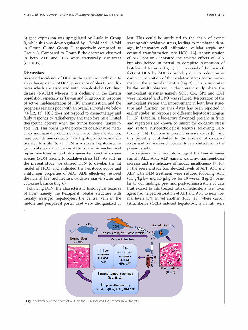
DiscussionIncreased incidence of HCC in the west are partly due toan earlier epidemic of HCV, prevalence of obesity and dia-betes which are associated with non-alcoholic fatty liverdisease (NAFLD) whereas it is declining in the Easternpopulation especially in Taiwan and Singapore in responseof active implementation of HBV immunization, and theprognosis remains poor with an overall survival rate below9% [12, 13]. HCC does not respond to chemotherapy andfairly responds to radiotherapy and therefore have limitedtherapeutic options when the tumor becomes unresect-able [12]. This opens up the prospects of alternative medi-cines and natural products or their secondary metabolites,have been demonstrated to have hepatoprotective and an-ticancer benefits [6, 7]. DEN is a strong hepatocarcino-genic substance that causes disturbances in nucleic acidrepair mechanisms and also generates reactive oxygenspecies (ROS) leading to oxidative stress [13]. As such inthe present study, we utilized DEN to develop the ratmodel of HCC, and evaluated the hepatoprotective andantitumour properties of ADE. ADE effectively restoredthe normal liver architecture, oxidative marker status andcytokines balance (Fig. 6).Following DEN, the characteristic histological features
of liver, namely the hexagonal lobular structure withradially arranged hepatocytes, the central vein in themiddle and peripheral portal triad were disorganized or
lost. This could be attributed to the chain of eventsstarting with oxidative stress, leading to membrane dam-age, inflammatory cell infiltration, cellular atypia andeventual transformation into HCC [14]. Administrationof ADE not only inhibited the adverse effects of DENbut also helped in partial to complete restoration ofhistological features (Fig. 1). The reversal of the toxic ef-fects of DEN by ADE is probably due to reduction orcomplete inhibition of the oxidative stress and improve-ment in the antioxidant status (Fig. 2). This is supportedby the results observed in the present study where, theantioxidant enzymes namely SOD, GR, GPx and CATwere increased and LPO was reduced. Restoration of theantioxidant system and improvement in both liver struc-ture and function by ajwa dates has been reported inearlier studies in response to different hepatocarcinogens[5, 15]. Luteolin, a bio-active flavonoid present in fruitsand vegetables are known to inhibit the oxidative stressand restore histopathological features following DENtoxicity [14]. Luteolin is present in ajwa dates [8], andthis probably contributed to the reversal of oxidativestress and restoration of normal liver architecture in thepresent study.In response to a hepatotoxic agent the liver enzymes
namely ALT, AST, ALP, gamma glutamyl transpeptidaseincrease and are indicative of hepatic insufficiency [7, 16].In the present study too, elevated levels of ALT, AST andALP with DEN treatment were reduced following ADE(0.5 g/kg bw and 1.0 g/kg bw for 10 weeks) (Fig. 3). Simi-lar to our findings, pre- and post-administration of datefruit extract to rats treated with dimethoate, a liver toxicagent had helped restoration of ALT and AST to near nor-mal levels [17]. In yet another study [18], where carbontetrachloride (CCl4) induced hepatotoxicity in rats were
Fig. 6 Summary of the effect of ADE on the DEN-induced liver cancer in Wistar rats
Khan et al. BMC Complementary and Alternative Medicine (2017) 17:418 Page 8 of 10

used, the elevated levels of the liver enzymes ALT, ASTand ALP returned close to their baseline values upontreatment with date seed extracts (1 g/kg bw for 4 weeks).The polyphenolic compounds comprising of phenolics,
flavonoids, stilbenes and lignins present in variousplants, fruits and vegetables are known to exert hepato-protective effects [7]. The total phenolic contentmeasured as from date palm flesh is approximately290 mg/100 g and in date seeds it is 38.8 mg/g [8],expressed as gallic acid equivalent. Therefore, theobserved hepatoprotective effect following ADE treat-ment in DEN-induced HCC model (Fig. 6) in thepresent study could probably be due to the high phen-olic contents in the palm date flesh.Cytokines are the pleiotropic hormones of the immune
system that play a pivotal role in the initiation, mainten-ance and progression of tumors [19]. Neoplastic cellsand tumor-associated macrophages (TAM) secrete IL-6,CSF-1, IL-10, TGF-β, TNF-α, IL-1α, angiogenic and lym-phangiogenic growth factors that promote tumor devel-opment [20, 21]. In our study, DEN administrationspecifically increased the serum levels of proinflamma-tory cytokines, such as IL-1α, IL-1β, GM-CSF, and treat-ment with ADE effectively attenuated these cytokines(Fig. 4). Studies have shown that flavonoids have anti-tumor and anti-inflammatory properties and are capableof reducing the serum levels of Th1 cytokines [22]. Thepresence of major flavonoids such as apigenin, quercetin,and luteolin in ajwa could be one of the reason forreduced serum levels of proinflammatory cytokines (IL-1β, TNF-α and IL-6), and hepatoprotection observed inDEN-induced HCC in rats [23, 24]. Furthermore, lowerlevels of GM-CSF by itself effectively reduces theconcentrations of other proinflammatory cytokines IL-1β and TNF-α [25].In the present study, that the levels of both IL-2 and
IL-12 were reduced significantly with DEN treatmentbut increased following ADE administration. Anotherpro-inflammatory cytokine IL-4, which is implicated intumor promoting activities however, remained un-affected with DEN treatment compared to untreatednormal. The cytokines IL-2 and IL-12 although consid-ered to be pro-inflammatory, they negatively regulatetumor behavior [26, 27], and are currently in clinicaluse/trials [26]. Proanthocyanidins were reported todecrease the inflammation associated with 2,4-dinitro-fluorobenzene and ultraviolet B UVB treatment in mice,by increasing the IL-12 concentration [28]. Proanthocya-nidins are found in dietary fruits including ajwa datesand this could be attributed to the observed increase inthe levels of IL-12 [7, 28].Cancer is associated with inflammation and some of
the inflammatory markers such as TNF-α, IL1, IL-6 be-come increased in HCC and after DEN administration
[29–31], as both are associated with inflammation. Inaddition, cancer diagnosis or prognosis depends on iden-tification of biomarkers. AFP is a commonly usedmarker in HCC and its serum [32, 33]. AFP and itsreceptor have been implicated in HBV mediated malig-nant transformation of hepatocytes and also in theirmetastasis via PI3K/AKT-mediated signaling [33]. In thepresent study we observed that both AFP and IL-6 geneexpression were downregulated in response to ADEtreatment (Fig. 5). Administration of luteolin (20 μg/kgbw, intraperitoneally) in mice decreased the plasmalevels of AFP that was initially increased after DENtreatment [22]. Similarly, luteolin administration(30 mg/kg bw, orally) decreased the gene expressionlevels of IL-6 in DEN and ethanol-mediated hepaticinflammation in mice [34]. Ajwa dates or its constituentsnamely luteolin thus appears to have beneficial proper-ties against HCC.
ConclusionsADE demonstrated significant beneficial effects againstDEN induced HCC in rats. The anti-inflammatory,hepatoprotective and anticancer properties observed inthe present study could be due to the presence of flavo-noids such as luteolin, apigenin, quercetin and proantho-cyanidins in dates which are rich in polyphenoliccompounds. As such dates and or its constituents canbe taken together with conventional chemotherapeutics,for synergistic benefits against HCC.
AbbreviationsADE: Aqueous Extract of ajwa Dates; AFP: Alpha-feto protein; AHU: Animal holdingunit; ALP: Alkaline phosphatase; ALT: Alanine aminotransferase; AST: Aspartateaminotransferase; bw: Body weight; CAT: Catalase; DEN: Diethylnitrosamine;g: Gram; GM-CSF: Granulocyte-macrophage colony-stimulating factor;GPx: Glutathione peroxidase; GR: Glutathione reductase; HCC: Hepatocellularcarcinoma; hrs: Hours; IL-1α: Interleukin-1alfa; KFMRC: King fahd medical researchcenter; kg: Kilogram; KSA: Kingdom of Saudi Arabia; LPO: Lipid peroxidation;MDA: Malondialdehyde; mg: Milligram; min: Minute; nm: Nanometer; no: Number;PCR: Polymerase chain reaction; RNA: Ribonucleic acid; rpm: Rotation per minute;SEM: Standard error mean; SOD: Superoxide dismutase
AcknowledgementsWe thank Prof. Firoze Anwar for his assistance during animal modeldevelopment. The financial support by the King Abdulaziz City for Scienceand Technology (KACST) through postgraduate grant funding [AT-34-237]and applied medical research grant [AT-34-330] are greatly acknowledged.
FundingThe authors would like to acknowledge the financial support provided byKing Abdulaziz City for Science and Technology (KACST) under thepostgraduate grant AT-34-237 and applied medical research grant AT-34-330.
Availability of data and materialsAll the data generated and analyzed for the current study is included inthe article.
Authors’ contributionsFK and TJK carried out the experiments, analyzed the results and preparedthe manuscript. GK and PNP were involved in conceptualization, intellectualcontribution, statistical evaluation and manuscript editing. AA, TK, EB andMAQ were involved in the overall co-ordination of the work, as well as
Khan et al. BMC Complementary and Alternative Medicine (2017) 17:418 Page 9 of 10

reviewed and edited the manuscript. All authors read and approved the finalmanuscript.
Ethics approvalAnimal experiments and protocols were performed as suggested by theethical approval committee of Faculty of Medicine, King Abdulaziz University(HA-02-J-008).
Consent for publicationNot applicable.
Competing interestsThe authors declare that they have no competing interests.
Publisher’s NoteSpringer Nature remains neutral with regard to jurisdictional claims inpublished maps and institutional affiliations.
Author details1Biochemistry Department, Faculty of Science, King Abdulaziz University,Jeddah, Saudi Arabia. 2Center of Excellence in Genomic Medicine Research(CEGMR), King Abdulaziz University (KAU), PO BOX 80216, Jeddah 21589,Kingdom of Saudi Arabia. 3Center of Innovation in Personalized Medicine,King Abdulaziz University, Jeddah, Saudi Arabia. 4King Fahd Medical ResearchCenter, King Abdulaziz University, Jeddah, Saudi Arabia. 5Department ofAgriculture, Faculty of Agricultural and Food Sciences, American University ofBeirut (AUB), Beirut, Lebanon.
Received: 7 April 2017 Accepted: 14 August 2017
References1. Siegel RL, Miller KD, Jemal A. Cancer statistics, 2017. CA Cancer J Clin.
2017;67:7–30.2. Saudi Cancer Registry. In: Cancer Incidence Report Saudi Arabia 2013. Saudi
Health Council. 2016. http://www.chs.gov.sa/Ar/HealthCenters/NCC/CancerRegistry/CancerRegistryReports/2013.pdf. Accessed 07 Apr 2017.
3. American Cancer Society. In: Cancer Facts & Figures 2016. American CancerSociety. 2016. https://www.cancer.org/content/dam/cancer-org/research/cancer-facts-and-statistics/annual-cancer-facts-and-figures/2016/cancer-facts-and-figures-2016.pdf. Accessed 07 Apr 2017.
4. Gordaliza M. Natural products as leads to anticancer drugs. Clin TranslOncol. 2007;9:767–76.
5. Ragab AR, Elkablawy MA, Sheik BY, Baraka HN. Antioxidant and tissue-protective studies on Ajwa extract: dates from al-Madinah al-Monwarah, SaudiaArabia. J Environ Anal Toxicol. 2013;3:163. doi:10.4172/2161-0525.1000163
6. Baliga MS, Baliga BRV, Kandathil SM, Bhat HP, Vayalil PK. A review of thechemistry and pharmacology of the date fruits (Phoenix dactylifera L.). FoodRes Int. 2011;44:1812–22.
7. Yasin B, El-Fawal H, Mousa S. Date (Phoenix dactylifera) Polyphenolics and otherbioactive compounds: a traditional Islamic Remedy’s potential in prevention ofcell damage, cancer therapeutics and beyond. Int J Mol Sci. 2015;16:26210.
8. Eid N, Enani S, Walton G, Corona G, Costabile A, Gibson G, Rowland I,Spencer JP. The impact of date palm fruits and their componentpolyphenols, on gut microbial ecology, bacterial metabolites and coloncancer cell proliferation. J Nutr Sci. 2014;3:e46.
9. Ishurd O, Kennedy JF. The anti-cancer activity of polysaccharide preparedfrom Libyan dates (Phoenix Dactylifera L.). Carbohydr Polym. 2005;59:531–5.
10. Khan F, Ahmed F, Pushparaj PN, Abuzenadah A, Kumosani T, Barbour E,AlQahtani M, Gauthaman K. Ajwa date (Phoenix dactylifera L.) extract inhibitshuman breast Adenocarcinoma (MCF7) cells in vitro by inducing apoptosisand cell cycle arrest. PLoS One. 2016;11:e0158963.
11. Bagi CM, Andresen CJ. Models of hepatocellular carcinoma and biomarkerstrategy. Cancers (Basel). 2010;2:1441–52.
12. Bruix J, Sherman M. Management of hepatocellular carcinoma. Hepatology.2005;42:1208–36.
13. Choo SP, Tan WL, Goh BK, Tai WM, Zhu AX. Comparison of hepatocellularcarcinoma in eastern versus western populations. Cancer. 2016;122:3430–46.
14. Loeppky RN. Nitrosamine and N-Nitroso compound chemistry and biochemistryadvances and perspectives. ACS Symp Ser. 1994; doi:10.1021/bk_1994_0553.ch001
15. Balamurugan K, Karthikeyan J. Evaluation of luteolin in the prevention of N-nitrosodiethylamine-induced hepatocellular carcinoma using animal modelsystem. Indian J Clin Biochem. 2012;27:157–63.
16. Sheikh BY, Elsaed WM, Samman AH, Sheikh BY, Ladin A-MMB. Ajwa dates asa protective agent against liver toxicity in rat. Eur Sci J. 2014;3:358–68.
17. King PD, Perry MC. Hepatotoxicity of chemotherapy. Oncologist. 2001;6:162–76.18. Saafi EB, Louedi M, Elfeki A, Zakhama A, Najjar MF, Hammami M, Achour L.
Protective effect of date palm fruit extract (Phoenix dactylifera L.) on dimethoateinduced-oxidative stress in rat liver. Exp Toxicol Pathol. 2011;63:433–41.
19. Abdelaziz DH, Ali SA. The protective effect of Phoenix dactylifera L. seeds againstCCl4-induced hepatotoxicity in rats. J Ethnopharmacol. 2014;155:736–43.
20. Dranoff G. Cytokines in cancer pathogenesis and cancer therapy. Nat RevCancer. 2004;4:11–22.
21. Coussens LM, Werb Z. Inflammation and cancer. Nature. 2002;420:860–7.22. Torisu H, Ono M, Kiryu H, Furue M, Ohmoto Y, Nakayama J, Nishioka Y, Sone
S, Kuwano M. Macrophage infiltration correlates with tumor stage andangiogenesis in human malignant melanoma: possible involvement ofTNFα and IL-1α. Int J Cancer. 2000;85:182–8.
23. Zhang Q, Yang J, Wang J. Modulatory effect of luteolin on redoxhomeostasis and inflammatory cytokines in a mouse model of liver cancer.Oncol Lett. 2016;12:4767–72. doi: 10.3892/ol.2016.5291
24. Brito AF, Ribeiro M, Abrantes AM, Mamede AC, Laranjo M, Casalta-Lopes JE,Gonçalves AC, Sarmento-Ribeiro AB, Tralhão JG, Botelho MF. New approachfor treatment of primary liver tumors: the role of Quercetin. Nutr Cancer.2016;68:250–66.
25. Leyva-López N, Gutierrez-Grijalva EP, Ambriz-Perez DL, Heredia JB.Flavonoids as cytokine modulators: a possible therapy for inflammation-related diseases. Int J Mol Sci. 2016;17:921.
26. Hamilton JA, Anderson GP. Mini review GM-CSF biology. Growth Factors.2004;22:225–31.
27. Rosenberg SA. IL-2: the first effective immunotherapy for human cancer. JImmunol. 2014;192:5451–8.
28. Tugues S, Burkhard S, Ohs I, Vrohlings M, Nussbaum K, Vom Berg J, Kulig P,Becher B. New insights into IL-12-mediated tumor suppression. Cell DeathDiffer. 2015;22:237–46.
29. Vaid M, Singh T, Li A, Katiyar N, Sharma S, Elmets CA, Xu H, Katiyar SK.Proanthocyanidins inhibit UV-induced Immunosuppression through IL-12–dependent stimulation of CD8+ Effector T cells and inactivation of CD4+ Tcells. Cancer Prev Res. 2011;4:238–47.
30. Grivennikov SI, Karin M. Inflammatory cytokines in cancer: tumour necrosisfactor and interleukin 6 take the stage. Ann Rheum Dis. 2011;70:i104–8.
31. Berasain C, Castillo J, Perugorria M, Latasa M, Prieto J, Avila M. Inflammationand liver cancer. Ann N Y Acad Sci. 2009;1155:206–21.
32. Sakurai T, He G, Matsuzawa A, Yu G-Y, Maeda S, Hardiman G, Karin M.Hepatocyte necrosis induced by oxidative stress and IL-1α release mediatecarcinogen-induced compensatory proliferation and liver tumorigenesis.Cancer Cell. 2008;14:156–65.
33. Clark P. Serum markers for hepatocellular carcinoma. Clin Liver Dis. 2016;8:29–33.34. Li M, Li H, Li C, Wang S, Jiang W, Liu Z, Zhou S, Liu X, McNutt MA, Li G.
Alpha-fetoprotein: a new member of intracellular signal molecules inregulation of the PI3K/AKT signaling in human hepatoma cell lines. Int JCancer. 2011;128:524–32.
Khan et al. BMC Complementary and Alternative Medicine (2017) 17:418 Page 10 of 10