Embed Size (px)
Citation preview

Brit. J. Ophthal. (i 969) 53, I 74
Dacryocystography of normal andpathological lacrimal passages
S. R. K. MALIK*, A. K. GUPTA*, S. CHATERJEE*,0. P. BHARDWAJ**, AND M. SAHA**Departments of Ophthalmology* and Radiology*", Maulana Azad Medical College and AssociatedHospitals, New Delhi, India
Dacryocystography is a method of visualizing the lacrimal passages by the use of radio-opaque contrast media. Ewing (I909) suggested the use of bismuth subnitrate in liquidpetroleum as the contrast medium. Von Szily (i92o) described the pathology of thelacrimal passages as seen by roentgenography. Lipoidol was used as the contrast mediumby Bollack (1924), lipoidol with olive oil by Spackman (I938), iodized oil by Fox (I947)and Blankstein (I952), ethyl iodophenyl undecylate (Pantopaque) by Milder and Demorest(I954) and Demorest and Milder (I955) and neohydriol by Agarwal (I96I) and Nahata(i 964) .
Material
The present study was carried out on two groups of individuals:L 37 control subjects These were out-patients with no complaint of epiphora. The lacrimalpassages were found to be patent on syringing.
IL I69 patients complaining of epiphora The condition of the lacrimal passages was assessed bysyringing and the results were correlated with those obtained by dacryocystography.
Technique
0o5 to I ml. concentrated dionosil solution (Saha, Bhardwaj, Malik, and Jain, I967) was injectedinto the lacrimal sac through the lower punctum with a 2 ml. syringe and cannula. Skiagrams weretaken immediately after the injection in the following positions:(a) Postero-anterior view The patient lay prone in the nose-chin position with the orbit at an angleof 40o to the horizontal. The central ray was directed through the infraorbital margin of theinjected side, about half an inch lateral to the midline.
(b) Lateral view The patient's head was turned to the injected side; the central ray was directed asin the postero-anterior view.
In each case a Potter-Bucky diaphragm was used.
Observations
(I) APPEARANCE OF NORMAL DACRYOCYSTOGRAM
In all the normal subjects the lower canaliculus, lacrimal sac, and nasolacrimal duct wereoutlined. The following observations were made:(a) Canaliculi The lower punctum and the lower canaliculi were visible in all cases.
Received for publication June 4, i968Address for reprinits: Departments of Ophthalmology and Radiology, Maulana Azad Mledical College anid Associated Hospitals,New Delhi, India
on 18 April 2019 by guest. P
rotected by copyright.http://bjo.bm
j.com/
Br J O
phthalmol: first published as 10.1136/bjo.53.3.174 on 1 M
arch 1969. Dow
nloaded from

Dacryocystography
The canaliculi opened into a common ampulla, the sinus of Maier, in 94-6 per cent. ofcases, and separately into the sac in the remainder.(b) Lacrimal sac This was found to have a smooth outline. A shallow constriction wasseen at the junction of the sac and the duct and indicated the location of the valve ofKrause.(c) Nasolacrimal duct In Io-8 per cent. of cases, constrictions in the column of contrastmedium in the nasolacrimal duct were seen. These were possibly due to mucosal folds.In the other cases the valves were absent and the duct looked like a smooth tube. In twocases small single diverticula were seen in the lateral wall of the duct. In one case the ductwas tortuous.(d) Measurements of lacrimal sac and nasolacrimal duct The dimensions are summarized inTable I.
Table I Measurements of normal lacrimal passages in 37 cases
Dimension Lacrimal sac (mm.) Nasolacrimal duct (mm.)
(i) Vertical diameterMean 1110 20*97S.D. + 1'97 ± 3-37S.E. ± 0-33 ± O-56
(2) Lateral diameterMean 2-43 2 30S.D. ± o095 + o*83S.E. ± o-i6 + O*14
(3) Antero-posterior diameterMean 4-0O 2.84S.D. + 1-49 ± O°79S.E. ± 020 + OI3
(2) CASES WITH PATHOLOGY OF
(a) Clinical particularsLACRIMAL PASSAGES
Sex Females were affected more commonly than males (Table II).
Table II Sex incidence of lacrimalpassage pathology
Sex Number of cases Percentage
Male 31 I8.3Female I38 8i.7
Age The average age in females was 35 -9 years (range i!2-80). The incidence rosesteadily to a maximum in the fourth decade. This was followed by a gradual decline.The average age in males was 23-8 (range 11-72). The highest incidence occurred in
the late twenties.The age incidence in the two sexes is shown in Fig i (overleaf).
I175
on 18 April 2019 by guest. P
rotected by copyright.http://bjo.bm
j.com/
Br J O
phthalmol: first published as 10.1136/bjo.53.3.174 on 1 M
arch 1969. Dow
nloaded from

S. R. K. Malik, A. K. Gupta, S. Chaterjee, 0. P. Bhardwaj, and M. Saha
Malesm Females
FIG. I Age incidence oj
pathological findings, by sex
1st. 2nd. 3rd. 4th. 5th. 6th. 7th.Decade of age
Side involved The left side (99; 58-8 per cent.) was more commonly involved than theright (70; 41-2 per cent.).
(b) Radiological appearances (Figs 2 to 6)
FIG. 2 Normal dacryocy.stogi-ami jor conmpatitoni with F1(G1. 3 BlocA at level oJ ofI aw,
Obstruction This was complete in I35 cases (8o per cent.) and incomplete in fifteen(8-8 per cent.). In nineteen cases (i l2 per cent.) there was no obstruction. Theseobservations agree with those of Nahata (I964).The commonest site of obstruction was the junction of the lacrimal sac and the naso-
lacrimal duct (ninety cases; 53-2 per cent.), and the next commonest site was the sinus ofMaier (4I cases; 24-3 per cent.). This observation agrees with that of Campbell (I964).
Condition of the sac In ten cases the dimensions were nearly normal, nineteen hadsmall irregular sacs, and seventy showed dilatation (49 in all dimensions including the
40
Z-30,>11u0
u 20
.f-
3 10z
0
176
k
on 18 April 2019 by guest. P
rotected by copyright.http://bjo.bm
j.com/
Br J O
phthalmol: first published as 10.1136/bjo.53.3.174 on 1 M
arch 1969. Dow
nloaded from
Dacryocystography
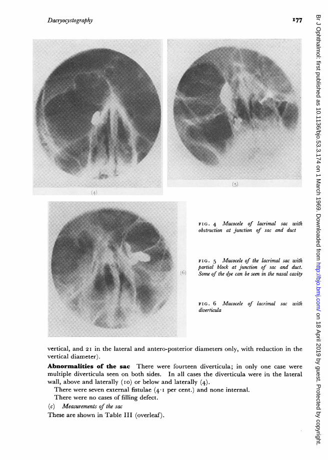
FIG. 4 Mucocele of lacrimal sac withobstruction at junction of sac and duct
FIG. 5 Mucocele of the lacrimal sac withpartial block at junction of sac and duct.Some of the dye can be seen in the nasal cavity
FIG. 6 Mucocele of lacrimal sac withdiverticula
vertical, and 2I in the lateral and antero-posterior diameters only, with reduction in thevertical diameter).Abnormalities of the sac There were fourteen diverticula; in only one case weremultiple diverticula seen on both sides. In all cases the diverticula were in the lateralwall, above and laterally (io) or below and laterally (4).There were seven external fistulae (4.1 per cent.) and none internal.There were no cases of filling defect.
(c) Measurements of the sacThese are shown in Table III (overleaf).
I177
on 18 April 2019 by guest. P
rotected by copyright.http://bjo.bm
j.com/
Br J O
phthalmol: first published as 10.1136/bjo.53.3.174 on 1 M
arch 1969. Dow
nloaded from

178 S. R. K. Malik, A. K. Gupta, S. Chaterjee, 0. P. Bhardwaj, and M. Saha
Table III Measurements of the lacrimal sac in normal controls and pathological conditions
Normal control Cases with nearnormaldimensionsIO (II-IO// )
Cases with dilatations 70 (77-8%0)Dilatations in Dilatation in Cases withall dimensions antero-posterior small shrunken49 (54.40) and transverse sacs
diameters IO (IIIo )2I (23*4% )
Vertical diameter
Lateral diameter
Antero-posteriordiameter
MeanS.D.S.E.
MeanS.D.S.E.
MeanS.D.S.E.
II,10 mm.±I397i° 33
2-43 mm.±0°95± O *I6
8-9± I*5I± 050
I*95±0i52±o i6
4oo mm. 2-9± I 49 i±1P04±0'20 0° 34
Discussion
Dacryocystography is a valuable aid in the diagnosis and management of lacrimal passage
pathology. It reveals the living anatomy of the passages, changes due to disease, and therelationship of functional impairment to structural abnormalities.One of the chief problems discussed by workers in this field has been the selection of ideal
radio-opaque contrast media. Oily media have the disadvantage of globule formationand may produce artefacts, and aqueous material is usually too thin to be retained whilethe radiological procedure is completed. Saha and others (I967) overcame this difficultyby using a very high concentration of dionosil in an aqueous base.Our study of dacryocystograms in normal subjects enabled us to establish standard
normal radiological appearances and measurements. These closely coincided with thedimensions reported in the text-books of anatomy (Duke-Elder, I96I) (Table IV).
Table IV Relationship of normal lacrimal passage dimensions in the presentseries to those given in anatomical text books
Area Dimension (mm.) Present study Anatomical text booksMean Range Mean Range
Sac Vertical diameter 1 u*I 6-14 12Lateral diameter 2-43 I-4 2-3Antero-posterior diameter 400 i -6 4-8
Nasolacrimal Vertical diameter 20-97 I3-26 17.7duct Lateral diameter 2-30 I-4
Antero-posterior diameter 2.84 I-4
We observed an obstruction at the level of the sinus of Maier in 24-3 per cent. of cases,
but Nahata (I964) and Castren and Korhonen (I964) found this in only 5.7 and I5 per
cent. respectively.The information obtained about the site of the obstruction, the size and shape of the sac,
and the presence of diverticula and fistulae was a great help in case management.
Dimensions
I2-9± 2 -92±0o42
5.98±2-48±0-36
7 02±2-59±0-36
7-0
±0o25
4.4I o-82O*I8
5.'
± I2I7±o-26
4.5± 2.91
2 -6±o-91±0o30
2-6±o-66±0-22
on 18 April 2019 by guest. P
rotected by copyright.http://bjo.bm
j.com/
Br J O
phthalmol: first published as 10.1136/bjo.53.3.174 on 1 M
arch 1969. Dow
nloaded from

Dacryocystography
Other observations were made in the course of the study. The left side was involvedmore commonly than the right, and the dacryocystitis was more common in women thanin men (approximately 4: i). The latter is probably due to the narrower lumen of thelacrimal passages in females.The peak incidence for females occurred in the forties while males showed a
preponderance in the late twenties. The difference may be caused by the fact that specificinfections are more common in males while females may suffer from chronic irritationsuch as that caused by smoke, etc., while cooking.
Summary
Dacryocystography was carried out in 37 normal subjects and I69 patients complainingof epiphora. Concentrated dionosil aqueous was found to give satisfactory results. Thenormal measurements for the lacrimal passages for Indian subjects have been determined.The value of routine dacryocystography before surgery is discussed.
References
AGARWAL, M. L. (I96I) Amer. J. Ophthal., 52, 245BLANKSTEIN, s. s. (I 952) A.M.A. Arch. Ophthal., 48, 322BOLLACK, J. (I924) Ann. Oculist., I6I, 321 (cited by Duke-Elder, I96I)CAMPBELL, W. (I964) Brit. J. Radiol., 37, ICASTREN, J. A., and KORHONEN, M. (1964) Acta ophthal. (Kbh.), 4, i88DEMOREST, B. H., and MILDER, B. (I955) A.M.A. Arch. Ophthal., 54, 410DUKE-ELDER, S. (I96I) "System of Ophthalmology", vol. 2, p. 572. Kimpton, LondonEWING, A. E. (I909) Amer. J. Ophthal., 26, IFOX, S. A. (I947) Ibid., 30, 878MILDER, B., and DEMOREST, B. H. (1954) A.M.A. Arch. Ophthal., 51, i8oNAHATA, M. C. (I964) Amer. J. Ophthal., 58, 490SAHA, M., BHARDWAJ, 0. P., MALIK, S. R. K.,and JAIN, S. K. (I967) Indian J. Radiol., 2I, No. I, p. 13SPACKMAN, E. W. (I 938) Amer. J. Ophthal., 21, 5 I 8SZILY, A. VON (I920) Klin. Mbl. Augenheilk., 64 3'
I179
on 18 April 2019 by guest. P
rotected by copyright.http://bjo.bm
j.com/
Br J O
phthalmol: first published as 10.1136/bjo.53.3.174 on 1 M
arch 1969. Dow
nloaded from