Embed Size (px)
Citation preview

Uniformed Services University of the Health Sciences
Manuscript/Presentation Approval or Clearance
INITIATOR
1. USU Principal Author/Presenter: CPT Sandhu, Rohtaz, K
2. Academic Title: Comparison of Bite Registration Material Accuracy on in-office Laser Scanned Digital Model Occlusion.
3. School/Department/Center: Two Year Advanced Education in General Dentistry. Fort Bragg,NC
4. Phone:910-429-6626
5. Type of clearance: _X_Paper _Article_ Book _Poster _Presentation _Workshops _Abstract _Other
6. Title: Comparison of Bite Registration Material Accuracy on in-office Laser Scanned Digital Model Occlusion
7. Intended publication/meeting: None
8. "Required by" date: 01 April 2014
9. Date of submission for USU approval: 14 May 2014
CHAIR/PROGRAM DIRECTOR OR DEPARTMENT HEAD APPROVAL
1. Name: Co L Ge_or~ ~. 8·)'\_,b~
2. School/Dept.: !\ 'cGD "') /d'(. r==-i-- '3~· N (_, 3. Date:
*Note: It is DoD policy that clearance of information or material shall be granted if classified areas are not jeopardized, and the author accurately portrays official policy, even if the author takes issue with that policy. Material officially representing the view or position of the University, DoD, or the Government is subject to editing or modification by the appropriate approving authority.
Chair/Department Head Approval: f'J /( f7 ~ Date'' "J..,. ... e ''<-/

COMMANDER APPROVAL
1. Name: ()oL- L-~ G._, r2o+~s
2. School (if applicabl)/Location: ~(j) 2-~~ '[4-8·~ 1 f{l~
3. Date: 11.~ l~
4. Higher approval clearance required (for University-, DoD- or US Gov't-level policy, communication systems or weapons issues review).
*Note: It is DoD policy that clearance of information or material shall be granted if classified areas are not jeopardized, and the author accurately portrays official policy, even if the author takes issue with that policy. Material officially representing the view or position of the University, DoD, or the Government is subject to editing or modification by the appropriate approving a ority.
Commander A12p roval: ----"-=~'l+-'----1--'----4"1---=-"-£_-LJ\ R RY G. ROTHFUSS COL, DC Commanding
SERVICE DEAN APPROVAL
Date: I :2. :Jr/ ;V ;t? Ir
1. Name: COL PRISCILLA H. HAMILTON, DMD, MHA, MSS
2. School (if applicable): ARMY POSTGRADUATE DENTAL SCHOOL
3. Date: 1 JULY 2014
4. Higher approval clearance required (for University-, DoD- or US Gov't-level policy, communications systems or weapons issues review).
*Note: It is DoD policy that clearance of information or material shall be granted if classified areas are not jeopardized, and the author accurately portrays official policy, even if the author takes issue with that policy. Material officially representing the view or position of the University, DoD, or the Government is subject to editing or modification by the appropriate approving authority.
Service Dean Approval fht.~ JI~ Date: Iµ Zo/'1:

PDC DEAN APPROVAL
1. Name:
2. School (if applicable):
3. Date:
4. Higher approval clearance required (for University-, DoD- or US Gov't-level policy, communications systems or weapons issues review).
*Note: It is DoD policy that clearance of information or material shall be granted if classified areas are not jeopardized, and the author accurately portrays official policy, even if the author takes issue with that policy. Material officially representing the view or position of the University, DoD, or the Government is subject to editing or modification by the appropriate approving authority.
Dean Approval. ____________ _ Date: ________ _
VICE PRESIDENT FOR EXTERNAL AFFAIRS ACTION
1. Name:
2. Date:
3. _USU Approved OR_DoD Approval/Clearance required
4. _Submitted to DoD (Health Affairs) on (date):
OR _Submitted to DoD (Public Affairs) on (date):
5. _DoD approved/cleared (as written) OR _DoD approved/cleared (with changes)
6. DoD clearance/date:
7. DoD Disapproval/date:
*Note: It is DoD policy that clearance of information or material shall be granted if classified areas are not jeopardized, and the author accurately portrays official policy, even if the author takes issue with that policy. Material officially representing the view or

position of the University, DoD, or the Government is subject to editing or modification by the appropriate approving authority.
External Affairs Approval _____________ Date: ______ _

The author hereby certifies that the use of any copyrighted material in the thesis manuscript entitled:
COMPARISON OF BITE REGISTRATION MATERIAL ACCURACY ON IN-OFFICE LASER SCANNED DIGITAL MODEL OCCLUSION
Is appropriately acknowledged and, beyond brief excerpts, is with the permission of the copyright owner.
43
~/)~ Rohtaz Kaur Sandhu 2-Year AEGD Program, Ft. Bragg Uniformed Services University Date: 0512012014

COMPARISON OF BITE REGISTRATION MATERIAL ACCURACY ON IN-OFFICE
LASER SCANNED DIGITAL MODEL OCCLUSION
BY
ROHTAZ KAUR SANDHU
A THESIS
Submitted in partial fulfillment of the requirements
for the degree of Master of Science in the Department of Oral Biology In the Graduate School of
The Uniformed Services University of the Health Sciences
FORT BRAGG, NC
2014

ii
Submitted by Rohtaz Kaur Sandhu in partial fulfillment of the requirements for
the degree of Master of Science specializing in Oral Biology.
Accepted on behalf of the Faculty of the Graduate School by the thesis
committee:
Date Russell Weaver, DDS, MS
Research Director/Mentor
Date George Barber, DMD AEGD Program Director

iii
TABLE OF CONTENTS
LIST OF ABBREVIATIONS………………………………………………………………iv
ACKNOWLEDGMENTS………………………………………………………………….v
LIST OF TABLES…………………………………………………………………………vi
LIST OF FIGURES……………………………………………………………………….vii ABSTRACT………………………………………………………………………………..viii INTRODUCTION………………………………………………………………………….1 MATERIALS AND METHODS…………………………………………………………..10 SAMPLE SIZE AND POWER DETERMINATION…………………………………….14 STATISTICAL ANALYSIS……………………………………………………….............15 RESULTS………………………………………………………………………………….27 DISCUSSION……………………………………………………………………………...32 CONCLUSIONS…………………………………………………………………………..35 APPENDIX: RAW DATA…………………………………………………………………36 BIBLIOGRAPHY…………………………………………………………………………..40 COPYRIGHT CERTIFICATION………………………………………………………….43

iv
LIST OF ABBREVIATIONS 3D…………...Three dimensional ABO…………American Board of Orthodontics AAO…………American Association of Orthodontics ANOVA……..Analysis Of Variance CBCT……….Cone Beam Computerized Tomography CO…………..Centric Occlusion g……………..Gram In…………….Inch ICC………….Intraclass Correlation Coefficient LLC………….Limited Liability Corporation ml……………Milliliters mm…………..Millimeter OBJ…………Object module format PLY…………Polygon format file PVS…………Polyvinylsiloxane SD…………...Standard Deviation STL file……...StereoLithography format file

v
ACKNOWLEDGMENTS
First and foremost, I would like to thank Dr. Russell Weaver for being the foundation
to this research project. His background knowledge was very critical in building this
project, especially, in designing the study. Dr. Dickinson was very supportive in
providing help with statistical analysis. Last but not least, a big thanks to my
husband, parents and sisters for taking care of my daughter, Kunooz. Without their
help, I would have not been able to go through the Masters’ process. They have
been very encouraging and supportive of me in continuing my education during the
past two years.

vi
LIST OF TABLES Table 1. Ortho Insight 3D recommended exposure settings…………………...12 Table 2. Summary rm one-way ANOVA results………………………………….29 Table 3. Intraclass correlation coefficients each material and for all tests…….31

vii
LIST OF FIGURES
Figure 1. Images of Stone casts……………………………………………………16
Figure 2. Image of Stone casts mounted on Galetti Articulator…………………17
Figure 3. Image of Three Different Bite Registration Materials………………….18
Figure 4. Screen Image of Ortho Insight 3D Removal of Tray/base………….....19
Figure 5. Screen image of Ortho Insight 3D Level the Occlusal Plane………....20
Figure 6. Screen Image of Ortho Insight 3D Bite Registration Markings……….21
Figure 7. Screen Image of Ortho Insight 3D Draw Midline……………………….22
Figure 8. Screen Image of Ortho Insight 3D Finished Model…………………….23
Figure 9. Screen Image of Ortho Insight 3D Linear Measurements…………….24
Figure 10. Image of Linear Measurements on Stone Models (control)…………..25
Figure 11. Flow diagram of Study…………………………………………………….26
Figure 12. Histogram of occlusion measurement data……………………………..27 Figure 13A. Interaction plots of Distance (mm) versus Material by Site……………28 Figure 14B. Interaction plots of Distance (mm) versus Site measured by Material.28

viii
ABSTRACT
Objective: To compare the accuracy of the digital occlusions created by three bite
registration materials scanned using an in-office laser surface scanner, Ortho Insight
3D
Materials and Methods: Fifteen stone dental models from an orthodontic practice
were impressed and bite registrations were obtained using the three separate
impression materials; Copper Wafer Wax, Blu-Moose and Byte Right. The
impressions and bite registrations were then scanned into the Ortho Insight 3D
system creating digital models. The digital dental models were then digitally
occluded using the three separate scanned bite registrations. Eight inter-arch
measurements were made on the stone models (control) and the three digitally
occluded models.
Results: Statistical Analysis utilizing 2 way ANOVA revealed small but statistically
significant variance due to the materials and interaction of the materials with the site.
Stone models and Blu-Moose tend to give lower values than the Copper Wax and
Byte Right. Blu-Moose and Byte Right have significantly lower variances, more
precise measurements (narrower distributions) than Copper Wax.
Conclusion: Virtual digital models constructed from impressions and bite
registration are clinically acceptable when compared to traditional stone models for
inter-arch measurement based on selection of bite registration materials:
Blu-Moose≥ Byte Right>Copper Wax

1
INTRODUCTION
According to American Association of Orthodontics (AAO), pretreatment and
post treatment records should include extraoral and intraoral photographs, dental
models, intraoral and/or panoramic radiographs, cephalometric radiographs, as well
as any additional indicated tests or procedures. Accurate records are essential for
effective orthodontic diagnostic and treatment planning. Thus, study models are an
essential part of an orthodontic record (White, Fallis, & Vandewalle, 2010).
Information obtained from study models helps the orthodontist to classify
malocclusions, identify aberrations and formulate treatment objectives. Additionally,
for the purposes of education, evaluation, and research, models are used to present
treatment results to colleagues and patients (Peluso, Josell, Levine, & Lorei, 2004).
Furthermore, a number of measurements and analyses such as tooth size-
arch length discrepancy and prediction of permanent tooth size can be obtained
from plaster study models. Measurements of tooth size-arch length discrepancies
are recorded more accurately on the study models, hence eliminating the need to
estimate the amount of crowding intraorally (Peluso, Josell, Levine, & Lorei, 2004).
However, these gypsum-based study models are heavy and bulky, pose storage and
retrieval problems, are liable to damage and can be difficult and time consuming to
measure. Legislation relating to the retention of patient records after the completion
of treatment has also led to huge demands on space for storage. This has prompted
the development of alternative methods of recording occlusal relationships and
electronic storage of records (Keating, Knox, Bibb, & Zhurov, 2008).

2
Transitioning to digital study models
In the 1990s, digital radiographs, photographs, and electronic charts were
introduced in orthodontic practice (Whetten, Williamson, Heo, C, & Major, 2006).
The introduction of “virtual study models” may allow the use of a fully electronic
patient record for the orthodontic patient (Joffe, 2004). 3D images are a reliable way
to archive study models without any fear of loss or damage to the original casts
(Hajeer, Millet, Ayoub, & Siebert, 2004). Digital study models were introduced
commercially in 1999 by OrthoCad (Cadent, Carlstadt, NJ, USA) and in 2001 by
Emodels (GeoDigm, Chanhassen, MN, USA). OrthoCad uses “destructive
scanning” with multiple scans of a model in thin slices whereas Emodels scans the
surface of a complete plaster model. Emodels has software to slice through the
image whereas OrthoCad actually slices through the model and images it (Fleming,
V, & Johal, 2011).
Digital models have been shown to be an acceptable substitute for stone
casts. Some studies have shown no statistical difference between the
measurements made on digital models and stone casts, whereas other studies have
found some statistically significant differences, but none that are clinically significant.
However, even with statistically significant differences, diagnosis and treatment
decisions are not statistically different when using digital models rather than stone
casts (Horton, Miller, Gaillard, & Larson, 2010). Thus, the virtual counterparts are
beneficial to orthodontist in the following areas: (1) patient records instantly
accessible on the computer screen; (2) saving money on the monthly cost of

3
storage space needed for plaster models; (3) accurate and efficient measurements
of tooth and arch sizes and dental crowding; (4) perform accurate and simple
diagnostic set ups of various extraction patterns; (5) ability to send virtual images
anywhere in the world for instant referral or consultation and (6) objective model
grading analysis for American Board of Orthodontics (ABO) certification (Stevens, C,
Nebbe, Raboud, & Major, 2006)
3D laser scanners in orthodontics
With new advances in 3D dental and orthodontic software, the orthodontist
can examine intra- and inter-arch relationships with much more precision (Hajeer,
Millet, Ayoub, & Siebert, 2004). A device for recreating three-dimensional (3D)
objects on a computer is the surface laser scanner. By triangulating distances
between the reflecting laser beam and the scanned surface, the surface laser
scanner can detect not only an object’s length and width but also its depth (Kusnoto
& Evans, 2002). The major advantages of laser scanners are high speed, accuracy,
and reproducibility. Transverse relationships between upper and lower arches are
also assessed when 3D models are viewed in occlusion from different angles on the
screen (Hajeer, Millet, Ayoub, & Siebert, 2004). One way to obtain digital models is
for the orthodontist to send alginate impressions and wax bites through overnight
service to an outside company such as OrthoCad for processing. Within a week,
models are ready to be downloaded via the Internet so the orthodontist can store,
retrieve, diagnose and communicate their cases electronically. However, the
production of digital models by these companies is associated with problems such

4
as time required to ship impressions, processing times and also the dimensional
stability of the impression material (Marcel, 2001).
Ortho Insight 3D by Motion View Software, LLC is an in-house high resolution
3D robotic laser scanner that scans full arch impressions, plaster models, and bite
registrations to make virtual 3D models in the computer. The scanner uses three
calibrated laser lines that allow the camera to sense depth via triangulation and
determine the 3D surface of an object. Three motors are used to move the object
and provide a complete view of the object to the camera and lasers without manually
rotating or moving the object, which allows for the scanning of the undercuts. This
results in a point cloud of hundreds of thousands of individual 3D coordinates that
represent the surface of the object. These points are then processed and used to
reconstruct the surface of the object and generate a mesh that can be saved in
standard STL, PLY or OBJ file format. Trimmed models are ready to use
immediately, however, untrimmed models and impressions can be placed in
occlusion using bite registration materials and trimmed using custom virtual model
trimmer software for articulation. Moreover, Motion View software integrates two
dimensional radiographs with 3D models to determine the position of the incisors in
relation to skeletal and soft tissues (Motion View Software, LLC, 2013). An
additional advantage claimed by the manufacturer is the ability to scan impressions
directly without having to pour stone models. This requires good quality impressions
however and there are circumstances that make impression scanning difficult such
as severe crowding and severe proclination of incisors.

5
Impression Materials
Impression materials are used to make an accurate replica or mold of the
hard and soft oral tissues. Contact with tissues in the mouth and the needs of
clinical procedures dictate critical requirements for the physical properties of dental
impression materials. Some of the desirable properties of impression materials
include 1) adequate shelf life for storage and distribution, 2) acceptable consistency
and texture, 3) elastic properties with freedom from permanent deformation after
strain, 4) adequate strength so it will not tear when removed from the mouth, 5)
dimensional stability and 6) accuracy in clinical use (Powers & Sakaguchi, 2006)
Selection of a suitable impression material for the orthodontic records is very
important. Polyvinylsiloxane (PVS) and alginate (irreversible hydrocolloid) are the
two most commonly used materials for intraoral impressions. Due to its dimensional
stability and detail reproduction, PVS is an excellent intraoral impression material.
Dimensional stability was defined by Nicholls as “the ability (of material) to maintain
accuracy over time” (Alcan, Ceylanoglu, & Baysal, 2009). PVS is least affected by
pouring delays or second pours, maintaining its accuracy up to one week after the
impression has been taken. However, due to cost limitations, most practioners
prefer to use a material that has less dimensional stability over time. Therefore, the
cost and potential locking into undercuts of fixed appliances has prevented
polyvinylsiloxane from being a common impression material in orthodontics (White,
Fallis, & Vandewalle, 2010).

6
Alginate impressions are commonly used due to their low cost, ease of
manipulation, hydrophilic properties and ability to displace blood and body fluids.
Nevertheless, the primary disadvantage of alginate lies in its dimensional instability
over time (White, Fallis, & Vandewalle, 2010). Alginate impressions lose water by
evaporation and shrink standing in air. Impressions left for as short as 30 minutes
may become inaccurate enough to require remaking the impression (Powers &
Sakaguchi, 2006). Even, when stored in 100% humidity conditions, alginate
impressions will still contract, indicating that polymerization and syneresis are also
involved along with dehydration (Alcan, Ceylanoglu, & Baysal, 2009). For maximum
accuracy, alginate impressions should be poured (or laser scanned) as soon as
possible since there is greater chance for distortion the longer the impression is
stored (Powers & Sakaguchi, 2006). However, one study done by Alcan et.al
showed that storing three brands of alginate impressions (Cavex, Orthoprint,
Tropicalgin) in sealed plastic bags up to 4 days showed significant deformations and
differences among one another but were not clinically significant for orthodontic
analysis. To complete the record of the patients’ dentition, a bite registration must
be obtained no matter what material was used to take the impression (Alcan,
Ceylanoglu, & Baysal, 2009).
Occlusal Registration Materials
Orthodontic study models can provide a three-dimensional view of a patient’s
occlusion. This may help in evaluating malocclusion in more detail compared to
clinical examination (Quimby, Viq, Rashid, & Firestone, 2004). The patient’s jaw

7
relation can be determined either in maximum intercuspation given by occlusal
morphology or mandibular position related to the centric position of the condyles
(Utz, Muller, Luckerath, Fuss, & Koeck, 2002). According to Dawson, an accurate
bite record must meet the following criteria:
1. No movement of teeth or displacement of soft tissues
2. Verify the accuracy of the interocclusal record in the mouth
3. Fit the dental casts as it fits in the mouth,
4. Verify the accuracy of the bite record on the dental cast and
5. Must not distort during storage or transportation to the dental laboratory
(Dawson, 2007).
Waxes and polyvinylsiloxanes are the most commonly used recording
materials (Ockert-Eriksson, Eriksson, Lockowandt, & Eriksson, 2000).
Polyvinylsiloxanes demonstrates high stiffness, low percent strain in compression,
low flow and low dimensional change even after 7 days (Powers & Sakaguchi,
2006). Conversely, the properties of waxes limit their accuracy since wax records
can be distorted upon removal, may change dimensions by release of internal
stresses, have high flow properties and can undergo large dimensional changes on
cooling from mouth to room temperature (Powers & Sakaguchi, 2006). Various
studies have shown significant variability between occlusal registrations of the same
material but clinically these differences would not be apparent (Gross, Nemcovsky,
Y, & Gazit, 1998). Companies such as OrthoCad strongly recommend using fast
8
setting polyvinylsiloxane as the bite registration material even though a wax bite is
considered to be acceptable.
Ortho Insight 3D manufactures a product called “Byte Right” as their bite
registration material of choice to record patient’s occlusion with their laser scanner.
The bite registration can then be scanned and computer oriented to the digitized
maxillary and mandibular casts. Byte Right is made out of polystyrene, one of the
most widely used plastic. Polystyrene is a synthetic aromatic polymer made from
the monomer styrene, and a liquid petrochemical. Its ability to be cast into molds
with fine detail is due to its temperature behavior. As a thermoplastic polymer,
polystyrene is in a solid (glassy) state at room temperature but flows if heated above
about 100 degrees Celsius, which is its glass transition temperature (Common
Plastic Resins Used in Packaging, 2013). Ortho Insight 3D selected Byte Right as
the material of choice because it exhibits rigidity after setting, less distortion,
opaqueness and consistent light color. Ortho Insight 3D claims the light color
reflects the laser light better therefore creating a better scanned bite registration
(Motion View Software, LLC, 2013).

9
Present Study Focus
A Clinician has multiple choices of bite registration materials available to
them. Each has associated pros and cons and the decision will be made based
upon the clinicians’ own preferences for cost and handling. However, the bite
registration material must be able to re-create an accurate digital occlusion in the
software no matter what material they choose. Each material scans differently in the
laser scanner producing virtual occlusions of differing quality. Therefore, the
purpose of the present in-vitro study is to evaluate the accuracy of three bite
registration materials scanned using an in-office laser surface scanner, Ortho Insight
3D, to create a virtual occlusion. Specifically the aim of this study is to measure and
compare interarch linear dimensions on a source model and the created digital
model.
Specific aims of the present study include:
1. Compare the accuracy of bite registration materials in generating an inter-
arch digital occlusion when compared to the stone model occlusion control.
2. Determine the intra-observer accuracy of repeated inter-arch measurements.

10
MATERIALS AND METHODS
The following study was approved by the Womack Army Medical Center
Institutional Review Board for Research, Fort Bragg, NC and by the Uniformed
Services University of the Health Sciences, Bethesda, MD. Funding for this study
was provided by the United States Army Dental Activity, Fort Bragg, NC, USA. No
commercial/financial relationship, interest, or association that might pose a conflict of
interest has been present.
DESIGN:
Fifteen casts from previously treated orthodontic patients were selected for
this study. The casts were being used for another study and patient information
(name, identification number) were already removed for that study to maximize
patient privacy. Selection criteria for the casts included a full dentition and minimal
crowding (0-2mm) or moderate crowding (3-4mm). Each stone model was scored
with number 2 round bur on the buccal or labial alveolar region apical to each
maxillary and mandibular tooth crown (Figure 1).
Once the stone model set (maxillary/mandibular) was scored, an alginate
impression (Henry Schein, Melville, NY) of each model set was taken. For the
alginate impressions, liquid tray adhesive (Henry Schein, Melville, NY) was sprayed
on stock impression trays (OrthoTechnology, Tampa, FL). The alginate material
was utilized per manufacturer guidelines with a 7g alginate to 19ml water ratio. The
material was spatulated in an Alginator (DUX Dental, Oxnard, CA) for 45 seconds
11
and loaded into the impression tray. Each impression was inspected for distortion
and voids and remade if unacceptable. The transfer of impressions for each model
set to scanner and scanning occurred within 15 minutes to reduce the potential for
dimensional changes. To scan, the impression was secured to the table inside the
scanner using a double sided tape to hold the adjustable impression tray holder. A
preliminary image was viewed to see if the impression scan was outside the camera
view and adjusted if necessary to avoid any potential undercuts. The impression
tray holder can also be rotated in the vertical axis to minimize the undercut area.
For each set of study models, three different bite registrations were made with
the casts in maximum intercuspation using the plasterless, fixed plane Galetti
Articulator (Kerr, Orange County, CA) (Figure 2). The three bite registration
materials used were Copper wafer wax (OrthoTechnology, Tampa, FL), Blu-Moose
(Henry Schein, Melville, NY), and Byte Right (Motion View Software LLC, Hixson,
TN). Whip Mix Water bath (Whip Mix, Louisville, KY) was utilized to maintain
temperatures in the range of 140F for softening the Copper Wafer Wax for 15
seconds and 170F to soften the Byte Right for 20-25 seconds, thus resulting in
accurate bite registration imprint (Figure 3). A hook shaped metal stand known as
the bite registration clip was used to secure the bite registration vertically on the
impression tray holder. The bite registration must be as vertical as possible without
leaning to one side and should not move during the scanning process. If the scan
appeared distorted or displayed a blurred image, the bite registration was
rescanned. Both the alginate impression material and the bite registration material

12
were scanned per the software recommended preview exposure and the scan
exposure settings according to the type of the material used (Table 1). Since, the
exposure settings can affect details of the image; strict adherence to the
manufacturer guidelines was maintained to minimize distortion. For example, the
exposure setting especially the Scan exposure, meaning the intensity of light
determines how long the shutter stays open on the camera. Thus, a longer time will
give more light to the camera. Therefore, a lower exposure produces less detail and
higher exposure produces more detail (Motion View Software, LLC, 2013). Hence, it
is very critical to follow the manufacturer guidelines.
Recommended Settings
Material Type Preview Exposure Scan Exposure
White Alginate Impressions 70 2-3
Blu-Moose Bite Registration 70 3-4
Byte Right Bite Registration 70 2.5
Wax Bite Registration 70 20
Table 1: Ortho Insight 3D recommended exposure settings according to the type of the material.

13
Once the bite registration and the alginate impression for each model set
were scanned, instructions were followed under the Digital “Model Trimmer” to
create finished models with trim angles utilizing impression and bite registration
(Figure 4- Figure 8).
The following eight linear interarch measurements (Site 1-Site8) were made
on the three digitally occluded models (Figure 9):
• Maxillary right 1st molar to mandibular right 1st molar
• Maxillary right canine to mandibular right canine
• Maxillary left 1st molar to mandibular left 1st molar
• Maxillary left canine to mandibular left canine
• Maxillary right 1st molar to mandibular right canine
• Mandibular right 1st molar to maxillary right canine
• Maxillary left 1st molar to mandibular left canine
• Mandibular left 1st molar to maxillary left canine
The above eight interarch measurements were also completed manually on
the stone casts (control) using a 4in Digital Caliper (Pittsburgh Model, 47256,
Harbor Freight Tools, Camarillo, CA) (Figure 10). Measurements for both the
stone casts and digital models were recorded to the nearest 0.01mm. 25% of the
samples (4 models) were resurveyed to illicit any intra-investigator variability.
Refer to Figure 11 for the flow chart related to the study.

14
SAMPLE SIZE AND POWER DETERMINATION
Based on the desired power (1-beta) of 80%, the significance level of 5%
(alpha), the expected within group standard deviation (SD) of 0.185mm from
previous articles, and the expected size difference of 0.2mm between the 2 groups
to be compared, a sample size of 15 sets of study casts per group was used.
Probability of Type I Error (alpha): 0.05
Power (1-beta): 0.8
Difference to be detected: 0.2
Within group SD: 0.185
Sample size required (per group): 15
15
STATISTICAL ANALYSIS
All data was managed using a Microsoft Excel spreadsheet (Version 14.3,
Microsoft, Redmond, WA) and analyzed using Graphpad Prism 6.0 software
(Version 6.0, Graphpad, La Jolla, CA). Intra-observer reliability of 4 samples for
Stone (control) and bite registration materials was assessed using an intraclass
correlation coefficient (Shrout & Fleiss, 1979). The PAST: Paleontological Statistics
software package was used for education and data analyses for the Intraclass
Correlation Coefficient determination of reliability (Hammer, Harper, & Ryan, 2001).
Comparison was made between the stone models and three different bite
registration materials. The mean of inter-arch measurements for all the 15 sets was
calculated for each data collection method (three bite registration materials and
stone models (control)). Two way ANOVA was used to test the relationship between
measured occlusion distance and material at each model position. Two way
ANOVA was cross checked with one way ANOVA and Tukey’s post test. Non-
parametric Friedman test was used for general comparison of measurements
between the three groups and the control. If one or more materials significantly
differed from others, spread of the values about the mean was evaluated.

16
Image of a Stone Cast
Figure 1. Images of Stone casts scored with Number 2 round bur on the buccal and labial alveolar region apical to the selected maxillary and mandibular teeth.

17
Image of Casts Mounted on Galetti Articulator
Figure 2. Image of casts in maximum intercuspation using the plasterless, fixed plane Galetti Articulator

18
Three Different Bite Registration Materials
Figure 3.Three bite registration materials; Top Left: Byte Right, Top Right: Blu-Moose, Bottom Center: Copper Wafer Wax
19
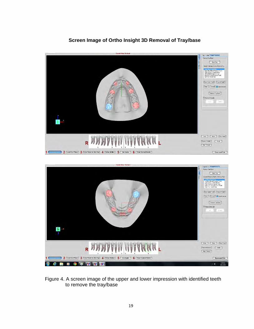
Screen Image of Ortho Insight 3D Removal of Tray/base
Figure 4. A screen image of the upper and lower impression with identified teeth to remove the tray/base

20
Screen image of Ortho Insight 3D Level the Occlusal Plane
Figure 5. Screen image displaying the leveling of the occlusal plane under the model Trimmer section.

21
Screen Image of Ortho Insight 3D Bite Registration Markings
Figure 6. Screen image displaying the CO bite registration that needs to be identified to align the upper cast with the lower cast to check occlusion.

22
Screen Image of Ortho Insight 3D Draw Midline
Figure 7. Screen image displaying the midline.

23
Screen Image of Ortho Insight 3D Finished Model
Figure 8. Screen image of the finished occluded virtual model with trim angles.

24
Screen Image of Ortho Insight 3D Linear Measurements
Figure 9. Screen image displaying different interarch linear measurements made on the virtual digitized models utilizing the scored round bur marks on the teeth.

25
Image of Linear Measurements on Stone Models (control)
Figure 10. Image of Linear Measurements on the stone casts (control) using a 4 in Digital Caliper.

26
Figure 11. Flow diagram of study. Laser scanning performed with Ortho Insight 3D Scanner. Virtual model measurement performed with accompanying software.

27
RESULTS
Research Question 1: Compare the accuracy of bite registration materials in
generating an inter-arch digital occlusion when compared to the stone model
occlusion control.
We started by evaluating the distribution of values in the dataset, therefore,
looking at the properties of raw data. Measurements at each site for each material
were compiled into a single group, and a histogram was plotted. Each material gave
a similar distribution of measurements (Figure 12), but the data is bimodal (different
oral sites), thus, not normally distributed.
Figure 12. Histogram of occlusion measurement data. Data from all sites for the occlusion length (mm) (15 bins of equal size) in each model
were plotted as a histogram for each material.
Subsequently, interaction plots were used to evaluate patterns of relationship
between the mean of the dependent variable distance for the 15 models and the site

28
(as the independent variable) or material independent variable (Figure 11A, B). All
three materials and the control gave very similar mean values for the 15 models at
each site.
A B
Figure 13A, B: Interaction plots of Distance (mm) versus Material by Site measured (A) or versus Site measured by Material (B). Means from the 15 models and 95% confidence intervals shown.
Hence, these observations indicate that within a site, there are no marked trends for
an effect of a material, and no trends for marked differences in variances.
Then, statistical analysis utilizing rm two way ANOVA was used to test the
relationship between measured occlusion distance and material at each model
position. The rm 2 way ANOVA revealed small but statistically significant variance (p
< 0.0001) due to the materials and interaction of the materials with the site
(p<0.0001). In this test, site variance was the bulk of the variance (p<0.0001) since
sites are different in size around the mouth. To crosscheck the 2 way ANOVA, a
parametric one way ANOVA was performed to compare the effect of materials at

29
each site since the 15 values determined at each of the eight sites broadly fit a
normal distribution (Table 2). All but positions 2, 6, 8 showed a significant difference
in mean occlusion distances between one or more materials. For eight ANOVA
tests, a Bonferroni correction for the usual p=0.05 was made and the calculated
value of p=0.00625 was used to reject the null hypothesis of no difference between
any materials.
Table 2. Summary rm one-way ANOVA results.
Next, Tukey’s multiple comparison test (alpha=0.05) was used to examine
differences between materials at each site. However, Tukey’s post test only found a
difference between Stone and Blu-Moose, therefore, raising suspicion about the
result.
The basic conclusion from the post tests with rm two-way ANOVA and cross-
check with rm one way ANOVA using each Sites dataset is that Stone tends to give
lower values than the three bite registration materials, which are indistinguishable.
As we saw earlier in histogram plots (Figure 10), the aggregate data from all
sites in each material group was non-normally distributed but each distribution had a
similar shape. Hence, to make a general comparison of measurements between the
rm ANOVA SITE1 SITE2 SITE3 SITE4 SITE5 SITE6 SITE7 SITE8
Mean (SD) 15.22 (0.35)
18.68 (0.28)
14.96 (0.40)
15.26 (0.39)
31.30 (0.33)
28.71 (0.40)
31.65 (0.41)
27.75 (0.16)
ANOVA p 0.0003 0.0287 0.0004 0.0023 0.0022 0.0078 0.0004 0.0391
Epsilon 0.754 0.776 0.567 0.819 0.794 0.565 0.715 0.838

30
groups, a non-parametric Friedman test was used. This gave a p-value of <0.0001,
indicating highly significant differences between the groups. These results clearly
indicate that Stone and Blu-Moose give lower measurements than Copper Wax and
Byte Right.
Lastly, we compared the spread of the values about the mean to evaluate if
one or more materials differ significantly from the others. The spread of the residual
distributions (the standard distribution, SD) is a relative measure of the precision of
the measurements for each material. Therefore, the lower values for the SD, the
more precise the measurement. Basically, Blu-Moose and Byte Right have
significantly lower variances (SD=0.3719 and SD=0.3811), thus more precise
measurements (narrower distributions) than Copper Wax (SD=0.5035).
Combining it all together, the order of the selection of the materials based on
the results and the above statistical analysis would be:
Blu Moose ≥ Byte Right>Copper Wax

31
Research Question 2: Determine the intra-observer accuracy of repeated inter-arch
measurements?
To test for this, an intraclass correlation coefficient (ICC) for one observer,
consistency was determined for data from each material. This coefficient quantifies
the degree of relatedness between two or more groups. An ideal test would show a
consistency of 1. All tests gave ICC values very close to 1, indicating highly
reproducible repeat measurements by the observer (Table 3).
Table 3. Intraclass correlation coefficients for each material and for all tests.
Material ICC Confidence interval
Stone 0.9994 0.9987 to 0.9997 Copper Wax 0.9986 0.9972, 0.9993 Blu-Moose 0.9985 0.997 to 0.9993 Byte Right 0.998 0.9959 to 0.999 All tests 0.9942 0.9924 to 0.9958

32
DISCUSSION
The main study was designed to evaluate the accuracy of bite registration
materials scanned using an in-office laser surface scanner, Ortho Insight 3D, to
create a virtual occlusion. There are various materials available on the market used
for bite registration of the patient’s dentition. However, each material scans
differently in the laser scanner producing virtual occlusions of differing quality. In
this study, linear inter-arch measurements were made on virtual models created by
three bite registration materials: Blu-Moose, Byte Right and Copper Wafer Wax.
Linear inter-arch measurements were also made directly on the stone models
utilizing digital calipers and served as a gold standard control. Statistical Analysis
utilizing 2 way ANOVA and 1 way ANOVA concludes that Stone (control) gives lower
values compared to the three bite registration materials, since less error is involved
in performing linear measurements using digital calipers on the defined bur marks on
the stone models.
On the other hand, significant differences were found among the three
different bite registration materials. Non-parametric Friedman test concluded that
Blu-Moose gave lower values compared to Copper Wafer Wax. As a result, Blu-
Moose is significantly different than Copper Wafer Wax. Additionally, Blu-Moose
and Byte Right have significantly lower variances, thus more precise measurements
(narrower distributions) than Copper Wax. It is believed that these interarch mean
differences are statistically but not clinically significant and suggest that bite
registration method used for the digital modeling system plays a huge role in
creating accurate virtual models. In a study done by White et. al, dimensional

33
stability of the bite registration material, digital manipulation of the scanned wax bite
registration material or a combination of both was suggested to be the source of
error in accurately reproducing the interarch relationships. White et. al attributed the
errors in dimensional stability of the wax bite to environmental conditions during
shipment, deformation on removal from the mouth, or a distortion on placement in
the flat storage dish for shipment. However, the present study was performed under
controlled environment in which bite registration was scanned after it is taken on the
articulated stone models, thus minimizing the distortion related to shipment
conditions.
Furthermore, there are user errors associated with the digital manipulation of
the scanned bite registration material. All three bite registration materials have
different exposure settings for scanning. The user has to be well aware of the laser
scanner software and should follow the manufacturer’s guidelines for laser scanning
the bite registration materials. Moreover, the user has to be aware of the different
physical and handling properties of the bite registration materials. In the present
study, it was shown that Copper Wax digital occlusion was significantly different from
the other two bite registration materials and the stone (control). This could be
ascribed to the handling properties of Wax since it can be distorted and have large
dimensional changes associated with it. If the clinician chooses to use the wax bite
registration due to its low cost, he or she needs to make sure that the staff is well
trained in registering and laser scanning the bite in in-office laser scanner to
minimize the inaccuracy related to digital representation of the interarch
relationships.

34
Nevertheless, Blu-Moose and Byte Right are both dimensionally stable, rigid
and a bit more expensive compared to Copper Wafer Wax. Therefore, it depends on
the clinician in determining which bite registration material to use.
Also, in the study performed by White et. al, DigiModel software used by
OrthoProofUSA includes a Collision Mapping Tool, which was utilized in the
correction of bite inaccuracies in 6 degrees of freedom. Comparing to the present
study, no manipulation or correction of the occlusion was performed after the digital
models were occluded. An accurate method of recording the patient’s occlusion is
an integral part of any digital system designed to provide accuracy comparable with
traditional plaster models (White, Fallis, & Vandewalle, 2010).
In comparison, in the present study, bur marks were placed on the
labial/alveolar mucosa of the maxillary and mandibular teeth on the stone models.
Impressions of the stone models were then laser scanned along with bite registration
materials. It is suggested that some of the inaccuracies related to interarch mean
measurements can be attributed to the investigator not fully capturing the bur marks
in the laser scanned digital model. For future studies, it is recommended to either
change the width, shape or place some material in marks for it to be definitively
captured.

35
CONCLUSIONS
The present study clearly demonstrated a significant difference between
linear interarch measurements collected from stone models and virtual models.
However, the clinical significance was determined to be minimal depending on the
choice of the bite registration material. Based on the results and the statistical
analysis, the order of the selection of the materials would be: Blu-Moose≥ Byte Right
> Copper Wax. We concluded that dimensional stability and digital manipulation of
the bite registration material by the user/operator plays a huge role in accurate
digital representation of the virtual models. If the operator is fully trained and
knowledgeable about the in-office laser scanner, the user can confidently analyze
and accurately treatment plan a patient based on impression/bite registration
scanned virtual models. In conclusion, Ortho Insight 3D is capable of efficiently
collecting and analyzing patient record data in regards to inter-arch linear
measurements.