Embed Size (px)
Citation preview

Loyola University ChicagoLoyola eCommons
Master's Theses Theses and Dissertations
1967
A Cephalometric Study of Prognathism in theNorth American Negro Male with NormalOcclusionRoland F. ThomasLoyola University Chicago
This Thesis is brought to you for free and open access by the Theses and Dissertations at Loyola eCommons. It has been accepted for inclusion inMaster's Theses by an authorized administrator of Loyola eCommons. For more information, please contact [email protected].
This work is licensed under a Creative Commons Attribution-Noncommercial-No Derivative Works 3.0 License.Copyright © 1967 Roland F. Thomas
Recommended CitationThomas, Roland F., "A Cephalometric Study of Prognathism in the North American Negro Male with Normal Occlusion" (1967).Master's Theses. Paper 2067.http://ecommons.luc.edu/luc_theses/2067

A CEPHALOMETRIC STUDY OF
PROGNATHISM IN THE NORTH AMERICAN NEGRO MALE
WITH NORMAL OCCLUSION
BY
ROLAND F. THOMAS
A Thesis Submitted to the Faculty of the Graduate School
of Loyola University in Partial Fulfillment of
the Requirements for the Degree of
Master of Science
JUNE
1967

LIFE
Roland F. Thomas was born in Duluth, Minnesota on
April 22, 1929.
He was graduated from Morgan Park High School in
June 1946. He attended the University of Minnesota for
three years, then entered the United States Air Force where
he served for five years.
He entered the dental school of Meharry Medical
College, Nashville, Tennessee, in 1956, and received his
Doctor of Dental Surgery degree in June, 1960. During the
following year he served as an intern at Michael Reese
Hospital in Chicago, Illinois.
Upon completion of his internship, he practiced
general dentistry in Chicago until June 1965, after which
he began his graduate studies at Loyola University,
Chicago, Illinois.
ii

ACKNOWLEDGEMENTS
My sincere appreciation is extended to all who have
assisted in making this study possible:
To Joseph R. Jarabak, D.D.S., M.S., Ph.D., Professor
of Orthodontics, who as adviser and teacher, directed my
graduate studies in Orthodontics.
To my colleague and co-worker, Clarence ~ed, D.D.S.,
without whose valuable assistance this work could not have
been completed.
To my wife and children whose encouragement, patience,
and untiring devotion were beyond measure.
iii

--
TABLE OF CONTENTS
CHAPTER PAGE
I. INTRODUCTION AND STATEMENT OF THE PROBLEM: A. Introduction. • • • • • • • • • • • • • • 1 S. Statement of the P~oblem • • • • • • • • •
II. REVIEW OF THE LITERATURE: 4
III. METHODS AND MATERIALS: A. Selection of the Sample •• • • • • • •• 12 B. Collection of the Data •••••••••• C. Measurement Procedures • • • • • • • • •• D. Description of the Selected Landmarks E. Spotting Procedure • • • • • • • • • • • • F. Measurement of the Craniofacial Skeleton • G. Measurement of Prognathism • • • • • • • •
IV. FINDINGS... • • • • • • • • • • •.• • • •• 29 A. Morphology of the Cranial Base • • • • • • B. Measurements of Facial Height • • • • • • C. Measurements of Facial Length • • • • • • D. Measurement of Vertical Facial Angles •• E. Measurement of Horizontal Facial Angles • F. Measurement of the Incisive Angles •••• G. Measurement of the Profile Angles ••••
V. DISCUSSION • • • • • • • • . . • • . . . . • •
VI. SUMMARY AND CONCLUSIONS . . . . . . . . . . . BIBLIOGRAPHY • • • • • • • . . . . . . • •
iv
52
70
75

FIGURE
I.
II.
LIST OF FIGURES
Location of Landmarks • • • • • • • • • • . . Planes used for Angular Measurement • . . . .
PAGE
17
22
III. Measurement of Vertical Height and Maxillary Protrusion • • • • • • • • • • • • • • • •• 25
IV. Facial Diagram Constructed From Mean Values
V.
of Entire Sample •• • • • • • • • • • • •• 51
Comparison of Facial Morphology • (Swedish and Negro)
v
• • • • • • 69

--
TABLE
I.
II.
III.
IV.
V.
VI.
VII.
VIII.
IX.
X.
XI.
LIST OF TABLES
Measurements Reported by Downs and Altemus
Angular Measurements from Bjork's Analysis
Linear Measurements from Bjork's Analysis
Angular Measurements from Lindegard's Study
· .
•
Linear Measurements from Lindegard's Study ••
Linear Craniofacial Skeletal Assessment (Negro sample)
Angular Craniofacial Skeletal Assessment (Negro Sample)
. . .
. . . Posterior Cranial Base Intercorrelations • • •
Posterior Cranial Base Intercorrelations • • •
Cranial Base Correlations with Angular Changes
Dentoalveolar Intercorrelations . . . . . . .
vi
PAGE
32
34
35
36
37
49
50
53
54
55
56

CHAPTER I
INTRODUCTION AND STATEMENT OF THE PROBLEM
A. Introduction:
The morphologic history of mankind is mirrored in the
evolutionary changes of the crainofacial skeleton from the
primitive tree shrew and lemur to modern man. Physical
anthropologists recognized the diverse morphologic patterns
of fossil skulls and used them to classify and delineate
primitive types. Craniometry, a branch of physical anthro
pology, has become an indispensable tool in the comparative
study of primates and in the description and analysis of
fossil remains. The skull has offered more anthropologic
information than all other entities.
Anthropologists have studied skulls for generations
endeavoring to reconstruct the morphologic history of man
and to trace the relationships between the races. Many
believed that skull form remained constant in each race,
and racial classification could be made on the basis of a
calculated cranial index. Campers adhered to this belief
when he established his angle for crainal measurement.
According to Campers, the ancient Greeks had a facial angle
measuring 1000 , the Europeans 800 , and the Negroes 700 •
1
2
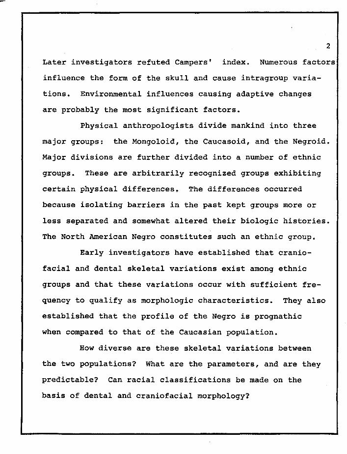
Later investigators refuted Campers' index. Numerous factors
influence the form of the skull and cause intragroup varia
tions. Environmental influences causing adaptive changes
are probably the most significant factors.
Physical anthropologists divide mankind into three
major groups: the Mongoloid, the Caucasoid, and the Negroid.
Major divisions are further divided into a number of ethnic
groups. These are arbitrarily recognized groups exhibiting
certain physical differences. The differences occurred
because isolating barriers in the past kept groups more or
less separated and somewhat altered their biologic histories.
The North American Negro constitutes such an ethnic group.
Early investigators have established that cranio
facial and dental skeletal variations exist among ethnic
groups and that these variations occur with sufficient fre
quency to qualify as morphologic characteristics. They also
established that the profile of the Negro is prognathic
when compared to that of the Caucasian population.
How diverse are these skeletal variations between
the two populations? What are the parameters, and are they
predictable? Can racial classifications be made on the
basis of dental and craniofacial morphology?
B. Statement of the Problem:
The purpose of this investigation is to study the
variations that occur in the craniofacial skeleton of the
North American Negro, and to examine the normal variations
in prognathism in this racial group.
3

...
CHAPTER II
REVIEW OF THE LITERATURE
Anthropologists have always been interested in the
facial profile of man; however, their studies centered on
variations in the dentoskeletal profile as they occur among
races. Early investigations of prognathism were unrelated
to prognathisms which concerned the dentists. The anthro
pologists looked upon prognathism as a characteristic present
only in the upper jaw. The lower end of the face was limited
by the border of the maxillary alveolar process. Since
the Greek word "gnathos" strictly signifies the lower jaw,
the term may be applied to the mand~ble as well.
Prognathism was always associated with a form of
dysplasia until Oppenheim (1928) cited a distinction between
racial and pathological prognathism. In the former the
teeth are in normal occlusion, however both jaws are relative
ly forward to the base of the skull. This is a harmonious
relationship. Pathologic prognathism is not a harmonious
condition. Hitherto no anthropologist had made this dis
tinction.
This study will use the anthropologic interpretation
of prognathism; prominence of the facial skeleton in relation
4

to the brain case.
Numerous reasons have been offered in an effort to
explain and measure the individual and group variations of
prognathism. Campers (1768) made one of the earliest re
corded attempts to analytically assess the facial form and
profile. He established a base line, Campers' line, drawn
from the lower part of the nose (anterior nasal spine) to
the orifice of the ear. A line drawn from the most pro
minent point of the forehead to the alveolar margin of the
maxillary incisor teeth was designated as the facial plane.
The angle formed by Campers' line with the facial plane was
designated the facial angle. According to Campers, the
various races of man and the higher forms of animals could
be identified by the relative degree of prognathism measured
by changes in the facial angle. Although Campers' angle has
been abandoned, it was a first attempt at giving some quan
titative expression to facial variations.
5
The International Congress on Prehistoric Anthro
pology and Archeology in 1882 accepted the Frankfort hori
zontal line as a standard for craniometric measurement. This
line was drawn from the superior margin of the acoustic
meatus to the lowest margin of the left orbit. The facial
angle formed by this plane supplanted Campers' angle as a

measure of racial variation and differentiation.
Investigators defined and measured prognathism in
numerous ways using both linear and angular measurements.
Angle (1899) presented his concept of ideal occlusion pre
dicated on the constancy of the position of the maxillary
first molar. He reasoned that ideal occlusion normally
occurs with a harmonious facial profile. Angle noted that
the so-called "perfect" profile of Apollo, the mythological
god, was in harmony with a straight line at three points;
6
the most prominent points of the frontal and mental eminences,
and the middle of the ala of the nose. He called this line
the "line of harmony". Angle assessed protrusion and re
trusion of the facial parts in relation to this line.
Klaatsch (1909) following Angle1s concept of the
constancy of the first molar, investigated the position of
the molar from a morphologic standpoint. He observed a
more or less constant relationship of this tooth to the key
ridge. He noted that this relationship varied in different
races. In prognathic races the distal roots of the maxillary
first molars are usually on the ridge, and with orthognathism
usually the mesial root. Pfaff (1923) and Oppenheim (1927)
made the same observations.

Hemley (1953) observed that in the prognathous indi-
vidual, the distal curvature of the mesial buccal root of
the maxillary first molar is exaggerated as compared to the
non-prognathous but entirely in harmony with the forward
curve of the key ridge and facial profile. The direction
of the key ridge and the curvature of the tooth roots har-
monize and are true manifestations of individual types.
Simon (1922) rejected Angle's concept of ideal oc-
clusion because it isolated the denture from its cranial
relationships. He related the denture to three planes using
gnathostatic methods. These three planes stand at right
angles to each other; the Frankfort horizontal, the orbital
7
plane, and the raphe-median plane. In an orthognathic denture
the orbitale plane passes through the maxillary canine tooth.
However, in prognathism, the orbital plane passes distal to
the maxillary canine tooth.
Oppenheim (1928) refuted Simon's orbitale canine law.
His anthropologic study of prognathism revealed the position
of the canine to be inconstant, with no definite position
relative to other anatomic structures.
Krogman (1934) corroborated the work of Oppenheim.
He measured 355 adult skulls to assess the reliability of
presumed craniofacial relationships. • He found no dependable

relationship between any facial point, plane, or tooth, but
rather extreme variability.
8
Todd (1932) concluded that prognathism results from
facial growth in excess of cranial extension. He observed
that forward facial extension is partly passive and partly
active. The distinction between the two is registered by the
cuspid which maintains a stationary relationship to the skull
as long as facial extension is passive, but moves forward
with active forward growth of the face itself. It is this
active forward facial growth which is the true prognathism.
Todd contends there is no true prognathism in Negroes or
whites. The face and cranium maintain an equal place in
horizontal forward growth. In whites the cuspid doesn't
maintain its position relative to the skull. It moves
erratically backward as though the forward growth of the
cranium outdistances that of the face, especially during the
second decade.
The studies of variations in the facial build prior
to 1931 were focused on racial differences. The measurements
were mainly craniometric. Roentgenometric methods applied
to living subjects had not been used to any noteworthy extent.
Broadbent designed an x-ray cephalometer (193l) simplifying
accurate measurement of living subjects and opened a new era

9
for research and study.
Bjork (1947) made an anthropological x-ray investi
gation of Swedish children and conscripts using a modification
of the Broadbent cephalometer. Only sketchy morphologic
x-ray studies of the facial skeleton of racial groups had
hitherto been made. He found that individual variations in
the occurrence of prognathism resulted from an interaction
of skeletal parts. Angular changes, especially reductions
in the saddle angle and the articular angle, or an increment
in the chin angle are most noteworthy. Changes in dimensions,
especially a shortening of the cranial base or an extension
of the mandibular body, increase the prognathism.
Cotton (1949) made a comparative analysis of the
facial relationships of North American Negroes. Using t~e
Downs analysis on twenty subjects varying in age from eleven
to thirty-four years, he found measureable differences in
the facial configurations of Negroes and Caucasians with
Negroes having a greater variability from the mean. The most
pronounced differences occurred in the dentures. The skeletal
patterns, on the other hand, showed considerable similarity.
Sassouni (1959) analysed the face from five dimensions,
three of space and two in time (ontogenetic and phylogenetic).
He used four horizontal planes of reference - the supra-

10
orbital plane, the palatal plane, the occlusal plane, and
the mandibular plane. These planes, when extended posteriorly
in a well proportioned face, will converge toward a common
center. An arc drawn from this common center will pass through
nasion,anterior nasal spine, the incisal edge of the maxillary
central incisors, and pogonion in a well balanced orthognathic
face. Prognathic areas of the face will project anterior
to the arc. Sassouni compared composite cephalometric tracings
of white, Negro, and Chinese subjects at eight and twelve
years of age and at the adult level. He found marked racial
differences in the teeth and circumdental areas. Comparing
Negroes with whites he found that Negroes have a shorter
anterior cranial base, the palate has a steeper upward in
clination anteriorly, the mandible is longer, the anterior
lower facial height is larger and the incisor teeth are
more protrusive.
Altemus (1960) used the analyses of Downs and Sassouni
in a cephalo-facial study of North American Negro children.
He compared his findings with three other North American
racial groups, Caucasian, Chinese and Japanese. Comparisons
disclosed that the over-all size of the heads and faces of
the Negro children were larger, the lower facial heights

were relatively larger than the upper facial hei~hts, the
teeth and lips were more protrusive.
11

CHAPTER III
METHODS AND MATERIALS
A. Selection of the Sample:
Fifty Negro males between the ages of twelve and
sixteen years were selected for this investigation. Selection
was 'based on their harmonious facial balance and excellence
of occlusion. Approximately two thousand potential subjects
were examined. Those selected satisfied the following
criteria:
1. Pleasing harmonious facial appearance.
2. Neutroclusion of the teeth when in centric
occlusion.
3. No missing teeth (except third molars).
4. The soft tissues of the oral cavity free of
pathology.
5. No pronounced asymmetry in either dental arch.
6. Overjet and/or overbite of five millimeters or
less.
7. Adjacent teeth in good contact, or with diastemas
not in excess of two millimeters.
8. No axial rotations in the dental arches exceeding
12

twenty degrees.
9. Absence of excessive caries or extremely large
restorations.
13
10. No displacement of any tooth exceeding two milli
meters from the arch.
11. No previous orthodontic treatment.
12. No functional or pathological disorders of the
temporomandibular joints.
13. No speech impediments.
Subjects were selected from all sections of the city
of Chicago to assure a random sampling of the Negro popula
tion in this city.
B. Collection of the Data:
Records consisting of lateral cephalograms, intraoral
roentgenograms, facial photographs, and dental impressions
were taken on sixty-two potential subjects from which fifty
were selected for the final sample.
1. Lateral Cephalograms:
A standard cephalometric unit (Universal) incorporating
a rotating anode was used for the lateral head exposures. Its
high voltage generator was set for exposures at 90KVP and 45
milliamperes. The distance from the focal point of the

roentgen tube to the midsagittal plane was fixed at sixty
inches. The exposures were made with the subject standing,
the median plane of the head parallel to the plane of the
film and at right angles to the central ray. The head was
orientated so that the projected Frankfort horizontal plane
was parallel to the floor. The ear rods, orbital marker,
and nasal guide aided in stabilizing the head in this posi
tion. The teeth were in full occlusion.
Kodak medical x-ray film, eight by ten inch, Blue
Brand, high speed type, was used in cassettes with double
high speed intensifying screens. The exposed films were
processed using the time-temperature method recommended by
the Eastman Kodak Company.
2. Intraoral Roentgenograms:
14
Fourteen periapical x-rays were taken using a stan
dard x-ray machine operated at 65 KVP and 10 milliamperes.
The short cone technique was used with all exposures made at
one-quarter of a second on Kodak ultraspeed dental peri
apical films. The films were processed using the time
temperature method recommended by the Eastman Kodak Company.
3. Plaster Casts:
Plaster casts of the maxillary and mandibular arches
were made for each subject. The impressions were taken with

15
alginate impression material (Supergel) used in a manner
suggested by the manufacturer. Kerr's snow white plaster was
mixed using a motor driven vacuum spatulator and then vibrated
into the impressions. The models were trimmed, soaped and
polished ~o a glossy finish. These were used in a companion
study done by Clarence Red.
4. Facial Photographs:
Frontal and profile views were taken on each subject.
The head was orientated with the Frankfort horizontal plane
parallel to the floor. Exposures were made using a Polaroid
land camera (model 9SA) with a close-up lens (#1) attached.
A Polaroid wink light was used for illumination.
C. Measurement Procedures:
A total of twenty-two reference points for linear
and angular measurements were marked on the cephalograms.
The accuracy of these points has been well documented in the
literature.
Measurements were tested by a process of double
determination carried out by this investigator and one corro
borator. The object of the double determination was to
establish which points give the most accurate measurements,
and to calculate the reliability of the measurements. The

systematic error inherent in the method and the accidental
error can then be determined and reduced.
D. Description of the Selected Landmarks:
16
The selection of reference points was based on the
ease and accuracy with which they could be located, and their
clinical intere~ Midsagittal plane structures were used
when possible. When double projections occurred the mid
point of the two images was selected. Most of the points
correspond to craniometric points; while the remainder are
constructed points (at the point of intersection of lines).
The selected landmarks are shown in Figure I.
1. "A" point (Jarabak) - A point measured two milli
meters anterior to the intersection of a line
drawn from the apex perpendicular to a line
tangent to the root and parallel to the long axis
of the teeth.
2. Anterior Nasal Spine (ANS) - the most anterior
point on the hard palate.
3. Apex of 1 (TR 1) - ~he apex of the root of the
most prominent maxillary central incisor.
4. Apex of r (TR 1) - The apex of the most prominent
mandibular central incisor.

Por ~
rus
an
, :rmtJRE I
n o-r Landmsrks
17
18
5. Articulare (Ar) - The point of intersection of
the dorsal contour of the articular process of
the mandible and the os temporale. The midpoint
was used when double projections gave rise to
two points.
6. "B" Point - The deepest point on the contour of
the alveolar projection, between infrandentale
and pogonion.
7. Gnathion (Gn) - The deepest point on the chin
(the lowest point on the symphysis of the mandi
ble).
8. Gonion (Go) - The lowest posterior and most
outward point of the angle of the mandible. The
point of intersection between the base and ramus
tangents to the mandible was used. The mid
point was used when double projections gave rise
to two points.
9. Infrandentale (In) - The point of transition from
the crown of the most prominent mandibular central
incisor to the alveolar projection.
10. Incision inferius (Ii) - The··incisal point of
the most prominent medial mandibular incisor.
11. Incision super ius (Is) - The incisal point of

the most prominent medial maxillary incisor.
12. Mesial of Crown of b (Mi) - The most prominent
point on the mesial curvature of the mandibular
left first molar projected to the plane of
occlusion.
~3. Mesial of Crown of ~ (Ms) - The most prominent
point of the mesial curvature of the maxillary
left first molar projected to the plane of
occlusion.
14. Nasion eN) - The anterior limit of the fronto
nasal suture.
19
15. Orbitale (Or) - The deepest point on the infra
orbital margin. The midpoint was used when double
projections gave rise to two points.
16. Pogonion (Pg) - The most anterior, prominent
point on the chin.
17. Porion (Por) - The midpoint on the superior margin
of the external auditory meatus, located by means
of the ear rods on the cephalometer. This is a
cephalometric reference point.
18. Posterior Nasal Spine (PNS) - The most posterior
point on the hard palate.
19. Prosthion (Pr) - The transition point between

20
the crown of the most prominent medial maxillary
incisor and the alveolar projection.
20. Sella-Turcia (S) - The midpoint of the horizontal
axis of sella turcia.
E. Spotting Procedure:
The spotting was performed on a transilluminated
tracing table in a darkened room. The cephalograms were
marked by puncturing the reference points with a phonograph
needle test probe. A sheet of cellulose acetate paper was
placed between the tracing table and the headfilm to protect
the table surface and to aid in maintaining a uniform size
perforation at each reference point. The needle probe was
replaced periodically to assure uniformly small perforations.
Spotting sessions were limited to forty-five minute increments
followed by rest periods. This reduced eye fatigue thereby
reducing spotting errors.
All reference points were located by this investi
gator, and then corroborated by two others. Landmarks not
readily identified were determined on a two-out-of-three vote
basis with this investigator and two corroborators parti
cipating.

21
F. Measurement of the Craniofacial Skeleton:
Twenty-two planes were selected for linear and angular
measurements. All planes are at right angles to the film
surface and are defined by two points in the plane of the
film (Figure II). The linear measurements were made by
placing the sharpened points of the vernier caliper into
the reference perforations, in the manner described by Wall
and Grimson (1965). They demonstrated that the ninety-five
percent confidence limits of this technique are plus or
minus one-half millimeter. The angular measurements were
done by drawing planes passing through the reference per
forations on the film. The planes were extended as long as
possible to permit measurement of the angles with a larger
protractor, and thereby increased the accuracy of the mea
surements. All angles were measured to the nearest five
tenths of a degree, the linear measurements to the nearest
one-tenth of a millimeter.
The various parts of the body are contiguous one to
another. There must be a natural correlation of the parts
if the body is to function as an integral unit. If this
premise is valid, a complicated interaction must exist to
maintain this integrity. No structure, no dimension, no

22
No. Points 1 N-S
2 S-Ar
:3 N-Ar
4 N-A.~S
5 N-Pr 6 N_tt A" Pt
7 U-In 8 N-"Bttpt
9 N-Pg 10 N-Gn 11 N-R-TS
12 ANS-~TS
13 Or-Por 14 1s-6 15 Ar-Go 16 Go-nn 17 1n-Pg 18 Is-TRI 19 li-TRI 20 Ar-Pr 21 Pr-Pg 22 "A"Pt-Pg
(PIBncs 21 end 22 not shown)
FIGURE II
PLANZS USED FOR ETGULAR t1EASURE~"Z'TTS

23
spatial relationship exist independently. The entire skele
tal and dentoalveolar arrangement of the cranium, teeth and
jaws exhibit this interaction. Bjork (1947), studying the
Swedish conscripts, concluded that the correlation of parts
may be manifested in angular and/or linear relationships.
Linear extensions may correlate with angular changes, and
conversely, angular spatial relationships with linear adjust
ments.
An assessment of the craniofacial skeleton must
necessarily include both linear and angular measurements.
The reference points and planes selected for this study
satisfy these requirements.
The craniofacial skeleton is divisible morphologically
into numerous components. Cephalometric measurements can
be grouped arbitrarily according to investigative needs.
For this study, it was divided into four parts: the cranial
base assessment, the upper face assessment, the lower face
assessment, and the total face assessment.
1. Cranial Base Assessment:
Nasion was selected as the anterior end-point of the
cranial base and articulare as the posterior end-point.
Nasion to sella (N-S) denotes the length of the anterior
cranial base. Sella-articulare (S-Ar) represents the

posterior cranial base. The cranial base angle N-S-Ar
(saddle-angle) expresses the shape of the cranial base.
2. Upper Face:
The nasion-sella plane defines the upper limits of
the upper face, and the palatal planes defines the lower
border.
3. Lower Face:
24
The lower face includes the area between the palatal
plane and the mandibular plane. This portion is divided
into maxillary and mandibular divisions by the occlusal
plane.
4. Total Face:
The total face includes the combined upper and lower
face dimensions, from the sella nasion plane to the mandibular
plane.
The anterior height of the face was measured between
points nasion and gnathion. The anterior upper face height,
and the maxillary and mandibular lower face heights were
measured on this line (Figure III).
The posterior vertical face heights were measured
using constructed planes (Figure III). This method was used
by Lindegard (1953) in a cephalometric investigation on
Scandinavian adults. A vertical line is drawn perpendicular
· 25
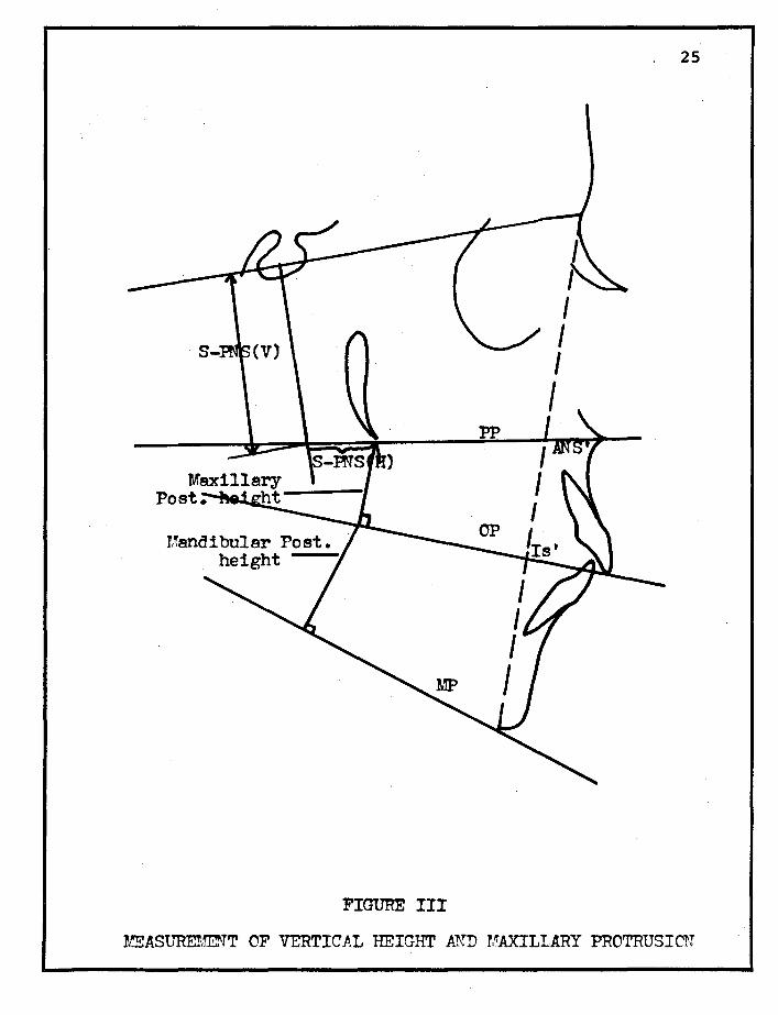
FIGURE III
~ASUREr/~rT OF VERTICAL HEIGHT A.~D MAXILLARY PROTRUSICN
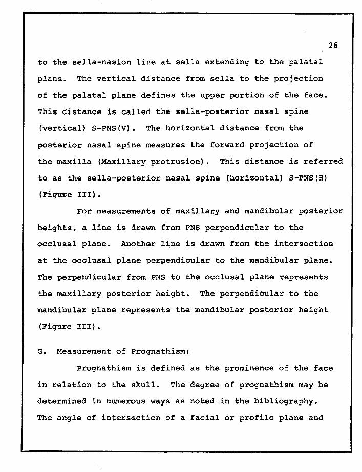
to the sella-nasion line at sella extending to the palatal
plane. The vertical distance from sella to the projection
of the palatal plane defines the upper portion of the face.
This distance is called the sella-posterior nasal spine
(vertical) S-PNS(V). The horizontal distance from the
posterior nasal spine measures the forward projection of
26
the maxilla (Maxillary protrusion). This distance is referred
to as the sella-posterior nasal spine (horizontal) S-PNS(H)
(Figure III).
For measurements of maxillary and mandibular posterior
heights, a line is drawn from PNS perpendicular to the
occlusal plane. Another line is drawn from the intersection
at the occlusal plane perpendicular to the mandibular plane.
The perpendicular from PNS to the occlusal plane represents
the maxillary posterior height. The perpendicular to the
mandibular plane represents the mandibular posterior height
(Figure III).
G. Measurement of Prognathism:
prognathism is defined as the prominence of the face
in relation to the skull. The degree of prognathism may be
determined in numerous ways as noted in the bibliography.
The angle of intersection of a facial or profile plane and

27
the Frankfort plane was most commonly used. Such measure
ments could be used both on living subjects and on skulls.
This was perhaps the most precise and logical index of measure
prior to the advent of x-ray cephalometry. However, roent
genometric measurements from the cranial base can be made
with a greater degree of accuracy than those from the
Frankfort plane.
This study will use the method employed by Bjork
(1947) in his investigation of Swedish children and con
scripts. The method is precise and practical from a technical
aspect, and will permit direct comparison of our sample
with his population.
Denture base or skeletal and alveolar prognathism
will be differentiated and measured. The former defines the
anterior sagittal extension of the maxillary body or mandi
bular base in relation to the cranium; while alveolar prog
nathism is an expression of the protrusion of the alveolar
process in relation to the maxillary and mandibular denture
bases.
Maxillary protrusion is another entity of importance
that was measured in this study. This is a measure of the
position of the maxilla in relation to the anterior cranial
base. Maxillary protrusion is measured by the projection

S-PNS(H). Maxillary denture base prognathism, which will
hereafter be called "basal maxillary prognathism", is an
expression of the proportion between the lengths of the
anterior cranial base (S-N) and of the upper face (the sum
of the maxillary protrusion and the length of the maxillary
body) Lindegard (1953).
The S-N plane (plane 1) was arbitrarily selected as
28
the reference plane for measurement of the profile angles
(Figure II). The N-A plane (plane 3) would be equally suitable
and will be used for the purpose of comparing results with
the Bjork study. The former plane is used more extensively
and may be more meaningful to clinical orthodontists.
Maxillary basal prognathism was measured by the angle
between planes 1-6 (S-N-"A"Pt).
Maxillary alveolar prognathism was measured by the
angle between planes 1-5 (S-N-Pr) in relation to the angle
formed between planes 1-6 (S-N-"A"Pt).
Mandibular alveolar prognathism was measured by the
angles formed by planes 1-7 (S-N-Pg).
Mandibular alveolar prognathism was measured by the
angles formed by planes 1-7 (S-N-In) in relation to 1-9
(S-N-Pg). Mandibular alveolar prognathism may also be ex
pressed as the angle between planes 16-17 (chin angle).

CHAPTER IV
FINDINGS
Any single study of a broad population is necessarily
of finite scope yielding a limited sample of data. The data,
as with data in all fields of science, are to some extent
variable. If the same investigation were repeated using
another sample from the same population, a somewhat different
set of data could be obtained. The most feasible way to
obtain information about populations is to make observations
on samples. Our sample must then be truly representative
of the population from which it was drawn if it is to serve
as an estimator of our population parameters.
Subjects were selected for this investigation pri
marily on the basis of their harmonious facial balance and
excellence of occlusion. Descriptive statistics including
the means, standard deviations, and ninety-five percent
confidence limits were calculated from the measurements taken.
These statistics serve as parameters that characterize this
population.
Statistics are descriptive measures of samples. They
serve only as estimates of population parameters. To qualify
29

our estimate and to assure that it will impart a maximum
amount of information about this selected Negro population,
we include the standard deviation and ninety-five percent
confidence limits. The standard deviation describes the
30
distribution of a sample about the mean. The ninety-five
percent confidence limits attest to the accuracy of the esti
mate. At this degree of confidence we are ninety-five percent
sure that our limits contain the true mean of the population
with normal occlusion.
For this sample the ninety-five percent confidence
limits are given by this expression:
X - 1.96'V ~ <M < X + 1.96~ X = Arithmetic mean
S = Standard deviation
n = Sample size
M = True mean
Measurements of this sample were compared with measure-
ments reported by Altemus (1960) on a similar population of
North American Negro males in the same age group. Altemus
used the Downs analysis in his dental and skeletal assessment
of his sample. We applied the same assessment methods to the
Negro subjeots used in this study and found that our measure-
ments were very similar. These measurements are listed in

31
Table I.
One important objective of this thesis was to observe
and evaluate general relationships between various anatomic
landmarks in the craniofacial skeleton. These relationships
are extremely complex but essential to the understanding of
facial morphology. The inter-relationships between variables
are expressed as coefficient correlation (r) values, and can
be used to give more meaningful information than simple angular
and linear stUdies reported earlier. The electronic computer
is invaluable for problem solving of this type and was used
for this part of the statistical computation. Higher values
usually indicate strong relationships and inter-relationships
between craniofacial and dental components; however, as with
all statistical estimates, there is always the possibility
that the results occurred by chance. Levels of significance
are listed with the r values (Tables VII-X).
Most of the cephalometric measurements used in this
study have been thoroughly tested and are widely used. A
few are of no clinical value1 however all contribute to
the total assessment of the craniofacial skeletal complex.
Some landmarks can not be easily located and therefore show
a wide range of natural variation. Most of them were elimi
nated. It is very difficult to delineate some landmarks,

32
TABLE I
MEASUREMENTS REPORTED BY DOWNS AND ALTEMUS
(Downs' data was obtained from 12 to 17 year old Caucasians Altemus' data was obtained from 12 to 16 year old Negroes)
Facial Angle This angle indicates the degree of recession or protrusion of the mandible in relation to the upper face.
Antle of Convexity (NA-Pg) Th s angle measures the degree of protrusion of the maxillary basal arch in relation toth~·total profile.
A-B Plane This is a measure of the relationship·of the anterior limits of the denture base to -each other' and,~to tbe profile.
Frankfort Mandibular Plane This angle measures the skeletal degree of existing facial divergence.
Interincisal An&le This measures t e degree of procumbency of the incisor teeth.
r to Occlusal Plane This relates the mandibular incisors to their functioning surface at the occlusal plane.
r to Mandibular Plane This expresses the axial relationship of the mandibular incisors to the mandibular plane.
Downs Altemus This Study
Downs Altemus This Study
Downs Altemus
.This Study
Downs Altemus This Study
Mean
87.5 85.5 87.1
0.0 +9.7 +7.7
-4.6 -6.3 -4.1
21.9 28.8 27.8
Downs 135.4 Altemus 119.2 This Study 114.6
Downs Altemus This Study
14.5 27.3 28.7
Downs 91.4 Altemus 99.8 This Study 100.2
Range
82.5 to 77.0 to 80.5 to
+10.0 to +23.5 to +18.5 to
95.0 94.·5 94.0
-8.5 -5.0 -8.5
0.0 to -9.0 +5.5 to -12.0 +4.0 to- ~11 .. 0
17.0 to 12.0 to 17.0 to
28.0 42.5 37.5
130.0 to 150.5 99.5 to 141.5
101.5 to 138.0
3.5 to 12.0 to 15.0 to
20.0 39.5 40.0
81.5 to 97.0 84.5 to 114.5 91.0 to 112.0

33
but these show low coefficients of variation when used to
define a horizontal or vertical plane. The points ANS and PNS
used to determine the length of the palatal planes fall in
this category. These two points are more reproducible ver
tically than horizontally. The images of the bone in these
areas are obscure when followed horizontally, but the out
line of the nasal floor indicates their vertical positions.
The descriptive statistics for this sample are compared
with results published by other investigators. These mea
surements are listed in Tables II through V.
The linear measurements from our study are listed
with both the twelve year old boys and the adult samples
published by Bjork and Lindegard.
The investigation by Lindegard (1953) was done on
the same subjects originally collected by Bjork for his study
of prognathism in 12 year old boys (Bjork 1947). The subjects
were between 20 and 21 years of age at the time of the Linde
gard investigation. Reference will be made to the Lindegard
sample, to the Bjork sample, and to the Swedish sample.
These distinctions are made only because their measurement
methods were different in certain areas. The "Swedish sample"
is used when both investigators report

34
TABLE II
ANGULAR MEASUREMENTS FROM BJORK'S ANALYSIS
(Bjork's data is obtained from 12 year old Swedish boys and 20 year old Swedish conscripts)
Bjork This Study
Angle bet.Slleen Boy Conscripts Planes Nos. Mean S.D. Mean. S.D. Mean. S.D.
1-2 122.9 4.9 123.0 5.3 125.9 5.9 1-3 18.0 1.8 18.5 2.1 17.3 1.9 1-4 85.8 3.8 88.2 4.2 88.5 3.4 3-5 65.5 3.2 66.4 3.7 71.3 3.4
13-5 91.5 3.7 90.8 3.6 96.3 3.5 1-5 83.7 3.7 84.8 4.1 88.3 3.4 1-7 80.0 3.7 82.3 4.4 83.9 2.9 1-9 78.9 3.6 81.7 4.4 79.6 3.6 1-13 5.0 3.1 4.6 2.6 8.0 3.5 1-12 7.8 2.9 7.8 3.4 5.7 3.3 1-14 19.4 3.9 16.3 4.4 18.0 3.5 1-16 37.0 5.2 36.5 6.5 35.7 4.8
12-16 29.2 5.4 28.9 6.4 30.1 4.6 12-14 11.7 3.8 8.8 4.0 12.5 3.4 14-16 17.5 4.8 20.4 5.6 17.6 3.5 18.19 128.5 8.8 137.4 11.8 114.6 8.5 14-18 . 58.0 5.0 64.0 6.6 53.2 5.9 14-19 70.5 6.2 73.6 7.9 61.5 6.3
5-21 166.9 6.0 171.3 7.8 157.5 6.4 6-22 173.9 5.3 177.0 7.0 172.7 6.4 2-15 143.0 6.2 143.3 6.9 146.3 5.9
15-16 131.1 6.1 130.9 7.3 123.8 5.4 16-17 68.6 5.4 64.2 6.4 81.6 7.3
Mass angle 397.0 6.2 397.3 6.2 395.9 4.9

35
TABLE III
LINEAR MEASUREMENTS FROM BJORK r S ANALYSIS
(Bjork r s data is obtained from 12 year old Swedish boys and 20 year old Swedish conscripts)
Bjork This Study
Measurement Boy Conscripts Points Mean S.D. Mean S.D. Mean S.D.
N-S 68.7 3.0 73.2 3.3 72.6 3.7
S-Ar 34.4 2.9 37.0 3.3 36.2 3.3
N-Ar 91.9 3.9 98.1 4.4 98.9 5.6
N-ANS 50.0 2.7 55.5 3.1 54.0 3.1
N-PNS 69.6 2.9 75.2 3.1 74.6 3.7
"" N-Pr 65.9 3.5 74.4 4.2 73.1 3.9
Ar-Go 42.1 3.6 53.2 5.2 46.1 4.4
N-Gn 113.1 5.4 128.3 6.7 127.6 6.2
N-In 84.7 3.9 93.6 5.0 94.7 4.1
ANS-PNS 51.7 2.8 56.8 3.2 55.4 3.1
Ar-Pr 88.0 4.4 96.2 4.7 102.4 5.1
Is-6 29.7 2.8 28.0 2.9 34.9 2.5
Ii-b 24.3 2.3 22.7 2.4 28.5 2.8
Pg-Go 72.8 4.1 80.6 5.2 85.1 6.6

TABLE IV
ANGULAR MEASUREMENTS FROM LINDEGARD'S STUDY
(Lindegard's data was obtained from Bjork's boy sample at 21 years of age)
36
Angle between Lindegard This Study Planes Nos. Mean S.D. Mean S.D.
1-6 82.4 3.6 82.84 3.7
1-9 80.7 3.9 79.58 3.6
1-12 7.6 3.1 5.66 3.3
1-16 35.4 6.2 35.73 4.8
12-16 28.0 5.8 30.14 4.6
12-14 8.7 3.6 12.46 3.5
14-16 19.6 5.3 17.64 3.6
18-19 130.3 9.0 114.64 8.5
15-16 127.7 6.7 123.79 5.4
16~17 65.6 6.4 81.57 7.3 r

Points Measured
N-S
N-ANS
ANS'-Is'
PP-OP
Is'-Gn
OP-MP
N-Gn
TABLE V
LINEAR MEASUREMENTS FROM LINDEGARD'S STUDY
(Lindegard's data was obtained from Bjork's boy sample at 21 years of age)
(Millimeters)
37
Lindegard This Study Mean S.D. Mean S.D.
73.9 3.3 72.60 3.7
56.3 3.0 53.97 3.1
30.8 2.9 28.69 2.6
22.7 2.7 18.58 2.5
72.8 5.6 43.67 3.1
27.0 3.5 26.65 4.3
128.9 6.3 127.61 6.2
S-PNS(H) 14.0 3.1 16.04 3.6
S-PNS (V) 48.3 2.5 46.84 3.4
Pg-Go 81.9 5.2 85.11 6.6

38
Accurate comparisons of the linear morphologic chara
cteristics of this sample to those in the Lindegard and
Bjork study cannot be made for the following reasons: there
is no way of assessing accurately what growth changes contri
buted to the linear differences. The lower age limit of our
sample coincides with the onset of the pubertal growth spurt;
and at the upper limit, most can be expected to achieve some
additional growth. The ages of our subjects were as follows:
Subject's Age No.
12 years 3
13 years 16
14 years 11
15 years 11
16 years 9
Angular comparisons are less critical. Growth patterns
or directions, once established, change very little Brodie
(1941). There were no significant angular differences between
the Bjork boy and adult samples with the exception of the
incisive angles. The mandibular incisors became less prog
nathic with mandibular growth. This was found in the re
ducbion in the incisive angles.

39
COMPARATIVE ASSESSMENT OF CRANIOFACIAL MORPHOLOGY
A. Morphology of the Cranial Base:
The posterior cranial base angles measured in this
sample are greater than those observed by Bjork. The mean
values for the saddle and articulare angles exceed the values
of the Bjork sample by approximately three degrees. Bjork
found a value of 123.0 degrees for the saddle angle, the
value in this sample was 125.9 degrees. The articulare angle
values are 143.3 degrees in the Bjork sample and 146.3 degrees
for this study. The angle formed by the S-N plane with the
N-Ar plane measured 18.5 degrees in the Bjork study and
17.3 degrees in this sample. This cranial base angle changes
inversely with changes in the saddle angle.
The linear measurements of the cranial base cannot be
accurately compared. The additional length that growth may
contribute must be considered.
The gonial angle, though not a cranial base angle, is
generally considered in the assessment of the posterior
cranial base profile. This angle expresses the relationship
of the ramus to the body of the mandible. Bjork reported
a value of 130.9 degrees, this study 123.8 degrees. The
difference is highly significant. It indicates that the
relationship between ascending ramus and body is more acute
in our sample.
B. Measurements of Facial Height:
1. The Upper Face:
The upper face was defined as that portion bounded
by the sella-nasion plane superiorly and the palatal plane
inferiorly. Measurements from N to ANS and N to PNS are
respectively. 54.0 millimeters and 74.6 millimeters in this
study. Bjork reported the N to ANS measurement 55.5 milli
meters and S to PNS 75.2 millimeters for his adult sample.
40
The N-ANS dimension represents the anterior upper face height.
The mean value in this sample was 1.5 millimeters le'ss'
than the adult sample measured by Bjork.
The posterior upper face height was measured in the
manner described by Lindegard (1953). Values reported by
Lindegard for the posterior upper face height were 48.3
millimeters, in this study the height was 46.8 millimeters.
The posterior face height is 1.5 millimeters shorter in our
sample when measured from the cranial base.
2. The Lower Face:
The palatal plane limits the upper border of the
lower face and the mandibular plane the lower.

The assessment of this portion of the face was made
in the manner described by Lindegard (1953). Lindegard
reported a total lower anterior face height (ANS'-Gn) of
72.8 millimeters. The maxillary and mandibular anterior
face heights were 30.8 millimeters and 42.3 millimeters,
respectively. The lower anterior face height in this study
was 72.4 millimeters; the maxillary anterior height being
28.7 millimeters, and the mandibular anterior face height
being 43.7 millimeters. Definitive conclusions can not be
made here; however, we can expect an increase in lower face
height in this Negro sample as the faces reach maturity.
41
The 72.8 millimeters value reported by Lindegard on his adult
sample will undoubtedly be exceeded in this sample. A com
parative study using Negro adults is indicated for an
accurate assessment of this dimension.
Lindegard found mean values of 22.7 millimeters for
the posterior maxillary height (PP-OP) and 27.0 millimeters
for the posterior mandibular heights (OP-MP). This research
reports mean values of 18.6 millimeters and 26.7 millimeters,
respectively, for the maxillary and mandibular posterior
heights.
The maxillary lower posterior height is four milli
meters smaller in this sample. Comparisons again are difficult

because of the age differences. Perhaps what is most im
portant is the relative proportions of the anterior to the
posterior facial heights in each of the samples. The pro
portions will change little with growth and can probably
be more accurately compared with the horizontal angular
assessment of the face.
3. The Upper and Lower Face:
42
This dimension measured on the N-Gn plane was re
ported to be 128.3 millimeters for the Swedish sample; it is
127.6 millimeters in this sample. The measurements from
nasion to the maxillary and mandibular alveolar margins
were 74.4 millimeters (N-Pr), and 93.6 millimeters, (N-In),
respectively, for the Swedish sample. This study reports
values of 73.1 millimeters (N-Pr), and 94.7 millimeters
(N-tn), respectively. In the assessment of this facial
entity the horizontal angular comparisons will be more en
lightening.
c. Measurements of Facial Length:
The anterior cranial base lengths (the upper limits
of ~he face) were strikingly similar in the two samples.
The maxillary body length, defined as distance ANS-PNS,
measured 56.8 millimeters as reported by Bjork; the mean

43
for this group was 55.4 millimeters. As stated earlier the
error of measurement for this dimension is large because the
exact perimeters of the landmarks are poorly defined.
The plane Ar-Pr measures the facial skeleton at one
of its broadest antero-posterior dimensions. The Bjork
sample gives values of 96.1 millimeters, those in this sample
102.4 millimeters. This difference of 6.3 millimeters is very
significant since it lies at the point at which maxillary
alveolar prognathism is measured.
The protrusion of the maxilla as measured by S-PNS{H)
had a mean value of 14.0 millimeters as reported by Linde
gard; the mean value in this sample was 16.0 millimeters.
Maxillary prognathism is the sum total of maxillary pro
trusion and the projected length of the maxilla. The max
illary protrusion is two millimeters greater in this sample;
however, the maxillary body length is 1.4 millimeters shorter.
Again we can only speculate as to what affect growth will
have on this dimension.
The measurement from incision inferius to the mesial
of the mandibular first molar was 22.7 millimeters in the
Bjork sample; this sample was 28.5 millimeters. The incision
superius to maxillary first molar measurements were 28.0
millimeters and 34.9 millimeters, respectively, for the

44
Bjork sample and this study. These are highly significant
differences. This can be attributed to the fact that the
teeth in the Negro population have greater mesial-distal
diameters. These are a subject of a thesis by Clarence Red.
The mandibular body length also differs significantly
between the two samples. Bjork reported the body length
from Pg-Go 72.8 millimeters and 80.6 millimeters, respective
ly, for his boy and adult samples. This sample establishes
values of 85.1 millimeters. Considering expected growth,
the corpus of the mandible in this sample is significantly
larger now with a future increment increase. expected.
D. Measurement of Vertical Facial Angles:
All of the vertical angles along the skeletal profile
express the antero-posterior relationship of the face. to
the cranium. Morphologic differences in the two populations
become readily apparent upon examinations of these angular
values. The Negro sample has larger mean values in the
dentoalveolar areas but shows close similarity in the denture
bases. The Swedish sample has a mean value of 84.4 degrees
for the angle of maxillary alveolar prognathism (1-5), and a
mean maxillary basal prognathism angle (1-6) of 82.4 degrees.
The angle of maxillary alveolar prognathism determined in
this study was 88.3 degrees, the angle of maxillary basal

45
prognathism was 82.3 degrees.
Mandibular basal prognathism was measured at the
intersection of the cranial base plane and the nasal pogonion
plane (1-9). Supramentale is generally considered the anterior
limit of the mandibular denture base; however, in our con
sideration of prognathism we are concerned with the most
prominent contour on the profile in relation to the cranium.
Pogonion then becomes the logical choice. The basal mandi
bular prognathism angle for the Bjork sample was 81.7 degrees
(1-9), and the alveolar mandibular prognathism angle (1-7)
was 82.3 degrees. This study established angular values of
79.6 degrees and 83.9 degrees, respectively, for mandibular
basal and alveolar angles of prognathism. It is interesting
to note that for this study the basal prognathism is less
than that reported by Bjork while the alveolar prognathism
was greater. Again there is the realization of future growth
which will, if directed horizontally, diminish the difference.
Two factors will influence the alveolar prognathism values
between the samples, future anticipated growth of the mandi
ble, and the concomitant uprighting of the mandibular incisor
teeth. It is. not pO$sible to accurately predict the amount
of change that will occur, but some increase in the angle
(1-7) can be expected.

46
The chin angle probably expresses the alveolar pro
cumbency more accurately than measurements from the cranial
base. Very significant differences are apparent here. Bjork
reports a chin angle of 64.2 degrees for his adult sample;
the measurement for the Negro sample was Sl.S degrees. This
sample has a mandibular alveolar angle of prognathism which
greatly exceeds the Swedish sample. Alveolar prognathism
on the basis of comparisons between the Swedish and Negro
populations sets out the Negro population as being definitely
more prognathic.
E. Measurement of Horizontal Facial Angles:
Bjork reported measurements of S~S degrees between
the palatal and mandibular planes (12-16), and 20.4 degrees
between the occlusal and mandibular planes (14-16). He found
the mandibular plane angle (1-16) to be 36.5 degrees. The
measurements for this sample were 12.5 degrees between the
palatal and occlusal planes, 30.1 degrees between palatal
and mandibular planes, and 17.6 degrees between the occlusal
and mandibular planes. The mandibular plane angle was 35.7
degrees. The horizontal angles reflect the relative height
proportions between the anterior and posterior aspects of the
face. These relationships change little as comparisons

47
between the Bjork boy and Lindegard adult measurements show.
We recall that both investigators measured the sarne subjects
but at different ages.
F. Measurement of the Incisive Angles:
The incisive angles also mirror the alveolar promi
nence of this'sample'~ .Bjork.reported values of 137.4
degrees for the interincisal angle (18-19), 64.0 degrees
between the maxillary incisors and the occlusal plane (18-14),
and 73.6 degrees between the mandibular incisors and the
occlusal plane (19-14). The angles in this sample are more
acute, measuring 114.6 degrees for the interincisal angle,
53.2 degrees between the maxillary incisors and the occlusal
planes and 61.5 degrees between the mandibular incisors and
the occlusal plane. These statistics are consistent with
earlier comparisons of alveolar procurnbency.
G. Measurement of the Profile Angles:
The profile angles show considerable differences
between the two samples. The angle N-Pr-Pg (5-21) was 171.3
degrees in the Bjork sample and 157.5 degrees in this study.
The angle N-"A"Pt-Pg (6-22) was 177.0 degrees in the Bjork
sample and 172.7 degrees in this study. The most significant
difference here is the alveolar measurement at prosthion,
a difference of 13.8 degrees. This is another indication
of the alveolar prominence of the Negro sample. Linear
and angular measurements for the entire sample are listed
in Tables VI and VII.
48

49
TABLE VI
LINEAR CRANIOFACIAL SKELETAL ASSESSMENT (Millimeters)
Standard 95% Confidence Limits Measurement Mean Deviation Low High
N-S 72.60 3.7 71.58 73.62 S-Ar 36.15 3.3 35.24 37.07 N-Ar 98.92 5.6 97.38 100.46 N-ANS 53.97 3.1 53.12 54.83 N-PNS 74.59 3.7 73.56 75.61 N-Pr 73.07 4.0 71.96 74.47 N-"A"Pt 59.52 4.3 58.32 60.72
ANS'-Is' 28.69 2.6 27.98 29.41 S-PNS (V) 46.84 3.4 45.90 47.78
pp-op 18.58 2.5 17.88 19.28 Ar-Go 46.13 4.4 44.92 47.34
Is'-Gn 43.67 3.1 42.81 44.53 OP-MP 26.65 4.34 25.45 27.85
N-Gn 127.61 6.2 125.88 129.34 N-In 94.70 4.1 93.55 95.84 N-tlB"Pt 107.36 5.7 105.77 108.95 N-Pg 120.58 5.73 118.99 122.17
ANS-PNS 55.44 3.1 54.60 56.29 Ar-Pr 102.39 5.1 100.99 103.80 Is-6 34.92 2.5 34.2 35.62
S-PNS (H) 16.04 3.6 15.03 17.05 1-NA 9.14 3.5 8.17 10.11
Ir-'6 28.50 2.82 27.72 29.28 Pg-Go 85.11 6.6 83.28 86.94
1-NB 10.97 2.8 10.20 11.73 Pg-NB -0.4 1.62 -0.80 0.09

50
TABLE VII
ANGULAR CRANIOFACIAL SKELETAL ASSESSMENT (Degrees)
Standard 95% Confidence Limits Planes Mean Deviation Low High
1-2 125.88 5.9 124.26 127.50 1-3 17.31 1.9 16.79 17.83 1-4 88.46 3.4 87.51 89.41 3-5 71.34 3.4 70.39 72.29
13-5 96.31 3.5 95.35 97.28 1-5 88.31 3.4 87.37 89.24 1-6 82.84 3.7 81.82 83.86 1-7 83.87 2.9 83.06 84.68 1-8 79.63 2.9 78.81 80.45 1-9 79.58 3.6 78.59 80.57 1-10 76.13 3.2 75.25 77.01 1-11 40.42 2.5 39.73 41.11 1-13 8.01 3.5 7.04 8.89 1-12 5.66 3.3 4.74 6.58 1-14 17.97 3.6 16.98 18.96 1-16 35 ~.73 4.8 34.40 37.06
12-16 30.14 4.6 28.87 31.41 12-14 12.46 3.5 11.50 13.43 14-16 17.64 3.6 16.65 18.63 13-16 27.75 5.0 26.37 29.13 18-19 114.64 8.5 112.28 117.00 14-18 53.18 5.9 51.56 54.80 14-19 61.47 6.3 59.72 63.22
6-18 25.90 7.1 23.92 27.88 8-19 35.97 5.2 34.52 37.42 1-18 109.12 6.3 107.37 110.86
13-18 117.18 5.8 115.57 118.79 16-19 100.17 5.3 98.70 101.64
5-21 157.50 6.4 155.27 159.28 6-22 172.73 6.5 170.94 174.52 2-15 146.29 5.94 144.64 147.94
15-16 123.79 5.4 122.29 125.29 16-17 81.57 7.3 79.54 83.60
Mass angle 395.93 4.9 394.57 397.30

51
FIGUF.E IV
FACT AL DIAG!Wf. CONSTRUCTED FRm{ MEAlIT VALUES OF mTIRE SAMPLE

CHAPTER V
DISCUSSION
Evaluation of the data revealed measurable differences
between the craniofacial skeletons of the Swedish and Negro
samples. These differences, however, are outnumbered by
the numerous similarities. Broad variations occur within
each group, and generally certain mean values for one group
will fall within the normal range of the other.
The variations that occur among the Swedish population
and their effect on the craniofacial skeletal morphology
have been reported. We now center our discussion on the
variations observed in this investigation.
In assessing the variations and the interactions of
the total craniofacial skeleton, we use a statistical tool
ncoefficient of correlationn discussed in Chapter III. The
coefficient of correlation values are listed in Tables VIII-X.
As reported earlier, the saddle and articulare angles
each have mean values three degrees larger than those in the
Swedish group. This angular difference of six degrees is
also consistent with the longer cranial base and facial length
seen in our sample.
52

53
TABLE VIII
POSTERIOR CRANIAL BASE INTERCORRELATIONS
Arti-Angles and Saddle cu1are Gonia1 Angle
Planes Angle Angle Ang!e Sum S-N
Saddle angle 1.000 -0.718 -0.112 0.204 -0.094
Articu1are angle -0.718 1.000 -0.271 0.052 0.111
Gonia1 angle -0.112 -0.271 1.000 0.636 -0.266
Angle Sum 0.204 0.0518 0.636 1.000 -0.268
S-N -0.095 0.111 -0.266 -0.268 1.000
S-Ar 0.236 -0.130 -0.511 -0.434 0.207
N-Ar 0.435 -0.273 -0.461 oloO.321 0.724
Ar-Go 0.120 -0.121 -0.364 -0.403 0.210
Go-Pg 0.305 0.056 -0.705 -0.340 0.445
Chin angle 0.123 0.039 0.016 0.083 -0.033
Level of Significance
.05 .01
.275 .365

54
TABLE IX
POSTERIOR CRANIAL BASE INTERCORRELATIONS
Angles and Chin Planes S-Ar N-Ar Ar-Go GO-Pg Angle
Saddle angle 0.236 0.298 0.120 0.305 0.123
Articu1are angle -0.130 -0.277 -0.121 0.056 -0.039
Gonia1 angle -0.511 -0.461 -0.364 -0.705 -0.016
Angle Sum -0.434 -0.321 -0.403 -0.340 0.083
S-N 0.207 0.724 0.210 0.445 -0.033
S-Ar 1.000 0.704 0.051 0.602 -0.047
N-Ar 0.704 1.000 0.201 0.688 -0.024
Ar-Go -0.045 0.201 1.000 0.290 0.080
Go-Pg 0.019 0.688 0.290 1.000 0.138
Chin angle -0.047 0.024 0.080 -0.138 1.000
Level of Significance
.05 .01
.275 .365

55
TABLE X
CRANIAL BASE CORRELATIONS WITH ANGULAR CHANGES
Angles and Planes 1-5 1-9 1-14 1-16 16-19 5-21
Mass angle -0.343 -0.669 0.696 0.979 -0.203 -0.444
Articu1are angle 0.301 0.202 -0.024 0.038 -0.475 -0.199
Saddle angle -0.538 -0.405 0.298 0.161 0.075 0.172
Chin angle 0.232 -0.124 0.114 0.028 0.528 -0.467
Gonia1 angle -0.060 -0.393 0.339 0.679 -0.194 -0.374
S-N -0.202 -0.012 -0.022 0.282 0.201 0.194
S-Ar -0.027 0.230 -0.166 0.450 0.054 0.302
Ar-Go -0.230 0.321 -0.261 -0.373 0.293 0.056
Go-Pg -0.064 0.322 -0.182 -0.403 -0.146 0.475
Level of Significance
.05 .01
.275 .365

56
TABLE XI
DENTOALVEOLAR INTERCORRELATIONS
Angles and Planes Is-6 Ii-6
ANS-PNS 0.342 0.194
Is-6 1.000 0.756
S-PNS(H) -0.100 0.134
1-NA 0.310 0.283
Ii-6 0.756 1.000
Pg-Go -0.100 -0.177
r-NB 0.502 0.471
Pg-NB -0.422 -0.410
1-5 0.055 -0.099
1-6 -0.031 -0.143
1-7 -0.077 -0.150
1-9 -0.220 -0.336
18-19 -0.496 -0.507
6-18 0.416 0.420
13-18 0.230 0.239
16-19 0.353 0.364
5-21 -0.424 -0.283
6-22 -0.258 -0.163
16-17 0.372 0.325

57
The saddle and articulare angles have a highly signi
ficant negative correlation, but the saddle angle assumes
the dominant role in craniofacial skeletal morphology. The
articulare angle has significant correlations with maxillary
protrusion and maxillary prognathism. The saddle angle,
however, is highly correlated with the cranial and mandibular
bases and the total craniofacial skeletal profile (Tables
VIII-X). It is negatively correlated with the angles of
prognathism. As this angle decreases, the face becomes more
prognathic. Bjork first reported this observation in 1947.
Maxillary basal prognathism, as pointed out earlier,
is a function of maxillary protrusion. Thus maxillary pro
trusion, as measured by S-PNS(H) , is also negatively corre
lated with the saddle angle.
The cranial base length (N-Ar) increases with the
saddle angle. This can be observed intuitively; however, the
anterior cranial base is not even probably correlated with
changes in the saddle angle.
Neither the articulare angle nor the saddle angle
has a probable correlation with the mass angle summation.
The gonial angle in this investigation was 123.8
degrees, 7.1 degrees less than that reported by Bjork. Our
measurement is close to the value of 122.4 degrees as reported

58
by Ashley (1966) cin his study of adult Caucasian males.
The gonia1 angle is very significantly correlated with
the morphology of the lower face (Table VIII). A large gonia1
angle is seen in association with a large mandibular plane
angle, a high FMA, and a steep occlusal plane. The mandibular
alveolar height increases in the anterior region, and the
mandibular profile becomes more retrognathic.
There is a very high negative correlation between the
gonia1 angle and the mandibular body length. This is in con
tradiction to measurements made by Lindegard (1953) who found
no such correlation in his sample. The correlation coef
ficient of -0.705 is very significant. It indicates that
the mandibular body length gets shorter as the gonia1 angle
increases. Conversely, as the gonia1 angle decreases the
mandibular length increases. This shortening of the mandi
bular body length contributes to the retrognathic mandibular
profile associated with a large gonia1 angle.
Significant negative correlations exist between the
gomal angle and the posterior cranial base and ramus lengths.
The facial length, as measured from articu1are to prosthion,
also shows significant negative correlation to the gonia1
angle. This in effect means that the posterior facial height
decreases as the gonia1 angle increases, and the facial length

59
shortens. These interactions contribute to the retrognathic
mandibular profile.
It must be pointed out that with a decrease in the
gonial angle the mandible becomes more prognathic. The above
structures that decreased in length with retrognathism in
crease in length changing the mandibular facial profile.
The mass angle is the summation of the posterior cranial
base angles (saddle angle, articular angle and gonial angle).
The sum of these three angles is an indicator of the direct
ion of growth of the posterior cranial base. The Negro
sample had a mass angle value similar to that of the Swedish
group (Table II).
The summation of the posterior cranial base angles is
significantly and positively correlated with the gonial
angle. The gonial angle has a weighty influence on the
craniofacial morphology of this sample.
The mass angle value relates more information about
the craniofacial morphology of this sample than all other
measurements investigated. Three angles are involved here;
but only two, the saddle and gonial angles, have significant
correlations with other facial entities (~able VIII)~
The mass angle total of the Negro sample is similar
to that of the Swedish group, but the angles contributing to

the mass angle vary significantly. The differences have
already been discussed. A large mass angle is seen in
connection with a steep mandibular plane angle, a large
60
gonial angle, a large FMA, steep occlusal and palatal plane
angles, and a long anterior face height. The posterior cranial
base, the mandibular body length, the ramus length, and
the vertical angles of prognathism are significantly and
negatively correlated to changes in the mass angle value.
Conversely, a small mass angle would be associated with facial
prognathism, a reduced anterior and posterior facial height
and sagittal extension of the facioskeleton.
Bjork reported that an inverse relationship exists
between the sagittal and vertical extensions of the face.
That relationship is strongly evident here. The mass angle
value has momentous import and is truly a mirror of the
craniofacial profile.
Tbe chin angle (16-19) was used in the manner described
by Bjork and Lindegard to measure mandibular alveolar progna
thism. An important distinction must be made here. Mandi
bular alveolar prognathism, as measured by the chin angle,
is a measure of the prominence of the mandibular alveolar
process in relation to its base. This is a dentoalveolar
protrusion unrelated to the prognathism as earlier defined.

61
Dentoalveolar protrusion is very pronounced in our
sample. This protrusion is not correlated with mandibular or
maxillary prognathism; however, it is very positively
correlated with measurements of the dental units (Table XI).
As measurements from the first molars to the incision points
increase, there is a concomitant decrease in the incisal
angles expressing their procumbency. There is a strong
probability that this dentoalveolar procumbency is a function
of the relationship of tooth size to denture base. Our
earlier comparisons with the Swedish sample pointed out very
significant dental arch length differences as measured from
the mesial of the first molars to the incision points. The
tooth morphology of this sample will be discussed in greater
detail in a companion thesis by Clarence Red.
The inverse relationship between the sagittal and
vertical extensions of the face is readily apparent upon
examination of the mandibular plane angle (1-16). Changes
in this angle correlate positively with vertical extensions
of the face and negatively with sagittal extensions. A large
mandibular plane angle is associated with a large gonial
angle, a large mass angle, increased vertical height of the
face, and smaller vertical angles of prognathism. There is
a reduction in the cranial base length, body length, ramus

62
height, and maxillary protrusion. The palatal, occlusal, and
mandibular planes become more divergent. Diminution of the
mandibular plane angle reverses this morphologic pattern.
It is interesting to note, however, that no correlation exists
between the saddle angle and facial height (N-Gn), even though
both are related to prognathism. Lindegard observed this
lack of correlation in his study.
The Frankfort plane, as measured from the sella nasion
plane (1-13), had a mean value of 8.0 degrees in this study.
Bjork reported 5.0 degrees and 4.6 degrees, respectively,
for his boy and conscript samples. This difference of three
degrees is in agreement with the saddle angle difference
between the two samples. The anterior cranial base appears
to have an upward inclination anteriorly. Such an inclina
tion would in effect reduce the angular measurement of facial
prognathism since the naso-glabellar area would move upward
and backward; but the facial profile would appear more
prominent.
Sassouni (1953) found that the cant of the palate in
Negroes had a steeper inclination anteriorly when compared
to that of Caucasians. A comparison of measurements in this
report corroborates the Sassouni findings.

63
It is very important to note that angular observations
of the palatal plane and anterior cranial base inclinations
cannot be confirmed by our linear measurements in this study.
We would expect to find proportional differences in anterior
height, but the additional growth expected in this sample
cannot be accurately assessed. Further study on adult sub
jects will be necessary before definite comparisons can be
made.
Positive correlations exist between upper and lower
sagittal extensions of the face. A large anterior cranial
base is associated with a large mandibular body, a large
maxilla, and a long facial length as measured from articulare
to prosthion. Such a face is not prognathic. This face has
a large saddle angle, a long anterior vertical face height,
and relatively horizontal palatal, occlusal, and mandibular
planes.
Prognathism (prominence of the face in relation to
the skull) has been measured in much the same manner for
centuries. Todays roentgenographic techniques allow some
modifications, but the concept is the same. The angle formed
by the intersection of a facial plane with a horizontal
plane still prevails as the best expression of facial
prominence.

64
Bjork measured maxillary prognathism at the inter
section of the cranial base plane with the nasion-prosthion
plane (S-N-Pr). He measured mandibular prognathism at the
intersection of the cranial base plane and the nasion-pogonion
plane (S-N-Pg). Maxillary prognathism, as reported by Bjork,
was 83.7 degrees for the boy sample and 84.8 degrees for the
conscripts. Mandibular prognathism was 78.9 degrees for the
boy samples, and 81.7 degrees for the conscripts.
The angle of maxillary prognathism for the Negro sample
was appreciably higher, 88.5 degrees; however, the angle of
mandibular prognathism was very similar, 79.6 degrees. The
position of the chin is also established by measurement of
the facial angle (Table I). The mean value for this Negro
sample is similar to that established by Downs for the
Caucasion face. The prognathism found in the Negro facial
skeleton is not basal mandibular. The chin point has the
same position relative to the cranial base in both groups,
but in the Negro the chin lacks prominence because the alveolar
processes are forward.
DuBurl and Sicher stated that "the chin is but a blob
of bone subject to all the laws of bone and musculature and
is the result of the shrinkage of the dentition." The chin
became more prominent as the teeth became less prominent in

the face because of their mesial distal reduction in size.
The measurements in this study agree with this relationship
between the dentition and the prominence of the chin
(Table XI).
65
The morphologic interactions associated with facial
prominence can be appraised by studying the correlation
coefficients associated with the angle of prognathism. When
the angle of prognathism increases the following correlations
(r) obtain:
N-Ar -0.3712
Saddle Angle -0.5381
The saddle angle decreases causing a decrease in the
cranial base, as measured from articulare to nasion.
Articulare angle 0.3007
The articulare angle, as noted earlier, has a highly
negative correlation with the saddle angle (Table VIII).
Changes here compensate somewhat for the reduction in the
saddle angle.
S-PNS(H) 0.5271
This is a measure of maxillary protrusion (Figure III).
It measures the position of the maxillary body in relation
to the cranial base. Maxillary protrusion is pronounced in
the prognathic face.

66
vertical Angles of Prognathism:
13-5 (FH-Pr) 0.4249
1-6 (S-N-"A"Pt) 0.9014
1-7 (S-N-In) 0.8996
1-8 (S-N-"B"Pt) 0.7586
1-9 (S-N-Pg) 0.6310
1-10 (S-N-Gn) 0.4613
All of the vertical angles of prognathism have high
positive correlations with the maxillary angle of prognathism.
This relationship is expected since we are studying normal
occlusions. In a well balanced face, whether orthognathic
or prognathic, the denture bases will be normally related.
Downs used the A-B plane to assess this relationship
(Table I). Our measurements of the A-B plane were similar
to that of Downs:
Horizontal Plane Angles:
1-12 (Palatal plane) -0.5179
1-13 (Frankfort plane) -0.5125
1-14 (Occlusal plane) -0.5084
1-16 (Mandibular plane) -0.3325
The above negative angular relationships are highly
significant. The horizontal planes become more nearly parallel
as the sagittal extension of the face increases. This follows

the inverse relationship observed earlier between sagittal
and vertical facial morphology.
Profile Angles:
5-21 (N-Pr-Pg) -0.4320
6-22 (N-"A"-Pg) -0.3931
67
The profile angles are a measure of the relationship
of the denture bases to each other. These angles become more
acute as the angle of maxillary prognathism increases.
Mass Angle -0.3433
It was established earlier that prognathism is asso
ciated with a low value for the mass angle. This negative
correlation is highly significant. The gonial angle has no
probable correlation with the maxillary angle of prognathism,
even though the mass angle and gonial angle are highly corre
lated with each other. The gonial angle and the mass angle
value are both highly correlated with mandibular prognathism.
It must be pointed out that this discussion of prog
nathism concerns only that which occurs in a well balanced,
normal occlusion. The observations were made on normal
dentitions. Obviously the same morphologic interactions
would not obtain in severe prognathic faces; they would not
be harmonious, especially those in which one jaw is more
prominent. The saddle angle and the horizontal plane angles

can be expected to diminish, as reported by Bjork, but the
vertical angles of prognathism and the profile angles will
show disharmonies.
68
A comparative facial diagram of the Swedish and Negro
samples is shown in Figure v.

..... - --
Negro ___ _
Swedish_
FIGURE V
Cm,"PARATIVE FACIAL DIAG-qAT~
, , ,
(Superimposed 'on the S-~T Plane)
69

CHAPTER VI
SUMMARY AND CONCLUSIONS
This cephalometric investigation of Negro males with
norm3l occlusions was designed with two objectives in view.
The first was to study normal craniofacial skeletal morphology
and the variations that occur. The second was to study the
morphologic variations and interactions associated with
prognathism.
Studies of prognathism in the past were centered on
racial variations. Bjork was the first investigator to report
on a systematic study of prognathism within a racial group.
Our investigative design was similar to that used by
Bjork with some modifications. This similarity in design
was planned to facilitate comparisons since no similar study
on prognathism has been reported.
Approximately two-thousand boys were examined before
a sample of fifty was selected. Those chosen fully satisfied
the requirements for normal occlusion as set forth in
Chapter II.
Cephalometric records were taken and measurements were
recorded using most of the same landmarks used by Bjork.
70
j

71
Descriptive statistics were compiled and correlation co
efficients were determined with the use of an electronic
computer. It was possible to intercorrelate all sixty mea
surements on each headplate with all measurements on all the
other headplates. A considerable amount of information con
cerning the inter-relationships of the craniofacial skeletal
parts was obtained.
The following may be concluded from this study:
1. There are definite and measurable differences
between the craniofacial skeletons of the Swedish
and Negro populations.
2. The greatest variations between the two populations
occur in the dentoalveolar 'area1 the Negro has a
pronounced dentoalveolar protrusion.
3. Measurements of the basal profile along the facial
plane are strikingly similar for both populations.
4. Broad variations in morphology exist in both
populations.
5. The fac10skeleton of the Negro is larger than that
of the Swedish male in both the sagittal and
vertical dimensions.
6. The facial angle (Downs) for Negroes is the same
as that for the orthognathic face of Caucasions.

72
7. The tooth size in Negroes is larger than that of
the Swedish population.
S. There is a significant correlation between tooth
size and dentoalveolar procumbency.
9. There is an inverse relationship between the
vertical and sagittal extensions of the face.
Large horizontal plane angles (palatal, mandibular,
and occlusal) are associated with increased facial
height and retrognathism, while more acute cranial
base angles and relatively horizontal planes are
associated with prognathism.
10. The summation of the posterior cranial base angles
of the two populations is similar; however, the
mean angle values differ. The Negro has a more
acute gonial angle and more obtuse articulare and
saddle angles.
11. The mass angle value has more significant cor
relations with craniofacial skeletal parts than
any other measurements studied in this investiga
tion.
12. The saddle angle has no correlation with anterior
face heights as determined here.
13. The articulare angle has a significant negative

73
correlation with the saddle angle but not even a
probable correlation with prognathism.
14. The gonial angle in this investigation was found
to have a weighty import on the facial configura
tions of Negroes. This angle is highly correlated
with the mandibular body length. Lindegard
reported no such correlation. The ramus length,
the cranial base length, the mass angle summation,
and the horizontal plane angles are all significant
ly correlated with the gonial angle.
15. The mass angle value has a significantly positive
correlation with the gonial angle and is almost
a function thereof.
16. The anterior cranial base and the palatal plane
appear to have a greater upward inclination an
teriorly in this sample.
17. There is a need for a study using Negro adults for
definitive comparisons of linear morphologic
relationships. Direct comparisons of sagittal and
vertical dimens.ions can then be made, and prog
nathism more accurately compared.
18. 'Normal values have been established as an aid for
assessment of the dentoskeleton of Negroes.

74
19. Normal values established for one racial group,
for the purpose of analysis, cannot be applied to
Negroes without some modifications.
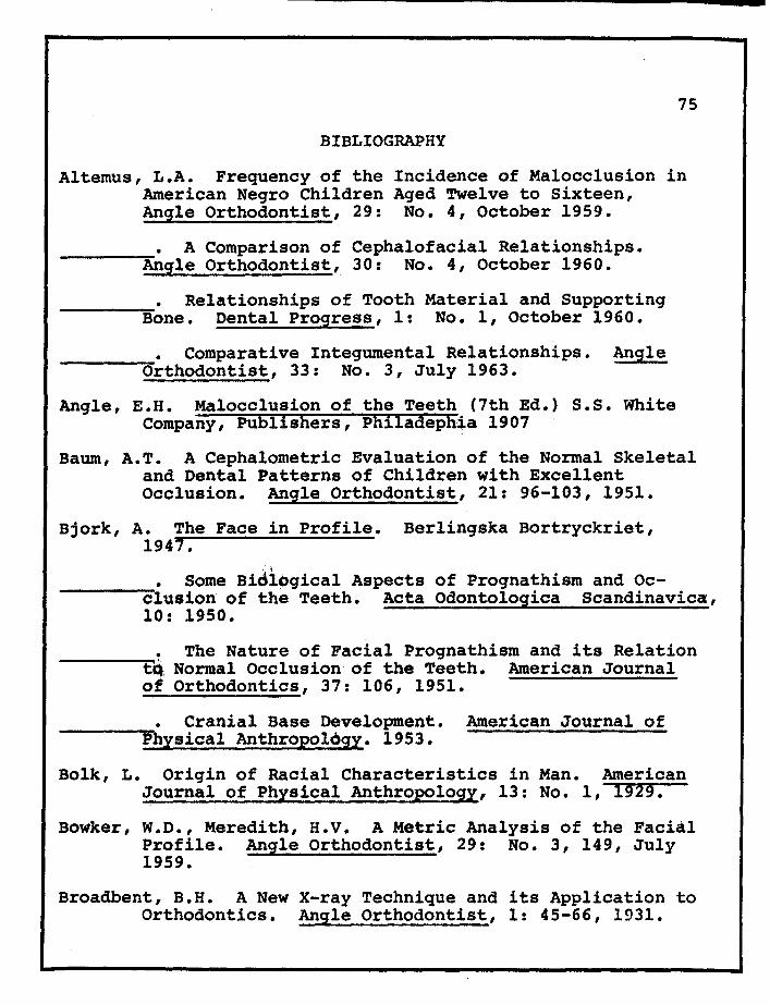
75
BIBLIOGRAPHY
Altemus, L.A. Frequency of the Incidence of Malocclusion in American Negro Children Aged Twelve to Sixteen, Angle Orthodontist, 29: No.4, October 1959.
A Comparison of Cephalofacial Relationships. Angle Orthodontist, 30: No.4, October 1960.
______ ~~. Relationships of Tooth Material and Supporting Bone. Dental Progress, 1: No.1, October 1960.
________ ~. Comparative Integumental Relationships. Angle Orthodontist, 33: No.3, July 1963.
Angle, E.H. Malocclusion of the Teeth (7th Ed.) S.S. White Company, Publishers, Philadeph4a 1907
Baum, A.T. A Cephalometric Evaluation of the Normal Skeletal and Dental Patterns of Children with Excellent occlusion. Angle Orthodontist, 21: 96-103, 1951.
Bjork, A. The FaCe in Profile. Berlingska Bortryckriet, 1947.
,1
• Some Biological Aspects of Prognathism and Oc--------c~lusion of the Teeth. Acta Odontologica Scandinavica,
10: 1950.
• The Nature of Facial Prognathism and its Relation --------t~Q Normal Occlusion of the Teeth. American Journal
of Orthodontics, 37: 106, 1951.
• Cranial Base Development. American Journal of ---~P:::o.hysical Anthropology. 1953.
Bolk, L. Origin of Racial Characteristics in Man. American Journal of Physical Anthropology, 13: No.1, 1929.
Bowker, W.O., Meredith, H.V. A Metric Analysis of the Facial Profile. Angle Orthodontist, 29: No.3, 149, July 1959.
Broadbent, B.H. A New X-ray Technique and its Application to Orthodontics. Angle Orthodontist, 1: 45-66, 1931.

76
Brodie, A.G. On the Growth Pattern of the Human Head from the Third Month to the Eighth Year of Life. American Journal of Anatomy, 68: 209-262, 1941.
Burstone, C.J. Integumental Contour and Extension Patterns. Angle Orthodontist, 29: 93-104, April 1959.
Campers, Petrus. Works on Connexion Between Anatomy and Drawings. Published Dutch Ams~erdam, 1794.
Coben, S.E. The Intergration of Facial Skeletal Variants. American Journal of Orthodontics, 41: 407-434, June 1955.
Cotton, W.N. Variations in Facial Relationships in American Negroes of the San Francisco Say ~ea. Unpublished Report, University of California College of Dentistry, 1949.
Downs, W.B. Variations in Facial Relationship: Their Significance in Treatment and Prognosis, American Journal of Orthodontics, 34: 812-840, October 1948.
________ ~. Analysis of the Dentofacial Profile. Angle 6rthodontist, 26: 191-212, 1956.
Elsasser, William A. A Cephalometric Method for the Linear Analysis of the Human Profile. American Journal of Orthodontics, 43: 192-209, March, 1957. ,
Freund, J.E. Modern Elementary Statistics, Prentice-Hall, Inc., New Jersey, 1952.
Goldstein, A. Biostatistics: An Introductory Text. Macmillian Company, New York, 1964.
The
Graber, T.M. Orthodontics princi1als and Practice. Saunders Company, Phi1ade phia, 1961.
W.B.
Grimson, K.J. A Linear Cephalometric Analysis: Its description and Application in Assessing Changes in the Mandible After Orthodontic Treatment. Master's thesis. Loyola University, Chicago, 1965.

77
Harris, J.E., Johnston, L., and Moyers, R.E. A Cephalometric Template: Its Construction and Clinical Significance. American Journal of Orthodontics, 49: 249-263, April 1963.
Hellman, M. Changes in the Human Face Brought About by Development. International Journal of Orthodontics, 13: 475, 1927.
• The Face and Teeth of Man. Journal of Dental --------~Research, 9: 177, 1929.
Hemley, Samuel. Orthodontic Theory and Practice. Grune and Stratton, Inc., 2nd Edition, 1953.
Herskovitz, M.J. The American Negro. Alfred A. Knopf, New York, 1930.
Jarabak, J.R. and Fizzell, J.A. Technique and Treatment With the Light-Wire Appliance. C.V. Mosby Company, Inc. St. Louis, 1963.
Klaatsch, H. Kraniomorphologie und Kranietrigonometric. Arch. f. Anthropol. 8, 1909.
Krogman, W.M. rt U.S. Public Health Grant 0-87 (C • P 1 a e p 1a Center or Researc 1n Child Growth, Philadelphia, 1958.
Lindegard, B. variations in Human Body-Build. Acta Psychiatrica Et Neurologica Scandinavica, supp1ementa, 84-86, 1953.
Lischer, B.E. Photography for Orthodontist. International Journal of Orthodontics, 12: 191-199, 1926.
McCall, J.O., and Wald, S.S. Clinical Dental Roentgenography. W.B. Saunders Company, Philadelphia, 1952.
Oppenheim, A. Prognathism from the Anthropological and Orthodontic View Point. Dental Cosmos 20: 1170-1184, 1928.

Pfaff, C.L. Rassen Krainologische untersuchungen uber die Stellun1des oberen im Kiefergerust. Dtsch. ZahOhk, 61, 192 •
78 .
Reidel, R.A. The Relation of Maxillary structures to Cranium in Malocclusion and in Normal Occlusion. Angle Orthodontist, 22: 142-145, 1952.
Salzman, J.A. Orthodontics, princieles and Prevention. J.B. Lippincott Company, Ph11adelphia, 1957.
• Orthodontics, Practice and Technigue. --------~Lippincott Company, Philadelphia, 1957.
J.B.
Sassouni, V. The Face in Five Dimensions. University of Pittsburgh, Pittsburgh, Pennsylvania.
• A Roentgenographic Cephalometric Analysis of --------=Cephalo-Facio-Dental Relationships. American Journal
of Orthodontics, 41: 735-764, October 1953.
Schudy, F.F. The Cant of the Occlusal Plane and the Axial Inclination of Teeth as Related to Diagnosis and Treatment. Angle Orthodontist, 33: 69-81, April 1963.
Simon, Paul W. Fundamental Principles ofa Systematic Diagnosis of Dental Anomo1ies. The Stratlord Company, Boston, 1926.
Steiner, C.C. Cephalometries For You and Me. American Journal of Orthodontics, 39: 729-755, 1953.
Strang, H.W. and Thompson, W.M. A Textbook of Orthodontia. Leo and Febiger, Philadelphia 1958.
Todd, T.W. The Orthodontic Value of Research and Observations in Developmental Growth of the Face. Angle Orthodontist, 1: 67, 1931.
________ =. Prognathism: A Study in Development of the Face. Journal of the American Dental Association, 19: 2172-2184, 1932.

Vorhies, J.M. and Adams, J.W. Polygonic Interpretation of Cephalometric Findings. Angle orthodontist, 21: 194, 1951.
Weinmann, J. and Sicher, H. Bone and Bones. C.V. Mosby Company, St. Louis, 1955.
79
Wylie, W.L. and Johnson, E.L. in the Vertical Plane. 165-181, 1952.
Evaluation of Facail Dysplasia Angle Orthodontist, 22:

APPROVAL SHEET
The thesis submitted by Dr. Roland F. Thomas has been
read and approved by members of the Department of Oral Biology.
The final copies have been examined by the director
of the thesis and the signature which appears below verifies
the fact that any necessary changes have been incorporated,
and that the thesis is now given final approval with reference
to content, form, and mechanical accuracy.
The thesis is therefore accepted in partial ful-
fillment of the requirements for the Degree of Master of
Science.
i:J!! ~ Ir.? ~ t:d.c.. 'J; / , I ./. " ATE


![Post-Orthodontic Cephalometric Variations in Bimaxillary ...fac.ksu.edu.sa/.../post-orthodontic_cephalometric... · analysis in accordance with cephalometric norms.[20] Soft tissue](https://img.dokumen.tips/doc/110x75/5ec5a1ed69d7b460ea09abc8/post-orthodontic-cephalometric-variations-in-bimaxillary-facksuedusapost-orthodonticcephalometric.jpg)