Embed Size (px)
Citation preview
Inde
xed
in
Inde
x Med
icus®
MED
LINE
VOL. 7, NO. 42005
EditorMartin Quan, MD
Prevention ofThromboembolic EventsGuest EditorsGeno J. Merli, MD, FACPThomas Jefferson UniversityPhiladelphia, PennsylvaniaDeepak L. Bhatt, MD, FACC, FSCAI, FESCCleveland Clinic FoundationCleveland, Ohio
Venous Thromboembolism: Epidemiology, Characteristics, and ConsequencesFranklin Michota, MD
Clinical Trials of Deep Vein Thrombosis Prophylaxis in Medical PatientsWalter Ageno, MDAlexander G.G. Turpie, MD
Venous Thromboembolism in Medically Ill Patients:Identifying Risk and Strategies for Prevention Arthur Wheeler, MD
Venous Thromboembolism Prophylaxis Guidelines: Use by Primary Care PhysiciansGeno J. Merli, MD, FACP
Pharmacologic Therapy for the Management of Thrombosis: Unfractionated Heparin or Low-Molecular-Weight Heparin?Alex C. Spyropoulos, MD, FACP, FCCP
Prevention of Thrombosis with Warfarin, Aspirin, and Mechanical Methods Geno J. Merli, MD, FACP
Also—Dialogues with the facultyLiterature Reviews
ClinicalCornerstone
This program was supportedby an educational grant from
Sanofi-Synthelabo Inc., a member of
The sanofi-aventis Group
This activity has been jointly sponsored by the
Elsevier Office of ContinuingMedical Education andExcerpta Medica, Inc.
UP TO 8 FREE
AMA PRA
CATEGORY 1 CREDITS
www.ClinicalCornerstone.com
cvr_1-4_00704_CCV7N4 3/6/06 1:41 PM Page cvr1
EDITORMartin Quan, MDProfessor of Clinical Family Medicine and
Director, Office of CMEDavid Geffen School of Medicine at UCLALos Angeles, California
ASSOCIATE EDITORRichard A. Johnson, MDClinical Professor of Family Medicine
and RadiologyDavid Geffen School of Medicine at UCLALos Angeles, California
EDITORIAL BOARDDennis Cope, MDProfessor of Medicine, Chair of MedicineOlive View – UCLA Medical CenterSylmar, California
Howard A. Miller, MDProfessor of Medicine and Executive Vice ChairDepartment of MedicineDrexel University College of MedicinePhiladelphia, Pennsylvania
Jane Murray, MDClinical Professor, Family & Community MedicineUniversity of Missouri-Kansas CityMedical Director, Sastun Center of Integrative
Health CareMission, Kansas
Aly Rashid, MDDe Montfort UniversityLeicester, United Kingdom
George T. Smith, MD, MSConsultant PhysicianPalm Beach, Florida
PUBLISHING AND EDITORIAL STAFFJoan Parker, Senior Vice President, Publishing908-547-2080
Cindy H. Jablonowski, Publisher908-547-2090
Theresa Salerno, Manuscript Production Manager
Vanessa Fendt, Editorial Project Manager
Annette Ulecka, Senior Proofreader
Richard T. Class, Director, US Production
Gail M. Gallo, Reprints Manager908-547-2069; fax 908-547-2204
Susan Polacheck, Subscriptions908-547-2072; fax 908-547-2204
Editorial and Subscription Offices685 Route 202/206Bridgewater, NJ 08807Telephone: 800-533-8763Fax: 908-547-2204E-mail: [email protected]
CONTINUING MEDICAL EDUCATIONMark J. Flanick, MS, Director
BUSINESS DEVELOPMENTJohn Elduff, Senior Vice President, Sales908-547-2150
Michael Moran, Director, Business Development
Clinical Cornerstone (ISSN 1098-3597) (GST #128741063) is published quarterly by Excerpta Medica, an Elsevier business.Indexed/abstracted in Index Medicus®/MEDLINE, EMBASE/Elsevier, and Cinahl Information Systems®.
Copyright: Copyright 2005 by Excerpta Medica, an Elsevier business. All rights reserved under the United States, Internationaland Pan American Copyright Conventions. No part of this publication may be reproduced, stored in a retrieval system, or trans-mitted in any form or by any means, mechanical, computer, photocopying, electronic recording or otherwise, without the prior writ-ten permission of Excerpta Medica, Inc. The copyright law of the United States (Title 17, U.S.C., as amended) governs the mak-ing of photocopies or other reproductions of copyrighted material.
Opinions: Opinions expressed in articles are those of the authors and do not necessarily reflect those of Excerpta Medica, Inc., theeditors, Editorial Board, sponsors, or grantors. Excerpta Medica, Inc. assumes no liability for any material published herein.
Subscriptions: 2005 US subscription rates: Individuals $79. Institutional $195. Subscription rates include supplements.
This paper meets the requirements of ANSI/NISO Z39.48-1992 (Permanence of Paper).
Release Date: December 2005.
ClinicalCornerstone
cvr_1-4_00704_CCV7N4 3/6/06 1:41 PM Page cvr2
PREVENTION OF THROMBOEMBOLIC EVENTS
GUEST EDITORS
Geno J. Merli, MD, FACPThomas Jefferson UniversityPhiladelphia, Pennsylvania
Deepak L. Bhatt, MD, FACC, FSCAI, FESCCleveland Clinic Foundation
Cleveland, Ohio
CONTRIBUTORS
Clinical Cornerstone
Clinical Cornerstone � PREVENTION OF THROMBOEMBOLIC EVENTS � Vol. 7, No. 4
1
Walter Ageno, MDDepartment of Clinical Medicine
Ospedale di CircoloUniversity of Insubria
Varese, Italy
Franklin Michota, MDAssociate Professor of Medicine
Department of General Internal MedicineCleveland Clinic Foundation
Cleveland, Ohio
Alex C. Spyropoulos, MD, FACP, FCCPMedical Director
Clinical Thrombosis CenterLovelace Sandia Health Systems
Clinical Associate ProfessorDepartment of Medicine
Associate Professor of PharmacyCollege of Pharmacy
University of New Mexico Health Sciences CenterAlbuquerque, New Mexico
Alexander G.G. Turpie, MDHamilton Health Sciences Corporation
McMaster UniversityHamilton, Ontario, Canada
Arthur Wheeler, MDAssociate Professor of Medicine
Vanderbilt University Medical CenterDivision of Allergy,
Pulmonary and Critical Care MedicineNashville, Tennessee
Clinical Cornerstone
Clinical Cornerstone � PREVENTION OF THROMBOEMBOLIC EVENTS � Vol. 7, No. 4
5
PREVENTION OF THROMBOEMBOLIC EVENTS
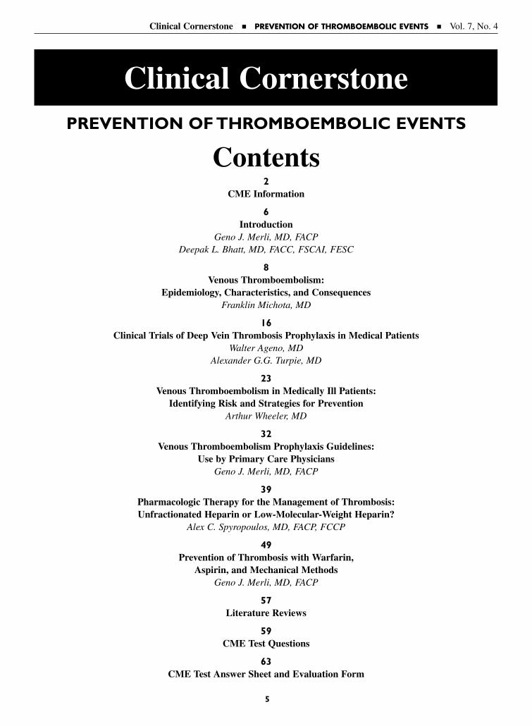
Contents2
CME Information
6Introduction
Geno J. Merli, MD, FACPDeepak L. Bhatt, MD, FACC, FSCAI, FESC
8Venous Thromboembolism:
Epidemiology, Characteristics, and ConsequencesFranklin Michota, MD
16Clinical Trials of Deep Vein Thrombosis Prophylaxis in Medical Patients
Walter Ageno, MDAlexander G.G. Turpie, MD
23Venous Thromboembolism in Medically Ill Patients:
Identifying Risk and Strategies for PreventionArthur Wheeler, MD
32Venous Thromboembolism Prophylaxis Guidelines:
Use by Primary Care PhysiciansGeno J. Merli, MD, FACP
39Pharmacologic Therapy for the Management of Thrombosis:Unfractionated Heparin or Low-Molecular-Weight Heparin?
Alex C. Spyropoulos, MD, FACP, FCCP
49Prevention of Thrombosis with Warfarin,
Aspirin, and Mechanical MethodsGeno J. Merli, MD, FACP
57Literature Reviews
59CME Test Questions
63CME Test Answer Sheet and Evaluation Form
Venous thromboembolism (VTE) and its manifesta-tions—including deep vein thrombosis (DVT) and pul-monary embolism (PE)—are serious medical conditionsassociated with high rates of morbidity and mortality.1
An estimated 300,000 patients are hospitalized each yearin the United States due to VTE.2 Furthermore, patientshospitalized for reasons other than VTE typically have atleast 1 risk factor for VTE.3 Depending on the surgicalprocedure, DVT occurs in 10% to 60% of hospitalizedpatients who do not receive prophylaxis. Studies havefound that 10% of hospital deaths are attributed to PE,with the majority of these deaths occurring in patientswho have not recently undergone surgery.3 The incidenceof VTE also is associated with enormous financial cost;an estimated $1.5 billion is spent each year on expensesassociated with DVT in the United States alone.4
VTE is a life-threatening illness that has multiple caus-es but few warning signs. Symptoms of DVT may includepain, erythema, tenderness, and swelling of the affectedlimb, whereas PE often presents as sudden breathlessnesswith chest pain or collapse with shock in the absence ofother causes.5 Yet, VTE prophylaxis is often underuti-lized. This issue of Clinical Cornerstone presents 6 arti-cles that focus on the need for greater awareness of VTEand encourage health care professionals to take appropri-ate preventive measures to reduce the incidence of VTEand its potentially life-threatening manifestations.
The first article by Franklin Michota, MD, describesthe epidemiology of VTE, its consequences, and how toidentify patients at risk for VTE. Dr. Michota also high-lights the need for prompt and accurate recognition ofrisk factors that can lead to the implementation of effec-tive VTE prophylaxis.
The second article by Walter Ageno, MD, andAlexander G.G. Turpie, MD, reviews the findings of sev-eral studies that investigated the use and effectiveness of thromboprophylaxis among medical patients. Theyfound that these studies support the evidence-based rec-ommendations for the systematic use of pharmacologicagents for VTE prevention in patients at risk for VTE.
The third article by Arthur Wheeler, MD, takes a closelook at the updated guidelines for the prevention of VTE
established by the Seventh American College of ChestPhysicians (ACCP) Conference on Antithrombotic andThrombolytic Therapy and discusses the high prevalenceof VTE among medically ill patients. Dr. Wheeler alsodescribes various strategies that can be used for VTEprophylaxis, highlighting relevant clinical studies thatsupport the use of antithrombotic therapy.
Practical applications of the ACCP revised guidelineson VTE prevention for primary care physicians is thefocus of the fourth article by Geno J. Merli, MD, FACP.Physicians are encouraged to assess VTE risk factors foreach patient—as well as the underlying illness or surgi-cal procedure of each patient and the benefits and risks ofpossible therapies—to determine the appropriate courseof action for each patient. This article also discusses theneed for hospitals to adopt clinical guidelines for VTEprevention, recognizing the growing impact of an agingpopulation that experiences more surgical procedureswith shorter durations of hospital stays.
The fifth article by Alex C. Spyropoulos, MD, FACP,FCCP, reports on the findings of several studies regard-ing pharmacologic therapy options for thrombosis man-agement in VTE and non–ST-elevation acute coronarysyndrome. Dr. Spyropoulos discusses various efficacy,safety, and pharmacoeconomic considerations relating tothe selection of a low-molecular-weight heparin versusunfractionated heparin for the prevention of VTE events.
The sixth article by Geno J. Merli, MD, FACP,describes specific strategies that can be used to preventVTE, including anticoagulant therapy with heparin (low-molecular-weight heparin or unfractionated heparin), directthrombin inhibitors, oral anticoagulants (such as war-farin), and mechanical methods. Dr. Merli also discussesthe role of aspirin therapy in patients at risk for VTE.
Greater awareness of VTE and the need for VTE pro-phylaxis can lead to widespread use of effective interven-tions to help reduce the incidence of thromboembolicevents in patients who receive health care services through-out the United States.
Geno J. Merli, MD, FACP, and Deepak L. Bhatt, MD, FACC, FSCAI, FESCGuest Editors
Introduction
Clinical Cornerstone � PREVENTION OF THROMBOEMBOLIC EVENTS � Vol. 7, No. 4
6
REFERENCES1. Ost D, Tepper J, Mihara H, et al. Duration of anticoagulation following venous thromboembolism: A meta-analysis. JAMA.
2005;294:706–715.
2. Hirsh J, Anand SS, Halperin JL, Fuster V. Guide to anticoagulant therapy: Heparin. A statement for healthcare professionals
from the American Heart Association. Circulation. 2001;103:2994–3018.
3. Geerts WH, Pineo GF, Heit JA, et al. Prevention of venous thromboembolism: The Seventh ACCP Conference on
Antithrombotic and Thrombolytic Therapy. Chest. 2004;126:338S–400S.
4. Spyropoulos AC, Hurley JS, Ciesla GN, et al. Management of acute proximal deep vein thrombosis: Pharmacoeconomic eval-
uation of outpatient treatment with enoxaparin versus inpatient treatment with unfractionated heparin. Chest. 2002;122:
108–118.
5. Turpie AG, Chin BS, Lip GY. Venous thromboembolism: Pathophysiology, clinical features, and prevention. Br Med J.
2002;325:887–890.
Clinical Cornerstone � PREVENTION OF THROMBOEMBOLIC EVENTS � Vol. 7, No. 4
7
Clinical Cornerstone � PREVENTION OF THROMBOEMBOLIC EVENTS � Vol. 7, No. 4
8
Venous thromboembolism (VTE) and its manifestations, including deep vein thrombosis (DVT) and pulmonaryembolism (PE), pose a life-threatening health problem for thousands of people each year. The diagnosis of VTEis frequently missed, however, because few signs and symptoms are recognized. Symptoms of DVT may includepain, erythema, tenderness, and swelling of the affected limb, whereas PE often presents as sudden breathless-ness with chest pain, or collapse with shock in the absence of other causes. Greater awareness of the epidemi-ology of VTE, the consequences of VTE, and the risk factors for VTE can help health care providers take appro-priate preventive measures to reduce the incidence of VTE. (Clinical Cornerstone. 2005;7[4]:8–15) Copyright ©2005 Excerpta Medica, Inc.
FRANKLIN MICHOTA, MDAssociate Professor of MedicineDepartment of General Internal MedicineCleveland Clinic FoundationCleveland, Ohio
Venous Thromboembolism:Epidemiology, Characteristics, andConsequences
8
Venous thromboembolism (VTE) is a silent, preventablekiller. VTE encompasses several manifestations of thesame disease process, including deep vein thrombosis(DVT) and pulmonary embolism (PE). In VTE, abnor-malities in blood flow, the blood vessel wall, or bloodclotting components contribute to formation of a throm-bus.1 DVT refers to a thrombus in the deep veins, mostoften in the lower extremities. The thrombus can embolizeand become lodged in the pulmonary arteries, resultingin PE, which frequently occurs without warning and isoften fatal.
Despite recent consensus guidelines on the preventionof VTE2 and the availability of antithrombotic agentssuch as the low-molecular-weight heparins, incidence ofVTE and its associated manifestations remains high.3,4
Incidence of VTE is 10% to 40% in general medical andsurgical patients and 40% to 60% in orthopedic surgerypatients.2 In the United States, PE accounts for more than250,000 hospitalizations annually.4 Although the risks ofVTE are well recognized in orthopedic and surgicalpatients, the majority of all in-hospital deaths due to PEare in nonsurgical patients5,6; however, most medicalpatients do not receive any form of VTE prophylaxis.7
Recent articles have highlighted the need for greaterawareness of VTE and its manifestations.8–10 Becausethere are few signs and symptoms of VTE, the diagnosisof DVT or PE is frequently missed.11 In one study of2388 autopsies in a general hospital population, PEaccounted for 10% of all in-hospital deaths. Of thesepatients, 83% had DVT in the legs at autopsy, includingonly 19% who had symptoms of DVT before death.12
Symptoms of DVT can include pain, erythema, tender-ness, and swelling of the affected limb.1 PE often pre-sents as sudden breathlessness with chest pain, or collapsewith shock in the absence of other causes.1 A thoroughunderstanding of VTE, awareness of the signs and symp-toms, and recognition of factors that place a patient atrisk will help physicians take appropriate preventivemeasures to reduce the incidence of VTE and its poten-tially life-threatening manifestations.
EPIDEMIOLOGY OF VENOUS THROMBOEMBOLISMOf all patients with VTE, about two thirds have DVTalone, whereas one third also have PE.13,14 The annualincidence of diagnosed, objectively confirmed VTE in
Clinical Cornerstone � PREVENTION OF THROMBOEMBOLIC EVENTS � Vol. 7, No. 4
9
the general population is about 1 to 2 per 1000 people,with ~66% of cases being first episodes.11 The annualincidence of PE is slightly under 1 per 1000 people.15
Postthrombotic syndrome (PTS) occurs in one third ofpatients after a symptomatic episode of DVT.1,16
About 37% of all cases of VTE occur in patients whoare either hospitalized or recently placed in a nursinghome.17 A study of 51,645 hospitalized patients over a 21-month period showed the prevalence of acute diagnosedPE to be 1% (526 of 51,645).18 PE was observed at autop-sy in 15% (59 of 404) of all patients on whom autopsieswere performed.18 Among those patients with PE at autop-sy, PE contributed to death in 22 (37%) of 59 patients.Among those patients who died from PE, the diagnosis wasunsuspected in 14 (70%) of 20 patients.18 About 75% of allin-hospital deaths from PE occur in nonsurgical patients.5
VTE is uncommon in patients younger than 20 years;most children diagnosed with VTE have a serious under-lying primary illness, such as cancer, chronic total par-enteral nutrition dependency, or congenital heart dis-ease.19 The incidence of VTE increases with increasingage, doubling with each decade after age 40 years.11,17 Inaddition, PE accounts for a higher proportion of diag-nosed episodes of VTE as age advances.11,17 In a studyof 342,000 inhabitants of Western France, the annualincidence of VTE reached 1 per 100 for individuals overthe age of 75 years.17 In this same study, 63% of patientswere at home when VTE occurred; however, 16% had been
hospitalized within the previous 3 months. Approximatelytwo thirds of these patients had been in a medical unit, andone third had been in a surgical unit.
Autopsy studies show that PE may cause or contribute toa high percentage (0.2%–20.3%) of deaths in hospitalizedmedical and surgical patients.12,18,20 –22 In a retrospectiveanalysis of 391 surgical patients in Malmo, Sweden, from1951 to 1988 in whom PE was found at autopsy, PE wasconsidered to be the cause of death in 113 (29%) patients, acontributor to death in 104 (27%) patients, and incidental in174 (45%) patients.20 In another study of 4881 general sur-gical patients in a Denmark hospital, fatal postoperativePE was found in 9% of patients who had an autopsy per-formed.21 The incidence of fatal postoperative PE amongpatients who received prophylaxis was 3.5% compared with11.2% in patients who did not receive prophylaxis.21
A retrospective analysis of 2427 autopsies from 1985through 1989 confirmed PE as the cause of death in 92(3.8%) patients, with no difference in incidence betweenmedical and surgical patients.22 In this study, classicsymptoms of PE were often absent in patients beforedeath, with dyspnea present in 59% of patients, chest painin 17% of patients, and hemoptysis in 3% of patients.Only 32% of patients had PE correctly assigned as thecause of death on the death certificate.
In a 5-year retrospective study of all autopsy reports ina general hospital patient population, PE was thought tobe the cause of death in 239 (10%) of 2388 patients, with15% of patients <60 years of age and 83% having DVT inthe legs. Only 19% of patients had symptoms of DVTbefore death, and only 3% had undergone investigationfor DVT, again illustrating the silent nature of this condi-tion in many patients.12
CONSEQUENCES OF VENOUS THROMBOEMBOLISMVTE is a common hospital-associated complication, with10% of hospital deaths attributed to PE.12 Death due to
9
K E Y P O I N T
The annual incidence of diagnosed,
objectively confirmed VTE in the general
population is about 1 to 2 per 1000 people.
K E Y P O I N T
About 75% of all in-hospital deaths from
PE occur in nonsurgical patients.
K E Y P O I N T
The incidence of VTE increases with
increasing age, doubling with each decade
after age 40 years.
Clinical Cornerstone � PREVENTION OF THROMBOEMBOLIC EVENTS � Vol. 7, No. 4
10
PE is frequently the first sign of VTE and often occurssuddenly, before effective intervention can be provided.In a prospective registry of 5451 patients with ultrasound-confirmed DVT in the United States, concomitant PEwas identified in 793 (14.5%) of patients.23 PE accountsfor 60,000 to 100,000 deaths in the United States annu-ally.23,24 Although generally uncommon in younger peo-ple and children, VTE is becoming recognized as a sig-nificant cause of morbidity and mortality in children.19
VTE results in serious long-term as well as short-termcomplications, which can significantly affect a patient’squality of life (QOL).9,25 Long-term recurrent VTE canresult in thromboembolic pulmonary hypertension anddamage to the venous valves, as well as PTS.24 PTS canbe extremely debilitating and is characterized by chronicleg symptoms, including persistent pain, swelling,cramping, skin discoloration, necrosis, and ulcerationof the affected limb due to venous hypertension.24 Inextreme cases of PTS, amputation of the limb may benecessary.9 In a study of 528 patients with a first episodeof symptomatic DVT, the cumulative incidence of recur-rent VTE after 2, 5, and 8 years was 17%, 24%, and 30%,respectively.26 In the same study, the cumulative inci-dence of PTS after 2, 5, and 8 years was 25%, 30%, and30%, respectively. Survival after 8 years was 69%.
A health-related QOL study of 359 patients after DVTdiagnosis showed that, on average, QOL improved dur-ing the 4 months following diagnosis.25 Mean scores onall physical and mental health surveys and disease-specific symptom questionnaires improved significantly(P < 0.001) during the 4-month period. However, QOLdeteriorated in about one third of patients, with a wors-ening PTS score associated with worsening QOL.25 Inanother QOL study,27 45 patients with VTE scored sig-nificantly lower on all subscales of a questionnaire com-pared with the norms associated with the general USpopulation (P < 0.05). Patients with PTS also had more
impairment of QOL than patients without PTS. In addi-tion to affecting an individual’s QOL, the direct and indi-rect medical costs associated with VTE are a burden topatients and society.
RISK FACTORS FOR VENOUS THROMBOEMBOLISMThe high prevalence, silent nature, significant financialand personal burden, and potentially rapid mortality ofVTE all underscore the importance of recognizing therisk factors and initiating appropriate prophylactic mea-sures. Early identification of patients at risk for VTE andits manifestations should substantially reduce morbidityand mortality from this disease.
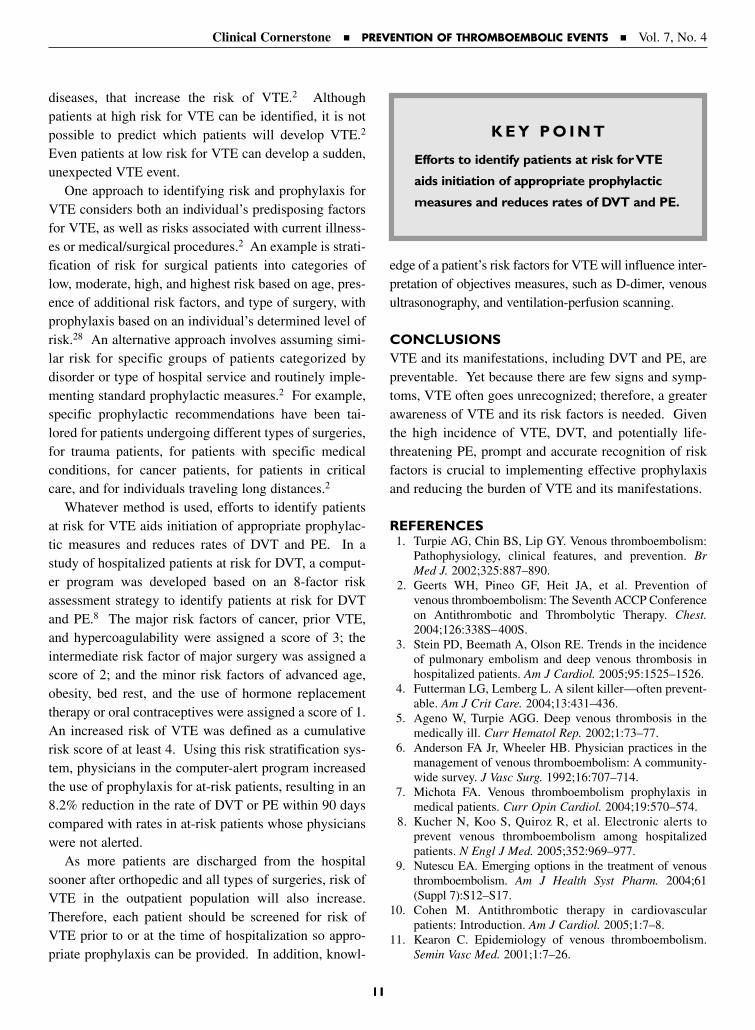
As shown in the table, risk factors for VTE have beenidentified, including age >40 years, immobilization orconditions resulting in venous stasis, conditions resultingin vessel wall damage, abnormalities in circulating coag-ulation elements, and previous DVT or PE.2,11,24 Riskfactors tend to be cumulative, so that older individualsare more likely than younger individuals to have otherfactors, such as immobility, malignancy, obesity, or other
10
K E Y P O I N T
VTE is a common hospital-associated
complication, with 10% of hospital deaths
attributed to PE.
TABLE. RISK FACTORS FOR VENOUS THROMBOEMBOLISM (VTE).2
Surgery
Trauma (major or lower extremity)
Immobility, paresis
Malignancy
Cancer therapy (hormonal, chemotherapy, or radiotherapy)
Previous VTE
Increasing age
Pregnancy and the postpartum period
Estrogen-containing oral contraception or hormone replacementtherapy
Selective estrogen receptor modulators
Acute medical illness
Heart or respiratory failure
Inflammatory bowel disease
Nephrotic syndrome
Myeloproliferative disorders
Paroxysmal nocturnal hemoglobinuria
Obesity
Smoking
Varicose veins
Central venous catheterizationInherited or acquired thrombophilia
Reprinted with permission.2
Clinical Cornerstone � PREVENTION OF THROMBOEMBOLIC EVENTS � Vol. 7, No. 4
11
diseases, that increase the risk of VTE.2 Althoughpatients at high risk for VTE can be identified, it is notpossible to predict which patients will develop VTE.2
Even patients at low risk for VTE can develop a sudden,unexpected VTE event.
One approach to identifying risk and prophylaxis forVTE considers both an individual’s predisposing factorsfor VTE, as well as risks associated with current illness-es or medical/surgical procedures.2 An example is strati-fication of risk for surgical patients into categories oflow, moderate, high, and highest risk based on age, pres-ence of additional risk factors, and type of surgery, withprophylaxis based on an individual’s determined level ofrisk.28 An alternative approach involves assuming simi-lar risk for specific groups of patients categorized by disorder or type of hospital service and routinely imple-menting standard prophylactic measures.2 For example,specific prophylactic recommendations have been tai-lored for patients undergoing different types of surgeries,for trauma patients, for patients with specific medicalconditions, for cancer patients, for patients in criticalcare, and for individuals traveling long distances.2
Whatever method is used, efforts to identify patientsat risk for VTE aids initiation of appropriate prophylac-tic measures and reduces rates of DVT and PE. In astudy of hospitalized patients at risk for DVT, a comput-er program was developed based on an 8-factor riskassessment strategy to identify patients at risk for DVTand PE.8 The major risk factors of cancer, prior VTE,and hypercoagulability were assigned a score of 3; theintermediate risk factor of major surgery was assigned ascore of 2; and the minor risk factors of advanced age,obesity, bed rest, and the use of hormone replacementtherapy or oral contraceptives were assigned a score of 1.An increased risk of VTE was defined as a cumulativerisk score of at least 4. Using this risk stratification sys-tem, physicians in the computer-alert program increasedthe use of prophylaxis for at-risk patients, resulting in an8.2% reduction in the rate of DVT or PE within 90 dayscompared with rates in at-risk patients whose physicianswere not alerted.
As more patients are discharged from the hospitalsooner after orthopedic and all types of surgeries, risk ofVTE in the outpatient population will also increase.Therefore, each patient should be screened for risk ofVTE prior to or at the time of hospitalization so appro-priate prophylaxis can be provided. In addition, knowl-
edge of a patient’s risk factors for VTE will influence inter-pretation of objectives measures, such as D-dimer, venousultrasonography, and ventilation-perfusion scanning.
CONCLUSIONSVTE and its manifestations, including DVT and PE, arepreventable. Yet because there are few signs and symp-toms, VTE often goes unrecognized; therefore, a greaterawareness of VTE and its risk factors is needed. Giventhe high incidence of VTE, DVT, and potentially life-threatening PE, prompt and accurate recognition of riskfactors is crucial to implementing effective prophylaxisand reducing the burden of VTE and its manifestations.
REFERENCES1. Turpie AG, Chin BS, Lip GY. Venous thromboembolism:
Pathophysiology, clinical features, and prevention. BrMed J. 2002;325:887–890.
2. Geerts WH, Pineo GF, Heit JA, et al. Prevention ofvenous thromboembolism: The Seventh ACCP Conferenceon Antithrombotic and Thrombolytic Therapy. Chest.2004;126:338S–400S.
3. Stein PD, Beemath A, Olson RE. Trends in the incidenceof pulmonary embolism and deep venous thrombosis inhospitalized patients. Am J Cardiol. 2005;95:1525–1526.
4. Futterman LG, Lemberg L. A silent killer—often prevent-able. Am J Crit Care. 2004;13:431–436.
5. Ageno W, Turpie AGG. Deep venous thrombosis in themedically ill. Curr Hematol Rep. 2002;1:73–77.
6. Anderson FA Jr, Wheeler HB. Physician practices in themanagement of venous thromboembolism: A community-wide survey. J Vasc Surg. 1992;16:707–714.
7. Michota FA. Venous thromboembolism prophylaxis inmedical patients. Curr Opin Cardiol. 2004;19:570–574.
8. Kucher N, Koo S, Quiroz R, et al. Electronic alerts to prevent venous thromboembolism among hospitalizedpatients. N Engl J Med. 2005;352:969–977.
9. Nutescu EA. Emerging options in the treatment of venousthromboembolism. Am J Health Syst Pharm. 2004;61(Suppl 7):S12–S17.
10. Cohen M. Antithrombotic therapy in cardiovascularpatients: Introduction. Am J Cardiol. 2005;1:7–8.
11. Kearon C. Epidemiology of venous thromboembolism.Semin Vasc Med. 2001;1:7–26.
11
K E Y P O I N T
Efforts to identify patients at risk for VTE
aids initiation of appropriate prophylactic
measures and reduces rates of DVT and PE.
Clinical Cornerstone � PREVENTION OF THROMBOEMBOLIC EVENTS � Vol. 7, No. 4
12
12. Sandler DA, Martin JF. Autopsy proven pulmonaryembolism in hospital patients: Are we detecting enoughdeep vein thrombosis? J R Soc Med. 1989;82:203–205.
13. Heit JA. Venous thromboembolism epidemiology: Impli-cations for prevention and management. Semin ThrombHemost. 2002;28(Suppl 2):3–13.
14. White RH. The epidemiology of venous thromboem-bolism. Circulation. 2003;107(Suppl 1):14–18.
15. Silverstein MD, Heit JA, Mohr DN, et al. Trends in theincidence of deep vein thrombosis and pulmonaryembolism: A 25-year population-based study. Arch InternMed. 1998;158:585–593.
16. Kahn SR, Solymoss S, Lamping DL, Abenhaim L. Long-term outcomes after deep vein thrombosis: Postphlebiticsyndrome and quality of life. J Gen Intern Med. 2000;15:425–429.
17. Oger E. Incidence of venous thromboembolism: A community-based study in Western France. ThrombHaemost. 2000;83:657–660.
18. Stein PD, Henry JW. Prevalence of acute pulmonaryembolism among patients in a general hospital and atautopsy. Chest. 1995;108:978–981.
19. Anton N, Massicotte MP. Venous thromboembolism inpediatrics. Semin Vasc Med. 2001;1:111–122.
20. Lindblad B, Eriksson A, Bergqvist D. Autopsy-verified pul-monary embolism in a surgical department: Analysis of theperiod from 1951 to 1988. Br J Surg. 1991;78:849–852.
21. Rasmussen MS, Wille-Jorgensen P, Jorgensen LN. Post-operative fatal pulmonary embolism in a general surgicaldepartment. Am J Surg. 1995;169:214–216.
22. Morgenthaler TI, Ryu JH. Clinical characteristics of fatalpulmonary embolism in a referral hospital. Mayo ClinProc. 1995;70:417–424.
23. Goldhaber SZ, Tapson VF, for the DVT FREE SteeringCommittee. A prospective registry of 5,451 patients withultrasound-confirmed deep vein thrombosis. Am JCardiol. 2004;93:259–262.
24. Haines ST. Venous thromboembolism: Pathophysiologyand clinical presentation. Am J Health Syst Pharm. 2003;60(Suppl 7):S3–S5.
25. Kahn SR, Ducruet T, Lamping DL, et al. Prospective eval-uation of health-related quality of life in patients with deep venous thrombosis. Arch Intern Med. 2005;165:1173–1178.
26. Prandoni P, Villalta S, Bagatell P, et al. The clinical courseof deep-vein thrombosis. Prospective long-term follow-upof 528 symptomatic patients. Haematologica. 1997;82:423–428.
27. van Korlaar IM, Vossen CY, Rosendaal FR, et al. Theimpact of venous thrombosis on quality of life. ThrombRes. 2004;114:11–18.
28. Geerts WH, Heit JA, Clagett GP, et al. Prevention ofvenous thromboembolism. Chest. 2001;119:132S–175S.
12
Clinical Cornerstone � PREVENTION OF THROMBOEMBOLIC EVENTS � Vol. 7, No. 4
13
EDITORIAL BOARDGiven the relatively high prevalence of venous throm-boembolism (VTE) in hospitalized patients, has anythought been given to just screening patients withvenous duplex scans?
MICHOTAThis strategy has been investigated and found to be
inadequate. Duplex scanning, even with high operator
skill, only has an 80% sensitivity for asymptomatic
deep vein thromboembolism (DVT). This would fail to
detect too large a number to be a reliable strategy, not to
mention the cost and the technical issues of trying to do
this on a regular basis. Ultimately, required technology
for such a strategy may be developed, but we don’t have
it now. Ideally, we would have some serologic marker of
thrombogenicity that could be measured in everybody
so that when you fell into the thrombogenic range,
you’d get prophylaxis.
EDITORIAL BOARDWhy is DVT prophylaxis not as effective as onemight think?
MICHOTAFirst let me say that we do have over 30 years of ran-
domized clinical trial evidence that DVT prophylaxis
reduces thromboembolic morbidity and mortality.
However, variability exists in individual patients in
terms of how they respond to our current prophylactic
measures. Although we clearly have effective pharma-
cologic prophylactic measures, there are some patients
whose thrombogenicity is high enough that they may be
more resistant to even our most intense pharmacologic
strategy. Today, our biggest problem lies in the fact that
many patients at risk for DVT still don’t get any prophy-
laxis at all. In the DVT-FREE registry, less than half of
patients who were hospitalized and developed a symp-
tomatic event received any form of prophylaxis. So
regardless of the risk reduction possible for any given
prophylaxis strategy, if we don’t use it, there is no
chance for it to be effective.
EDITORIAL BOARDSince prophylactic measures would not be effectivein patients with established DVT, is it possible thatsome of the failures seen with prophylactic therapyin clinical trials were due to asymptomatic VTEbeing already present at the time of entry into thestudy?
MICHOTANo, that was generally controlled for. In fact, some stud-
ies actually used venograms or other sensitive modali-
ties to exclude DVT in patients prior to enrolling them
into study populations.
EDITORIAL BOARDIs immobility the primary cause for the increasedthrombogenicity we see in hospitalized patients, ordo you think something else is going on?
MICHOTAOther factors have to be involved, particularly in the
medically ill population. To support this, one needs to
look no further than the situation seen in patients with
Lou Gehrig disease (ie, amyotrophic lateral sclerosis).
Have you ever seen or heard a case report of somebody
with amyotrophic lateral sclerosis dying of pulmonary
embolism (PE)? You’d scour the literature and discov-
er there’s no association between Lou Gehrig disease
and PE.
EDITORIAL BOARDAnd they have no muscle tone.
MICHOTAThe way I’ve looked at this is that ambulation is proba-
bly a marker of health, and thus I’m not so certain that
walking prevents DVT any more than that the state of
immobility is clearly a causative factor in and of itself.
It seems more likely that the ability to walk is just a
marker of somebody whose thrombogenicity is low, which
is closer to what we would expect in the community
population; there is an 8-fold lower incidence of VTE in
Dialogue Box
14
Clinical Cornerstone � PREVENTION OF THROMBOEMBOLIC EVENTS � Vol. 7, No. 4
the community than in a hospitalized population. Thus,
when you look at a nursing home population that’s
chronically immobile or bed-bound, their actual throm-
bogenicity may be low, provided nothing acute is going
on. If that same patient develops a urinary tract infec-
tion, becomes confused, and perhaps less mobile
(requiring more assistance to get out of bed and so
forth), that is when their thrombogenicity goes up—not
because they can’t get out of bed anymore, but because
of their acute systemic illness. Certainly, we know that
ambulation can improve venous flow and decrease sta-
sis and so forth. But that may not be where the action
is. The action may be in the thrombogenic state from
whatever made them unable to walk anymore.
EDITORIAL BOARDPlease comment on the future biochemical markersfor looking at thrombogenicity.
MICHOTAWell, at this point, that’s the holy grail. Although every-
body is looking for markers that would allow us to quan-
titatively measure dysregulation of the coagulation sys-
tem and thus increased thrombogenicity, at the moment,
there isn’t any. We don’t even have one on the drawing
board.
EDITORIAL BOARDAny role for D-dimers?
MICHOTAThey have no role for “ruling-in” a diagnosis of DVT or
PE in any patient population. D-dimers do have a role in
excluding VTE in an outpatient or emergency room popu-
lation. Enzyme-linked immunosorbent assay (ELISA)–
based assays are required, and in a patient with low or
moderate clinical suspicion, a negative D-dimer can rule
out DVT. However, once a patient is admitted to the
hospital, virtually all patients will have some elevation
of D-dimer in that setting. Thus, the specificity drops
so low that the negative predictive value, even if they’re
ELISA-based assays, are too low to be clinically useful.
EDITORIAL BOARDIn what patients would you launch a workup for anunderlying thrombophilia?
MICHOTAThe patient with idiopathic DVT. This would generally
be someone less than 45 years of age or someone more
than 45 years of age who has no identifiable risk factors
for clotting. We would also look for a thrombophilia
in a patient who had what we consider to be minor
risk factors but yet had a surprisingly high clot burden
(meaning their whole leg was packed with clot from the
ankle to the thigh), as well as the patient with a clot in
a very atypical location, such as the upper extremity
without a cervical rib issue or a central catheter access.
EDITORIAL BOARDWhat would be your workup?
MICHOTALet me begin by saying we categorically treat people for
6 months before beginning a workup. Although you can
do some genetic testing, such as Factor V Leiden muta-
tion and prothrombin gene mutation, even if a patient is
on anticoagulation therapy, unless there is some com-
pelling reason to know their genetic status (such as to
counsel a sibling who smokes or who is on the birth con-
trol pill), we generally wait at least 6 months. At that
point, we counsel the patient as to what we are going to
do if we find an abnormal result. For example, if we
find a positive result, would the patient be willing to
receive anticoagulation drugs for extended periods of
time? If not, we may not even test this patient. Since
there is no certainty of the absolute implications in any
given individual, the likelihood is that, erring on the side
of caution, we would recommend extended anticoagula-
tion for patients we know have positive results; if the
latter would not be an option, we may simply not want
to even know the result.
In direct response to your question, our initial battery
of tests would check for the prothrombin gene muta-
tions, Factor V Leiden mutation, and homocysteine since
Dialogue Box

Clinical Cornerstone � PREVENTION OF THROMBOEMBOLIC EVENTS � Vol. 7, No. 4
15
they are the most common thrombophilic abnormalities.
If these tests came back negative, we would then look for
protein C and S deficiencies, antithrombin-III deficien-
cy, and anticardiolipin and antiphospholipid antibodies.
It’s important to recognize that you should never test for
the factor deficiencies within 4 weeks of an acute clot
because your results will be difficult to interpret and
may lead to a false-positive result. Such patients do not
have thrombophilia; they’ve just had consumption of
these factors because of the acute clot.
Dialogue Box
Clinical Cornerstone � PREVENTION OF THROMBOEMBOLIC EVENTS � Vol. 7, No. 4
16
Autopsies and clinical studies have shown that venous thromboembolism (VTE) is a common cause of morbid-ity and mortality in medical patients. Prophylaxis of VTE has been less extensively studied in medical patientsthan in surgical patients, and the results of recent practice audits indicate that the use of thromboprophylaxisis uncommon in medical patients. In the past few years, 3 large randomized clinical trials have demonstratedthe efficacy and safety of prophylaxis of VTE in the medical setting. The prophylaxis in MEDical patients withENOXaparin (MEDENOX), Prospective Evaluation of Dalteparin Efficacy for PREVENTion of VTE in Immobi-lized Patients Trial (PREVENT), and ARixta for ThromboEmbolism Prevention in a Medical Indications Study(ARTEMIS) studies have compared the low-molecular-weight heparins enoxaparin and dalteparin, and thespecific factor Xa inhibitor fondaparinux, respectively, with placebo in acutely ill medical patients hospitalizedwith heart failure, respiratory failure, infectious disease, or inflammatory disease. All studies showed both astatistically significant reduction in the rate of venous thromboembolic events (as assessed by venography orcompression ultrasonography) and a rate of major bleeding events that were comparable to placebo. The resultsof these studies support the evidence-based recommendations for systematic use of thromboprophylaxis in thissetting. (Clinical Cornerstone. 2005;7[4]:16–22) Copyright © 2005 Excerpta Medica, Inc.
WALTER AGENO, MDDepartment of Clinical MedicineOspedale di CircoloUniversity of InsubriaVarese, Italy
ALEXANDER G.G.TURPIE, MDHamilton Health Sciences CorporationMcMaster University Hamilton, Ontario, Canada
Clinical Trials of Deep Vein ThrombosisProphylaxis in Medical Patients
16
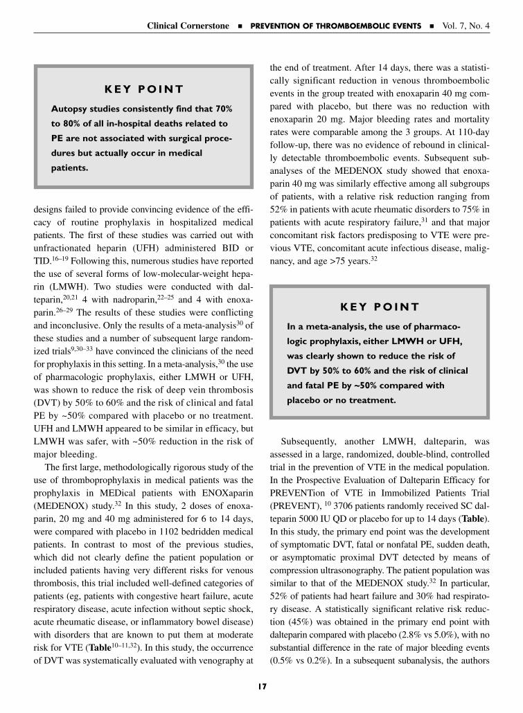
Pulmonary embolism (PE) is a leading cause of mortal-ity in hospitalized patients, accounting for ~10% of allin-hospital deaths.1 In the last 3 decades, great attentionhas been given to the prevention of venous thromboem-bolism (VTE) in patients undergoing surgery, and manygood-quality clinical trials have placed the routine use ofprophylaxis in such patients on a firm scientific footing.However, autopsy studies consistently find that 70% to80% of all in-hospital deaths related to PE are not asso-ciated with surgical procedures but actually occur inmedical patients.1–5 Others have reported that 50% to70% of symptomatic venous thromboembolic eventsrelated to hospitalization occur in medical patients.6,7
More recently, Monreal et al8 reported a more severepresentation and a significantly worse outcome inpatients who developed VTE after an acute medical dis-ease than in patients who developed VTE after surgery.Overall, prevention of VTE has been less extensively stud-ied in medical patients than in surgical patients. However,in recent years a number of landmark studies9–11 have con-
sistently found that pharmacologic prophylaxis of VTEwas indeed both safe and effective in the medical setting.As a result, evidence-based practice guidelines began tostrongly recommend the use of pharmacologic prophy-laxis in patients with acute medical diseases (eg, heartfailure, acute respiratory disease, sepsis, cancer, andinflammatory bowel disease).12 Despite this evidence,recent practice audits indicate significant underuse ofthromboprophylaxis in medical patients.13–15 Reasons forsuch underuse include, among others, the great hetero-geneity of the population of medical patients that makesthis group more difficult to target than surgical patients,concern about bleeding risk, and the lack of perceptionthat VTE is a real issue.
THE EVIDENCEUntil 5 to 6 years ago, there was only limited evidence tosupport the use of thromboprophylaxis in medicalpatients. A number of small clinical trials conducted in aheterogeneous population and with heterogeneous study
Clinical Cornerstone � PREVENTION OF THROMBOEMBOLIC EVENTS � Vol. 7, No. 4
17
designs failed to provide convincing evidence of the effi-cacy of routine prophylaxis in hospitalized medicalpatients. The first of these studies was carried out withunfractionated heparin (UFH) administered BID orTID.16–19 Following this, numerous studies have reportedthe use of several forms of low-molecular-weight hepa-rin (LMWH). Two studies were conducted with dal-teparin,20,21 4 with nadroparin,22–25 and 4 with enoxa-parin.26–29 The results of these studies were conflictingand inconclusive. Only the results of a meta-analysis30 ofthese studies and a number of subsequent large random-ized trials9,30–33 have convinced the clinicians of the needfor prophylaxis in this setting. In a meta-analysis,30 the useof pharmacologic prophylaxis, either LMWH or UFH,was shown to reduce the risk of deep vein thrombosis(DVT) by 50% to 60% and the risk of clinical and fatalPE by ~50% compared with placebo or no treatment.UFH and LMWH appeared to be similar in efficacy, butLMWH was safer, with ~50% reduction in the risk ofmajor bleeding.
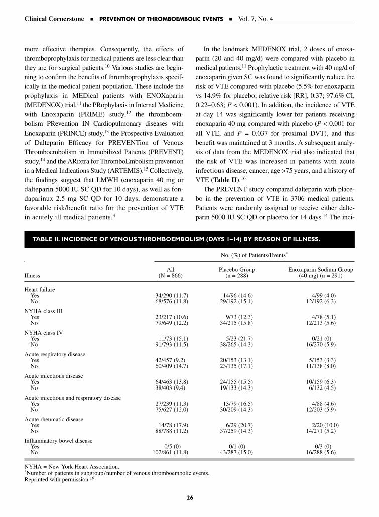
The first large, methodologically rigorous study of theuse of thromboprophylaxis in medical patients was theprophylaxis in MEDical patients with ENOXaparin(MEDENOX) study.32 In this study, 2 doses of enoxa-parin, 20 mg and 40 mg administered for 6 to 14 days,were compared with placebo in 1102 bedridden medicalpatients. In contrast to most of the previous studies,which did not clearly define the patient population orincluded patients having very different risks for venousthrombosis, this trial included well-defined categories ofpatients (eg, patients with congestive heart failure, acuterespiratory disease, acute infection without septic shock,acute rheumatic disease, or inflammatory bowel disease)with disorders that are known to put them at moderaterisk for VTE (Table10–11,32). In this study, the occurrenceof DVT was systematically evaluated with venography at
the end of treatment. After 14 days, there was a statisti-cally significant reduction in venous thromboembolicevents in the group treated with enoxaparin 40 mg com-pared with placebo, but there was no reduction withenoxaparin 20 mg. Major bleeding rates and mortalityrates were comparable among the 3 groups. At 110-dayfollow-up, there was no evidence of rebound in clinical-ly detectable thromboembolic events. Subsequent sub-analyses of the MEDENOX study showed that enoxa-parin 40 mg was similarly effective among all subgroupsof patients, with a relative risk reduction ranging from52% in patients with acute rheumatic disorders to 75% inpatients with acute respiratory failure,31 and that majorconcomitant risk factors predisposing to VTE were pre-vious VTE, concomitant acute infectious disease, malig-nancy, and age >75 years.32
Subsequently, another LMWH, dalteparin, wasassessed in a large, randomized, double-blind, controlledtrial in the prevention of VTE in the medical population.In the Prospective Evaluation of Dalteparin Efficacy forPREVENTion of VTE in Immobilized Patients Trial(PREVENT), 10 3706 patients randomly received SC dal-teparin 5000 IU QD or placebo for up to 14 days (Table).In this study, the primary end point was the developmentof symptomatic DVT, fatal or nonfatal PE, sudden death,or asymptomatic proximal DVT detected by means ofcompression ultrasonography. The patient population wassimilar to that of the MEDENOX study.32 In particular,52% of patients had heart failure and 30% had respirato-ry disease. A statistically significant relative risk reduc-tion (45%) was obtained in the primary end point withdalteparin compared with placebo (2.8% vs 5.0%), with nosubstantial difference in the rate of major bleeding events(0.5% vs 0.2%). In a subsequent subanalysis, the authors
17
K E Y P O I N T
Autopsy studies consistently find that 70%
to 80% of all in-hospital deaths related to
PE are not associated with surgical proce-
dures but actually occur in medical
patients.
K E Y P O I N T
In a meta-analysis, the use of pharmaco-
logic prophylaxis, either LMWH or UFH,
was clearly shown to reduce the risk of
DVT by 50% to 60% and the risk of clinical
and fatal PE by ~50% compared with
placebo or no treatment.
Clinical Cornerstone � PREVENTION OF THROMBOEMBOLIC EVENTS � Vol. 7, No. 4
18
of the PREVENT study assessed the efficacy and safety ofdalteparin in the subgroups of obese patients, in whompotential decreased efficacy of the fixed-dose regimen washypothesized, and of patients aged >75 years, in whompotential decreased safety was hypothesized.23 Theresults of the analysis showed a nonstatistically significantrelative risk reduction (0.64) in the primary end point infavor of dalteparin in the subgroup of obese patients and astatistically significant relative risk reduction (0.52) (95%CI, 0.31–0.87) in the subgroup of elderly patients. No dif-ference in the rate of major bleeding events was observedbetween dalteparin and placebo in either group.
Finally, the ARixta for ThromboEmbolism prevention ina Medical Indications Study (ARTEMIS)11 evaluated theefficacy and safety of fondaparinux, a synthetic inhibitor offactor Xa, in the prevention of VTE in medical patients. Inthis study conducted in 849 patients aged ≥60 years whowere admitted because of heart failure, acute or chronic res-piratory disease, acute infection, or acute inflammatory dis-ease (Table), fondaparinux (2.5 mg/d SC) significantlyreduced the rate of VTE compared with placebo, includingboth DVT and fatal PE, without increasing major bleedingevents. The composite measure of venographically provenDVT and symptomatic DVT and/or PE was at baseline10.5% to 5.6% at the end of study. The rate of major bleed-ing events was 0.2% in both groups.
Following the convincing results of these randomizedcontrolled trials, international guidelines such as the
Seventh American College of Chest Physicians (ACCP)Conference on Antithrombotic Therapy9 are now strong-ly recommending the use of pharmacologic prophylaxis,either low-dose UFH or LMWH, in patients with acutemedical diseases (eg, heart failure or acute respiratoryfailure) and in patients who are bedridden and have 1 ofthe following risk factors: sepsis, active cancer, previousVTE, acute neurologic disease, or inflammatory boweldisease.9
CONCLUSIONSVTE is an important cause of morbidity and mortalityin hospitalized medical patients. The results of recent
18
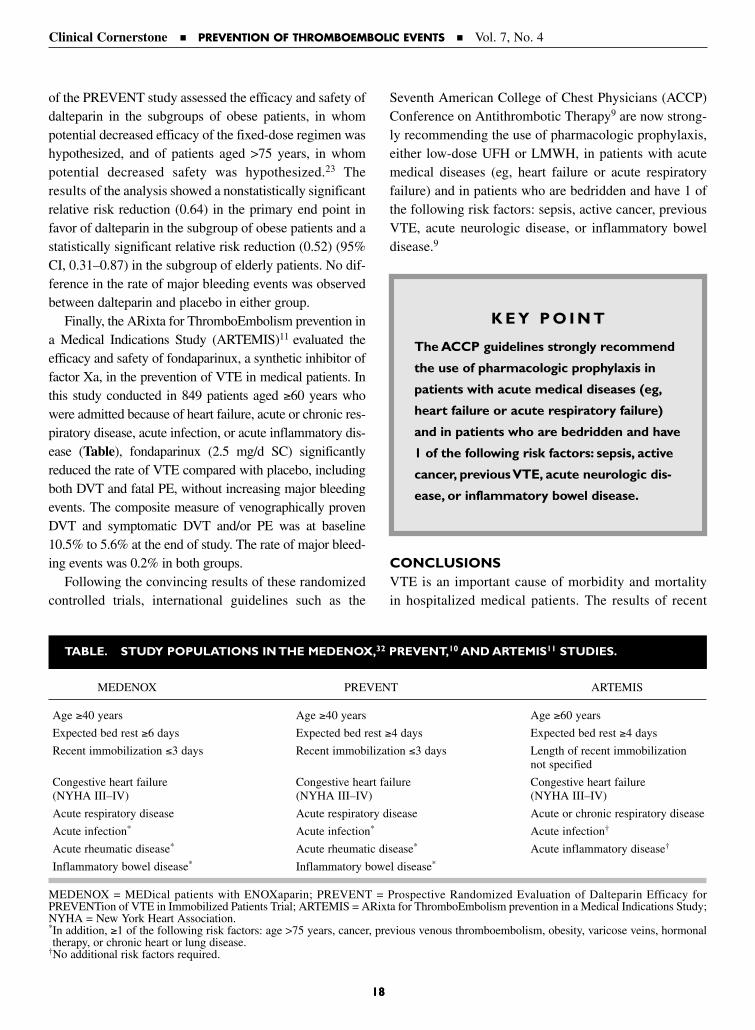
TABLE. STUDY POPULATIONS IN THE MEDENOX,32 PREVENT,10 AND ARTEMIS11 STUDIES.
MEDENOX PREVENT ARTEMIS
Age ≥40 years Age ≥40 years Age ≥60 years
Expected bed rest ≥6 days Expected bed rest ≥4 days Expected bed rest ≥4 days
Recent immobilization ≤3 days Recent immobilization ≤3 days Length of recent immobilizationnot specified
Congestive heart failure Congestive heart failure Congestive heart failure(NYHA III–IV) (NYHA III–IV) (NYHA III–IV)
Acute respiratory disease Acute respiratory disease Acute or chronic respiratory disease
Acute infection* Acute infection* Acute infection†
Acute rheumatic disease* Acute rheumatic disease* Acute inflammatory disease†
Inflammatory bowel disease* Inflammatory bowel disease*
MEDENOX = MEDical patients with ENOXaparin; PREVENT = Prospective Randomized Evaluation of Dalteparin Efficacy forPREVENTion of VTE in Immobilized Patients Trial; ARTEMIS = ARixta for ThromboEmbolism prevention in a Medical Indications Study;NYHA = New York Heart Association.*In addition, ≥1 of the following risk factors: age >75 years, cancer, previous venous thromboembolism, obesity, varicose veins, hormonaltherapy, or chronic heart or lung disease.
†No additional risk factors required.
K E Y P O I N T
The ACCP guidelines strongly recommend
the use of pharmacologic prophylaxis in
patients with acute medical diseases (eg,
heart failure or acute respiratory failure)
and in patients who are bedridden and have
1 of the following risk factors: sepsis, active
cancer, previous VTE, acute neurologic dis-
ease, or inflammatory bowel disease.
Clinical Cornerstone � PREVENTION OF THROMBOEMBOLIC EVENTS � Vol. 7, No. 4
19
clinical trials show that VTE in medical patients is apreventable disease. Furthermore, these trials have betterdefined the target population and have provided impor-tant information on the risk of bleeding in thesepatients. The results were consistent among the studies,despite some differences in the designs and despite thedifferent compounds assessed. Patient populations weresimilar in the MEDENOX and the PREVENT studiesand slightly different in ARTEMIS, in which inclusioncriteria were simplified. However, the same disease cate-gories were represented in all studies. The primary endpoints of the studies were based on the results of venogra-phy in the MEDENOX study and in ARTEMIS and on theresults of compression ultrasonography in the PREVENTstudy. These differences in the methods used to detectDVT explain why the rates of events differed among tri-als. However, the clinical relevance of asymptomaticDVT, either detected by venography or by compressionultrasonography, is well established. In all studies, therate of major bleeding events was minimal and it wassimilar between the active drug and placebo. However,caution must be taken in the presence of impaired renalfunction and other risk factors for bleeding. Thus, theresults of clinical studies of VTE prevention in medicalpatients clearly support the use of thromboprophylaxis inthis setting.
REFERENCES1. Sandler DA, Martin JF. Autopsy proven pulmonary
embolism in hospital patients: Are we detecting enoughdeep vein thrombosis? J R Soc Med. 1989;82:203–205.
2. Goldhaber SZ, Savage DD, Garrison RJ, et al. Risk factorsfor pulmonary embolism: The Framingham study. Am JMed. 1983;74:1023–1028.
3. Anderson FA, Wheeler HB, Goldberg RJ, et al. A population-based perspective of the hospital incidence andcase-fatality rates of deep vein thrombosis and pulmonaryembolism. The Worcester DVT study. Arch Intern Med.1991;151:933–938.
4. Lindblad B, Sternby NH, Bergqvist D. Incidence of venousthromboembolism verified by necropsy over 30 years. Br Med J. 1991;302:709–711.
5. Leizorovicz A, Mismetti P. Preventing venous thrombo-embolism in medical patients. Circulation. 2004;110(Suppl IV):IV13–IV19.
6. Bouthier J. The venous thrombotic risk in nonsurgicalpatients. Drugs. 1996;52(Suppl):16–29.
7. Goldhaber SZ, Dunn K, MacDougall RC. New onset ofvenous thromboembolism among hospitalized patients atBrigham and Women’s Hospital is caused more often byprophylaxis failure than by withholding treatment. Chest.2000;118:1680–1684.
8. Monreal M, Kakkar AK, Caprini JA, et al, and the RIETEInvestigators. The outcome after treatment of venousthromboembolism is different in surgical and acutely illmedical patients. Findings from the RIETE Registry.J Thromb Haemost. 2004;2:1892–1898.
9. Samama MM, Cohen AT, Darmon JY, et al, for theProphylaxis in Medical Patients with Enoxaparin StudyGroup. A comparison of enoxaparin with placebo for theprevention of venous thromboembolism in acutely illmedical patients. N Engl J Med. 1999;341:793–800.
10. Leizorovicz A, Cohen AT, Turpie AG, et al, for the PREVENT Medical Thromboprophylaxis Study. Ran-domized, placebo-controlled trial of dalteparin for the pre-vention of venous thromboembolism in acutely ill medicalpatients. Circulation. 2004;110:874–879.
11. Cohen AT, Davidson B, Gallus AS, et al. Fondaparinux forthe prevention of VTE in acutely ill medical patients.Artemis Study. Blood. 2003;102:15a.
12. Geerts WH, Pineo GF, Heit JA, et al. Prevention of venousthromboembolism: The Seventh ACCP Conference onAntithrombotic and Thrombolytic Therapy. Chest. 2004;126:338S–400S.
13. Ageno W, Squizzato A, Ambrosini F, et al. Thrombosis pro-phylaxis in medical patients: A retrospective review of clin-ical practice patterns. Haematologica. 2002;87:746–750.
14. Anderson FA Jr, Tapson VF, Decousus H, et al.IMPROVE, a multinational observational cohort study ofpractices in prevention of venous thromboembolism inacutely ill medical patients: A comparison with clinicalstudy patient populations. Blood. 2003;102:1146. Abstract.
15. Rahim SA, Panju A, Pai M, Ginsberg J. Venous throm-boembolism prophylaxis in medical inpatients: A retro-spective chart review. Thromb Res. 2003;111:215–219.
16. Belch JJ, Lowe GD, Ward AG, et al. Prevention of deepvein thrombosis in medical patients by low-dose heparin.Scott Med J. 1981;26:115–117.
17. Halkin H, Goldberg J, Modan M, Modan B. Reduction inmortality in general medical in-patients by low-doseheparin prophylaxis. Ann Intern Med. 1982;96:561–565.
18. Cade JF. High risk of the critically ill for venous throm-boembolism. Crit Care Med. 1982;10:448–450.
19. Gardlund B, for the Heparin Prophylaxis Study Group.Randomised, controlled trial of low dose heparin for pre-vention of fatal pulmonary embolism in patients withinfectious diseases. Lancet. 1996;347:1357–1361.
20. Poniewierski M, Barthels M, Kuhn M, Poliwoda H.Effectiveness of low molecular weight heparin (Fragmin)in the prevention of thromboembolism in internal medi-cine patients. A randomized double-blind study. Med Klin.(Munich). 1988; March 31;83(7):241–245, 278
21. Harenberg J, Kallenbach B, Martin U, et al. Randomizedcontrolled study of heparin and low molecular weightheparin for prevention of deep vein thrombosis in medicalpatients. Thromb Res. 1990;59:639–650.
22. Forette B, Wolmark Y. Calcium nadroparin in the preven-tion of thromboembolic disease in elderly subjects. Studyof tolerance. Press Med. 1995;24:567–571.
23. Harenberg J, Roebruck P, Heene DL. Subcutaneous lowmolecular weight heparin versus standard heparin and theprevention of thromboembolism in medical inpatients.The Heparin Study in Internal Medicine (HESIM) Group.Haemostasis. 1996;26:127–139.
19
Clinical Cornerstone � PREVENTION OF THROMBOEMBOLIC EVENTS � Vol. 7, No. 4
20
24. Manciet G, Vergnes C, Vaissié JJ, Boisseau MR. Studyof the efficacy and tolerance of Fraxipauni administeredlong term to older subjects, randomized blended study.Bounameaux H, Samama MM, Ten Cate JW, eds.Fraxiparine, 2nd International Symposium. Recent phar-macological and clinical data. Stuttgart, NY: Schattauer;1990:55–59.
25. Bergmann JF, Caulin C. Heparin prophylaxis in bedriddenpatients. Lancet. 1996;348:205–206.
26. Dahan R, Houlbert D, Caulin C, et al. Prevention of deepvein thrombosis in elderly medical in-patients by a lowmolecular weight heparin: A randomized double-blindtrial. Haemostasis. 1986;16:159–164.
27. Lechler E, Schramm W, Flosbach CW. The venous throm-botic risk in non-surgical patients: Epidemiological data andefficacy/safety profile of a low molecular weight heparin(Enoxaparin). Haemostasis. 1996;26(Suppl 2):49–56.
28. Bergmann JF, Neuhart E. A multicenter randomized double-blind study of enoxaparin compared with unfractionatedheparin in the prevention of venous thromboembolic dis-ease in elderly in-patients bedridden for an acute medicalillness. The Enoxaparin in Medicine Study Group. ThrombHaemost. 1996;76:529–534.
29. Kleber FX, Witt C, Vogel G, et al. Randomized compari-son of enoxaparin with unfractionated heparin for the pre-vention of venous thromboembolism in medical patientswith heart failure or severe respiratory disease. Am Heart J.2003;145:614–621.
30. Mismetti P, Laporte-Simitsidis S, Tardy B, et al. Preventionof venous thromboembolism in internal medicine withunfractionated or low molecular weight heparins: A meta-analysis of randomised clinical trials. Thromb Haemost.2000;83:14–19.
31. Alikhan R, Cohen AT, Combe S, et al. Prevention ofvenous thromboembolism in medical patients with enoxa-parin: A subgroup analysis of the MEDENOX study.Blood Coagul Fibrinolysis. 2003;14:341–346.
32. Alikhan R, Cohen AT, Combe S, et al. Risk factors forvenous thromboembolism in hospitalized patients withacute medical illness: Analysis of the MEDENOX Study.Arch Intern Med. 2004;164:963–968.
33. Kucher N, Leizorovicz A, Vaitkus PT, et al. Efficacy andsafety of fixed low-dose dalteparin in preventing venousthromboembolism among obese or elderly hospitalizedpatients: A subgroup analysis of the PREVENT trial. ArchIntern Med. 2005;165:341–345.
20
Address correspondence to: Walter Ageno, MD, Department of Clinical Medicine, Ospedale di Circolo, Universityof Insubria, Viale Borri 57, 21100 Varese, Italy. E-mail: [email protected]. Alexander G.G. Turpie, MD, HamiltonHealth Sciences Corporation, McMaster University, 237 Barton Street East, Hamilton, Ontario, Canada L8L 2X2. E-mail: [email protected]
EDITORIAL BOARDAlthough the meta-analysis by Mismetti et aldemonstrated that unfractionated heparin (UFH)and low-weight-molecular heparin (LMWH) werecomparable in preventing deep vein thrombosis(DVT) in medical patients, it also showed thatLMWH was associated with a 50% lower risk ofmajor bleeding. Is LMWH really that much saferthan UFH?
TURPIENo, I don’t view the meta-analysis as conclusive in that
regard. It is important to recognize that the studies
included in the meta-analysis used varying dosages of
LMWH. These doses included doses that were low
enough to be ineffective against thrombosis; such doses,
in turn, would naturally be associated with a lower risk
for bleeding. The bottom line is that this may cause
LMWHs to falsely appear safer in comparison to UFH
than they actually are when used for prophylaxis.
EDITORIAL BOARDWhat about in patients being treated for establishedDVT?
TURPIEThat’s a different story. Pretty solid evidence exists that
LMWHs are safer than UFH in patients with venous
thromboembolism (VTE) since the doses used in such
patients are much higher across the board.
EDITORIAL BOARDAs prophylactic agents, are UFH and LMWH com-parable in terms of efficacy?
TURPIEThe meta-analysis certainly supports that. In addition, if
you examine studies comparing them in specific set-
tings, such as heart failure and respiratory failure, no
statistically significant difference has been demonstrat-
ed between LMWH versus UFH given at a dosage of
5000 U SC TID. In patients who experienced a stroke,
however, LMWH was found to be more effective than
UFH in one study.
EDITORIAL BOARDCan the higher percentage of symptomatic VTEcases seen among medical patients be attributablesimply to there being more medical patients who arehospitalized than surgical patients?
TURPIEThat’s an interesting thought. However, in point of fact,
there are at least the same, if not more, surgical patients
in hospitals than medical patients. Yet there are 4 times
as many medical patients dying from pulmonary
embolism (PE) than surgical patients. The higher risk
seen in medical patients likely results from medical
patients simply being sicker. A lot more surgical patients
who are hospitalized are in good health since a number
of young people come in for elective procedures.
Overall, if one looks in terms of at-risk patients, fewer
surgical patients are at risk for VTE than medical
patients in the hospital.
EDITORIAL BOARDWouldn’t that also explain why worse clinical out-comes are seen in medical patients with VTE?
AGENOExactly. Monreal et al simply confirmed the observation
that medical patients in the hospital tend to be sicker
than surgical patients. Such patients are at high risk for
bleeding as well as at higher risk for DVT. In addition,
when VTE occurs, the consequences are more severe.
EDITORIAL BOARDDo medical patients require higher doses for DVTprophylaxis than surgical patients?
TURPIEYes, they do. The MEDENOX study compared 2 doses
of LMWH, 20 mg and 40 mg, as prophylaxis against
VTE. Although the 20-mg daily dose of enoxaparin has
Clinical Cornerstone � PREVENTION OF THROMBOEMBOLIC EVENTS � Vol. 7, No. 4
Dialogue Box
21
been shown to be effective in moderate-risk surgical
patients, this dose proved ineffective in medical patients
in the MEDENOX trial. Instead, a 40-mg dose was
required to be effective.
EDITORIAL BOARDIs there a higher risk of converting a thromboticstroke to a hemorrhagic one in the setting of anacute stroke without evidence of hemorrhage on CTscan of the brain?
AGENONo. Prophylactic doses of LMWH or UFH appear safe
in such patients. Therapeutic doses, on the other hand,
would significantly increase the risk of bleeding in such
patients.
EDITORIAL BOARDWhat is meant by “rebound” when used in the set-ting of DVT prophylaxis?
TURPIEThe term rebound is somewhat a misnomer. Although
there is evidence that there’s an ongoing risk in medical
patients, there’s no evidence that the risk becomes
greater in patients after DVT prophylaxis. What is seen
is an ongoing or continuing risk, and the big question is:
How long should we keep prophylaxis going? This issue
is being looked at in a study called EXCLAIM, which
is investigating the use of enoxaparin 1 month versus
1 week in medical patients.
EDITORIAL BOARDThe PREVENT trial seemed to suggest that thedosage may need to be higher in obese patients.What are your thoughts?
TURPIEIt’s still unresolved. Most of the data do not support the
use of dose adjustment, but I don’t think the studies
have been sufficiently powered to give us a firm answer
to that one.
EDITORIAL BOARDHow does fondaparinux compare to LMWH in VTEprophylaxis in medical patients?
TURPIEAlthough it has not yet been published, the ARTEMIS
trial demonstrated a reduced risk of fatal PE in medical
patients treated with fondaparinux compared with place-
bo. ARTEMIS was a prospective, randomized, double-
blind trial and is the only study to show a reduced risk
of fatal PE in medical patients. However, one really
can’t conclude that fondaparinux is better than heparin
because there never has been a direct comparison
between the 2 agents in this setting. What we can say is
that they both are effective as DVT prophylactic agents
and that prophylaxis is good.
EDITORIAL BOARDWhat specific categories of medical patients warrantstrong consideration for DVT prophylaxis duringhospitalization?
TURPIEA good reference would be a recent publication byAnder Cohen in the Journal of Thrombosis and
Haemostasis, which really pinpoints the at-risk medicalpatients. Such patients include those with heart failure,respiratory failure, and acute infections.
AGENOStroke patients also should be added to that list.
22
Clinical Cornerstone � PREVENTION OF THROMBOEMBOLIC EVENTS � Vol. 7, No. 4
Dialogue Box
Venous thromboembolism (VTE) is a condition that has multiple causes but few warning signs. Consequently,the 2 manifestations of VTE—pulmonary embolism and deep vein thrombosis—often go unpredicted. This isespecially true for medical patients. Treatment guidelines indicate that most hospitalized patients should receiveprophylaxis for VTE. This report discusses these guidelines, the high prevalence of VTE among medical patients,and clinical studies of thromboprophylaxis in medically ill patients.
VTE prophylaxis continues to be underutilized in medically ill patients. These patients are at significant risk ofVTE and require prophylaxis, an objective that is supported by the recent guidelines of the American College ofChest Physicians. In addition, several lines of clinical evidence support the use of prophylaxis in this subgroup ofpatients. Improved systems are needed in medically ill patients to help improve outcomes and compliance for theuse of VTE prophylaxis. (Clinical Cornerstone. 2005;7[4]:23–31) Copyright © 2005 Excerpta Medica, Inc.
ARTHUR WHEELER, MDAssociate Professor of MedicineVanderbilt University Medical CenterDivision of Allergy, Pulmonary and Critical Care MedicineNashville,Tennessee
Venous Thromboembolism in MedicallyIll Patients: Identifying Risk andStrategies for Prevention
Clinical Cornerstone � PREVENTION OF THROMBOEMBOLIC EVENTS � Vol. 7, No. 4
23
Venous thromboembolism (VTE) is a condition that hasmultiple causes but few warning signs. Consequently,pulmonary embolism (PE) and deep vein thrombosis(DVT), the 2 components of VTE, often go unpredicted.This is especially true for medical patients, in whomthromboembolic risk has been less clearly establishedthan for surgical patients.
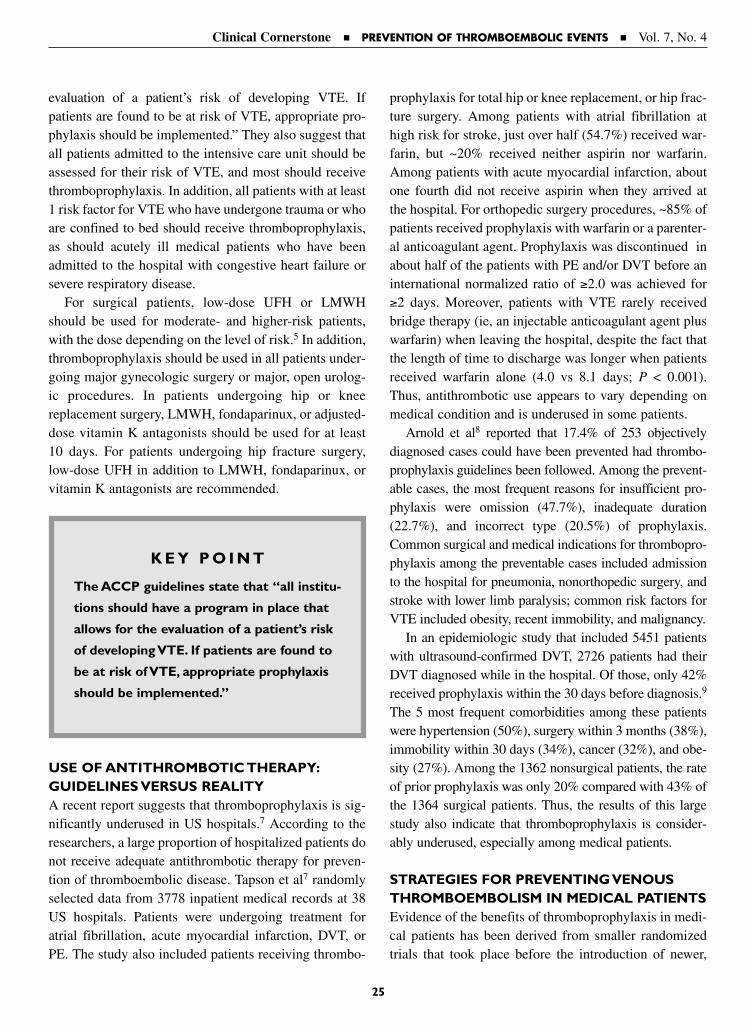
Several factors are thought to be associated with anincreased risk of VTE, including periods of reducedmobility, increasing age, major surgery, prior VTE, andchronic heart failure (Table I).1 Strong risk factors forVTE include hip or knee replacement and major generalsurgery; weaker risk factors include increasing age andlaparoscopic surgery. Given the wide range of risk asso-ciated with various factors, the decision to provide VTEprophylaxis should take into account the specific risk ofeach patient1; however, guidelines indicate that most hos-pitalized patients should receive prophylaxis for VTE.This report discusses these guidelines, the high preva-lence of VTE among medical patients, and clinical stud-ies of thromboprophylaxis in medically ill patients.
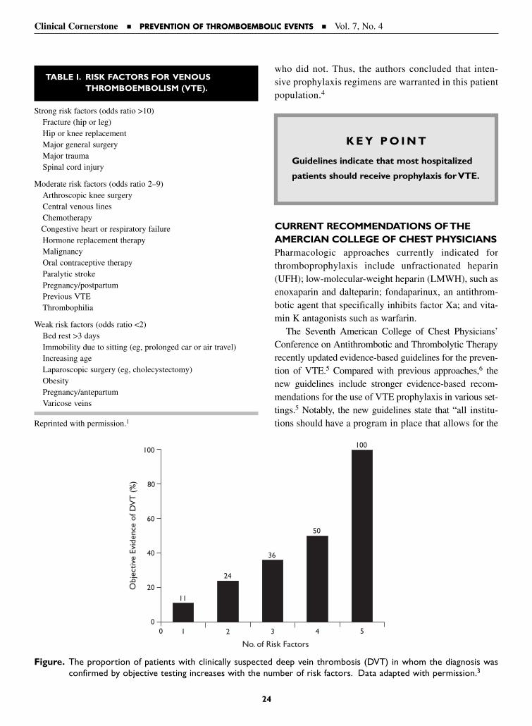
PREVALENCE OF VENOUS THROMBOEMBOLISM AMONG MEDICAL PATIENTS In a typical hospital population, 78% of patients have ≥1 risk factors for VTE, and ~20% of patients have at least3 risk factors.2 Overall, the incidence of VTE among medi-cally ill patients is estimated to be ~18% to 23% (Figure).3
The benefits of treating and preventing VTE in surgi-cal patients are well established; however, many at-riskhospitalized medical patients do not appear to be receiv-ing adequate prophylaxis. In 1995, Hirsch et al4 reportedthat the rate of DVT was unexpectedly high in medicalpatients in the intensive care unit, despite the fact thatprophylaxis was administered to 61% of the patients.DVT was detected with ultrasound in one third of 100eligible patients over an 8-month period. About half of the cases involved proximal lower-extremity DVT. Fur-thermore, no difference in risk factors—including age, sex,body mass index, diagnosis of cancer, recent surgery, orduration of hospitalization before DVT detection—wasobserved among those who developed DVT versus those
who did not. Thus, the authors concluded that inten-sive prophylaxis regimens are warranted in this patientpopulation.4
CURRENT RECOMMENDATIONS OF THEAMERCIAN COLLEGE OF CHEST PHYSICIANSPharmacologic approaches currently indicated forthromboprophylaxis include unfractionated heparin(UFH); low-molecular-weight heparin (LMWH), such asenoxaparin and dalteparin; fondaparinux, an antithrom-botic agent that specifically inhibits factor Xa; and vita-min K antagonists such as warfarin.
The Seventh American College of Chest Physicians’Conference on Antithrombotic and Thrombolytic Therapyrecently updated evidence-based guidelines for the preven-tion of VTE.5 Compared with previous approaches,6 thenew guidelines include stronger evidence-based recom-mendations for the use of VTE prophylaxis in various set-tings.5 Notably, the new guidelines state that “all institu-tions should have a program in place that allows for the
Clinical Cornerstone � PREVENTION OF THROMBOEMBOLIC EVENTS � Vol. 7, No. 4
24
Figure. The proportion of patients with clinically suspected deep vein thrombosis (DVT) in whom the diagnosis wasconfirmed by objective testing increases with the number of risk factors. Data adapted with permission.3
100
20
40
60
0
Obj
ectiv
e Ev
iden
ce o
f DV
T (
%)
0
11
36
50
100
5431 2
80
24
No. of Risk Factors
K E Y P O I N T
Guidelines indicate that most hospitalized
patients should receive prophylaxis for VTE.
TABLE I. RISK FACTORS FOR VENOUS THROMBOEMBOLISM (VTE).
Strong risk factors (odds ratio >10)Fracture (hip or leg)Hip or knee replacementMajor general surgeryMajor traumaSpinal cord injury
Moderate risk factors (odds ratio 2–9)Arthroscopic knee surgeryCentral venous linesChemotherapyCongestive heart or respiratory failureHormone replacement therapyMalignancyOral contraceptive therapyParalytic strokePregnancy/postpartumPrevious VTEThrombophilia
Weak risk factors (odds ratio <2)Bed rest >3 daysImmobility due to sitting (eg, prolonged car or air travel)Increasing ageLaparoscopic surgery (eg, cholecystectomy)ObesityPregnancy/antepartumVaricose veins
Reprinted with permission.1
evaluation of a patient’s risk of developing VTE. Ifpatients are found to be at risk of VTE, appropriate pro-phylaxis should be implemented.” They also suggest thatall patients admitted to the intensive care unit should beassessed for their risk of VTE, and most should receivethromboprophylaxis. In addition, all patients with at least1 risk factor for VTE who have undergone trauma or whoare confined to bed should receive thromboprophylaxis,as should acutely ill medical patients who have beenadmitted to the hospital with congestive heart failure orsevere respiratory disease.
For surgical patients, low-dose UFH or LMWHshould be used for moderate- and higher-risk patients,with the dose depending on the level of risk.5 In addition,thromboprophylaxis should be used in all patients under-going major gynecologic surgery or major, open urolog-ic procedures. In patients undergoing hip or kneereplacement surgery, LMWH, fondaparinux, or adjusted-dose vitamin K antagonists should be used for at least10 days. For patients undergoing hip fracture surgery,low-dose UFH in addition to LMWH, fondaparinux, orvitamin K antagonists are recommended.
USE OF ANTITHROMBOTIC THERAPY:GUIDELINES VERSUS REALITYA recent report suggests that thromboprophylaxis is sig-nificantly underused in US hospitals.7 According to theresearchers, a large proportion of hospitalized patients donot receive adequate antithrombotic therapy for preven-tion of thromboembolic disease. Tapson et al7 randomlyselected data from 3778 inpatient medical records at 38US hospitals. Patients were undergoing treatment foratrial fibrillation, acute myocardial infarction, DVT, orPE. The study also included patients receiving thrombo-
prophylaxis for total hip or knee replacement, or hip frac-ture surgery. Among patients with atrial fibrillation athigh risk for stroke, just over half (54.7%) received war-farin, but ~20% received neither aspirin nor warfarin.Among patients with acute myocardial infarction, aboutone fourth did not receive aspirin when they arrived atthe hospital. For orthopedic surgery procedures, ~85% ofpatients received prophylaxis with warfarin or a parenter-al anticoagulant agent. Prophylaxis was discontinued inabout half of the patients with PE and/or DVT before aninternational normalized ratio of ≥2.0 was achieved for≥2 days. Moreover, patients with VTE rarely receivedbridge therapy (ie, an injectable anticoagulant agent pluswarfarin) when leaving the hospital, despite the fact thatthe length of time to discharge was longer when patientsreceived warfarin alone (4.0 vs 8.1 days; P < 0.001).Thus, antithrombotic use appears to vary depending onmedical condition and is underused in some patients.
Arnold et al8 reported that 17.4% of 253 objectivelydiagnosed cases could have been prevented had thrombo-prophylaxis guidelines been followed. Among the prevent-able cases, the most frequent reasons for insufficient pro-phylaxis were omission (47.7%), inadequate duration(22.7%), and incorrect type (20.5%) of prophylaxis.Common surgical and medical indications for thrombopro-phylaxis among the preventable cases included admissionto the hospital for pneumonia, nonorthopedic surgery, andstroke with lower limb paralysis; common risk factors forVTE included obesity, recent immobility, and malignancy.
In an epidemiologic study that included 5451 patientswith ultrasound-confirmed DVT, 2726 patients had theirDVT diagnosed while in the hospital. Of those, only 42%received prophylaxis within the 30 days before diagnosis.9
The 5 most frequent comorbidities among these patientswere hypertension (50%), surgery within 3 months (38%),immobility within 30 days (34%), cancer (32%), and obe-sity (27%). Among the 1362 nonsurgical patients, the rateof prior prophylaxis was only 20% compared with 43% ofthe 1364 surgical patients. Thus, the results of this largestudy also indicate that thromboprophylaxis is consider-ably underused, especially among medical patients.
STRATEGIES FOR PREVENTING VENOUSTHROMBOEMBOLISM IN MEDICAL PATIENTS Evidence of the benefits of thromboprophylaxis in medi-cal patients has been derived from smaller randomizedtrials that took place before the introduction of newer,
Clinical Cornerstone � PREVENTION OF THROMBOEMBOLIC EVENTS � Vol. 7, No. 4
25
K E Y P O I N T
The ACCP guidelines state that “all institu-
tions should have a program in place that
allows for the evaluation of a patient’s risk
of developing VTE. If patients are found to
be at risk of VTE, appropriate prophylaxis
should be implemented.”
more effective therapies. Consequently, the effects ofthromboprophylaxis for medical patients are less clear thanthey are for surgical patients.10 Various studies are begin-ning to confirm the benefits of thromboprophylaxis specif-ically in the medical patient population. These include theprophylaxis in MEDical patients with ENOXaparin(MEDENOX) trial,11 the PRophylaxis in Internal Medicinewith Enoxaparin (PRIME) study,12 the thromboem-bolism PRevention IN Cardiopulmonary diseases withEnoxaparin (PRINCE) study,13 the Prospective Evaluationof Dalteparin Efficacy for PREVENTion of VenousThromboembolism in Immobilized Patients (PREVENT)study,14 and the ARixtra for ThromboEmbolism preventionin a Medical Indications Study (ARTEMIS).15 Collectively,the findings suggest that LMWH (enoxaparin 40 mg ordalteparin 5000 IU SC QD for 10 days), as well as fon-daparinux 2.5 mg SC QD for 10 days, demonstrate afavorable risk/benefit ratio for the prevention of VTEin acutely ill medical patients.3
In the landmark MEDENOX trial, 2 doses of enoxa-parin (20 and 40 mg/d) were compared with placebo inmedical patients.11 Prophylactic treatment with 40 mg/d ofenoxaparin given SC was found to significantly reduce therisk of VTE compared with placebo (5.5% for enoxaparinvs 14.9% for placebo; relative risk [RR], 0.37; 97.6% CI,0.22–0.63; P < 0.001). In addition, the incidence of VTEat day 14 was significantly lower for patients receivingenoxaparin 40 mg compared with placebo (P < 0.001 forall VTE, and P = 0.037 for proximal DVT), and this benefit was maintained at 3 months. A subsequent analy-sis of data from the MEDENOX trial also indicated thatthe risk of VTE was increased in patients with acuteinfectious disease, cancer, age >75 years, and a history ofVTE (Table II).16
The PREVENT study compared dalteparin with place-bo in the prevention of VTE in 3706 medical patients.Patients were randomly assigned to receive either dalte-parin 5000 IU SC QD or placebo for 14 days.14 The inci-
Clinical Cornerstone � PREVENTION OF THROMBOEMBOLIC EVENTS � Vol. 7, No. 4
26
TABLE II. INCIDENCE OF VENOUS THROMBOEMBOLISM (DAYS 1–14) BY REASON OF ILLNESS.
No. (%) of Patients/Events*
All Placebo Group Enoxaparin Sodium GroupIllness (N = 866) (n = 288) (40 mg) (n = 291)
Heart failureYes 34/290 (11.7) 14/96 (14.6) 4/99 (4.0)No 68/576 (11.8) 29/192 (15.1) 12/192 (6.3)
NYHA class IIIYes 23/217 (10.6) 9/73 (12.3) 4/78 (5.1)No 79/649 (12.2) 34/215 (15.8) 12/213 (5.6)
NYHA class IVYes 11/73 (15.1) 5/23 (21.7) 0/21 (0)No 91/793 (11.5) 38/265 (14.3) 16/270 (5.9)
Acute respiratory diseaseYes 42/457 (9.2) 20/153 (13.1) 5/153 (3.3)No 60/409 (14.7) 23/135 (17.1) 11/138 (8.0)
Acute infectious diseaseYes 64/463 (13.8) 24/155 (15.5) 10/159 (6.3)No 38/403 (9.4) 19/133 (14.3) 6/132 (4.5)
Acute infectious and respiratory diseaseYes 27/239 (11.3) 13/79 (16.5) 4/88 (4.6)No 75/627 (12.0) 30/209 (14.3) 12/203 (5.9)
Acute rheumatic diseaseYes 14/78 (17.9) 6/29 (20.7) 2/20 (10.0)No 88/788 (11.2) 37/259 (14.3) 14/271 (5.2)
Inflammatory bowel diseaseYes 0/5 (0) 0/1 (0) 0/3 (0)No 102/861 (11.8) 43/287 (15.0) 16/288 (5.6)
NYHA = New York Heart Association. *Number of patients in subgroup/number of venous thromboembolic events.Reprinted with permission.16
dence of VTE was 4.96% in the placebo group versus2.77% in the dalteparin group (RR reduction, 45%; P <0.002). Likewise, the ARTEMIS trial compared fonda-parinux with placebo in acutely ill, elderly medicalpatients.15 The incidence of VTE at day 15 was 10.5% inthe placebo group versus 5.6% in the fondaparinux group(RR reduction, 49.5%; P < 0.003).
Enoxaparin has also been compared in head-to-headtrials with UFH. The PRIME study, a randomized, double-blind controlled trial comparing enoxaparin 40 mg/dwith UFH TID in a high-risk group of 959 hospitalizedmedical patients, found that enoxaparin was comparableto UFH in preventing VTE, with fewer adverse events.12
Similarly, the PRINCE study compared the efficacy andsafety of 10 days of enoxaparin (40 mg/d SC) versusUFH (5000 IU TID SC) in 665 medical patients withsevere cardiopulmonary disease.13 As with the PRIMEstudy, the results indicated that enoxaparin was as effec-tive as UFH 5000 IU given TID.
Enoxaparin has also been found to be superior to UFHin medical patients who are at increased to high risk ofVTE.17 Harenberg et al17 analyzed data from patientswith severe respiratory disease, severe heart failure, oracute ischemic stroke. A total of 877 patients receivedeither 40 mg enoxaparin QD or 5000 IU UFH TID.Among the 630 patients eligible for the efficacy analysis,thromboembolic events with subsequent death occurredin 15.6% of the patients in the enoxaparin group com-pared with 22.1% in the UFH group. Risk of VTE washighest among patients with acute ischemic stroke, fol-lowed by patients with severe heart failure and severerespiratory disease. Bleeding events occurred in 1.8% ofthe enoxaparin group versus 3.2% of the UFH group.
Finally, the ongoing EXtended CLinical Prophylaxisin Acutely Ill Medical Patients with Prolonged Immo-
bilization Study18 will help clarify the benefits of extend-ed prophylaxis among medical patients. This study isassessing the role of extended prophylaxis with enoxa-parin among ~5800 medical patients. Patients willreceive enoxaparin 40 mg SC QD versus placebo for 28 days after 10 days of enoxaparin. Study end pointsinclude the incidence of ultrasound-confirmed VTE,major bleeding, and mortality.
CONCLUSIONSGuidelines indicate that most hospitalized patientsshould receive prophylaxis for VTE; however, prophy-laxis continues to be underutilized in medically illpatients. The decision to provide prophylaxis should takeinto account the specific risk of each patient and all insti-tutions should have a program in place that evaluates apatient’s risk of developing VTE.
REFERENCES1. Anderson FA Jr, Spencer FA. Risk factors for venous
thromboembolism. Circulation. 2003;107:I9–I16. 2. Anderson FA Jr, Wheeler HB, Goldberg RJ, et al. The
prevalence of risk factors for venous thromboembolismamong hospital patients. Arch Intern Med. 1992;152:1660–1664.
3. Gerotziafas GT, Samama MM. Prophylaxis of venousthromboembolism in medical patients. Curr Opin PulmMed. 2004;10:356–365.
4. Hirsch DR, Ingenito EP, Goldhaber SZ. Prevalence ofdeep venous thrombosis among patients in medical inten-sive care. JAMA. 1995;274:335–337.
5. Geerts WH, Pineo GF, Heit JA, et al. Prevention of venousthromboembolism: The Seventh ACCP Conference onAntithrombotic and Thrombolytic Therapy. Chest. 2004;126:338S–400S.
6. Geerts WH, Heit JA, Clagett GP, et al. Prevention ofvenous thromboembolism. Chest. 2001;119:132S–175S.
7. Tapson VF, Hyers TM, Waldo AL, et al. Antithrombotictherapy practices in US hospitals in an era of practiceguidelines. Arch Intern Med. 2005;165:1458–1464.
8. Arnold DM, Kahn SR, Shrier I. Missed opportunities forprevention of venous thromboembolism: An evaluation ofthe use of thromboprophylaxis guidelines. Chest. 2001;120:1964–1971.
9. Goldhaber SZ, Tapson VF, DVT FREE SteeringCommittee. A prospective registry of 5,451 patients withultrasound-confirmed deep vein thrombosis. Am J Cardiol.2004;93:259–262.
10. Gallus AS, Nurmohammed M, Kearon C, Prins M.Thromboprophylaxis in non-surgical patients: Who, whenand how? Haemostasis. 1998;28(Suppl 3):71–82.
Clinical Cornerstone � PREVENTION OF THROMBOEMBOLIC EVENTS � Vol. 7, No. 4
27
K E Y P O I N T
Collectively, study findings suggest that
LMWH, as well as fondaparinux, demon-
strate a favorable risk/benefit ratio for
the prevention of VTE in acutely ill medical
patients.
11. Samama MM, Cohen AT, Darmon JY, et al, for theProphylaxis in Medical Patients with Enoxaparin StudyGroup. A comparison of enoxaparin with placebo for theprevention of venous thromboembolism in acutely illmedical patients. N Engl J Med. 1999;341:793–800.
12. Lechler E, Schramm W, Flosbach CW, for the Prime StudyGroup. The venous thrombotic risk in non-surgical patients:Epidemiological data and efficacy/safety profile of a low-molecular-weight heparin (enoxaparin). Haemostasis.1996;26(Suppl 2):49–56.
13. Kleber FX, Witt C, Flosbach CW, et al. Study to comparethe efficacy and safety of the LMWH enoxaparin and stan-dard heparin in the prevention of thromboembolic eventsin medical patients with cardiopulmonary diseases. AnnHematol. 1998;76(Suppl 1):A93. Abstract.
14. Leizorovicz A, Cohen AT, Turpie AG, et al, for the PREVENT Medical Thromboprophylaxis Study Group.
Randomized, placebo-controlled trial of dalteparin for theprevention of venous thromboembolism in acutely illmedical patients. Circulation. 2004;110:874–879.
15. Cohen AT, Davidson BL, Gallus AS, et al. Fondaparinuxfor the prevention of VTE in acutely ill medical patients.Blood. 2003;102:42. Abstract.
16. Alikhan R, Cohen AT, Combe S, et al. Risk factors forvenous thromboembolism in hospitalized patients withacute medical illness: Analysis of the MEDENOX Study.Arch Intern Med. 2004;164:963–968.
17. Harenberg J, Schomaker U, Flosbach CW, et al. Enoxaparinis superior to unfractionated heparin in the prevention ofthromboembolic events in medical patients at increasedthromboembolic risk. Blood. 1999;9(Suppl 1):399a.
18. Clinical Trials. Gov. The EXCLAIM Trial. Available at:http://www.clinicaltrials.gov/ct/show/NCT00077753.Accessed September 13, 2005.
Clinical Cornerstone � PREVENTION OF THROMBOEMBOLIC EVENTS � Vol. 7, No. 4
28
Address correspondence to: Arthur Wheeler, MD, Vanderbilt University Medical Center, Division of Allergy,Pulmonary and Critical Care Medicine, 1161 21st Avenue South, Suite T-1218 MCN, Nashville, TN 37232–2650.
EDITORIAL BOARDTo what do you attribute the 18% to 23% incidenceof venous thromboembolism (VTE) seen in medical-ly ill patients in the hospital setting?
WHEELERA number of factors play a role. The medical population
has always been an underappreciated at-risk group.
Although historically they may have been less ill than
their surgical counterparts, this is clearly no longer the
case. These days medical patients who make it into
our hospitals are substantially more ill than they were
20 years ago, both acutely and chronically, since the
threshold for getting through the doors of most hospi-
tals has gone up. Another factor is we simply discharge
patients substantially sooner than we used to. By dis-
charging patients earlier, the level of severity of illness
in hospitalized medical patients is higher. Although
studies show varying risks of VTE across different
populations within the hospital (with probably some of
the highest-risk medical patients being those with con-
gestive heart failure, chronic obstructive lung disease,
and cancer), most medical patients in the hospital have
multiple risk factors, and the more risk factors you have,
the higher the risk.
EDITORIAL BOARDWhy the discrepancy between the baseline 18% to 23% figure you cited and the 5% to 10% inci-dence seen in placebo groups in the ARTEMIS and PREVENT trials?
WHEELERTwo possible differences are the populations studied and
the method used to detect deep vein thrombosis (DVT).
In trials such as these, subjects at the highest risk for the
disease are invariably excluded from study. For example,
some patients (such as those with advanced cancer or in
the intensive care unit) may be perceived to be at such
high risk that researchers determine it would be unethi-
cal to include them in a placebo-controlled trial. The
second factor lies in differences in the varying sensitivi-
ties of the tests used to detect DVT. For example, if ultra-
sound is used, you might find an overall incidence of the
disease somewhere between 5% and 10%. In those same
patients, when using a more sensitive test such as con-
trast venography, you might find an incidence between
15% and 20%. If you use an investigative tool capable of
detecting very small clots, such as radiolabeled fibrino-
gen scanning, you might find clots in 25% or 30% of
these patients.
EDITORIAL BOARDHow do the different DVT prophylactic options com-pare with regard to efficacy?
WHEELERIf all things were equal, the greatest risk reduction for
DVT has been shown with low-molecular-weight
heparin, followed closely by unfractionated heparin
that, when given TID in a fixed dose, reduces risk by
about two thirds. Next best would be pneumatic com-
pression devices, which fail roughly 50% of the time.
Elastic stockings provide a 25% to 30% risk reduction.
EDITORIAL BOARDSince none of these methods are wholly effective,should multiple prophylactic measures be consid-ered in patients felt to be at particularly high risk?
WHEELERThat is a consideration. Let’s say patients have a 60%
chance of getting a calf DVT from a hip replacement
procedure. With 1 intervention, I might be able to cut
that in half—down to 30%. Only half of these (15%)
will propagate into the thigh. Half of these (7%)
become pulmonary emboli. Most of these VTE cases
will be asymptomatic, with maybe 10% of them fatal,
which comes to 1 in 100 patients provided the prophy-
lactic measure has a fatality. Some people would say
that 1 in 100 is good enough. Others would instead
maintain that when you have a very high-risk patient
(such as patients undergoing joint replacement, hip
fracture surgery, or a quadriplegic patient following
Clinical Cornerstone � PREVENTION OF THROMBOEMBOLIC EVENTS � Vol. 7, No. 4
Dialogue Box
29
trauma), you should use 2 methods. Some data exist that
the so-called “belt and suspenders” strategy of adding a
mechanical device to chemical anticoagulation is more
effective than either method alone.
EDITORIAL BOARDPlease discuss further the rationale for continuingDVT prophylactic therapy in patients following hos-pital discharge.
WHEELERThere are 6 published studies in the orthopedic literature
looking at extending DVT prophylaxis after discharge in
patients following high-risk procedures such as knee
replacement, hip replacement, and hip-fracture surgery.
All of these studies were relatively consistent in showing
that extending prophylaxis, either for 3 or 4 weeks, re-
duced the number of symptomatic clots that occurred
and at least 1 study actually claimed a mortality benefit.
This would seem almost intuitive, seeing that in the past
these patients typically would have spent 2 weeks in the
hospital following hip replacement and would have been
offered DVT prophylaxis during that time. In my hospital,
a patient who receives a hip replacement now may be dis-
charged in 3 days, reducing the period of DVT prophylax-
is from 2 weeks down to 3 to 4 days. That’s likely where
the benefit comes—extending prophylaxis a little bit
longer into the recovery time at home, where the patient is
still relatively immobile with still-resolving thrombogenic
postsurgical changes. Another area of potential benefit is
in oncology patients after extensive intra-abdominal and
pelvic cancer resection. The ongoing EXCLAIM trial
may be more relevant to the average practitioner because
it’s looking at a group of patients who internists and fam-
ily doctors are much more likely to see—people in the
hospital with congestive heart failure, chronic obstructive
pulmonary disease, pneumonia, or rheumatologic disease
who are being discharged with the eventual hope that
someday they’re going to improve but who still aren’t
fully ambulatory. With regard to the latter, patients in the
study are being stratified by their levels of activity, such as
whether they can walk to the bathroom or not.
EDITORIAL BOARDIs there something about hospitalization that makeshospitalized medical patients at greater risk forDVT than the same patients in the nursing homesetting?
WHEELERThat’s a very common question I receive from internists
who have difficulty seeing the need for DVT prophylax-
is in a patient admitted from a nursing home, whether it
be for urinary tract infection with hypotension, aspira-
tion pneumonitis, or bed sores with cellulitis. Since
such patients look the same to them in the hospital as
they did in the nursing home, they sometimes have a
hard time seeing why they should be offered DVT pro-
phylaxis. My response is the patient must really not
look the same; otherwise, they wouldn’t have been
admitted to the hospital. There had to be something
different about the patient—whether it be the fever or
leukocytosis or dyspnea/hypoxia—that made you con-
cerned enough to put the patient in the hospital.
EDITORIAL BOARDAre you saying that hospitalization is an indicator ofa worsening of 1 or more components of Virchow’striad?
WHEELERAbsolutely. There was an article by John Heit a number
of years ago that provided a 25-year retrospective of all
cases of VTE disease diagnosed in Olmstead County.
He showed that the disease frequency was roughly 200
times higher among hospitalized patients than those
who were ambulatory. Interestingly, about half of all the
cases diagnosed outside the hospital occurred in
patients who had been in a hospital or a nursing home
in the preceding 90 days.
EDITORIAL BOARDFrom the standpoint of cancer as a risk factor, arethere some tumors that are so thrombogenic thatyou would be inclined to initiate DVT prophylaxis?
30
Clinical Cornerstone � PREVENTION OF THROMBOEMBOLIC EVENTS � Vol. 7, No. 4
Dialogue Box

WHEELERI would say any metastatic carcinoma. Of the cancers I see,
the ones most commonly associated with thromboembolic
disease are lung, breast, pancreas, and colon. I don’t think
there’s anything really special about these cancers except
possibly lung carcinoma. We tend to see a lot of patients
with lung carcinoma and thromboembolic disease, but this
may be a little bit unfair because, if you’ve got lung
cancer, there’s a good chance you’ve also got other risk
factors such as chronic obstructive lung disease with older
age and reduced mobility. These patients might also have
chemotherapy or radiation as risk factors. At least in my
practice, thromboembolic disease in patients who have
lymphoma or leukemia is a relatively uncommon event.
Clinical Cornerstone � PREVENTION OF THROMBOEMBOLIC EVENTS � Vol. 7, No. 4
Dialogue Box
31
Clinical Cornerstone � PREVENTION OF THROMBOEMBOLIC EVENTS � Vol. 7, No. 4
32
Venous thromboembolism (VTE), a prevalent, costly medical condition, is one of the most common causes ofdeath in the United States. Although risk factors for VTE are well known, thromboembolic events cannot bepredicted because patients are asymptomatic and screening methods have limitations. Anticoagulant therapy(eg, low-molecular-weight heparin, unfractionated heparin, selective factor Xa inhibitors) has proved effectivefor preventing thromboembolism, including deep vein thrombosis and pulmonary embolism. While quality carefor VTE entails prophylaxis for all relevant patients, many high-risk patients are undertreated or treated incor-rectly. Both primary and secondary prevention of VTE remain inadequate for several reasons, including lackof awareness of the American College of Chest Physicians guidelines, of the seriousness of VTE, of the benefitsof prophylaxis, and of the relatively low risk of bleeding complications. To provide appropriate treatment,physicians must assess the numbers and types of risk factors for each patient, the underlying illness or surgicalprocedure, and the benefits and risks of possible therapies. The problem of VTE will grow as the US popula-tion ages, as surgery is performed on increasingly sick patients, and as the length of hospital stays continues todecrease. (Clinical Cornerstone. 2005;7[4]:32–38) Copyright © 2005 Excerpta Medica, Inc.
GENO J. MERLI, MD, FACPProfessor of MedicineVice Chair for Clinical AffairsDirector, Division of Internal MedicineThomas Jefferson University Philadelphia, Pennsylvania
Venous Thromboembolism ProphylaxisGuidelines: Use by Primary CarePhysicians
Venous thromboembolism (VTE), a prevalent, costlymedical condition,1 is one of the most common causes ofdeath in the United States.2 VTE causes an estimated300,000 hospital admissions annually in the UnitedStates,3 and patients hospitalized for other reasons typi-cally have at least 1 risk factor for VTE.1,4 Deep veinthrombosis (DVT) occurs in 10% to 60% (depending onthe surgical procedure) of hospitalized patients who donot receive prophylaxis, and pulmonary embolism (PE)causes ~10% of all hospital mortality.1 The risk of throm-boembolic events continues after hospital discharge.1 Thecosts of diagnostic testing, hospitalization, and treatmentof the condition and its complications are enormous.1
Although risk factors for VTE are well known, throm-boembolic events cannot be predicted.1 Testing for DVT isproblematic because of asymptomatic cases and limitationsof various screening methods.1 Fatal PE usually occursunexpectedly,1 and at least 75% of these deaths involvepatients who have not recently undergone surgery.1,5,6
Anticoagulant therapy has proved effective for prevent-ing both venous and arterial thromboembolism. Multiplewell-designed studies have confirmed that thrombo-prophylaxis decreases the risk of DVT and PE withoutcausing clinically important bleeding.1 For example, a ret-rospective study of US hospitals showed a 70% reductionin the risk of VTE among inpatients treated with enoxa-parin (a low-molecular-weight heparin [LMWH]) com-pared with untreated patients.4 Likewise, a meta-analysisshowed that LMWH or unfractionated heparin (UFH)reduced the risk of DVT by 50% to 60%.6 Comparableor higher reductions in the risk of VTE were observedwith the use of UFH in patients with acute myocardialinfarction or stroke6 and with warfarin for the preventionof stroke.7
Accordingly, the American College of Chest Physi-cians (ACCP) revised its guidelines1 for VTE preventionin 2004. This article focuses on the practical applicationof these guidelines by primary care physicians.
Clinical Cornerstone � PREVENTION OF THROMBOEMBOLIC EVENTS � Vol. 7, No. 4
33
QUALITY CARE AND VENOUS THROMBOEMBOLISM PROPHYLAXISQuality care is defined in several ways. Physicians judgequality based on accurate diagnosis, appropriate treat-ment, and use of rigorous clinical trials to inform medi-cal decisions.8 A comprehensive review of the topic con-cluded that physicians, nurses, administrators, andpatients agree on several quality criteria.9 Among theseare competent staff; ample resources; cost-effective andefficient care; accurate diagnosis, treatment, and monitor-ing; adherence to best practices and protocols; patient-centered care; good health outcomes; and ongoing qualitycontrol.9 To promote best practices for VTE prevention,as for any medical condition, guidelines must be devel-oped, disseminated, tested, and implemented.10
Quality care for VTE entails prophylaxis for all rele-vant patients because the literature demonstrates theeffectiveness of prophylaxis, because standards of careexist,1 and because surgical and medical patients have ahigh risk of VTE.6 However, many acutely ill patients areundertreated during hospitalization2 and in communitypractice.4 Only about one third of high-risk patients areprescribed anticoagulants to prevent VTE.5 The reasonmay be a lack of diagnosis. Although VTE is suspectedbefore death in ~65% of postsurgical patients who die ofPE, VTE is suspected in only ~25% of medical (nonsur-gical) patients who die of PE.2,5 Autopsy studies confirmhigh rates of unsuspected PE.2
When thrombotic disease is confirmed, patients oftenreceive incorrect or insufficient prophylaxis.11 In a retro-spective study of antithrombotic therapy in 38 US hos-pitals between 2000 and 2003, only 55% of high-riskpatients with atrial fibrillation received warfarin and 21%received no anticoagulant or aspirin.11 In fact, treatmentof atrial fibrillation to prevent stroke may be least opti-mal in patients at highest risk for stroke—the elderly.7
In the hospital study, only 76% of patients with acutemyocardial infarction were treated with aspirin at admis-
sion.11 Among patients with DVT or PE, only 60%received LMWH or UFH and nearly half had heparin dis-continued before treatment goals were reached (interna-tional normalized ratio [INR] ≥2.0 for 2 days).11 VTEpatients were typically discharged without bridge thera-py (ie, parenteral anticoagulant plus warfarin). Amongorthopedic surgery patients, 86% received prophylaxis.11
Thus, both primary and secondary prevention of VTEremain inadequate.11 Undertreatment may be due to alack of awareness of the ACCP guidelines,1 of the impor-tance of VTE, of the benefits of prophylaxis, and of the relatively low risk of bleeding complications.11,12
Physicians must be educated about treatment guidelinesand the risks of therapy and must have adequateresources for prescribing and follow-up.6,11,12
PRIMARY CARE PHYSICIANS: PRACTICALAPPLICATION OF THE GUIDELINESTo provide appropriate treatment, physicians must assessthe numbers and types of risk factors in each patient, theunderlying illness or surgical procedure, and the benefitsand risks of possible therapies.1,2 Knowledge of the liter-ature and clinical judgment play important roles.1
Risk FactorsThe ACCP guidelines1 describe treatment recom-
mendations for patients in 8 high-risk groups: majorgeneral surgery (vascular, gynecologic, urologic, and
K E Y P O I N T
Multiple well-designed studies have con-
firmed that thromboprophylaxis decreases
the risk of DVT and PE without causing
clinically important bleeding.
K E Y P O I N T
Quality care for VTE entails prophylaxis
for all relevant patients because the litera-
ture demonstrates the effectiveness of
prophylaxis, because standards of care
exist, and because surgical and medical
patients have a high risk of VTE.
K E Y P O I N T
Both primary and secondary prevention of
VTE remain inadequate.
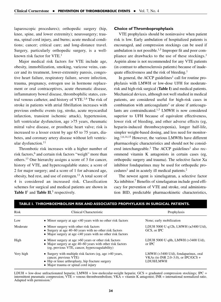
Clinical Cornerstone � PREVENTION OF THROMBOEMBOLIC EVENTS � Vol. 7, No. 4
34
laparoscopic procedures); orthopedic surgery (hip,knee, spine, and lower extremity); neurosurgery; trau-ma, spinal cord injury, and burns; acute medical condi-tions; cancer; critical care; and long-distance travel.Surgery, particularly orthopedic surgery, is a well-known risk factor for VTE.1
Major medical risk factors for VTE include age,obesity, immobilization, smoking, varicose veins, can-cer and its treatment, lower-extremity paresis, conges-tive heart failure, respiratory failure, severe infection,trauma, pregnancy, estrogen use in hormone replace-ment or oral contraceptives, acute rheumatic disease,inflammatory bowel disease, thrombophilic states, cen-tral venous catheter, and history of VTE.1,6 The risk ofstroke in patients with atrial fibrillation increases withprevious embolic events (eg, stroke, acute myocardialinfarction, transient ischemic attack), hypertension,left ventricular dysfunction, age >75 years, rheumaticmitral valve disease, or prosthetic heart valve; risk isincreased to a lesser extent by age 65 to 75 years, dia-betes, and coronary artery disease without left ventric-ular dysfunction.11
Thrombotic risk increases with a higher number ofrisk factors,6 and certain risk factors “weigh” more thanothers.13 One hierarchy assigns a score of 3 for cancer,history of VTE, and hypercoagulable states; a score of2 for major surgery; and a score of 1 for advanced age,obesity, bed rest, and use of estrogen.13 A total score of4 is considered an increased risk. Classificationschemes for surgical and medical patients are shown inTable I1 and Table II,5 respectively.
Choice of ThromboprophylaxisVTE prophylaxis should be noninvasive when patient
risk is low. Early ambulation of hospitalized patients isencouraged, and compression stockings can be used ifambulation is not possible.1,5 Improper fit and poor com-pliance are drawbacks to the use of these stockings.1
Aspirin alone is not recommended for any VTE patients(in contrast to atherosclerosis patients) because of inade-quate effectiveness and the risk of bleeding.1
In general, the ACCP guidelines1 call for routine pro-phylaxis with LMWH or low-dose UFH for moderate-risk and high-risk surgical (Table I) and medical patients.Mechanical devices, although not well studied in medicalpatients, are considered useful for high-risk cases incombination with anticoagulants1 or alone if anticoagu-lants are contraindicated.1,5 LMWH is now consideredsuperior to UFH because of equivalent effectiveness,lower risk of bleeding, and other adverse effects (eg,heparin-induced thrombocytopenia), longer half-life,simpler weight-based dosing, and less need for monitor-ing.1,6,14,15 However, the various LMWHs have differentpharmacologic characteristics and should not be consid-ered interchangeable.1 The ACCP guidelines1 also rec-ommend vitamin K antagonists in certain cases (eg,orthopedic surgery and trauma). The selective factor Xainhibitor fondaparinux may be used for orthopedic pro-cedures1 and in acutely ill medical patients.2
The newest agent is ximelagatran, a selective factorXa inhibitor.7 Benefits of ximelagatran include good effi-cacy for prevention of VTE and stroke, oral administra-tion BID, predictable pharmacokinetic characteristics,
TABLE I. THROMBOEMBOLISM RISK AND ASSOCIATED PROPHYLAXIS IN SURGICAL PATIENTS.
Risk Clinical Characteristic Prophylaxis
Low ● Minor surgery at age <40 years with no other risk factors None; early mobilization
Moderate ● Minor surgery with other risk factors LDUH 5000 U q12h, LMWH (≤3400 U/d),● Surgery at age 40–60 years with no other risk factors GCS, or IPC● Major surgery at age <40 years with no other risk factors
High ● Minor surgery at age >60 years or other risk factors LDUH 5000 U q8h, LMWH (>3400 U/d), ● Major surgery at age 40–60 years with other risk factors or IPC
(eg, previous VTE, cancer, hypercoagulability)
Very high ● Surgery with multiple risk factors (eg, age >40 years, LMWH (>3400 U/d), fondaparinux, oralcancer, previous VTE) VKAs (to INR 2.0–3.0), or IPC/GCS +
● Hip or knee arthroplasty, hip fracture surgery LDUH/LMWH● Major trauma or spinal cord injury
LDUH = low-dose unfractionated heparin; LMWH = low-molecular-weight heparin; GCS = graduated compression stockings; IPC =intermittent pneumatic compression; VTE = venous thromboembolism; VKA = vitamin K antagonist; INR = international normalized ratio.Adapted with permission.1
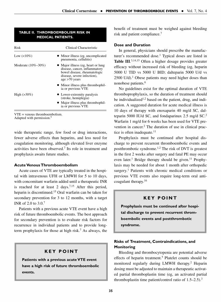
Clinical Cornerstone � PREVENTION OF THROMBOEMBOLIC EVENTS � Vol. 7, No. 4
35
wide therapeutic range, few food or drug interactions,fewer adverse effects than heparins, and less need forcoagulation monitoring, although elevated liver enzymeactivities have been observed.7 Its role in treatment andprophylaxis awaits future studies.
Acute Venous ThromboembolismAcute cases of VTE are typically treated in the hospi-
tal with intravenous UFH or LMWH for 5 to 10 days,with concomitant warfarin added until a therapeutic INRis reached for at least 2 days.7,15 After this period,heparin is discontinued.15 Oral warfarin can be taken forsecondary prevention for 3 to 12 months, with a targetINR of 2.0 to 3.0.7
Patients with a previous acute VTE event have a highrisk of future thromboembolic events. The best approachfor secondary prevention is to evaluate risk factors forrecurrence in individual patients and to provide long-term prophylaxis for those at high risk.7 As always, the
benefit of treatment must be weighed against bleedingrisk and patient compliance.7
Dose and DurationIn general, physicians should prescribe the manufac-
turer’s recommended dose.1 Typical doses are listed inTable III.3,14,15 Often a higher dosage provides greaterefficacy without increased risk of bleeding (eg, heparin5000 U TID vs 5000 U BID; dalteparin 5000 U/d vs2500 U/d).1 Obese patients may need higher doses thannonobese patients.6
No guidelines exist for the optimal duration of VTEthromboprophylaxis, so the duration of treatment shouldbe individualized1,2 based on the patient, drug, and indi-cation. A suggested duration for acute medical illness is10 days of therapy with enoxaparin 40 mg/d SC, dal-teparin 5000 IU/d SC, and fondaparinux 2.5 mg/d SC.2
Warfarin 1 mg/d for 6 weeks has been used for VTE pre-vention in cancer.2 The duration of use in clinical prac-tice is often inadequate.11
Prophylaxis must be continued after hospital dis-charge to prevent recurrent thromboembolic events andpostthrombotic syndrome.1,2 The risk of DVT is greatestin the first 2 weeks after surgery and fatal PE may occureven later.1 Bridge therapy should be given.11 Prophy-laxis may be needed for about 1 month after orthopedicsurgery.1 Patients with chronic medical conditions orprevious VTE events also require long-term oral anti-coagulant therapy.16
Risks of Treatment, Contraindications, andMonitoring
Bleeding and thrombocytopenia are potential adverseeffects of heparin treatment.5 Platelet counts should bemonitored regularly during LMWH therapy.2 Heparindosing must be adjusted to maintain a therapeutic activat-ed partial thromboplastin time (eg, an activated partialthromboplastin time patient/control ratio of 1.5–2.5).3
TABLE II. THROMBOEMBOLISM RISK IN MEDICAL PATIENTS.
Risk Clinical Characteristic
Low (<10%) ● Minor illness (eg, uncomplicatedpneumonia, cellulitis)
Moderate (10%–30%) ● Major illness (eg, heart or lungdisease, cancer, inflammatorybowel disease, rheumatologicdisease, severe infection), age >70 years
● Minor illness plus thrombophil-ia or previous VTE
High (>30%) ● Lower-extremity paralysis(stroke, hemiplegia)
● Major illness plus thrombophil-ia or previous VTE
VTE = venous thromboembolism.Adapted with permission.5
K E Y P O I N T
Patients with a previous acute VTE event
have a high risk of future thromboembolic
events.
K E Y P O I N T
Prophylaxis must be continued after hospi-
tal discharge to prevent recurrent throm-
boembolic events and postthrombotic
syndrome.
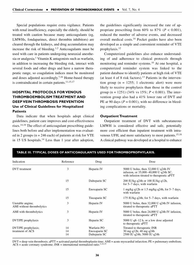
Clinical Cornerstone � PREVENTION OF THROMBOEMBOLIC EVENTS � Vol. 7, No. 4
36
Special populations require extra vigilance. Patientswith renal insufficiency, especially the elderly, should betreated with caution because many anticoagulants (eg,LMWHs, fondaparinux, direct thrombin inhibitors) arecleared through the kidneys, and drug accumulation mayincrease the risk of bleeding.1,2 Anticoagulants must beused with care in patients undergoing neuraxial anesthe-sia or analgesia.1 Vitamin K antagonists such as warfarin,in addition to increasing the bleeding risk, interact withseveral foods and other drugs and have a narrow thera-peutic range, so coagulation indices must be monitoredand doses adjusted accordingly.7,16 Home-based therapyis contraindicated in certain patients.11,15,17
HOSPITAL PROTOCOLS FOR VENOUSTHROMBOEMBOLISM TREATMENT ANDDEEP VEIN THROMBOSIS PREVENTIONUse of Clinical Guidelines for HospitalizedPatients
Data indicate that when hospitals adopt clinicalguidelines, patient care improves and cost-effectivenessrises.14,18 The effect of anticoagulant prescribing guide-lines both before and after implementation was evaluat-ed in 2 groups (n = 246 each) of patients at risk for VTEin 15 US hospitals.14 Less than 1 year after adoption,
the guidelines significantly increased the rate of ap-propriate prescribing from 60% to 87% (P < 0.001),reduced the number of adverse events, and decreasedoverall medical costs.14 Pocket guides have also beendeveloped as a simple and convenient reminder of VTEprophylaxis.12
Computerized guidelines also enhance understand-ing of and adherence to clinical protocols throughmonitoring and reminder systems.10 At one hospital, acomputerized reminder system was linked to thepatient database to identify patients at high risk of VTE(at least 4 of 8 risk factors).13 Patients in the interven-tion group (n = 1255; 1 electronic alert) were morelikely to receive prophylaxis than those in the controlgroup (n = 1251) (34% vs 15%; P < 0.001). The inter-vention group also had a 41% lower rate of DVT andPE at 90 days (P = 0.001), with no difference in bleed-ing complications or mortality.
Outpatient TreatmentOutpatient treatment of DVT with subcutaneous
LMWH is considered effective and safe, potentiallymore cost efficient than inpatient treatment with intra-venous UFH, and more satisfactory to most patients.15,18
A clinical pathway was developed at a hospital to enhance
TABLE III. TYPICAL DOSES OF ANTICOAGULANTS USED FOR THROMBOPROPHYLAXIS.
Indication Reference Drug Dose
DVT treatment 3 Heparin IV 5000 U bolus, then 32,000 U q24h IV infusion, or 35,000–40,000 U q24h SC,with infusion titrated to therapeutic aPTT
15 Dalteparin SC 200 IU/kg q24h or 100 IU/kg q12h, for 5–7 days, with warfarin
15 Enoxaparin SC 1 mg/kg q12h or 1.5 mg/kg q24h, for 5–7 days,with warfarin
15 Tinzaparin SC 175 IU/kg q24h, for 5–7 days, with warfarin
Unstable angina; 3 Heparin IV 5000 U bolus, then 32,000 U q24h IV infusion,AMI without thrombolytics titrated to therapeutic aPTT
AMI with thrombolytics 3 Heparin IV 5000 U bolus, then 24,000 U q24h IV infusion,titrated to therapeutic aPTT
DVT/PE prophylaxis 3 Heparin SC 5000 U q8–12 h, or a low dose adjustedto therapeutic aPTT
DVT/PE prophylaxis; 14 Warfarin PO Titrated to therapeutic INRtreatment of ACS 14 Enoxaparin SC 30 mg q12h; 40 mg q24h
14 Dalteparin SC 2500 IU q24h; 5000 IU q24h
DVT = deep vein thrombosis; aPTT = activated partial thromboplastin time; AMI = acute myocardial infarction; PE = pulmonary embolism;ACS = acute coronary syndrome; INR = international normalized ratio.3,14,15
Clinical Cornerstone � PREVENTION OF THROMBOEMBOLIC EVENTS � Vol. 7, No. 4
37
the use of outpatient LMWH with enoxaparin forpatients with DVT in an emergency department.17 Theprotocol included checklists for exclusion criteria, imple-mentation steps, laboratory tests, dosing guidelines forenoxaparin and warfarin, and follow-up recommenda-tions; patients received educational and clinical sup-port.17 Most of the eligible patients (74%) were startedon the outpatient therapy, but further education wasdeemed necessary for patients and clinicians to increaseenrollment in the program.17 Although LMWH costsmore than UFH,15 the home treatment saves money over-all by avoiding unnecessary hospitalization.15,17
Other studies show that patients receiving long-termoral anticoagulant therapy are capable of monitoring INRsand managing dosing at home, analogous to the self-carerequired for diabetes.16 Current coagulation monitors forhome use measure INR accurately, and home monitoringimproves quality of life and saves money over time.16
Disadvantages exist with home self-care, however.Many health plans do not reimburse for outpatient treat-ment.15 Patients must be selected carefully, educatedabout the proper use of LMWH and warfarin, warnedabout adverse effects, and monitored.15 Some patients areunable to self-inject SC LMWH.15,17 INR must be mea-sured during the transition to warfarin therapy, althoughmonitoring of anti-factor Xa activity is controversial.15
Home-based therapy with VTE prophylaxis is contraindi-cated in patients with low platelet count, active bleedingor risk of bleeding, heparin sensitivity, use of aspirin oranticoagulants, pregnancy or lactation, renal insufficien-cy, recent surgery, and severe hypertension.11,15,17
SUMMARYStudies show inadequate treatment of high-risk VTEpatients. Physicians must administer prophylaxis forVTE when appropriate for at least 3 reasons: VTE is aprevalent, preventable, and often silent condition; thefirst presentation of VTE may be a fatal PE; and treat-ment guidelines promote this as the standard of care.5
Programs should continue to educate physicians aboutthe necessity of prophylaxis and the relative safety ofanticoagulants. Hospitals should adopt clinical guide-lines to improve patient care and to monitor efficacy.The problem of VTE will only grow as the US popula-tion ages, as surgery is performed on increasingly sickerpatients, and as the length of hospital stays continues todecrease.
REFERENCES1. Geerts WH, Pineo GF, Heit JA, et al. Prevention of venous
thromboembolism: The Seventh ACCP Conference onAntithrombotic and Thrombolytic Therapy. Chest. 2004;126:338S–400S.
2. Gerotziafas GT, Samama MM. Prophylaxis of venousthromboembolism in medical patients. Curr Opin PulmMed. 2004;10:356–365.
3. Hirsh J, Anand SS, Halperin JL, Fuster V. Guide to anti-coagulant therapy: Heparin. A statement for healthcareprofessionals from the American Heart Association.Circulation. 2001;103:2994–3018.
4. McGarry LJ, Thompson D. Retrospective database analy-sis of the prevention of venous thromboembolism withlow-molecular-weight heparin in acutely ill medical inpa-tients in community practice. Clin Ther. 2004;26:419–430.
5. Rahim SA, Panju A, Pai M, Ginsberg J. Venous throm-boembolism prophylaxis in medical inpatients: A retro-spective chart review. Thromb Res. 2003;111:215–219.
6. Ageno W, Turpie AG. Deep venous thrombosis in themedically ill. Curr Hematol Rep. 2002;1:73–78.
7. Agnelli G. Current issues in anticoagulation. PathophysiolHaemost Thromb. 2005;34(Suppl 1):2–9.
8. English EJ. Quality care. All want it; let’s define it. MinnMed. 2002;85:7, 44.
9. Attree M. A study of the criteria used by healthcare pro-fessionals, managers and patients to represent and evalu-ate quality care. J Nurs Manag. 2001;9:67–78.
10. de Clercq PA, Blom JA, Korsten HH, Hasman A.Approaches for creating computer-interpretable guide-lines that facilitate decision support. Artif Intell Med.2004;31:1–27.
11. Tapson VF, Hyers TM, Waldo AL, et al, for the NABOR(National Anticoagulation Benchmark and OutcomesReport) Steering Committee. Antithrombotic therapypractices in US hospitals in an era of practice guidelines.Arch Intern Med. 2005;165:1458–1464.
12. Kakkar AK, Davidson BL, Haas SK, for the InvestigatorsAgainst Thromboembolism (INATE) Core Group.Compliance with recommended prophylaxis for venousthromboembolism: Improving the use and rate of uptakeof clinical practice guidelines. J Thromb Haemost.2004;2:221–227.
13. Kucher N, Koo S, Quiroz R, et al. Electronic alerts to pre-vent venous thromboembolism among hospitalizedpatients. N Engl J Med. 2005;352:969–977.
14. Schumock GT, Blackburn JC, Nutescu EA, et al. Impactof prescribing guidelines for inpatient anticoagulation.Ann Pharmacother. 2004;38:1570–1575.
15. American Society of Health-System Pharmacists. ASHPtherapeutic position statement on the use of low-molecular-weight heparins for adult outpatient treatment of acutedeep-vein thrombosis. Am J Health-Syst Pharm. 2004;61:1950–1955.
Clinical Cornerstone � PREVENTION OF THROMBOEMBOLIC EVENTS � Vol. 7, No. 4
38
16. Ansell J, Jacobson A, Levy J, et al. Guidelines forimplementation of patient self-testing and patient self-management of oral anticoagulation. International con-sensus guidelines prepared by International Self-Monitoring Association for Oral Anticoagulation. Int JCardiol. 2005;99:37–45.
17. Shapiro NI, Spear J, Sheehy S, et al. Barriers to the use ofoutpatient enoxaparin therapy in patients with deepvenous thrombosis. Am J Emerg Med. 2005;23:30–34.
18. Panis LJ, Kolbach DN, Hamulyák K, Prins MH. Identifyinginappropriate hospital stay in patients with venous throm-boembolism. Eur J Intern Med. 2004;15:39–44.
Address correspondence to: Geno J. Merli, MD, FACP, Thomas Jefferson University, Division of Internal Medicine,211 South 9th Street, Suite 300, Philadelphia, PA 19107. E-mail: [email protected]
Heparin and heparin-derived drugs play a major therapeutic role in thrombotic and cardiovascular disorders.Infusion of unfractionated heparin (UFH) followed by warfarin has traditionally been the standard pharmaco-logic therapy for treatment of venous thromboembolism (VTE), which includes both deep vein thrombosis andpulmonary embolism, and for initial therapy of non–ST-elevation (NSTE) acute coronary syndrome (ACS).More recently, low-molecular-weight heparins (LMWHs) have been shown to provide at least as good efficacyand safety outcomes as UFH regimens for prevention of these conditions. In addition to good efficacy outcomeswith LMWHs compared with UFH, LMWHs have other advantages over UFH, including improved bioavail-ability, QD administration, more predictable anticoagulant response, lack of the need for monitoring, and suit-ability for outpatient use, thereby increasing convenience, reducing cost, and improving cost-to-benefit ratios.In carefully screened and managed patients, LMWH offers a cost-effective, convenient, and safe alternative toUFH for thrombosis management. The aim of this article is to summarize efficacy, safety, and pharmacoeco-nomic considerations when selecting LMWH versus UFH for thrombosis management in VTE and NSTE ACS.(Clinical Cornerstone. 2005;7[4]:39–48) Copyright © 2005 Excerpta Medica, Inc.
ALEX C. SPYROPOULOS, MD, FACP, FCCPMedical DirectorClinical Thrombosis CenterLovelace Sandia Health SystemsAlbuquerque, New MexicoClinical Associate ProfessorDepartment of MedicineAssociate Professor of PharmacyCollege of PharmacyUniversity of New Mexico Health Sciences Center Albuquerque, New Mexico
Pharmacologic Therapy for theManagement of Thrombosis:Unfractionated Heparin or Low-Molecular-Weight Heparin?
Clinical Cornerstone � PREVENTION OF THROMBOEMBOLIC EVENTS � Vol. 7, No. 4
39
Heparin and heparin-derived drugs play a major thera-peutic role in thrombotic and cardiovascular disorders.1
Inpatient IV infusion of unfractionated heparin (UFH) fol-lowed by warfarin administration has traditionally beenthe standard pharmacologic therapy for the acute treat-ment of venous thromboembolism (VTE),2,3 a term thatincludes both deep vein thrombosis (DVT) and pulmonaryembolism (PE), and for non–ST-elevation (NSTE) acutecoronary syndrome (ACS).4 More recently, low-molecular-weight heparins (LMWHs) have been shown to provide atleast as good efficacy and safety outcomes as UFH regi-
mens for prevention and treatment of VTE5–8 and NSTEACS.4,9,10 LMWHs have the additional advantages of easeof administration, predictability of anticoagulant response,lack of the need for monitoring, and suitability for outpa-tient use, thereby increasing convenience, reducing cost,and improving cost-to-benefit ratios.5,11,12
The American College of Chest Physicians (ACCP)provides strong evidence-based guidelines on VTE pre-vention that support use of either LMWH or UFH formost medical and surgical patients and VTE treatmentthat supports the use of LMWH over UFH in outpatient-
eligible patients.13 The American College of Cardiologyand the American Heart Association recommend thatanticoagulation therapy with LMWH or UFH be addedto antiplatelet therapy in patients with unstable angina orNSTE ACS.14 This article summarizes efficacy, safety,and pharmacoeconomic considerations when selectingpharmacologic agents for thrombosis prevention and treat-ment in VTE and thrombosis management in NSTE ACS.
PHARMACOLOGIC THERAPY FOR PREVENTION OF VENOUS THROMBOEMBOLISMEfficacy and Safety in Surgical Patients
Several studies have compared use of LMWH withUFH for prevention of thrombosis after major surgicalinterventions. In a meta-analysis of studies, Koch et al6
analyzed data from 23 randomized, double-blind clinicaltrials comparing the effectiveness of LMWHs versus UFHfor thrombosis prophylaxis after different types of majorsurgery. For patients undergoing general surgery, LMWHsand UFH were equally effective in preventing DVT, butlow doses of LMWH were associated with reduced risk ofwound hematoma (P < 0.001). For patients undergoingorthopedic surgery, LMWH was more effective than UFH,with lower rates of proximal DVT in patients receivingLMWH compared with UFH (P = 0.002).
In a study designed to evaluate risk factors for VTE inorthopedic patients, multivariate analyses identified thetype of LMWH used as an independent predictor ofVTE. Ten percent of patients receiving prophylaxis withenoxaparin experienced VTE, compared with 18% ofpatients receiving dalteparin (odds ratio for enoxaparin0.4; 95% CI, 0.2–0.8; P = 0.009).15 Authors theorize thatthis difference may reflect differences in anti-Xa activity
between the 2 LMWHs, or differences in dosing, sinceenoxaparin was given BID and dalteparin QD.
Medical Patients and BridgingFew studies have compared the effects of LMWHs ver-
sus UFH in patients with underlying medical diseases. Amulticenter study of hospitalized patients with eithersevere respiratory disease or heart failure compared thesafety and efficacy of the LMWH enoxaparin with UFHfor prevention of VTE.16 In the 451 patients evaluated,enoxaparin was at least as effective as UFH, with an inci-dence of VTE of 8.4% in patients receiving enoxaparinand 10.4% in patients receiving UFH. Enoxaparin wasassociated with similar death and bleeding rates but sig-nificantly fewer adverse events (P = 0.044)
Risk for thromboembolism is higher in patients with mechanical heart valves. Traditionally, inpatientbridging therapy with UFH has been used when thesepatients require discontinuation of anticoagulation ther-apy prior to elective or invasive procedures. Studiescomparing the use of outpatient bridging therapy usingLMWH versus inpatient bridging therapy using UFHfor patients with mechanical heart valves shows out-patient LMWH therapy is as safe and effective as inpa-tient UFH therapy.17,18 In addition, patients receivingbridging therapy with LMWH had shorter hospitalstays and were less likely to undergo major surgery.In an integrated health maintenance organization basedin Albuquerque, New Mexico, with approximately200,000 members per year, use of the LMWH enoxa-parin as bridge therapy in patients—including thosewith mechanical heart valves undergoing elective sur-gical procedures—resulted in a cost savings of approxi-mately $212,475 per year from January 1998 to March2002.18
PHARMACOLOGIC THERAPY FOR TREATMENT OF VENOUS THROMBOEMBOLISMEfficacy and Safety
Several meta-analyses have analyzed the safety andefficacy of LMWHs compared with UFH in the treat-ment of DVT and VTE.5–8,19,20 These meta-analyses col-lectively show LMWHs are at least as effective as orsuperior to UFH for prevention of recurrent VTE andreduction of overall mortality and major bleeding(Table).
Clinical Cornerstone � PREVENTION OF THROMBOEMBOLIC EVENTS � Vol. 7, No. 4
40
K E Y P O I N T
In patients undergoing general surgery,
LMWHs and UFH were equally effective
in preventing DVT, but low doses of
LMWH were associated with reduced risk
of wound hematoma.
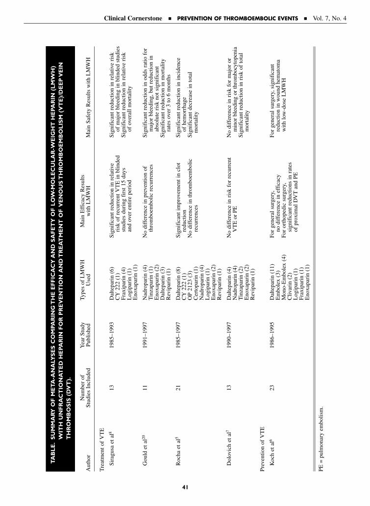
Clinical Cornerstone � PREVENTION OF THROMBOEMBOLIC EVENTS � Vol. 7, No. 4
41
TA
BL
E.
SU
MM
ARY
OF
ME
TA
-AN
ALY
SE
S C
OM
PAR
ING
TH
E E
FF
ICA
CY
AN
D S
AF
ET
Y O
F L
OW
-MO
LE
CU
LA
R-W
EIG
HT
HE
PAR
IN (
LM
WH
)W
ITH
UN
FR
AC
TIO
NA
TE
D H
EPA
RIN
FO
R P
RE
VE
NT
ION
AN
D T
RE
AT
ME
NT
OF
VE
NO
US
TH
RO
MB
OE
MB
OL
ISM
(V
TE
)/D
EE
P V
EIN
TH
RO
MB
OS
IS (
DV
T).
Num
ber
ofY
ear
Stud
yTy
pes
of L
MW
HM
ain
Eff
icac
y R
esul
tsA
utho
rSt
udie
s In
clud
edPu
blis
hed
Use
dw
ith L
MW
HM
ain
Safe
ty R
esul
ts w
ith L
MW
H
Tre
atm
ent o
f V
TE
Sira
gusa
et a
l813
1985
–199
3D
alte
pari
n (6
)Si
gnif
ican
t red
uctio
n in
rel
ativ
eSi
gnif
ican
t red
uctio
n in
rel
ativ
e ri
skC
Y 2
22 (
1)ri
sk o
f re
curr
ent V
TE
in b
linde
dof
maj
or b
leed
ing
in b
linde
d st
udie
sFr
axip
arin
(4)
stud
ies
duri
ng f
irst
15
days
Sign
ific
ant r
educ
tion
in r
elat
ive
risk
Log
ipar
in (
1)an
d ov
er e
ntir
e pe
riod
of o
vera
ll m
orta
lity
Eno
xapa
rin
(1)
Gou
ld e
t al20
1119
91–1
997
Nad
ropa
rin
(4)
No
diff
eren
ce in
pre
vent
ion
ofSi
gnif
ican
t red
uctio
n in
odd
s ra
tio f
orT
inza
pari
n (1
)th
rom
boem
bolic
rec
urre
nces
maj
or b
leed
ing,
but
red
uctio
n in
Eno
xapa
rin
(2)
abso
lute
ris
k no
t sig
nifi
cant
Dal
tepa
rin
(3)
Sign
ific
ant r
educ
tion
in m
orta
lity
Rev
ipar
in (
1)ra
tes
over
3 to
6 m
onth
s
Roc
ha e
t al5
2119
85–1
997
Dal
tepa
rin
(8)
Sign
ific
ant i
mpr
ovem
ent i
n cl
otSi
gnif
ican
t red
uctio
n in
inci
denc
eC
Y 2
22 (
1)re
duct
ion
of h
emor
rhag
e O
P 21
23 (
3)N
o di
ffer
ence
in th
rom
boem
bolic
Sign
ific
ant d
ecre
ase
in to
tal
Cer
topa
rin
(1)
recu
rren
ces
mor
talit
yN
adro
pari
n (4
)L
ogip
arin
(1)
Eno
xapa
rin
(2)
Rev
ipar
in (
1)
Dol
ovic
h et
al7
1319
90–1
997
Dal
tepa
rin
(4)
No
diff
eren
ce in
ris
k fo
r re
curr
ent
No
diff
eren
ce in
ris
k fo
r m
ajor
or
Nad
ropa
rin
(4)
VT
E o
r PE
min
or b
leed
ing
or th
rom
bocy
tope
nia
Tin
zapa
rin
(2)
Sign
ific
ant r
educ
tion
in r
isk
of to
tal
Eno
xapa
rin
(2)
mor
talit
yR
evip
arin
(1)
Prev
entio
n of
VT
E
Koc
h et
al6
2319
86–1
995
Dal
tepa
rin
(11)
For
gene
ral s
urge
ry,
For
gene
ral s
urge
ry, s
igni
fica
nt
Em
bole
x (3
)no
dif
fere
nce
in e
ffic
acy
redu
ctio
n in
wou
nd h
emat
oma
Mon
o-E
mbo
lex
(4)
For
orth
oped
ic s
urge
ry,
with
low
-dos
e L
MW
HC
livar
in (
2)si
gnif
ican
t red
uctio
ns in
rat
es
Log
ipar
in (
1)of
pro
xim
al D
VT
and
PE
Frax
ipar
in (
1)E
noxa
pari
n (1
)
PE =
pul
mon
ary
embo
lism
.
Two earlier meta-analyses noted benefits of LMWHsover UFH for the treatment of DVT and VTE.8,19
Leizorovicz19 analyzed the results of 20 randomized, con-trolled clinical studies comparing the safety and efficacyof LMWHs with UFH in patients with DVT. Significantreductions in overall mortality, major hemorrhage, andthrombus extension were observed in patients treatedwith LMWHs compared with UFH (P < 0.05). Recur-rence of VTE was lower, though not significantly, inpatients treated with LMWH. The authors concluded thatLMWHs have a higher benefit-to-risk ratio comparedwith UFH for the treatment of VTE.
Siragusa et al8 compared the effects of LMWHs andUFH on recurrent VTE and major bleeding in the treat-ment of acute VTE. The researchers analyzed data sepa-rately for studies that were or were not blinded. Forblinded studies, the relative risk of recurrent VTE wassignificantly lower in patients treated with LMWHs com-pared with UFH over the first 15 days of treatment (P <0.05) and over the entire period of anticoagulation ther-apy (P < 0.01). The relative risk of major bleeding wasalso significantly lower in patients treated with LMWHscompared with UFH in blinded studies (P = 0.01). Morerecent meta-analyses show potential benefits of LMWHscompared with UFH for treatment of DVT and VTE.5,20
Gould et al20 used clinical outcomes to compare theeffects of LMWHs and UFH on major bleeding, recur-rent thromboembolic events, and mortality rates over 3 to6 months. Results again showed a significant reductionin the overall mortality rate with LMWHs compared withUFH (P < 0.05). The odds ratio suggested risk of majorbleeding was lower in patients receiving LMWHs com-pared with UFH (P < 0.05), but the absolute risk reduc-tion was not statistically significant. LMWH and UFHwere equally effective in preventing thromboembolicrecurrences.
Rocha et al5 analyzed data from 4472 patients withDVT in 21 studies. Treatment with LMWHs was associ-ated with improvement in clot venography (P = 0.004)and reductions in total mortality (P = 0.012) and inci-dence of hemorrhage (P = 0.47). No significant differ-ences in recurrence of DVT were noted between groups.In this study, the effects of QD versus BID injections ofLMWHs were also compared. QD dosing of LMWHswas associated with reduced risk of major bleeding (P =0.025), whereas BID dosing was more effective for clotreduction (P = 0.004).
Another recent meta-analysis showed LMWHs were atleast as effective as UFH for treatment of VTE.7 In a meta-analysis of 13 randomized, blinded studies of patients withDVT treated with LMWHs or UFH, Dolovich et al7 foundno statistically significant differences between groups inrisk of recurrent VTE, PE, major bleeding, minor bleed-ing, or thrombocytopenia. Total mortality risk, however,was significantly lower in patients treated with LMWHscompared with UFH (relative risk, 0.76; 95% CI,0.59–0.98). Of the 13 studies included, 3 were consideredto be outpatient trials. Comparison of inpatient and outpa-tient trials found a reduction in major bleeding in inpatienttrials, highlighting the need for rigorous monitoring withoutpatient treatment.
These meta-analyses included studies using differenttypes of LMWH preparations. Because each LMWHhas distinct anticoagulation effects,1 results may havebeen different if only studies using 1 type of LMWH wereincluded.
Outpatient Studies for the Treatment of DVTThe majority of studies comparing the efficacy and
safety of LMWH with UFH for the treatment of VTEhave been inpatient studies. The vast majority of patientswith VTE are eligible for outpatient therapy withLMWH. Segal et al21 summarized 8 studies that com-pared the efficacy, safety, and cost of outpatient use of a LMWH with inpatient use of UFH. All of the studiesused enoxaparin, nadroparin, or dalteparin during inter-vention and follow-up. Results from a total of 3762patients showed similar rates of recurrent DVT andmajor bleeding between groups, but shortened hospitalstays and lower costs in individuals receiving LMWH asoutpatients.
Not all patients are appropriate candidates for outpa-tient therapy with LMWH, and each patient must beindividually assessed. Contraindications to outpatienttherapy with LMWH include active bleeding, car-diopulmonary instability, hereditary bleeding disorders,history of heparin-induced thrombocytopenia, andallergy to heparin or LMWH.2 Other potential con-traindications that require clinical judgment includeclinical factors such as gastrointestinal or genitouri-nary bleeding of recent onset, renal insufficiency, orsocioeconomic/psychosocial factors such as potentialnonadherence and geographic inaccessibility for follow-upcare.2,22
Clinical Cornerstone � PREVENTION OF THROMBOEMBOLIC EVENTS � Vol. 7, No. 4
42
Other Considerations and PharmacoeconomicEvaluations
LMWH offers several advantages over UFH, includ-ing improved bioavailability, prolonged and predictableanticoagulant response, QD administration, lack of theneed for monitoring, and convenience.1 These advan-tages make LMWH suitable for outpatient use and trans-late into cost savings. In the description of outpatientstudies by Segal et al,21 use of LMWH was associatedwith shorter hospitalizations and lower costs. Medianhospital stays were 2.7 days for patients receivingLMWH compared with 6.5 days for patients receivingUFH, resulting in a median cost savings of $1600 forpatients receiving LMWH.
A landmark Canadian study established the safety andefficacy of outpatient use of the LMWH enoxaparincompared with inpatient administration of UFH for treat-ment of DVT.23 A later economic evaluation of this studycompared the cost of treatment in a subset of 300 patientsfollowed for up to 3 months.24 Use of enoxaparin result-
ed in a cost savings of Can $3045. Spyropoulos et al25
retrospectively replicated the Canadian study in a USmanaged care setting. They found no significant differ-ence in the number of recurrent VTE or bleeding events,but again found substantial cost savings with outpatientenoxaparin administration compared with inpatient UFHadministration. The mean cost savings per patient withenoxaparin was $2583.
PHARMACOLOGIC THERAPY FOR NON–ST-ELEVATION ACUTE CORONARYSYNDROMEAntithrombin therapy is an integral part of managingNSTE ACS.14 Several studies have shown that use ofLMWH is at least as effective as or more effectivethan UFH in reducing the incidence of ischemicevents.4,9,10,26,27 The Efficacy and Safety of SubcutaneousEnoxaparin in Non–Q-Wave Coronary Events (ESSENCE)trial, a randomized, double-blind, placebo-controlled studyof 3171 patients with NSTE ACS, showed that antithrom-botic therapy with enoxaparin plus aspirin was more effec-tive than therapy with UFH plus aspirin.9 Risk of death,myocardial infarction (MI), or recurrent angina was sig-nificantly lower at 14 days (P = 0.019) and 30 days (P =0.016). The need for revascularization was also significant-ly less at 30 days (P = 0.001). Rates of major bleeding didnot differ significantly between groups.
A meta-analysis of data from the ESSENCE andThrombolysis in Myocardial Infarction (TIMI 11B) trialsfound that enoxaparin was associated with a 20% reduc-tion in death and serious cardiac ischemic events.10 Thisreduction became apparent within the first few days oftreatment and was still apparent after 43 days of treat-ment. Rates of minor bleeding were significantly higherin patients receiving enoxaparin (P = 0.0001), but no dif-ference was found between groups in rates of majorbleeding. Analysis of a subset of patients in theESSENCE or TIMI 11B trials showed that obesity didnot impact clinical outcomes of these trials.23 Patientswith severe renal impairment, however, had a higher risk
Clinical Cornerstone � PREVENTION OF THROMBOEMBOLIC EVENTS � Vol. 7, No. 4
43
K E Y P O I N T
Many patients with VTE are appropriate
candidates for outpatient therapy with
LMWH, but each patient must be individu-
ally assessed.
K E Y P O I N T
Antithrombin therapy is an integral part
of managing NSTE ACS.
K E Y P O I N T
In the analysis of outpatient studies by
Segal et al, use of LMWH was associated
with shorter hospitalizations and lower
costs. Median hospital stays were 2.7 days
for patients receiving LMWH compared
with 6.5 days for patients receiving UFH,
resulting in a median cost savings of $1600
for patients receiving LMWH.
of clinical events and hemorrhage than patients withoutsevere renal impairment.
The Superior Yield of the New Strategy of Enoxaparin,Revascularization, and GlYcoprotein IIb/IIIa Inhibitorstrial compared use of enoxaparin with UFH in high-riskpatients with ACS.27 Based on 6-month and 1-year follow-up data from 9978 patients in 487 hospitals in12 countries, patients receiving enoxaparin or UFH hadsimilar rates of death or MI. In patients receiving consis-tent therapy (only enoxaparin or UFH) during the hospi-talization period, the reduction in rate of death or nonfa-tal MI that occurred within the first 30 days in patientsreceiving enoxaparin compared with UFH was main-tained at 6 months (P = 0.006). One-year all-cause deathrates in the 2 treatment groups and in patients receivingconsistent therapy were similar.
In a recent review of 6 major trials that comparedenoxaparin with UFH for the treatment of NSTE ACS(including the ESSENCE and TIMI 11B trials), resultsfrom ~22,000 patients showed the use of enoxaparin wasassociated with modest reductions in the incidence of thecomposite of death and MI at 30 days.4 A larger effect wasseen in patients who did not receive antithrombin pretreat-ment before randomization. No significant differenceswere found in rates of major bleeding or transfusions.
In addition to potential efficacy and safety advantages,LMWH offers other advantages over UFH for the pre-vention of ischemic events in NSTE ACS. Limitations ofUFH include a high degree of protein binding, inactiva-tion by platelet factor 4, a variable dose response, stimu-lation of platelet aggregation, and risk of heparin-induced thrombocytopenia.4 In contrast, LMWHs havean effect higher in the coagulation cascade, with greaterinhibition of factor Xa than IIa.4
Economic assessment of results from the ESSENCEtrial showed cost savings with enoxaparin compared withUFH therapy.12 Hospital billing data were available for655 of the 936 US patients randomized to the ESSENCEtrial. Total medical costs for the initial hospitalizationwere $763 less for patients receiving enoxaparin than forpatients receiving UFH. At the end of 30 days, the totalcost savings in patients receiving enoxaparin versus UFHwas $1172.
CONCLUSIONSLMWH is at least as effective as UFH for prevention ofVTE events. In VTE treatment, compared with UFH,
LMWH has been shown to reduce overall mortality andrisk of major hemorrhage. For treatment of NSTE ACS,LMWH is associated with at least modest reductions indeath and incidence of MI, with no increase in majorbleeding. LMWH offers several advantages over UFHand yields cost savings in treatment of both VTE andNSTE ACS. In carefully screened and managed patients,LMWH offers a cost-effective, convenient, and safe alter-native to UFH for thrombosis prevention and treatment.
REFERENCES1. Bick RL, Frenkel EP, Walenga J, et al. Unfractionated
heparin, low molecular weight heparins, and pentasaccha-ride: Basic mechanism of actions, pharmacology, and clin-ical use. Hematol Oncol Clin N Am. 2005;19:1–51.
2. Merli G. Anticoagulants in the treatment of deep veinthrombosis. Am J Med. 2005;118(8A):13S–20S.
3. Nuteschu EA. Emerging options in the treatment ofvenous thromboembolism. Am J Health-Syst Pharm.2004;61(Suppl 7):512–517.
4. Califf RM, Petersen JL, Hasselblad V, et al. A perspectiveon trials comparing enoxaparin and unfractionated heparinin the treatment of non–ST-elevation acute coronary syn-dromes. Am Heart J. 2005;149:S91–S99.
5. Rocha E, Martínez-González MA, Montes R, Panizo C.Do the low molecular weight heparins improve efficacyand safety of the treatment of deep venous thrombosis? A meta-analysis. Haematologica. 2000;85:935–942.
6. Koch A, Ziegler S, Breitschwerdt H, Victor N. Lowmolecular weight heparin and unfractionated heparin inthrombosis prophylaxis: Meta-analysis based on originalpatient data. Thrombosis Res. 2001;102:295–309.
7. Dolovich LR, Ginsberg JS, Douketis JD, et al. A meta-analysis comparing low-molecular-weight heparins withunfractionated heparin in the treatment of venous throm-boembolism. Arch Intern Med. 2000;160:181–188.
8. Siragusa S, Cosmi B, Piovella F, et al. Low-molecular-weight heparins and unfractionated heparin in the treat-ment of patients with acute venous thromboembolism:Results of a meta-analysis. Am J Med. 1996;100:269–277.
9. Cohen M, Demers C, Gurfinkel EP, et al, for the Efficacyand Safety of Subcutaneous Enoxaparin in Non–Q-waveCoronary Events Study Group. A comparison of low-molecular weight heparin with unfractionated heparin forunstable coronary artery disease. N Engl J Med. 1997;337:447–452.
10. Antman EM, Cohen M, Radley D, et al, for the TIMI 11B(Thrombolysis In Myocardial Infarction) and ESSENCE(Efficacy and Safety of Subcutaneous Enoxaparin inNon–Q-wave Coronary Events) Investigators. Assessmentof the treatment effect of enoxaparin for unstableangina/non–Q-wave myocardial infarction. TIMI 11B—ESSENCE Meta-analysis. Circulation. 1999;100:1602–1608.
11. McGarry LJ, Thompson D, Weinstein MC, Goldhaber SZ.Cost effectiveness of thromboprophylaxis with a low-molecular-weight heparin versus unfractionated heparin in
Clinical Cornerstone � PREVENTION OF THROMBOEMBOLIC EVENTS � Vol. 7, No. 4
44
acutely ill medical inpatients. Am J Manag Care. 2004;10:632–642.
12. Mark DB, Cowper PA, Berkowitz SD, et al. Economicassessment of low-molecular-weight heparin (enoxaparin)versus unfractionated heparin in acute coronary syndromepatients. Results from the ESSENCE randomized trial.Circulation. 1998;97:1702–1707.
13. Geerts WH, Pineo GF, Heit JA, et al. Prevention of venousthromboembolism: The Seventh ACCP Conference onAntithrombotic and Thrombolytic Therapy. Chest.2004;126:338S–400S.
14. Braunwald E, Antman EM, Beasley JW, et al, for theCommittee on the Management of Patients with UnstableAngina, ACC/AHA Task Force on Practice Guidelines.ACC/AHA guidelines for the management of patientswith unstable angina and non–ST-segment elevationmyocardial infarction: Executive summary and recom-mendations. Circulation. 2000;102:1193–1209.
15. Schiff RL, Kahn SR, Shrier I, et al. Identifying orthopedicpatients at high risk for venous thromboembolism despitethromboprophylaxis. Chest. 2005;128:3364–3371.
16. Kleber F-X, Witt C, Vogel G, et al, for THE PRINCEStudy Group. Randomized comparison of enoxaparin withunfractionated heparin for the prevention of venousthromboembolism in medical patients with heart failure orsevere respiratory disease. Am Heart J. 2003;154:614–621.
17. Spyropoulos AC, Dunn AS, Graham A, et al. Peri-operative bridging therapy with unfractionated heparin orlow-molecular-weight heparin in patients with mechanicalheart valves on long-term oral anticoagulants: Resultsfrom the REGIMEN registry [abstract]. J Am CollCardiol. 2005;Feb 1:352A. Abstract 1016-47.
18. Spyropoulos AC, Jenkins P, Bornikova L. A disease man-agement protocol for outpatient perioperative bridge therapywith enoxaparin in patients requiring temporary interruptionof long-term oral anticoagulation. Pharmacotherapy.2004;24:649–658.
19. Leizorovicz A. Comparison of the efficacy and safety oflow molecular weight heparins and unfractionated heparin
in the initial treatment of deep venous thrombosis. Anupdated meta-analysis. Drugs. 1996;52(Suppl 7):30–37.
20. Gould MK, Dembitzer AD, Doyle RL, et al. Low-molecular-weight heparins compared with unfractionatedheparin for treatment of acute deep venous thrombosis: Ameta-analysis of randomized, controlled trials. Ann InternMed. 1999;130:800–809.
21. Segal JB, Bolger DT, Jenckes MW, et al. Outpatient ther-apy with low molecular weight heparin for the treatmentof venous thromboembolism: A review of efficacy, safety,and costs. Am J Med. 2003;115:298–308.
22. Spyropoulos AC. The use of a protocol for the treatment ofdeep vein thrombosis. Am J Manag Care. 2001;7:S553–S558.
23. Levine M, Gent M, Hirsh J, et al. A comparison of low-molecular-weight heparin administered primarily at homewith unfractionated heparin administered in the hospitalfor proximal deep-vein thrombosis. New Engl J Med.1996;334:677–681.
24. O’Brien B, Levine M, Willan A, et al. Economic evalua-tion of outpatient treatment with low-molecular-weightheparin for proximal vein thrombosis. Arch Intern Med.1999;159:2298–2304.
25. Spyropoulos AC, Hurley JS, Ciesla GN, de Lissovoy G.Management of acute proximal deep vein thrombosis:Pharmacoeconomic evaluation of outpatient treatmentwith enoxaparin vs inpatient treatment with unfractionat-ed heparin. Chest. 2002;122:108–114.
26. Spinler SA, Inverso SM, Cohen M, et al, for theESSENCE and TIMI 11B Investigators. Safety and effica-cy of unfractionated heparin versus enoxaparin in patientswho are obese and patients with severe renal impairment:Analysis from the ESSENCE and TIMI 11B studies. AmHeart J. 2003;146:33–41.
27. Mahaffey KW, Cohen M, Garg J, et al. High-risk patientswith acute coronary syndromes treated with low-molecular-weight or unfractionated heparin: Outcomes at 6 monthsand 1 year in the SYNERGY Trial. JAMA. 2005;294:2594–2600.
Clinical Cornerstone � PREVENTION OF THROMBOEMBOLIC EVENTS � Vol. 7, No. 4
45
Address correspondence to: Alex C. Spyropoulos, MD, FACP, FCCP, Lovelace Sandia Health Systems, 5400 GibsonBoulevard, SE, Albuquerque, NM 87108.
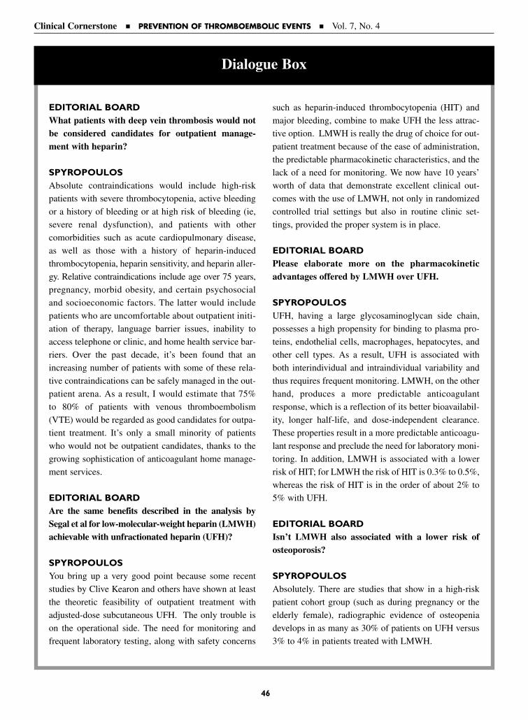
EDITORIAL BOARDWhat patients with deep vein thrombosis would notbe considered candidates for outpatient manage-ment with heparin?
SPYROPOULOSAbsolute contraindications would include high-risk
patients with severe thrombocytopenia, active bleeding
or a history of bleeding or at high risk of bleeding (ie,
severe renal dysfunction), and patients with other
comorbidities such as acute cardiopulmonary disease,
as well as those with a history of heparin-induced
thrombocytopenia, heparin sensitivity, and heparin aller-
gy. Relative contraindications include age over 75 years,
pregnancy, morbid obesity, and certain psychosocial
and socioeconomic factors. The latter would include
patients who are uncomfortable about outpatient initi-
ation of therapy, language barrier issues, inability to
access telephone or clinic, and home health service bar-
riers. Over the past decade, it’s been found that an
increasing number of patients with some of these rela-
tive contraindications can be safely managed in the out-
patient arena. As a result, I would estimate that 75%
to 80% of patients with venous thromboembolism
(VTE) would be regarded as good candidates for outpa-
tient treatment. It’s only a small minority of patients
who would not be outpatient candidates, thanks to the
growing sophistication of anticoagulant home manage-
ment services.
EDITORIAL BOARDAre the same benefits described in the analysis bySegal et al for low-molecular-weight heparin (LMWH)achievable with unfractionated heparin (UFH)?
SPYROPOULOSYou bring up a very good point because some recent
studies by Clive Kearon and others have shown at least
the theoretic feasibility of outpatient treatment with
adjusted-dose subcutaneous UFH. The only trouble is
on the operational side. The need for monitoring and
frequent laboratory testing, along with safety concerns
such as heparin-induced thrombocytopenia (HIT) and
major bleeding, combine to make UFH the less attrac-
tive option. LMWH is really the drug of choice for out-
patient treatment because of the ease of administration,
the predictable pharmacokinetic characteristics, and the
lack of a need for monitoring. We now have 10 years’
worth of data that demonstrate excellent clinical out-
comes with the use of LMWH, not only in randomized
controlled trial settings but also in routine clinic set-
tings, provided the proper system is in place.
EDITORIAL BOARDPlease elaborate more on the pharmacokineticadvantages offered by LMWH over UFH.
SPYROPOULOSUFH, having a large glycosaminoglycan side chain,
possesses a high propensity for binding to plasma pro-
teins, endothelial cells, macrophages, hepatocytes, and
other cell types. As a result, UFH is associated with
both interindividual and intraindividual variability and
thus requires frequent monitoring. LMWH, on the other
hand, produces a more predictable anticoagulant
response, which is a reflection of its better bioavailabil-
ity, longer half-life, and dose-independent clearance.
These properties result in a more predictable anticoagu-
lant response and preclude the need for laboratory moni-
toring. In addition, LMWH is associated with a lower
risk of HIT; for LMWH the risk of HIT is 0.3% to 0.5%,
whereas the risk of HIT is in the order of about 2% to
5% with UFH.
EDITORIAL BOARDIsn’t LMWH also associated with a lower risk ofosteoporosis?
SPYROPOULOSAbsolutely. There are studies that show in a high-risk
patient cohort group (such as during pregnancy or the
elderly female), radiographic evidence of osteopenia
develops in as many as 30% of patients on UFH versus
3% to 4% in patients treated with LMWH.
46
Clinical Cornerstone � PREVENTION OF THROMBOEMBOLIC EVENTS � Vol. 7, No. 4
Dialogue Box
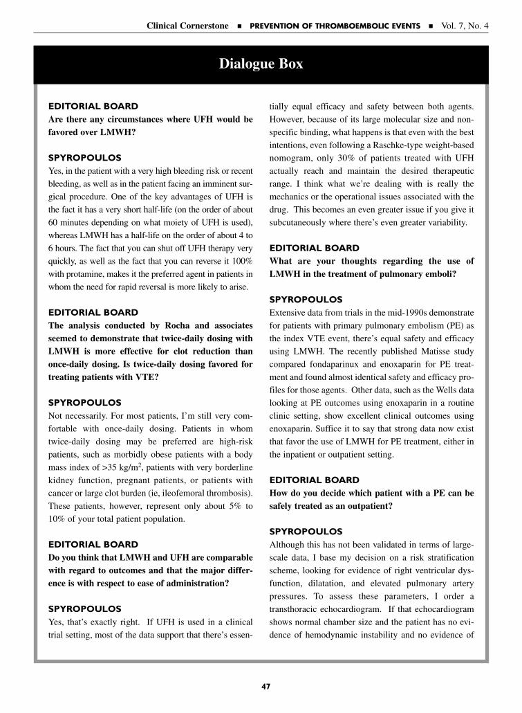
EDITORIAL BOARDAre there any circumstances where UFH would befavored over LMWH?
SPYROPOULOSYes, in the patient with a very high bleeding risk or recent
bleeding, as well as in the patient facing an imminent sur-
gical procedure. One of the key advantages of UFH is
the fact it has a very short half-life (on the order of about
60 minutes depending on what moiety of UFH is used),
whereas LMWH has a half-life on the order of about 4 to
6 hours. The fact that you can shut off UFH therapy very
quickly, as well as the fact that you can reverse it 100%
with protamine, makes it the preferred agent in patients in
whom the need for rapid reversal is more likely to arise.
EDITORIAL BOARDThe analysis conducted by Rocha and associatesseemed to demonstrate that twice-daily dosing withLMWH is more effective for clot reduction thanonce-daily dosing. Is twice-daily dosing favored fortreating patients with VTE?
SPYROPOULOSNot necessarily. For most patients, I’m still very com-
fortable with once-daily dosing. Patients in whom
twice-daily dosing may be preferred are high-risk
patients, such as morbidly obese patients with a body
mass index of >35 kg/m2, patients with very borderline
kidney function, pregnant patients, or patients with
cancer or large clot burden (ie, ileofemoral thrombosis).
These patients, however, represent only about 5% to
10% of your total patient population.
EDITORIAL BOARDDo you think that LMWH and UFH are comparablewith regard to outcomes and that the major differ-ence is with respect to ease of administration?
SPYROPOULOSYes, that’s exactly right. If UFH is used in a clinical
trial setting, most of the data support that there’s essen-
tially equal efficacy and safety between both agents.
However, because of its large molecular size and non-
specific binding, what happens is that even with the best
intentions, even following a Raschke-type weight-based
nomogram, only 30% of patients treated with UFH
actually reach and maintain the desired therapeutic
range. I think what we’re dealing with is really the
mechanics or the operational issues associated with the
drug. This becomes an even greater issue if you give it
subcutaneously where there’s even greater variability.
EDITORIAL BOARDWhat are your thoughts regarding the use ofLMWH in the treatment of pulmonary emboli?
SPYROPOULOSExtensive data from trials in the mid-1990s demonstrate
for patients with primary pulmonary embolism (PE) as
the index VTE event, there’s equal safety and efficacy
using LMWH. The recently published Matisse study
compared fondaparinux and enoxaparin for PE treat-
ment and found almost identical safety and efficacy pro-
files for those agents. Other data, such as the Wells data
looking at PE outcomes using enoxaparin in a routine
clinic setting, show excellent clinical outcomes using
enoxaparin. Suffice it to say that strong data now exist
that favor the use of LMWH for PE treatment, either in
the inpatient or outpatient setting.
EDITORIAL BOARDHow do you decide which patient with a PE can besafely treated as an outpatient?
SPYROPOULOSAlthough this has not been validated in terms of large-
scale data, I base my decision on a risk stratification
scheme, looking for evidence of right ventricular dys-
function, dilatation, and elevated pulmonary artery
pressures. To assess these parameters, I order a
transthoracic echocardiogram. If that echocardiogram
shows normal chamber size and the patient has no evi-
dence of hemodynamic instability and no evidence of
Clinical Cornerstone � PREVENTION OF THROMBOEMBOLIC EVENTS � Vol. 7, No. 4
Dialogue Box
47
right ventricular dysfunction, I’m very comfortable
with starting treatment or continued treatment in the
outpatient setting. The majority of those patients will
get at least a 24-hour observational period in the hospi-
tal so we can facilitate exams and watch the patient in a
controlled setting. In centers in Canada, outpatient PE
treatment is now becoming the standard of care.
EDITORIAL BOARDIn patients adequately anticoagulated on a continu-ous heparin infusion, how would you convert them toLMWH?
SPYROPOULOSOne would simply stop the infusion and then, after 4
to 6 hours, start the LMWH at its prespecified dosing
scheme.
EDITORIAL BOARDIs it true that in terms of timing, you have a 6-hourwindow following surgery to initiate prophylactictherapy?
SPYROPOULOSYes. Russell Hull has advocated a “just-in-time” dose
of using anticoagulants whether it be with UFH or
LMWH. I think that we’ve all surmised, the closer you
give that first dose postoperatively, the greater the effi-
cacy but at the expense of greater bleeding. As a result,
for most surgical procedures that just-in-time dose is
now moved outwards toward 8 to 12 hours instead.
What we’re seeing in the United States is that many sur-
geons like to give that first dose at 12 hours, if not later.
EDITORIAL BOARDIn a patient with a history of HIT associated withUFH use, is LMWH also contraindicated?
SPYROPOULOSAbsolutely. There’s a greater than 80% cross-reactivity
with any type of heparin. So any type of heparin (includ-
ing LMWH) would be contraindicated. In such patients,
alternative options would include lepirudin, argatroban,
and possibly fondaparinux.
EDITORIAL BOARDWhat about for patients with a history of thrombo-cytopenia unrelated to heparin use?
SPYROPOULOSIf it’s severe, such as a platelet count under 100,000,
I would use fondaparinux. If the platelet counts are over
100,000, without documented HIT, I’m comfortable with
the LMWHs.
48
Clinical Cornerstone � PREVENTION OF THROMBOEMBOLIC EVENTS � Vol. 7, No. 4
Dialogue Box
Venous thromboembolism (VTE) is a serious disorder and a major cause of morbidity and mortality amongacutely ill medical patients. However, despite the growing number of patients with acute medical illnesses whohave an associated risk of VTE, the widespread use of VTE prophylaxis does not yet occur for both surgical andnonsurgical patients. Although individuals at greatest risk for VTE include patients undergoing major ortho-pedic surgery and those with medical conditions that require prolonged immobilization, all patients who haveacute medical illnesses should be considered for VTE prophylaxis. Several strategies, including various mechan-ical and pharmacologic approaches, are currently used for VTE prophylaxis. Increased awareness about thefull range of options for VTE prophylaxis can help health care providers select the appropriate course of actionto help reduce the incidence of VTE among patients with acute medical illnesses. (Clinical Cornerstone.2005;7[4]:49–56) Copyright © 2005 Excerpta Medica, Inc.
GENO J. MERLI, MD, FACPProfessor of MedicineVice Chair for Clinical AffairsDirector, Division of Internal MedicineThomas Jefferson UniversityPhiladelphia, Pennsylvania
Prevention of Thrombosis withWarfarin, Aspirin, and MechanicalMethods
Clinical Cornerstone � PREVENTION OF THROMBOEMBOLIC EVENTS � Vol. 7, No. 4
49
Two million people each year are affected by venousthromboembolism (VTE) in the United States, making itthe third most common cardiovascular disease after coro-nary heart disease and stroke.1 VTE includes the devel-opment of deep vein thrombosis (DVT) and pulmonaryembolism (PE); of the estimated 600,000 Americanseach year who develop PE, 60,000 people will die of thiscomplication.2 DVT and PE can develop spontaneously,or they can result from medical circumstances such assurgery, prolonged bed rest, or trauma. Several strategies,including mechanical and pharmacologic approaches,are currently available for VTE prophylaxis (Table I).3
Recently, several new anticoagulants have been evalu-ated in Phase III trials. The THRombin Inhibitor in VenousthromboEmbolism (THRIVE) Treatment Study, for exam-ple, found that the oral direct thrombin inhibitor (DTI)ximelagatran was as effective as standard enoxaparin/warfarin treatment for the prevention of recurrent VTE.4
In this study of 2489 patients with acute DVT (of whichone third had concomitant PE), VTE recurred in 26 of the1240 patients assigned to receive 6 months of treatmentwith ximelagatran (estimated cumulative risk, 2.1%) and
in 24 of the 1249 patients assigned to receive 6 monthsof treatment with enoxaparin/warfarin (estimated cumu-lative risk, 2.0%).
Another randomized trial evaluated the efficacy and safety profile of the low-molecular-weight heparin (LMWH)fondaparinux compared with the LMWH enoxaparin followed by warfarin in patients with acute DVT.5 At 3 months, symptomatic VTE had recurred in 3.9% ofpatients who had received fondaparinux and in 4.9% ofpatients who had received enoxaparin and warfarin, demon-strating the noninferiority of fondaparinux in these patients.Major bleeding was recorded in about 1% of the patients inboth groups. Several other studies are now under way toassess newer agents indicated for the prevention of VTE.
Currently, the use of an LMWH followed by a vita-min K antagonist, such as warfarin, for up to 6 months isappropriate for use in preventing thrombus formation andembolism in most patients6; however, some patients,such as those at risk for heparin-induced thrombocytope-nia or excessive bleeding, may benefit from other ap-proaches. These alternative strategies are addressed in thecurrent discussion.
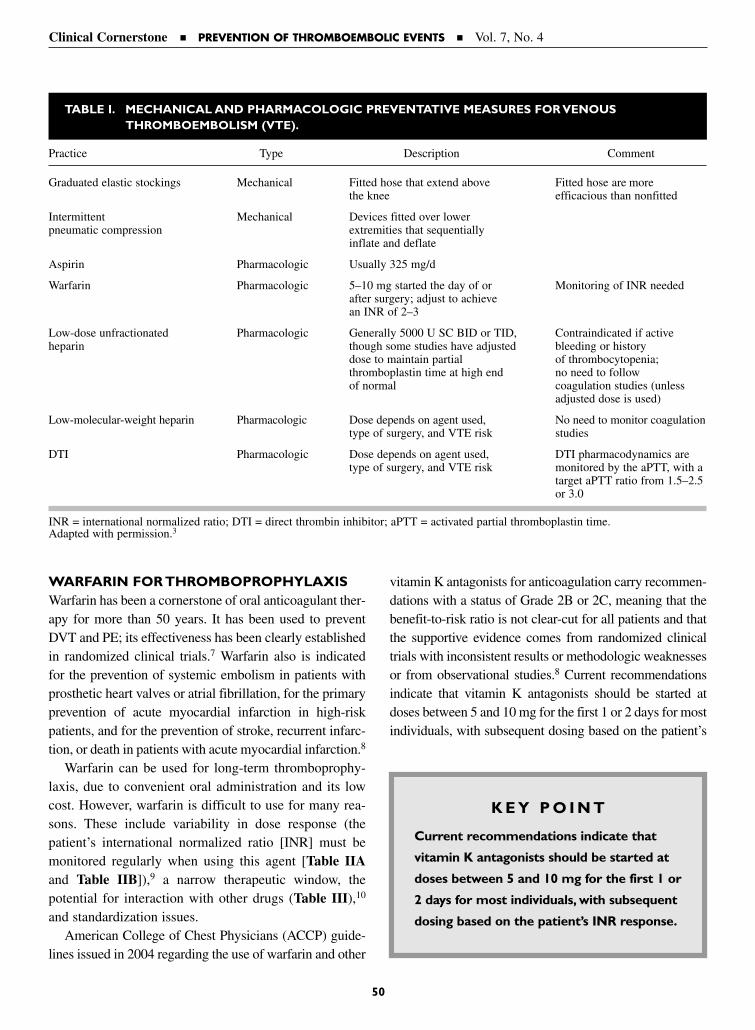
WARFARIN FOR THROMBOPROPHYLAXISWarfarin has been a cornerstone of oral anticoagulant ther-apy for more than 50 years. It has been used to preventDVT and PE; its effectiveness has been clearly establishedin randomized clinical trials.7 Warfarin also is indicatedfor the prevention of systemic embolism in patients withprosthetic heart valves or atrial fibrillation, for the primaryprevention of acute myocardial infarction in high-riskpatients, and for the prevention of stroke, recurrent infarc-tion, or death in patients with acute myocardial infarction.8
Warfarin can be used for long-term thromboprophy-laxis, due to convenient oral administration and its lowcost. However, warfarin is difficult to use for many rea-sons. These include variability in dose response (thepatient’s international normalized ratio [INR] must bemonitored regularly when using this agent [Table IIAand Table IIB]),9 a narrow therapeutic window, thepotential for interaction with other drugs (Table III),10
and standardization issues. American College of Chest Physicians (ACCP) guide-
lines issued in 2004 regarding the use of warfarin and other
vitamin K antagonists for anticoagulation carry recommen-dations with a status of Grade 2B or 2C, meaning that thebenefit-to-risk ratio is not clear-cut for all patients and thatthe supportive evidence comes from randomized clinicaltrials with inconsistent results or methodologic weaknessesor from observational studies.8 Current recommendationsindicate that vitamin K antagonists should be started atdoses between 5 and 10 mg for the first 1 or 2 days for mostindividuals, with subsequent dosing based on the patient’s
Clinical Cornerstone � PREVENTION OF THROMBOEMBOLIC EVENTS � Vol. 7, No. 4
50
TABLE I. MECHANICAL AND PHARMACOLOGIC PREVENTATIVE MEASURES FOR VENOUS THROMBOEMBOLISM (VTE).
Practice Type Description Comment
Graduated elastic stockings Mechanical Fitted hose that extend above Fitted hose are morethe knee efficacious than nonfitted
Intermittent Mechanical Devices fitted over lower pneumatic compression extremities that sequentially
inflate and deflate
Aspirin Pharmacologic Usually 325 mg/d
Warfarin Pharmacologic 5–10 mg started the day of or Monitoring of INR neededafter surgery; adjust to achieve an INR of 2–3
Low-dose unfractionated Pharmacologic Generally 5000 U SC BID or TID, Contraindicated if active heparin though some studies have adjusted bleeding or history
dose to maintain partial of thrombocytopenia;thromboplastin time at high end no need to followof normal coagulation studies (unless
adjusted dose is used)
Low-molecular-weight heparin Pharmacologic Dose depends on agent used, No need to monitor coagulationtype of surgery, and VTE risk studies
DTI Pharmacologic Dose depends on agent used, DTI pharmacodynamics aretype of surgery, and VTE risk monitored by the aPTT, with a
target aPTT ratio from 1.5–2.5 or 3.0
INR = international normalized ratio; DTI = direct thrombin inhibitor; aPTT = activated partial thromboplastin time.Adapted with permission.3
K E Y P O I N T
Current recommendations indicate that
vitamin K antagonists should be started at
doses between 5 and 10 mg for the first 1 or
2 days for most individuals, with subsequent
dosing based on the patient’s INR response.
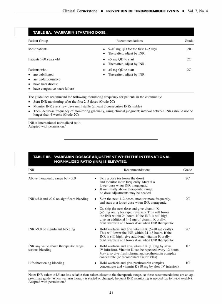
Clinical Cornerstone � PREVENTION OF THROMBOEMBOLIC EVENTS � Vol. 7, No. 4
51
TABLE IIA. WARFARIN STARTING DOSE.
Patient Group Recommendations Grade
Most patients ● 5–10 mg QD for the first 1–2 days 2B● Thereafter, adjust by INR
Patients >60 years old ● ≤5 mg QD to start 2C● Thereafter, adjust by INR
Patients who: ● ≤5 mg QD to start 2C● are debilitated ● Thereafter, adjust by INR● are undernourished ● have liver disease ● have congestive heart failure
The guidelines recommend the following monitoring frequency for patients in the community: ● Start INR monitoring after the first 2–3 doses (Grade 2C)● Monitor INR every few days until stable (at least 2 consecutive INRs stable)● Then, decrease frequency of monitoring gradually, using clinical judgment; interval between INRs should not be
longer than 4 weeks (Grade 2C)
INR = international normalized ratio.Adapted with permission.9
TABLE IIB. WARFARIN DOSAGE ADJUSTMENT WHEN THE INTERNATIONAL NORMALIZED RATIO (INR) IS ELEVATED.
INR Recommendations Grade
Above therapeutic range but <5.0 ● Skip a dose (or lower the dose) 2Cand monitor more frequently. Start at a lower dose when INR therapeutic.
● If minimally above therapeutic range, no dose adjustments may be needed.
INR ≥5.0 and <9.0 no significant bleeding ● Skip the next 1–2 doses, monitor more frequently, 2Cand start at a lower dose when INR therapeutic.
● Or, skip the next dose and give vitamin K (≤5 mg orally for rapid reversal). This will lower the INR within 24 hours. If the INR is still high, give an additional 1–2 mg of vitamin K orally. Start warfarin at a lower dose when INR therapeutic.
INR ≥9.0 no significant bleeding ● Hold warfarin and give vitamin K (5–10 mg orally). 2CThis will lower the INR within 24–48 hours. If the INR is still high, give additional vitamin K orally. Start warfarin at a lower dose when INR therapeutic.
INR any value above therapeutic range, ● Hold warfarin and give vitamin K (10 mg by slow 1Cserious bleeding IV infusion). Vitamin K can be repeated every 12 hours.
May also give fresh plasma and prothrombin complex concentrate (or recombinant factor VIIa).
Life-threatening bleeding ● Hold warfarin and give prothrombin complex 1Cconcentrate and vitamin K (10 mg by slow IV infusion).
Note: INR values >4.5 are less reliable than values closer to the therapeutic range, so these recommendations are an ap-proximate guide. When warfarin therapy is started or changed, frequent INR monitoring is needed (up to twice weekly).Adapted with permission.9
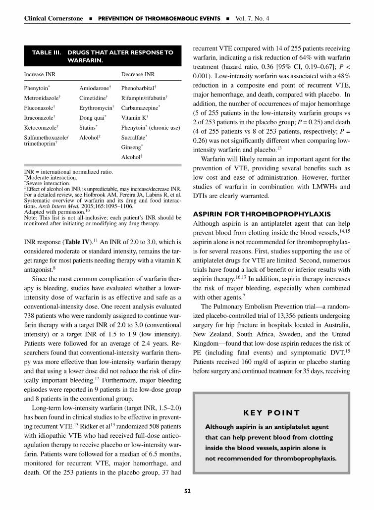
INR response (Table IV).11 An INR of 2.0 to 3.0, which isconsidered moderate or standard intensity, remains the tar-get range for most patients needing therapy with a vitamin Kantagonist.8
Since the most common complication of warfarin ther-apy is bleeding, studies have evaluated whether a lower-intensity dose of warfarin is as effective and safe as a conventional-intensity dose. One recent analysis evaluated738 patients who were randomly assigned to continue war-farin therapy with a target INR of 2.0 to 3.0 (conventionalintensity) or a target INR of 1.5 to 1.9 (low intensity).Patients were followed for an average of 2.4 years. Re-searchers found that conventional-intensity warfarin thera-py was more effective than low-intensity warfarin therapyand that using a lower dose did not reduce the risk of clin-ically important bleeding.12 Furthermore, major bleedingepisodes were reported in 9 patients in the low-dose groupand 8 patients in the conventional group.
Long-term low-intensity warfarin (target INR, 1.5–2.0)has been found in clinical studies to be effective in prevent-ing recurrent VTE.13 Ridker et al13 randomized 508 patientswith idiopathic VTE who had received full-dose antico-agulation therapy to receive placebo or low-intensity war-farin. Patients were followed for a median of 6.5 months,monitored for recurrent VTE, major hemorrhage, anddeath. Of the 253 patients in the placebo group, 37 had
recurrent VTE compared with 14 of 255 patients receivingwarfarin, indicating a risk reduction of 64% with warfarintreatment (hazard ratio, 0.36 [95% CI, 0.19–0.67]; P <0.001). Low-intensity warfarin was associated with a 48%reduction in a composite end point of recurrent VTE,major hemorrhage, and death, compared with placebo. Inaddition, the number of occurrences of major hemorrhage(5 of 255 patients in the low-intensity warfarin groups vs2 of 253 patients in the placebo group; P = 0.25) and death(4 of 255 patients vs 8 of 253 patients, respectively; P =0.26) was not significantly different when comparing low-intensity warfarin and placebo.13
Warfarin will likely remain an important agent for theprevention of VTE, providing several benefits such aslow cost and ease of administration. However, furtherstudies of warfarin in combination with LMWHs andDTIs are clearly warranted.
ASPIRIN FOR THROMBOPROPHYLAXISAlthough aspirin is an antiplatelet agent that can helpprevent blood from clotting inside the blood vessels,14,15
aspirin alone is not recommended for thromboprophylax-is for several reasons. First, studies supporting the use ofantiplatelet drugs for VTE are limited. Second, numeroustrials have found a lack of benefit or inferior results withaspirin therapy.16,17 In addition, aspirin therapy increasesthe risk of major bleeding, especially when combinedwith other agents.7
The Pulmonary Embolism Prevention trial—a random-ized placebo-controlled trial of 13,356 patients undergoingsurgery for hip fracture in hospitals located in Australia,New Zealand, South Africa, Sweden, and the UnitedKingdom—found that low-dose aspirin reduces the risk ofPE (including fatal events) and symptomatic DVT.15
Patients received 160 mg/d of aspirin or placebo startingbefore surgery and continued treatment for 35 days, receiving
Clinical Cornerstone � PREVENTION OF THROMBOEMBOLIC EVENTS � Vol. 7, No. 4
52
TABLE III. DRUGS THAT ALTER RESPONSE TOWARFARIN.
Increase INR Decrease INR
Phenytoin* Amiodarone† Phenobarbital†
Metronidazole† Cimetidine† Rifampin/rifabutin†
Fluconazole† Erythromycin† Carbamazepine*
Itraconazole† Dong quai* Vitamin K†
Ketoconazole† Statins* Phenytoin* (chronic use)
Sulfamethoxazole/ Alcohol‡ Sucralfate*
trimethoprim†
INR = international normalized ratio.*Moderate interaction.†Severe interaction.‡Effect of alcohol on INR is unpredictable, may increase/decrease INR.For a detailed review, see Holbrook AM, Pereira JA, Labiris R, et al.Systematic overview of warfarin and its drug and food interac-tions. Arch Intern Med. 2005;165:1095–1106.Adapted with permission.10
Note: This list is not all-inclusive; each patient’s INR should bemonitored after initiating or modifying any drug therapy.
K E Y P O I N T
Although aspirin is an antiplatelet agent
that can help prevent blood from clotting
inside the blood vessels, aspirin alone is
not recommended for thromboprophylaxis.
Ginseng*
Alcohol‡
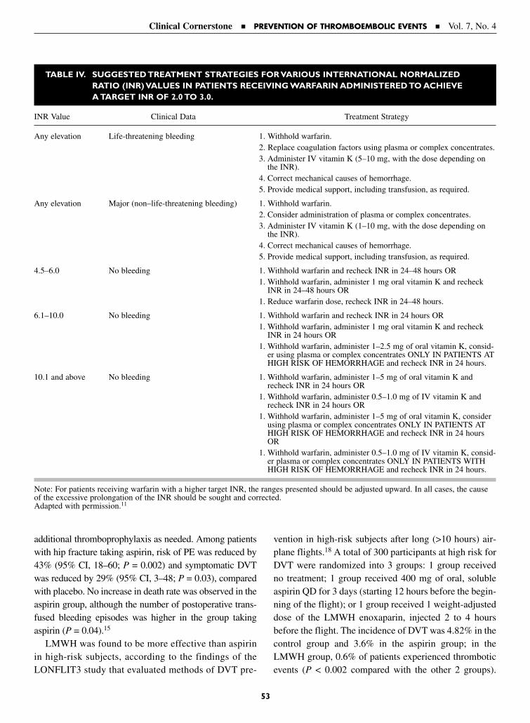
additional thromboprophylaxis as needed. Among patientswith hip fracture taking aspirin, risk of PE was reduced by43% (95% CI, 18–60; P = 0.002) and symptomatic DVTwas reduced by 29% (95% CI, 3–48; P = 0.03), comparedwith placebo. No increase in death rate was observed in theaspirin group, although the number of postoperative trans-fused bleeding episodes was higher in the group takingaspirin (P = 0.04).15
LMWH was found to be more effective than aspirinin high-risk subjects, according to the findings of the LONFLIT3 study that evaluated methods of DVT pre-
vention in high-risk subjects after long (>10 hours) air-plane flights.18 A total of 300 participants at high risk forDVT were randomized into 3 groups: 1 group receivedno treatment; 1 group received 400 mg of oral, solubleaspirin QD for 3 days (starting 12 hours before the begin-ning of the flight); or 1 group received 1 weight-adjusteddose of the LMWH enoxaparin, injected 2 to 4 hoursbefore the flight. The incidence of DVT was 4.82% in thecontrol group and 3.6% in the aspirin group; in theLMWH group, 0.6% of patients experienced thromboticevents (P < 0.002 compared with the other 2 groups).
Clinical Cornerstone � PREVENTION OF THROMBOEMBOLIC EVENTS � Vol. 7, No. 4
53
TABLE IV. SUGGESTED TREATMENT STRATEGIES FOR VARIOUS INTERNATIONAL NORMALIZED RATIO (INR) VALUES IN PATIENTS RECEIVING WARFARIN ADMINISTERED TO ACHIEVE A TARGET INR OF 2.0 TO 3.0.
INR Value Clinical Data Treatment Strategy
Any elevation Life-threatening bleeding 1. Withhold warfarin.2. Replace coagulation factors using plasma or complex concentrates.3. Administer IV vitamin K (5–10 mg, with the dose depending on
the INR).4. Correct mechanical causes of hemorrhage.5. Provide medical support, including transfusion, as required.
Any elevation Major (non–life-threatening bleeding) 1. Withhold warfarin.2. Consider administration of plasma or complex concentrates.3. Administer IV vitamin K (1–10 mg, with the dose depending on
the INR).4. Correct mechanical causes of hemorrhage.5. Provide medical support, including transfusion, as required.
4.5–6.0 No bleeding 1. Withhold warfarin and recheck INR in 24–48 hours OR1. Withhold warfarin, administer 1 mg oral vitamin K and recheck
INR in 24–48 hours OR1. Reduce warfarin dose, recheck INR in 24–48 hours.
6.1–10.0 No bleeding 1. Withhold warfarin and recheck INR in 24 hours OR1. Withhold warfarin, administer 1 mg oral vitamin K and recheck
INR in 24 hours OR1. Withhold warfarin, administer 1–2.5 mg of oral vitamin K, consid-
er using plasma or complex concentrates ONLY IN PATIENTS ATHIGH RISK OF HEMORRHAGE and recheck INR in 24 hours.
10.1 and above No bleeding 1. Withhold warfarin, administer 1–5 mg of oral vitamin K andrecheck INR in 24 hours OR
1. Withhold warfarin, administer 0.5–1.0 mg of IV vitamin K andrecheck INR in 24 hours OR
1. Withhold warfarin, administer 1–5 mg of oral vitamin K, considerusing plasma or complex concentrates ONLY IN PATIENTS ATHIGH RISK OF HEMORRHAGE and recheck INR in 24 hoursOR
1. Withhold warfarin, administer 0.5–1.0 mg of IV vitamin K, consid-er plasma or complex concentrates ONLY IN PATIENTS WITHHIGH RISK OF HEMORRHAGE and recheck INR in 24 hours.
Note: For patients receiving warfarin with a higher target INR, the ranges presented should be adjusted upward. In all cases, the causeof the excessive prolongation of the INR should be sought and corrected.Adapted with permission.11
Mild gastrointestinal symptoms were reported in 13% ofthe patients who received aspirin therapy.
MECHANICAL METHODS FOR VTE PROPHYLAXISMechanical methods of thromboprophylaxis are intendedto increase venous outflow from the legs and/or reduceblood stasis within leg veins and are attractive primarilydue to the absence of bleeding risk compared with pharma-cologic agents. As a result, these methods are particularlyuseful for patients at high risk of bleeding. They are alsosuitable for chronically bedridden patients, in whom pro-longed therapy with heparins may be problematic due tobleeding risk or osteoporosis. The Seventh ACCPConference on Antithrombotic and Thrombolytic Therapyrecommends mechanical prophylaxis in case of contraindi-cation to anticoagulant therapy in nonsurgical patients.19
Efficacy studies comparing the use of mechanical pro-phylaxis (such as graduated compression socks, pneu-matic compression devices, and venous foot pumps) ver-sus the use of pharmacologic anticoagulation approachesin nonsurgical patients are lacking to date.20 However,mechanical prophylaxis is often used with anticoagula-tion therapy to improve effectiveness.21,22 Several studieshave shown that the use of compression socks or elasticstockings is advantageous in patients with myocardialinfarction23 or in reducing DVT following acute stroke.24
A study that analyzed the outcomes of a total of 1892 patients who were treated with intermittent pneumat-ic compression following gynecologic surgery found thatpatients who were most likely to fail intermittent pneumat-ic compression prophylaxis included those patients withcancer, patients with a history of DVT, and patients at age ≥60 years.25 Researchers concluded that patients withmore than 1 of these independent risk factors should beconsidered for more intense prophylaxis regimens.25
The effectiveness and safety of mechanical versusLMWH approaches for DVT prophylaxis have been
compared in patients after total hip replacement.26 Atotal of 216 patients were randomized for DVT prophy-laxis management with either the use of a foot pump orLMWH, resulting in 200 patients available for analysis(16 patients were excluded because they either did nottolerate the continuous use of the foot pump or the use ofLMWH). DVT was detected by serial duplex sonographyin 3 of 100 patients in the foot-pump group and in 6 of100 patients in the LMWH group (P < 0.05). In addition,the mean postoperative drainage was lower in the foot-pump group (259 mL vs 328 mL; P < 0.05), and they hadless swelling of the thigh (10 mm compared with 15 mm;P < 0.05). Researchers concluded that mechanical pro-phylaxis of DVT after total hip replacement was effectiveand safe.26
The issue of adequate patient compliance withmechanical devices warrants consideration; some patientsfind these methods difficult to tolerate. For example, astudy by Westrich and Sculco16 showed that the degree of compliance among 122 patients who experienced totalknee arthroplasty was associated with the prevalence ofDVT after surgery—no DVT was present in patients withan average of 80% compliance with the use of pneumaticplantar compression, but a significant incidence of DVTwas observed in patients averaging 55% compliance.
A study by Robertson et al27 evaluated a consecutiveseries of patients undergoing total joint arthroplasty whowere sequentially treated with 2 mechanical devicesdesigned to prevent DVT. One group of 104 patientswore a thigh-high sequential compression device. Thesecond group of 120 patients wore a foot pump. Asmeasured by responses to a questionnaire, patient satis-faction among patients using the foot pump was signifi-cantly higher than for those using the sequential com-pression device (73% vs 55%, respectively). The studyalso found a higher degree of compliance among patientsusing the foot pumps as compared with patients using thesequential compression device.27
Filter placement within the inferior vena cava (IVC)can be an effective intervention for VTE prophylaxis incertain patients. Although IVC filters do not preventDVT, they can prevent PE.28 Two recent studies (a studyof 94 patients with multiple trauma who underwentplacement of temporary IVC filters in 2002/2003 andanother study of 88 multiple-trauma patients whoreceived temporary IVC filter placement at the intensivecare unit bedside from 2002 to 2004) found that this
Clinical Cornerstone � PREVENTION OF THROMBOEMBOLIC EVENTS � Vol. 7, No. 4
54
K E Y P O I N T
Mechanical prophylaxis is often used with
anticoagulation therapy to improve
effectiveness.
approach was simple and safe, prevented fatal PE, andserved as an effective bridge to anticoagulation therapyuntil other VTE prophylaxis measures could be taken.28,29
A decision to use IVC filters is based on several con-siderations, including the patient’s clinical condition, thetype of filter available, the alternative access sites avail-able, and the expertise of the physician.30 Approximately30,000 to 40,000 IVC filters are placed in patients eachyear in the United States, particularly in about 50% oftrauma patients.31 IVC filters are often considered whena clear contraindication to anticoagulant therapy exists orwhen VTE occurs despite adequate anticoagulation.31
However, vena cava interruption is often regarded as anincomplete treatment for VTE because, unlike anticoag-ulant therapy, it offers no beneficial effect on the preven-tion of DVT or the prevention of DVT extension, recur-rence, and subsequent postthrombotic syndrome.32
Therefore, physicians recommend the use of IVC filtersselectively in patients with contraindications to anticoag-ulants or those with recurrent PE despite adequate anti-coagulation. Whenever possible, pharmacologic antico-agulation is the preferred approach to VTE prophylaxisin patients at risk for VTE.
CONCLUSIONSThe use of VTE prophylaxis is justified by the often clin-ically silent presentation of VTE and its high prevalenceamong hospitalized patients. VTE is diagnosed in~170,000 incident cases among hospitalized patients inthe United States each year; >20,000 patients die of VTEbefore discharge.33 VTE prophylaxis is clearly the mostcost-effective strategy for managing DVT and PE.34 Atgreatest risk are patients undergoing major orthopedicsurgery and those with medical conditions that requireimmobilization for 5 days or longer. Strategies currentlyused to prevent VTE include anticoagulant therapy withheparin (LMWH or unfractionated heparin), DTIs, oral
anticoagulants such as warfarin, and mechanical meth-ods. For patients undergoing total hip or knee replace-ment, treatment with adjusted-dose warfarin, LMWH, orfondaparinux may be used. Warfarin has been the leadingoral anticoagulant agent for various thromboembolicevents. The DTI ximelagatran—the first clinically testedoral anticoagulant since the introduction of warfarin inthe early 1940s—is currently being evaluated for use inthe United States for the prevention and treatment ofVTE.
Several randomized clinical trials have evaluated theefficacy of pharmacologic prophylaxis, with only a lim-ited number of studies that have assessed the efficacy ofmechanical prophylaxis. More studies are needed toassess various approaches to achieve maximal preventionof VTE.
REFERENCES1. Hawkins D. The role of oral direct thrombin inhibitors in the
prophylaxis of venous thromboembolism. Pharmacotherapy.2004;24:179S–183S.
2. Hirsh J, Hoak J. Management of deep vein thrombosisand pulmonary embolism: A statement for healthcare pro-fessionals from the Council on Thrombosis (in consul-tation with the Council on Cardiovascular Radiology),American Heart Association. Circulation. 1996;93:2212–2245.
3. Kleinbart J, Williams MW, Rask K. Prevention of venousthromboembolism. In: Making Health Care Safer: ACritical Analysis of Patient Safety Practices. EvidenceReport/Technology Assessment: Number 43. AHRQ publication 01-E058, July 2001. Agency for HealthcareResearch and Quality, Rockville, Md. Available at:http://www.ahrq.gov/clinic/ptsafety/chap31b.htm.Accessed November 28, 2005.
4. Fiessinger JN, Huisman MV, Davidson BL, et al.Ximelagatran vs low-molecular-weight heparin and war-farin for the treatment of deep vein thrombosis: A ran-domized trial. JAMA. 2005;293:681–689.
5. Buller HR, Davidson BL, Decousus H, et al. Fonda-parinux or enoxaparin for the initial treatment of symp-tomatic deep venous thrombosis: A randomized trial. AnnIntern Med. 2004;140:867–873.
Clinical Cornerstone � PREVENTION OF THROMBOEMBOLIC EVENTS � Vol. 7, No. 4
55
K E Y P O I N T
VTE prophylaxis is clearly the most cost-
effective strategy for managing DVT and PE.
K E Y P O I N T
IVC filters are often considered when a
clear contraindication to anticoagulant
therapy exists or when VTE occurs despite
adequate anticoagulation.
6. Kyrle PA, Eichinger S. Deep vein thrombosis. Lancet.2005;365:1163–1174.
7. Hirsh J, Dalen JE, Anderson DR, et al. Oral anticoagulants:Mechanism of action, clinical effectiveness, and optimaltherapeutic range. Chest. 2001;199:8S–21S.
8. Ansell J, Hirsh J, Poller L, et al. The pharmacology andmanagement of the vitamin K antagonists: The SeventhACCP Conference on Antithrombotic and ThrombolyticTherapy. Chest. 2004;126(Suppl 3):204S–233S.
9. Antithrombotic therapy update. Available at: http://www.ratiopharm.ca/e/Articles/27.htm. Accessed November 30,2005.
10. White RH. Impatient inpatient dosing. Agency forHealthcare Research and Quality Morbidity & MortalityRounds on the Web. July/August 2005. Available at:http://www.webmm.ahrq.gov/case.aspx?caseID=101.Accessed November 29, 2005.
11. Ginsberg JA, Crowther MA, White RH, Ortel TL.Anticoagulation therapy. Hematology. 2001;1:339–357.
12. Kearon C, Ginsberg JS, Kovacs MJ, et al, for the ExtendedLow-Intensity Anticoagulation for Thrombo-EmbolismInvestigators. Comparison of low-intensity warfarin ther-apy with conventional-intensity warfarin therapy for long-term prevention of recurrent venous thromboembolism.N Engl J Med. 2003;349:631–639.
13. Ridker PM, Goldhaber SZ, Danielson E, et al. Long-term,low-intensity warfarin therapy for the prevention of recur-rent venous thromboembolism. N Engl J Med. 2003;348:1425–1434.
14. Lotke PA, Palevsky H, Keenan AM, et al. Aspirin andwarfarin for thromboembolic disease after total jointarthroplasty. Clin Orthop Relat Res. 1996;324:251–258.
15. Prevention of pulmonary embolism and deep vein throm-bosis with low dose aspirin: Pulmonary EmbolismPrevention (PEP) trial. Lancet. 2000;355:1295–1302.
16. Westrich GH, Sculco TP. Prophylaxis against deep venousthrombosis after total knee arthroplasty. Pneumatic plantarcompression and aspirin compared with aspirin alone. J Bone Joint Surg Am. 1996;78:826–834.
17. Gent M, Hirsh J, Ginsberg JS, et al. Low-molecular-weight heparinoid orgaran is more effective than aspirin inthe prevention of venous thromboembolism after surgeryfor hip fracture. Circulation. 1996;93:80–84.
18. Cesarone MR, Belcaro G, Nicolaides AN, et al. Venousthrombosis from air travel: The LONFLIT3 study—Prevention with aspirin vs low-molecular-weight heparin(LMWH) in high-risk subjects: A randomized trial.Angiology. 2002;53:1–6.
19. Geerts WH, Pineo GF, Heit JA, et al. Prevention of venousthromboembolism: The Seventh ACCP Conference onAntithrombotic and Thrombolytic Therapy. Chest. 2004;126(Suppl 3):338S–400S.
20. Imberti D, Prisco D. Venous thromboembolism prophy-laxis in medical patients: Future perspectives. Thromb Res.2005;116:365–375.
21. Levine MN, Lee AYY. Risk assessment and primary VTEprevention in the cancer patient. Pathophysiol HaemostThromb. 2003;33(Suppl 1):36–41.
22. Ramos R, Salem BI, De Pawlikowski MP, et al. The effi-cacy of pneumatic compression stockings in the preven-tion of pulmonary embolism after cardiac surgery. Chest.1996;109:82–85.
23. Kierkegaard A, Norgren L. Graduated compression stock-ings in the prevention of deep vein thrombosis in patientswith acute myocardial infarction. Eur Heart J. 1993;14:1365–1368.
24. Muir KW, Watt A, Baxter G, et al. Randomized trial ofgraded compression stockings for prevention of deep-veinthrombosis after acute stroke. QJM. 2000;93:359–364.
25. Clarke-Pearson DL, Dodge RK, Synan I, et al. Venousthromboembolism prophylaxis: Patients at high risk to failintermittent pneumatic compression. Obstet Gynecol.2003;101:157–163.
26. Pitto RP, Hamer H, Heiss-Dunlop W, Kuehle J.Mechanical prophylaxis of deep-vein thrombosis aftertotal hip replacement: A randomised clinical trial. J BoneJoint Surg Br. 2004;86:639–642.
27. Robertson KA, Bertot AJ, Wolfe MW, Barrack RL.Patient compliance and satisfaction with mechanicaldevices for preventing deep venous thrombosis after jointreplacement. J South Orthop Assoc. 2000;9:182–186.
28. Rosenthal D, Wellons ED, Levitt AB, et al. Role of pro-phylactic temporary inferior vena cava filters placed at theICU bedside under intravascular ultrasound guidance inpatients with multiple trauma. J Vasc Surg. 2004;40:958–964.
29. Rosenthal D. Use of bedside intravascular ultrasonographyto place and retrieve inferior vena cava filters. Presen-tation made at: Combined Sessions: Vascular Surgeryand Interventional Radiology. Available at: http://www.veithsymposium.org/pdf2004/270.pdf. AccessedDecember 8, 2005.
30. Grassi DJ, Swan TL, Cardella J, et al. Quality improve-ment guidelines for percutaneous permanent inferior venacava filter placement for the prevention of pulmonaryembolism. J Vasc Interv Radiol. 2003;14:S271–S275.
31. Kurtoglu M, Güloglu R, Alimoglu O, et al. The late out-comes of vena cava filters in the prevention of pulmonaryembolism. Ulus Travam Derg. 2003:9:114–119.
32. Girard P, Stern J-B, Parent F. Medical literature and venacava filters: So far so weak. Chest. 2002:122:963–967.
33. McGarry L, Thompson D. Retrospective database analysisof the prevention of venous thromboembolism with low-molecular-weight heparin in acutely ill medical inpatientsin community practice. Clin Ther. 2004;26:419–430.
34. Bergqvist D, Jendteg S, Johansen L, et al. Cost of long-term complications of deep venous thrombosis of thelower extremities: An analysis of a defined patient popu-lation in Sweden. Ann Intern Med. 1997;126:454–457.
Clinical Cornerstone � PREVENTION OF THROMBOEMBOLIC EVENTS � Vol. 7, No. 4
56
Address correspondence to: Geno J. Merli, MD, FACP, Thomas Jefferson University, Department of InternalMedicine, 211 South 9th Street, Suite 300, Philadelphia, PA 19107. E-mail: [email protected]
˘˘˘
57
Given the increased number of patients hospitalized foracute medical illnesses and the associated risk of venousthromboembolism (VTE), the use of prophylaxis hasbecome a public health matter. Thromboprophylaxis isnot widely practiced in acutely ill medical patients, duein part to the heterogeneity of this group and the per-ceived difficulty in assessing those who would mostbenefit from treatment. Nevertheless, the results ofrecent well-conducted clinical trials support the evidence-based recommendations for more widespreadsystematic use of low-dose low-molecular-weightheparin (LMWH) or unfractionated heparin (UFH) inthis population. Three large well-controlled studies
(MEDENOX, PREVENT, and ARTEMIS) in acutely illmedical patients confirm previous findings that differentat-risk patient populations show a consistent 50% reduc-tion in VTE events with LMWH and fondaparinux. Ameta-analysis in nearly 5000 patients in internal medi-cine comparing UFH and LMWH revealed a trend forreduction of deep vein thrombosis and pulmonaryembolism with LMWH. Based on duration of use inclinical trials in acutely ill medical patients, prophylac-tic treatment with UFH and LMWH is recommended for2 weeks.
Copyright © 2004 American Heart Association, Inc.
Clinical Cornerstone � PREVENTION OF THROMBOEMBOLIC EVENTS � Vol. 7, No. 4
Commentary on Current LiteraturePreventing Venous Thromboembolism in Medical Patients
Leizorovicz A, Mismetti P.Circulation. 2004;110(Suppl IV):IV-13-IV-19.
Richard A. Johnson, MD
COMMENTARYThis paper by Leizorovicz and Mismetti provides a comprehensive review of recent clinical trials and evidence-based recommendations regarding the use of VTE prophylaxis for patients with acute illnesses.Of the approximately 300,000 cases of VTE now estimated to develop each year in the United States, about75% of these VTE cases occur in acutely ill nonsurgical patients. However, despite an ever-growing popu-lation of patients with acute medical illness who have an associated risk of VTE, the widespread use of VTEprophylaxis does not yet occur in various groups of at-risk patients. Reasons for this limited use of VTEprophylaxis include lack of awareness of the risk of VTE in certain groups of patients; concern about bleed-ing; uncertain cost-to-benefit ratio, and lack of convenient risk-stratification tools for identifying thosepatients who can benefit from VTE prophylaxis. The findings of 3 large, placebo-controlled studies—MEDENOX, PREVENT, and ARTEMIS—found a reduction of VTE in the range of 50% with the use ofLMWH and fondaparinux for a maximum of 14 days, with a minimal risk of major bleeding. More wide-spread and systematic use of VTE prophylaxis can greatly reduce the risk of VTE in patients with acutemedical illnesses.
58
Clinical Cornerstone � PREVENTION OF THROMBOEMBOLIC EVENTS � Vol. 7, No. 4
Commentary on Current LiteratureRichard A. Johnson, MD
BACKGROUND: Clinical trials have demonstrated thatprophylaxis with low-molecular-weight heparin reducesthe occurrence of venous thromboembolism (VTE) amongacutely ill medical inpatients in the experimental setting.
OBJECTIVE: The goal of this retrospective databaseanalysis was to examine the outcomes of low-molecular-weight heparin thromboprophylaxis among acutely illmedical inpatients in community practice.
METHODS: Using a large, geographically diverse,multihospital US database, we identified persons aged≥40 years who had a hospital stay ≥6 days for an acutemedical condition (including selected circulatory disor-ders, respiratory disorders, infectious diseases, or neo-plasms) during calendar-year 2000. From these patients,those who received either enoxaparin thromboprophy-laxis or no thromboprophylaxis were identified. Surgicalpatients, patients with nonthrombotic conditions requir-ing anticoagulant therapy, those transferred from or dis-charged to another acute care facility, and those medical-ly ineligible for anticoagulation therapy were excluded.We compared the incidence of deep-vein thrombosis
(DVT), pulmonary embolism (PE), all VTE (ie, DVTand/or PE), and death during the hospital stay in the 2cohorts.
RESULTS: A total of 162 patients receiving enoxaparinthromboprophylaxis and 3557 receiving no thrombopro-phylaxis were identified. The risk of VTE over thecourse of hospitalization was 1.9% with enoxaparinthromboprophylaxis versus 6.2% with no thrombopro-phylaxis (relative risk = 0.30; P = 0.023); mortality wassimilar in the 2 groups (8.0% vs 7.3; P = NS).
CONCLUSIONS: Using hospital administrative data,we observed a 70% lower risk of VTE for hospitalizedacutely ill medical patients receiving low-molecular-weight heparin thromboprophylaxis versus those receiv-ing no thromboprophylaxis; these results are consistentwith findings from clinical trials of low-molecular-weight heparin versus placebo. We conclude that thelow-molecular-weight heparin enoxaparin is effective inreducing the risk of VTE in acutely ill medical inpatientsin community practice.
Copyright © 2005 Excerpta Medica Inc. All rights reserved.
Retrospective Database Analysis of the Prevention of Venous Thromboembolism with Low-Molecular-Weight Heparin in Acutely Ill Medical Inpatients in Community Practice
McGarry LJ,Thompson D.Clin Ther. 2004;26:419–430.
COMMENTARY
This analysis by McGarry and Thompson highlights the need to focus more attention on VTE prophylaxisamong acutely ill medical patients in community practice. In addition to surgical patients at elevated risk forVTE, nonsurgical patients who are hospitalized for various conditions (including congestive heart failure,chronic obstructive pulmonary disease, acute infections, or cancer) often have prolonged lengths of staywith periods of immobility, placing them at increased risk of VTE. This analysis of administrative data from23 acute care institutions throughout the United States for calendar-year 2000 examined the outcomes of2 groups of patients (1 group of 162 patients who received enoxaparin thromboprophylaxis and 1 group of3557 patients who received no thromboprophylaxis). Researchers observed a 70% lower risk of VTE amongpatients receiving enoxaparin thromboprophylaxis as compared with patients who received no thrombopro-phylaxis. In community practice, many patients with acute illnesses who can potentially benefit from throm-boprophylaxis are not receiving this care. Health care professionals are encouraged to identify risk factorsfor VTE (including a history of VTE, advanced age, obesity, varicose veins, and estrogen use) in all patientsand adopt a more proactive approach to help prevent VTE.