Embed Size (px)
Citation preview
of September 17, 2018.This information is current as
I TransactivatorExpression in Mice Deficient for Nlrc5/Class Cutting Edge: Impaired MHC Class I
S. KobayashiAmlan Biswas, Torsten B. Meissner, Taro Kawai and Koichi
http://www.jimmunol.org/content/189/2/516doi: 10.4049/jimmunol.12000642012;
2012; 189:516-520; Prepublished online 18 JuneJ Immunol
MaterialSupplementary
4.DC1http://www.jimmunol.org/content/suppl/2012/06/18/jimmunol.120006
Referenceshttp://www.jimmunol.org/content/189/2/516.full#ref-list-1
, 10 of which you can access for free at: cites 23 articlesThis article
average*
4 weeks from acceptance to publicationFast Publication! •
Every submission reviewed by practicing scientistsNo Triage! •
from submission to initial decisionRapid Reviews! 30 days* •
Submit online. ?The JIWhy
Subscriptionhttp://jimmunol.org/subscription
is online at: The Journal of ImmunologyInformation about subscribing to
Permissionshttp://www.aai.org/About/Publications/JI/copyright.htmlSubmit copyright permission requests at:
Email Alertshttp://jimmunol.org/alertsReceive free email-alerts when new articles cite this article. Sign up at:
Print ISSN: 0022-1767 Online ISSN: 1550-6606. Immunologists, Inc. All rights reserved.Copyright © 2012 by The American Association of1451 Rockville Pike, Suite 650, Rockville, MD 20852The American Association of Immunologists, Inc.,
is published twice each month byThe Journal of Immunology
by guest on September 17, 2018
http://ww
w.jim
munol.org/
Dow
nloaded from
by guest on September 17, 2018
http://ww
w.jim
munol.org/
Dow
nloaded from

Cutting Edge: Impaired MHC Class I Expression in MiceDeficient for Nlrc5/Class I TransactivatorAmlan Biswas,*,†,1 Torsten B. Meissner,*,†,1 Taro Kawai,‡,x andKoichi S. Kobayashi*,†
MHC class I and class II are crucial for the adaptive im-mune system. Although regulation of MHC class II ex-pression by CIITA has long been recognized, themechanism of MHC class I transactivation has beenlargely unknown until the recent discovery of NLRC5/class I transactivator. In this study, we show usingNlrc5-deficient mice that NLRC5 is required for bothconstitutive and inducible MHC class I expression.Loss of Nlrc5 resulted in severe reduction in the expres-sion of MHC class I and related genes such as b2-micro-globulin, Tap1, or Lmp2, but did not affect MHC classII levels. IFN-g stimulation could not overcome the im-paired MHC class I expression in Nlrc5-deficient cells.Upon infection with Listeria monocyogenes, Nlrc5-de-ficient mice displayed impaired CD8+ T cell activation,accompanied with increased bacterial loads. Thesefindings illustrate critical roles of NLRC5/class I trans-activator in MHC class I gene regulation and hostdefense by CD8+ T cell responses. The Journal ofImmunology, 2012, 189: 516–520.
Major histocompatibility complex class I and class IImolecules play key roles in the activation of theadaptive immune system. MHC class I molecules
present peptide Ags of intracellular origin such as tumor orviral Ags to CD8+ T cells, whereas MHC class II moleculespresent peptide Ags of extracellular sources to CD4+ T cells(1). The expression of both constitutive and inducible MHCclass II requires the master transcriptional coactivator CIITA(2). Although CIITA itself lacks a DNA binding domain,CIITA can activate the promoters of MHC class II genesby engaging in a nucleoprotein complex called the MHCenhanceosome (3, 4), together with promoter-resident tran-scription factors, including the trimeric regulatory factor Xprotein complex, CREB/activating transcription factor 1 familymembers, and the NF-Y protein (5, 6). CIITA can also trans-
activate MHC class I genes at least in vitro (7, 8), althoughboth bare lymphocyte syndrome patients with mutations inthe CIITA gene and CIITA-deficient mice retain intact MHCclass I expression, indicating that there is another mechanismfor the activation of MHC class I in vivo (2, 9–12).The recent discovery of a transactivator specific for MHC
class I genes, NLRC5, identified a new addition to the reg-ulators of MHC genes (13, 14). Both NLRC5 and CIITAbelong to the NLR or nucleotide binding domain (NBD),leucine-rich repeat family of proteins, and phylogeneticallythey are most closely related to each other (13, 15). NLRC5,or class I transactivator (CITA), is also an IFN-g–induciblenuclear protein, but NLRC5 specifically associates with andtransactivates MHC class I promoters, resulting in the ex-pression of MHC class I and related genes such as b2-microglobulin (b2m), TAP1, and large multifunctional pro-tease 2 (13, 14). The NBD is a critical domain for thefunction of NLRC5, as the NBD is required for both nuclearimport and transactivation of MHC class I genes (16). Inthe nucleus, NLRC5 participates in a MHC class I-specificenhanceosome, together with the regulatory factor X com-ponents and CREB/activating transcription factor 1 familytranscription factors to activate MHC class I gene promoters(17). In addition to its function as a MHC class I trans-activator, NLRC5 has been reported to be involved in theregulation of TLR and RIG-I–like receptor signaling, antiviralresponses, and inflammasome activation (15, 18–21). How-ever, those reports provide conflicting results, and their con-clusions are not supported by the data obtained from theinitial characterization of Nlrc5-deficient mice (14, 22).Although the identification of NLRC5 as a MHC class I
gene transactivator is significant, the role of NLRC5 in MHCclass I expression in vivo had not been elucidated. To addressthis question, we investigated the function of NLRC5 usingNlrc5-deficient mice. In this study, we show that Nlrc5 isrequired for both constitutive and inducible expression ofMHC class I. Nlrc5-deficient mice were more susceptible to
*Department of Cancer Immunology and AIDS, Dana-Farber Cancer Institute, Boston,MA 02215; †Division of Immunology, Department of Microbiology and Immunobiol-ogy, Harvard Medical School, Boston, MA 02215; ‡Laboratory of Host Defense, WorldPremier International Immunology Frontier Research Center, Osaka University, Osaka565-0871, Japan; and xDepartment of Host Defense, Research Institute for MicrobialDiseases, Osaka University, Osaka 565-0871, Japan
1A.B. and T.B.M. contributed equally to this work.
Received for publication January 10, 2012. Accepted for publication May 16, 2012.
This work was supported by grants from the National Institutes of Health and theCrohn’s and Colitis Foundation of America. K.S.K. is a recipient of the InvestigatorAward from the Cancer Research Institute and the Claudia Adams Barr Award. T.B.M.
has been a recipient of the European Molecular Biology Organization Long-Termfellowship, and A.B. is a recipient of Crohn’s Colitis Foundation of America fellowship.
Address correspondence and reprint requests to Dr. Koichi S. Kobayashi, Department ofCancer Immunology and AIDS, Dana-Farber Cancer Institute, Dana 1420A, 450Brookline Avenue, Boston, MA 02215. E-mail address: [email protected]
The online version of this article contains supplemental material.
Abbreviations used in this article: CITA, class I transactivator; Hsp, heat shock protein;b2m, b2-microglobulin; NBD, nucleotide binding domain; qPCR, quantitative PCR.
Copyright� 2012 by TheAmerican Association of Immunologists, Inc. 0022-1767/12/$16.00
www.jimmunol.org/cgi/doi/10.4049/jimmunol.1200064
by guest on September 17, 2018
http://ww
w.jim
munol.org/
Dow
nloaded from
infection with Listeria monocytogenes, as highlighted by im-paired CD8+ T cell activation and increased bacterial burdenin the infected organs, indicating a critical role of Nlrc5/CITA in MHC class I-dependent CD8+ T cell responses.
Materials and MethodsMice
Nlrc5-deficient mice were provided by T. Kawai and S. Akira (Osaka Uni-versity, Osaka, Japan) (22). Wild-type mice (F1 mice from 129SvEv andC57BL/6 mice) were obtained from Taconic. OT-1 TCR transgenic micewere a gift of H. Cantor (Dana-Farber Cancer Institute, Boston, MA). Micewere maintained under specific pathogen-free conditions and used in accor-dance with institutional and National Institutes of Health guidelines.
Cell culture and reagents
Generation and stimulation of bone marrow-derived macrophages and den-dritic cells have been described previously (23). Splenocytes and thymocyteswere cultured in RPMI 1640 supplemented with 10% FBS, 55 mM 2-ME(Life Technologies), and penicillin/streptomycin (Life Technologies). MurinerIFN-g was from BioLegend. Peritoneal cells were isolated without pre-treatment with thioglycolate.
RNA isolation and quantitative PCR
RNA isolation and quantitative PCR were performed, as previously described(13). Primer sequences are listed in Supplemental Table I.
Western blot analysis
Western blot analysis was performed, as described previously (13), usingthe following Abs: anti–H2-Kb, anti-b2m (gifts of T. Hansen, WashingtonUniversity), and anti-heat shock protein (Hsp) 90 Ab (F-8; Santa CruzBiotechnology).
Flow cytometric analysis
FACS analysis was performed, as previously described (13), using the followingAbs: FITC anti-mouse B220, FITC anti-mouse CD11c, allophycocyaninanti-mouse CD4, PE/Cy5 anti-mouse CD8, allophycocyanin anti-mouseF4/80, PE anti-mouse I-Ab/I-Ad (eBioscience), PE anti-mouse H2, andallophycocyanin anti-mouse IFN-g (BioLegend).
OT-1 cell coculture assay
MACS-sorted CD8+ T cells from OT-1 mice with CD45.1 background wereCFSE labeled and cocultured with SIINFEKL (1 mM) pulsed irradiatedsplenic B220+ cells from either Nlrc5+/2 or Nlrc52/2 mice at a ratio of 1:25(B:T) for 72 h. Proliferation of OT-1 T cells was determined by FACSanalysis of the CFSE dilution in CD45.1 gated cells.
L. monocytogenes infection
L. monocytogenes (strain 10403s, provided by D. Portnoy at University ofCalifornia, Berkeley) was grown to midlog phase (A600, 0.1), and injected intomice via the lateral tail vein. The bacterial burden in the spleen and liver ofinfected mice was determined by CFU count of serial dilution of lysates.
Statistical analysis
Data were subjected to one-way ANOVA for analysis of statistical significanceusing Prism (GraphPad). Results are given as the mean 6 SEM. A p value,0.05 was considered to be significant.
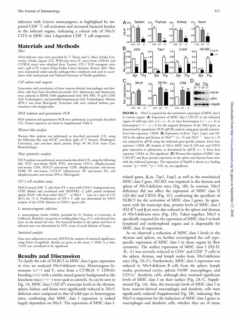
Results and DiscussionTo clarify the role of NLRC5 in MHC class I gene expressionin vivo, we analyzed Nlrc5-deficient mice. Heterozygous lit-termates (+/2) and F1 mice from a C57BL/6 3 129SvEvbreeding (+/+) with a similar mixed genetic background to theknockout mice (2/2) were used as controls. As can be seen inFig. 1A, MHC class I (H2-Kb) transcript levels in the thymus,spleen, kidney, and ileum were significantly reduced in Nlrc5-deficient mice compared to that of wild-type or heterozygousmice, confirming that MHC class I expression is indeedlargely dependent on Nlrc5. The expression of MHC class I-
related genes, b2m, Tap1, Lmp2, as well as the nonclassicalMHC class I gene, H2-M3, was impaired in the thymus andspleen of Nlrc5-deficient mice (Fig. 1B). In contrast, Nlrc5deficiency did not affect the expression of MHC class II(H2-Aa) and CIITA (Fig. 1C), confirming the specificity ofNLRC5 for the activation of MHC class I genes. In agree-ment with the transcript data, protein levels of MHC class I(H2-Kb) and b2m were also reduced in the spleen and thymusof Nlrc5-deficient mice (Fig. 1D). Taken together, Nlrc5 isspecifically required for the expression of MHC class I in bothlymphoid and nonlymphoid organs and is dispensable forMHC class II expression.As we observed a reduction of MHC class I levels in the
thymus and spleen, we further investigated the cell type-specific expression of MHC class I in those organs by flowcytometry. The surface expression of MHC class I (H2-D,-K, -L) was severely reduced in CD4+ and CD8+ T cells inthe spleen, thymus, and lymph nodes from Nlrc5-deficientmice (Fig. 2A–C). Furthermore, MHC class I expression wasreduced in Nlrc5-deficient B cells from the spleen, lymphnodes, peritoneal cavity, splenic F4/80+ macrophages, andCD11c+ dendritic cells, although they retained significantlevels of MHC class I on their surface (Fig. 2A–C, Supple-mental Fig. 1A). Also, the transcript levels of MHC class I inbone marrow-derived macrophages and dendritic cells weresignificantly reduced (Supplemental Fig. 1B), indicating thatNlrc5 is important for the induction of MHC class I genes inmacrophages and dendritic cells, whether they are of tissue
FIGURE 1. Nlrc5 is required for the constitutive expression of MHC class I
in various organs. (A) Expression of MHC class I (H2-Kb) in the indicated
organs of wild-type mice (+/+, n = 3), or mice heterozygous (+/2, n = 4) or
homozygous (2/2, n = 3) for the targeted disruption of the Nlrc5 gene, as
determined by quantitative PCR (qPCR) analysis using gene-specific primers.
Error bars represent 6SEM. (B) Expression of b2m, Tap1, Lmp2, and H2-M3 in the spleen and thymus of Nlrc5+/+ (n = 3) and Nlrc52/2 mice (n = 3)
was analyzed by qPCR using the indicated gene-specific primers. Error bars
represent 6SEM. (C) Analysis of Nlrc5, MHC class II (H2-Aa), and CIITA
gene expression in splenocytes, as determined by qPCR. n = 3. Error bars
represent6SEM. ns, Not significant. (D) Western blot analysis of MHC class
I (H2-Kb) and b2m protein expression in the spleen and thymus from mice
with the indicated genotype. The expression of Hsp90 is shown as a loading
control. *p , 0.05, **p , 0.01; ns, not significant.
The Journal of Immunology 517
by guest on September 17, 2018
http://ww
w.jim
munol.org/
Dow
nloaded from
origin or bone marrow derived. Interestingly, the surface ex-pression of MHC class I on these bone marrow-derived cellswas largely unaffected (Supplemental Fig. 1A), suggestingthat there appears to be a compensatory posttranscriptionalmechanism to rescue the MHC class I deficiency in culturedcells. Together, these findings suggest that Nlrc5 plays a majorrole in the regulation of MHC class I gene expression, albeitthe degree of the requirement for Nlrc5 in MHC class I ex-pression varies between different cell types.We have previously demonstrated that NLRC5 is required
for MHC class I gene expression upon IFN-g stimulation (13).Therefore, we examined the impact of Nlrc5 deficiency onIFN-g–inducible MHC class I expression by stimulatingsplenocytes from Nlrc5-deficient mice with IFN-g. As previ-ously shown (13, 15, 20, 21), IFN-g treatment resulted in theupregulation of Nlrc5 transcript, which correlated with theinduction of MHC class I gene (H2-Kb) expression in bothwild-type and heterozygous splenocytes (Fig. 3A) (13). InNlrc5-deficient splenocytes, however, IFN-g stimulationcould not rescue the impairment of MHC class I expression,when compared with the levels of MHC class I observed inthe controls (Fig. 3A). b2m expression was also only partiallyrescued (Fig. 3A). In contrast, upregulation of Stat1 tran-
scripts, which is induced by IFN-g stimulation in a Nlrc5-independent manner (13), was comparable among the threegenotypes, indicating that the JAK/STAT signaling cascadedownstream of the IFN-g receptor is intact in Nlrc5-deficientcells. Interestingly, there was a small, but distinct, inductionof MHC class I expression in Nlrc5-deficient splenocytesupon IFN-g stimulation, supporting the existence of a Nlrc5-independent mechanism(s) of MHC class I expression (Fig.3A). In agreement with the transcript data, IFN-g stimulationdid not restore the expression of MHC class I (H2-Kb) andb2m at the protein level in Nlrc5-deficient splenocytes andthymocytes (Fig. 3B). Moreover, flow cytometric analysisshowed that IFN-g stimulation did not rescue the reducedMHC class I surface expression in CD4+, CD8+ T cells,B cells, F4/80+ macrophages, and CD11c+ dendritic cellsobtained from the spleen of Nlrc5-deficient mice (Fig. 3C).To address whether the impaired MHC class I expression
caused by Nlrc5 deficiency has an impact on immuneresponses, OT-1 CD8+ T cells were cocultured with peptide-loaded B cells. OT-1 T cells cultured with Nlrc5-deficientB cells displayed impaired proliferation, indicating thatNLRC5 is indeed required for Ag-specific stimulation ofCD8+ T cells (Fig. 4A). The role of NLRC5 in MHC class I-
FIGURE 2. Reduced MHC class I expression in
various cell types of Nlrc5-deficient mice. Single-cell
suspension of spleen (A, left panel; D), thymus (B, leftpanel), and lymph node (C, left panel) from Nlrc5+/2
(gray line) and Nlrc52/2 (black line) mice was ana-
lyzed by flow cytometry for the expression of MHC
class I [H2-K/D/L, in (A), (B), and (C), left panel] orMHC class II (D) molecules (I-Ab/I-Ad), gated on
CD8+, CD4+, B220+, F4/80+, or CD11c+ cells.
Shaded region represents the isotype control. Data are
representative of two independent experiments. Bar
graphs (A–C, right panel) present the mean and SEM
of the corresponding mean fluorescence intensity (n =
3 for each genotype). *p , 0.05, **p , 0.01.
518 CUTTING EDGE: IMPAIRED MHC CLASS I EXPRESSION IN Nlrc5-DEFICIENT MICE
by guest on September 17, 2018
http://ww
w.jim
munol.org/
Dow
nloaded from

mediated immune responses was further investigated byinfection studies using an intracellular bacterium, L. mono-cytogenes, because a CD8+ T cell response is critical for thehost defense against this bacterium. Although Listeria infec-tion clearly induced Ag-specific CD8+ T cell activation in thespleen and liver of wild-type mice, as demonstrated by in-creased numbers of IFN-g–positive cells after ex vivo stimu-lation with heat-killed bacteria (Fig. 4B, Supplemental Fig.1C), in Nlrc5-deficient mice, Ag-specific CD8+ T cell acti-vation was impaired and the mice harbored increased num-bers of the bacterium in both spleen and liver (Fig. 4B, 4C).This Listeria-susceptible phenotype contradicts the previouslyproposed function of NLRC5 as a TLR inhibitor, as in thatcase the Nlrc5-deficient mice should be more protected ratherthan being more susceptible to infection. Indeed, Nlrc5-de-ficient macrophages expressed normal levels of IL-6, TNF-a,IL-12 p40, and IL-1b at both transcript and protein levelsupon LPS, CpG oligo, and poly(IC) stimulation, confirmingthe observations made in the previous study using Nlrc5-de-ficient mice (Supplemental Fig. 1D, 1E) (22). These datacollectively demonstrate a critical role of Nlrc5/CITA inMHC class I-mediated immune responses in vivo.
In addition to our previous study in which we used humanlymphoid and epithelial cell lines (13), the current studycompellingly demonstrates the critical role of NLRC5 in bothconstitutive and inducible MHC class I expression in vivousing Nlrc5-deficient mice. Moreover, we show that NLRC5is required for CD8+ T cell responses in an Ag-specificmanner. Ag peptide-loaded Nlrc5-deficient B cells failed toactivate OT-1 T cells efficiently. Strikingly, our Listeria in-fection study demonstrated that CD8+ T cells obtained fromNlrc5-deficient mice displayed impaired Ag-specific activa-tion, and the mice had increased bacterial loads in the spleenand liver. Therefore, we conclude that NLRC5 plays a criticalrole in MHC class I-mediated immune responses in vivo.This study also reveals that the requirement for NLRC5 inMHC class I gene expression varies between different celltypes. The reduced MHC class I phenotype was most promi-nent in CD4+ and CD8+ T cells and less prominent in B cells(Fig. 2A–C). Also, macrophages and dendritic cells retainedresidual MHC class I expression (Fig. 2A). This may suggestthat an alternative, NLRC5-independent mechanism of MHCclass I transactivation exists. Interestingly, similar residualexpression of MHC class II genes has been reported in CIITA-deficient mice; CIITA-deficient dendritic cells retained MHCclass II expression, although expression levels were signifi-cantly reduced (12). These observations indicate that althoughNLRC5/CITA and CIITA are critical for the expression ofMHC class I and class II, respectively, APCs may possess al-ternative mechanisms to ensure the efficient presentation of
FIGURE 3. IFN-g stimulation does not rescue the reduced MHC class I
expression observed in Nlrc5-deficient mice. (A) Splenocytes from Nlrc5+/+,Nlrc5+/2, and Nlrc52/2 mice (n = 3 for each genotype) were stimulated for 18 h
with IFN-g (100 U/ml), and transcript levels were analyzed by qPCR using
the indicated gene-specific primers. Error bars represent 6SEM. (B) MHC
class I (H2-Kb) and b2m protein expression in splenocytes and thymocytes
from mice with the indicated genotype. Cell extracts were prepared 18 h
poststimulation with IFN-g and analyzed by Western blotting. Hsp90 levels
are depicted as a loading control. (C) Splenocytes from Nlrc5+/+, Nlrc5+/2,and Nlrc52/2 mice (n = 3 for each genotype) were cultured overnight in the
presence of IFN-g and were analyzed by flow cytometry for the expression of
MHC class I molecules (H2-K/D/L), gated on CD8+, CD4+, B220+, F4/80+,
and CD11c+ cells. Bar graphs represent the mean and SEM of the corre-
sponding mean fluorescence intensity (MFI). *p , 0.05, **p , 0.01; ns, not
significant.
FIGURE 4. Nlrc5 is required for MHC class I-mediated immune responses.
(A) Nlrc5-deficient B cells display impaired Ag-specific stimulation of CD8+
T cells. CFSE-labeled OT-1 (CD45.1) CD8+ T cells were cocultured with
SIINFEKL (1 mM)-pulsed B220+ splenocytes from Nlrc5+/2 or Nlrc52/2
mice at a ratio of 1:25 (B:T) for 72 h. Proliferation of CD45.1-gated cells was
determined by flow cytometric analysis of CFSE dilution. Data are repre-
sentative of two independent experiments (n = 3). (B) Nlrc5+/+, Nlrc5+/2, orNlrc52/2 mice were infected with L. monocytogenes (1 3 104 CFU).
Splenocytes and hepatic leukocytes isolated from Nlrc5+/+ and Nlrc52/2 mice
at day 6 postinfection were cultured with heat-killed L. monocytogenes for 16 hand analyzed by flow cytometry for the expression of IFN-g gated on CD8+
cells. Numbers indicate percentage of cells. Data are representative of two
independent experiments (n = 3). Bar graphs (B, right panel) present the mean
and SEM of the percentage of CD8+ IFN-g+ cells (n = 3 for each genotype).
(C) Bacterial load in the spleen and liver of L. monocytogenes-infected Nlrc5+/+,Nlrc5+/2, or Nlrc52/2 mice was determined. Data are representative of two
independent experiments; *p , 0.05, **p , 0.01.
The Journal of Immunology 519
by guest on September 17, 2018
http://ww
w.jim
munol.org/
Dow
nloaded from

Ags to T cells. In summary, we demonstrated the critical roleof NLRC5/CITA in MHC class I expression and CD8+ T cellresponses in vivo. Further analysis of Nlrc5-deficient mice willcertainly extend our understanding of MHC class I biology,and, hence, may improve therapeutic interventions in the fieldof infectious diseases, transplantation, and cancer immuno-therapy.
AcknowledgmentsWe thank Drs. Peter Cresswell and Ted Hansen for providing reagents and
Yuen-Joyce Liu and Amy Li for critically proofreading the manuscript.
DisclosuresThe authors have no financial conflicts of interest.
References1. Germain, R. N., and D. H. Margulies. 1993. The biochemistry and cell biology of
antigen processing and presentation. Annu. Rev. Immunol. 11: 403–450.2. Steimle, V., L. A. Otten, M. Zufferey, and B. Mach. 1993. Complementation
cloning of an MHC class II transactivator mutated in hereditary MHC class IIdeficiency (or bare lymphocyte syndrome). Cell 75: 135–146.
3. Beresford, G. W., and J. M. Boss. 2001. CIITA coordinates multiple histoneacetylation modifications at the HLA-DRA promoter. Nat. Immunol. 2: 652–657.
4. Masternak, K., A. Muhlethaler-Mottet, J. Villard, M. Zufferey, V. Steimle, andW. Reith. 2000. CIITA is a transcriptional coactivator that is recruited to MHCclass II promoters by multiple synergistic interactions with an enhanceosomecomplex. Genes Dev. 14: 1156–1166.
5. Reith, W., and B. Mach. 2001. The bare lymphocyte syndrome and the regulationof MHC expression. Annu. Rev. Immunol. 19: 331–373.
6. Ting, J. P., and J. Trowsdale. 2002. Genetic control of MHC class II expression.Cell 109: S21–S33.
7. Martin, B. K., K. C. Chin, J. C. Olsen, C. A. Skinner, A. Dey, K. Ozato, andJ. P. Ting. 1997. Induction of MHC class I expression by the MHC class IItransactivator CIITA. Immunity 6: 591–600.
8. Gobin, S. J., A. Peijnenburg, V. Keijsers, and P. J. van den Elsen. 1997. Site alpha iscrucial for two routes of IFN gamma-induced MHC class I transactivation: the ISRE-mediated route and a novel pathway involving CIITA. Immunity 6: 601–611.
9. Benichou, B., and J. L. Strominger. 1991. Class II-antigen-negative patient andmutant B-cell lines represent at least three, and probably four, distinct genetic
defects defined by complementation analysis. Proc. Natl. Acad. Sci. USA 88: 4285–4288.
10. Chang, C. H., S. Guerder, S. C. Hong, W. van Ewijk, and R. A. Flavell. 1996. Micelacking the MHC class II transactivator (CIITA) show tissue-specific impairment ofMHC class II expression. Immunity 4: 167–178.
11. Itoh-Lindstrom, Y., J. F. Piskurich, N. J. Felix, Y. Wang, W. J. Brickey, J. L. Platt,B. H. Koller, and J. P. Ting. 1999. Reduced IL-4-, lipopolysaccharide-, and IFN-gamma-induced MHC class II expression in mice lacking class II transactivator dueto targeted deletion of the GTP-binding domain. J. Immunol. 163: 2425–2431.
12. Williams, G. S., M. Malin, D. Vremec, C. H. Chang, R. Boyd, C. Benoist, andD. Mathis. 1998. Mice lacking the transcription factor CIITA: a second look. Int.Immunol. 10: 1957–1967.
13. Meissner, T. B., A. Li, A. Biswas, K. H. Lee, Y. J. Liu, E. Bayir, D. Iliopoulos,P. J. van den Elsen, and K. S. Kobayashi. 2010. NLR family member NLRC5 isa transcriptional regulator of MHC class I genes. Proc. Natl. Acad. Sci. USA 107:13794–13799.
14. Meissner, T. B., A. Li, and K. S. Kobayashi. 2012. NLRC5: a newly discoveredMHC class I transactivator (CITA). Microbes Infect. 14: 477–484.
15. Benko, S., J. G. Magalhaes, D. J. Philpott, and S. E. Girardin. 2010. NLRC5 limitsthe activation of inflammatory pathways. J. Immunol. 185: 1681–1691.
16. Meissner, T. B., A. Li, Y. J. Liu, E. Gagnon, and K. S. Kobayashi. 2012. Thenucleotide-binding domain of NLRC5 is critical for nuclear import and trans-activation activity. Biochem. Biophys. Res. Commun. 418: 786–791.
17. Meissner, T. B., Y. J. Liu, K. H. Lee, A. Li, A. Biswas, M. C. van Eggermond,P. J. van den Elsen, and K. S. Kobayashi. 2012. NLRC5 cooperates with the RFXtranscription factor complex to induce MHC class I gene expression. J. Immunol.188: 4951–4958.
18. Cui, J., L. Zhu, X. Xia, H. Y. Wang, X. Legras, J. Hong, J. Ji, P. Shen, S. Zheng,Z. J. Chen, and R. F. Wang. 2010. NLRC5 negatively regulates the NF-kappaB andtype I interferon signaling pathways. Cell 141: 483–496.
19. Davis, B. K., R. A. Roberts, M. T. Huang, S. B. Willingham, B. J. Conti,W. J. Brickey, B. R. Barker, M. Kwan, D. J. Taxman, M. A. Accavitti-Loper, et al.2011. Cutting edge: NLRC5-dependent activation of the inflammasome. J.Immunol. 186: 1333–1337.
20. Kuenzel, S., A. Till, M. Winkler, R. Hasler, S. Lipinski, S. Jung, J. Grotzinger,H. Fickenscher, S. Schreiber, and P. Rosenstiel. 2010. The nucleotide-bindingoligomerization domain-like receptor NLRC5 is involved in IFN-dependent anti-viral immune responses. J. Immunol. 184: 1990–2000.
21. Neerincx, A., K. Lautz, M. Menning, E. Kremmer, P. Zigrino, M. Hosel,H. Buning, R. Schwarzenbacher, and T. A. Kufer. 2010. A role for the humannucleotide-binding domain, leucine-rich repeat-containing family member NLRC5in antiviral responses. J. Biol. Chem. 285: 26223–26232.
22. Kumar, H., S. Pandey, J. Zou, Y. Kumagai, K. Takahashi, S. Akira, and T. Kawai.2011. NLRC5 deficiency does not influence cytokine induction by virus and bac-teria infections. J. Immunol. 186: 994–1000.
23. Koh, Y. S., J. E. Koo, A. Biswas, and K. S. Kobayashi. 2010. MyD88-dependentsignaling contributes to host defense against ehrlichial infection. PLoS One 5:e11758.
520 CUTTING EDGE: IMPAIRED MHC CLASS I EXPRESSION IN Nlrc5-DEFICIENT MICE
by guest on September 17, 2018
http://ww
w.jim
munol.org/
Dow
nloaded from


![RESEARCH ARTICLE Open Access Comprehensive analysis of MHC ... · tide presentation by the classical MHC class II molecules [1,2]. A newly synthesized classical MHC class II mol-ecule,](https://img.dokumen.tips/doc/110x75/5f7f16d4b027dd7008560d94/research-article-open-access-comprehensive-analysis-of-mhc-tide-presentation.jpg)