Embed Size (px)
Citation preview

5/23/2015
1
Current Concepts in the Pathological Diagnosis of Pulmonary Carcinomas
Kirk D. Jones, MDUCSF Dept. of Pathology
Overview• New WHO Classification• Treatment update• Staging issues
New WHO Classification• Published in March 2015• Incorporates terminology
already widely used. • Makes a few changes to large
cell carcinoma reflected in other entities.
• Changes the name of sclerosing hemangioma.
http://apps.who.int/bookorders/anglais/home1.jsp
Adenocarcinoma• Based on 2011 IASLC/ATS/ERS classification.• Eliminates the word “Predominant” from
tumor type: • e.g. “Acinar predominant adenocarcinoma” is
now just “Acinar adenocarcinoma”• HOWEVER…predominant is appended in diagnostic
line (since most are heterogeneous)

5/23/2015
2
Adenocarcinoma Types• Lepidic• Acinar• Papillary• Solid• Micropapillary
• These are the major non-mucinous types
Lepidic: Surface alveolar growth of tumor cells Acinar: Round, oval, or irregular glands invading in a fibrous stroma

5/23/2015
3
Cribriform pattern included in acinar Papillary: Tumor cells grow on surface of fibrovascular cores
Solid: Sheets of tumor cells without gland formation

5/23/2015
4
Solid without mucin production – former large cell Micropapillary: Growth as small papillae without fibrovascular cores
Other Adenocarcinomas• Invasive mucinous adenocarcinoma• Colloid adenocarcinoma• Fetal adenocarcinoma
• Cribriform pattern (currently under acinar, but behaves like solid)

5/23/2015
5
Invasive mucinous adenocarcinoma: Often shows lepidic growth… Invasive mucinous adenocarcinoma: …admixed with acinar pattern
Colloid adenocarcinoma: Abundant pools of mucin replacing alveoli… Colloid adenocarcinoma: …with tumor cells floating as clusters and alveolar walls.

5/23/2015
6
Architecture as Grade• Lepidic = Grade 1• Acinar and Papillary = Grade 2• Solid and Micropapillary = Grade 3
• Mucinous, colloid, fetal = Grade 3
• There is an additional grading scheme using nuclear grade and mitoses that helps divide the 2’s Yoshizawa A, et al. Mod Pathol. 2011 May;24(5):653-64.
von der Thüsen JH, et al. J Thorac Oncol. 2013 Jan;8(1):37-44.
Yoshizawa A, et al. Mod Pathol. 2011May; 24(5): 653-64.
Prognosis by Pattern• Micropapillary type shows worse prognosis.
• Zhang J, et al. Histopathology. 2011 Dec;59(6):1204-14

5/23/2015
7
Any Micropapillary?
Lee G, et al. Am J Surg Pathol. 2015 May;39(5):660-6. PMID: 25724001.
Central scar tissue (red), Acinar (yellow), Papillary (blue), and Micropapillary (green).
Any Micropapillary?
Lee G, et al. Am J Surg Pathol. 2015 May;39(5):660-6. PMID: 25724001.
Semiquantitative Analysis• Divide into patterns based on 5% increments.
Then divide into predominant pattern.• “Weak recommendation, low-quality
evidence”
Adenocarcinoma Variants• Does it matter to the clinician? • What to put on the bottom line
- Adenocarcinoma with a comment. - ____-predominant adenocarcinoma.
• I mention if micropapillary pattern is present. • Lepidic pattern (AIS) has the same clinical
intrigue as BAC used to have.
5/23/2015
8
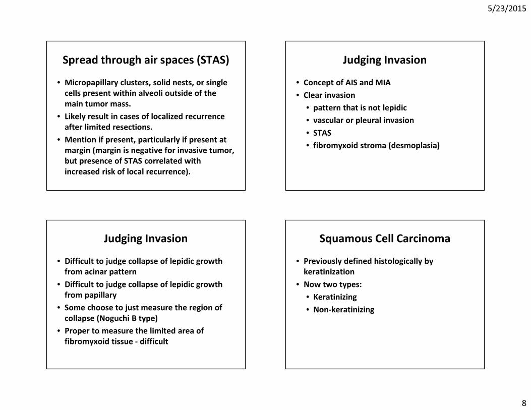
Spread through air spaces (STAS)• Micropapillary clusters, solid nests, or single
cells present within alveoli outside of the main tumor mass.
• Likely result in cases of localized recurrence after limited resections.
• Mention if present, particularly if present at margin (margin is negative for invasive tumor, but presence of STAS correlated with increased risk of local recurrence).
Judging Invasion• Concept of AIS and MIA• Clear invasion
• pattern that is not lepidic• vascular or pleural invasion• STAS• fibromyxoid stroma (desmoplasia)
Judging Invasion• Difficult to judge collapse of lepidic growth
from acinar pattern• Difficult to judge collapse of lepidic growth
from papillary• Some choose to just measure the region of
collapse (Noguchi B type)• Proper to measure the limited area of
fibromyxoid tissue - difficult
Squamous Cell Carcinoma• Previously defined histologically by
keratinization• Now two types:
• Keratinizing• Non-keratinizing

5/23/2015
9
p40

5/23/2015
10
Large Cell Carcinoma• Previously used when no morphologic
support for squamous cell or adenocarcinoma• Now use immunohistochemical stains to help
subclassify into:• Solid type adenocarcinoma• Non-keratinizing squamous cell carcinoma• Large cell carcinoma
Potential Pitfalls• TTF-1:
– Thyroid carcinoma– Entrapped pneumocytes– Gyn tumors (~80% ut. carcinosarcoma)– Neuroendocrine tumors
• Napsin-A: – Pulmonary macrophages (darker)– Renal cell carcinoma (~80%)– GI mucinous tumors (~80%)
Potential Pitfalls• p63:
– Entrapped basal layer– Urothelial tumors– Metastatic squamous tumors– Adenocarcinoma of lung
• Require >10% of nuclei to stain• p40:
– More specific, but similar pitfalls
Mystery Case• 64-year-old woman with right lower lobe
lung nodule. • CT-guided percutaneous fine needle
aspiration performed.

5/23/2015
11
Using the CT scanBone tumors, ILD, and now lung tumors
• Ground glass opacities versus solid masses- Determining extent of lepidic growth- Determining size of lesion
• Border of a lesion- Spiculated versus smooth- Typical adeno vs benign or fast-growing
• Multiplicity of lesions- Extrathoracic with met, lung met, synch primary

5/23/2015
12
The Argus (Melbourne, Australia) February 6, 1936, page 10
Sclerosing Pneumocytoma• Formerly sclerosing hemangioma
– “Sclerosing hemangioma (histiocytoma, xanthoma) of the lung”– A.A. Liebow and D.S. Hubbell, Cancer, 1953.
• Characteristic radiologic appearance– Rounded edges are often either really bad (fast
growing) or benign• Characteristic immunoprofile
– EMA positive, Keratin negative– TTF-1 positive, Napsin-A negative
• Immature pneumocytes with surface normal bronchiolar epithelium Keratin

5/23/2015
13
EMA Napsin-A
TTF-1 52
Treatment Options• Many tumors are typically treated with
standard chemotherapy• In recurrent and stage 4 tumors, and
increasingly as first line, targeted treatments being used: • EGFR• EML4-ALK• ROS-1
• BRAF• MET

5/23/2015
14
Resistance Mutations• EGFR TKI-treated tumors often develop
additional mutations– commonly T790M within EGFR – Novel TKI
• Targeting other pathways being activated– MET, AXL
Immunotherapy• PD-1 and PD-L1
– Programmed death 1 receptor and its ligands– PD-1 is an inhibitory checkpoint pathway in T cells– Some tumor cells have increased surface expression
of PD-L1 (35-95% of NSCLC)– Currently in trials (although already FDA approved),
most often for patients that have failed first and second line therapies
Staging Issues• Multiple nodules• Pleural invasion• Pleural drop metastases
Multiple Nodules• Sometimes difficult to determine if two
tumor nodules represent– Synchronous primary tumors– Intraparenchymal metastases

5/23/2015
15
Martini-Melamed• Tumors are synchronous primaries if:
1. Histologically different. 2. Histologically similar but…
A. Arise from CISB. No tumor in shared lymphaticsC. No extrapulmonary mets
• At the time, histologically different meant SqC vs adeno, and CIS was Sq.CIS
Comprehensive Histologic Assessment
• The “histologically different” component is expanded substantially– Percentage of adenocarcinoma subtype
becomes significant– Cytologic features, stromal components
also aid differentiation
• Additional concept of AIS/Lepidic growth• To be discussed in the new AJCC – next year?
Pleural invasion• The many definitions of pleural invasion
– What we want to think versus what there is data to support
– Research from Japan (lots more EVG staining going on overseas)
• The prominent elastic layer (the visceral pleural elastica, aka the external elastic layer)

5/23/2015
16

5/23/2015
17

5/23/2015
18
Pleural Invasion• EVG for all tumors approaching the pleura.• pT2a if external elastic layer is penetrated
(visceral pleural elastica).– Raises stage from IA to IB in small tumors.
• Elastica of chest wall is variable, and it is sometimes difficult to assess chest wall invasion.– Look for penetration into parietal fat.
• Can use PL designations if desired– Past elastica PL1, on pleural surface PL2, into
chest wall PL3
Pleural Drop Metastases• Tumor studding on pleural surface
• NOT direct extension (T2, PL2)• NOT subpleural lymphatic invasion with
spread to other areas of the lung (T3 or T4)• Similar prognosis as malignant pleural
effusion (M1a)

5/23/2015
19
Take Home Messages• No significant changes to adenocarcinoma
terminology since IASLC/ATS/ERS changes. • Splitting of large cell using IHC.• Sclerosing pneumocytoma. • Targeted therapy, targeting resistance,
immunotherapy.• Not all multiple lesions mean poor prognosis.• Treat the pleura with respect.