Embed Size (px)
Citation preview
T H E D E G E N E R A T I V E C H A N G E S I N P A N C R E A T I C A C I N A R
CELLS CAUSED BY D L - E T H I O N I N E
L A W R E N C E H E R M A N , Ph.D., and P A T R I C K J. F I T Z G E R A L D , M.D.
From the Department of Pathology, State University of New York, Downstate Medical Center, Brooklyn
ABSTRACT
Degeneration of pancreatic acinar cells in rats injected with ethionine was studied by electron microscopy. The most conspicuous morphologic lesions occurred in the ergasto- plasm. There was a widening of the endoplasmic reticulum, a decrease in number of membrane-associated ribosomes, and a development of fine and coarse vacuoles containing agranular disoriented membranes. Cytoplasmic ribosomes unassociated with membranes were less numerous. Nuclear changes consisted of a coarsening and clumping of the nuclear chromatin, chromatin margination, and increased osmiophilia and vacuolation of the nucleolus. Eight to ten days after the beginning of ethionine injections, changes in zymogen granules, mitochondria, and the Golgi apparatus appeared, but only after extensive damage to the acinar cell. The effects were consistent with ethionine's known interference with protein metabolism but also suggest disturbance in ribonucleic acid metabolism. The ergastoplasmic changes after ethionine were similar in some respects to the early lesions produced in liver parenchymal cells by fasting, to the changes occurring in animals on protein-free diets, or to some of the liver changes produced by azo dye carcinogens. The ribosomal and ergastoplasmic changes represent early morphologic expressions of the bio- chemical effect of ethionine.
I N T R O D U C T I O N
Within the past few years many reports have de- scribed the histologic and cytologic effects of ethio- nine, a methionine analogue, on various organs. In the pancreas, acinar cell necrosis is conspicuous and it may be followed by acinar cell regeneration (17, 30, 60, 11, 23, 18).
Although the fine structure of normal pancreas as revealed by electron microscopy has received considerable attention (50, 43, 12, 65, 57-59, 61, 62, 44, 14, 39, 8), only a few reports in the literature have described pathologically altered pancreatic cells. Fasting-refeeding changes and those changes following pilocarpine injections in mice (65, 12, 46), intracellular disturbances caused by neutral red injections (66), lesions in adult mice infected with a strain of pleurodynia
virus (51), islet cell changes in alloxan-treated rabbits (67), and the effects of cobalt on guinea pig pancreas (32) have all been recorded.
In an earlier study from this laboratory (16) a combination of ethionine injections and a protein- free diet caused destruction of most of the acinar ceils of the pancreas and decreased the regenera- tion of the gland to about 20 per cent of that found when a stock diet was introduced after ethionine injections. This regimen was used because it gave more marked ethionine lesions and a longer period of regeneration in which to study cellular and histologic changes than did other combinations of diet and ethionine dosage.
The intracellular degenerative changes in the pancreatic acinar and duct tissue following daily
277
on April 4, 2019jcb.rupress.org Downloaded from http://doi.org/10.1083/jcb.12.2.277Published Online: 1 February, 1962 | Supp Info:
ethionine adminis t ra t ion for 10 days is presented
in detail in this paper.
M A T E R I A L A N D M E T H O D S
One hundred and fifty male albino rats, Wistar strain (Carworth Farms, New York City), weighing 160 to 180 gm, were givcn water ad libitum and kept in individual cages in an air-conditioned room at 68°F. One hundred and twenty-five of thc animals were placed on a protein-free dict (Nutritional Bio- chemical Corp., Cleveland). The diet contained cornstarch 70 per cent, Alphacel (cellulose) 15 per cent, vegetable oil l0 per cent, salt mixture u.s.P. X I V 4 per cent, and cod liver oil 1 per cent. The othcr twenty-five rats werc placed on a stock diet of Purina chow checkers and water ad libitum. Seventy-five of the animals on the protein-free diet received daily intrapcritoncal injections for l0 days of DL-ethioninc (0.7 mg/gm body weight) dissolved in aqueous 0.9 pcr cent sodium chloride (cthioninc group). Fifty animals were kept on the protein-free dict.
Threc animals from each of the stock, protein-free, and cthioninc groups were sacrificed at days 5, 8, and l0 of the experiment. After day 10 thcrc were no further injections of ethionine, and thc animals of both the ethionine and protein-free groups were maintained on the protein-free diet through day 28. The expcrimcnt was rcpcated twice. For study of the earliest lesion, similar groupings were used and addi- tional animals were sacrificed at l, 2, 4, 6, 12, 18, 24, and 48 hours after injection. Only changes in the dcgenerative period, days 1 to 10, arc discussed here. Changes during the regenerative phase, days 12 through 28, are dcscribcd in a subsequent report.
Tissues cxcised for electron microscopy werc placed on sheets of dental wax, flooded with several drops of cold, veronal-acetatc-buffcred (pH 7.4) 1 per cent osmium tetroxidc (42) containing sucrose (6), and cut into 0.5 to 1.0 mm cubes. The tissue blocks werc fixed for 2 hours at 6°C. They werc washed in distilled water, rapidly dehydrated through a series of graded alcohol-water solutions, and in- filtrated with either n-butyl methacrylatc or mixtures of butyl and methyl mcthacrylate (a ratio of 9.0 to 0.4 was most often employed). Experimental and control tissues were processed together. Thc methac- rylate was catalyzed with 0.5 per cent or 2 per cent Lupcrco (50 per cent 2,5-dichlorobenzoyl pcroxide with dibutyl phthalate). Gelatin capsules containing tissue and catalyzed methacrylate were subjected to a N2 atmosphere (37), and were polymcrized at 80°C (5). In later experiments, tissues were embedded in an epoxy resin, Epon (33, 34).
Thin sections were cut on a Portcr-Blum microtomc (Servall) with glass knives and stained with lead hydroxidc or lead acetatc (63). The staincd sections were viewed with an RCA EMU-3C electron micro-
scope which had been fitted with either a 25 /z or a 50 # objective aperture. Micrographs were taken at original magnifications of 1800 to 9000 diameters and were subsequently enlarged photographically.
Tissues for light microscopy were fixed in phos- phate-buffered (pH 7.0) 10 per cent (u.s.p.) formalin and embedded in paraffin. Experimental and control tissues were embedded together, cut as one section, and stained. Deoxyribonucleic acid (DNA) was removed by incubating 4 /z sections in deoxyribo- nuclease (DNase) (Worthington Biochemical Co., Freehold, New Jersey) (0.01 per cent DNase in 0.003 M MgSO4), at pH 6.5, for 2 hours at room temperature. The sections were stained with McIlvain-buffered (pH 4.0) azure B for 2 hours, at 37°C (21).
O B S E R V A T I O N S
Stock Diet Group
The morphology of the normal acinar and duct cells of the pancreas has been described for the ra t (12, 14, 43, 61), guinea pig (44), mouse (39, 50, 57-59, 62, 65, 66), and h u m a n (8).
Protein-Free Ethionine Group
Acinar cell changes following various regimens of ethionine t r ea tmen t have been extensively studied by l ight microscopy (24, 17, 2, 60, 30). In our experiments, swelling of the cell, hydropic and fatty vacuolization, loss of cytoplasmic basophilia, and decreased staining of apical mass occurred dur ing days 1 to 5. This was followed by pyknosis, karyorrhexis, and, eventually, karyolysis with ul t imate destruct ion and dissolution of the cell. Most of the acinar cells had disappeared by days l0 to 12, a l though a few were surprisingly intact. Islet cells, duct tissue, connective tissue, and capil lary endothe l ium were relatively un- damaged and were conspicuous under the l ight microscope because of the decrease of acinar tissue. Regenera t ion of acinar cells occurred subsequently, bu t a t day 28 only abou t 25 per cent resti tution of the original weight of ac inar tissue
was apparent .
A. EARLY DEGENERATIVE CHANGES
T h e changes scan at the various t ime periods showcd considerable variat ion, not only from exper iment to exper iment bu t also from an imal to animal. We have given a composite por t ra i t of the common morphologic findings at the times
discussed below. A schematic d iag ram summar iz ing the clectron
278 THE JOURNAL OF CELL BIOLOGY • VOLUME 1~, 1962
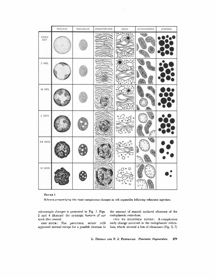
FIOURE 1
Schema summarizing the most conspicuous changes in cell organelles following ethionine injection.
microscopic changes is presented in Fig. 1. Figs. 2 and 4 illustrate the cytologic features of our stock diet control.
ONE HOUR: The pancreatic acinar cells appeared normal except for a possible increase in
the amount of smooth surfaced elements of the endoplasmic reticulum.
T W O TO E I G H T E E N H O U R S : A conspicuous early change occurred in the endoplasmic reticu- lum, which showed a loss of ribosomes (Fig. 3, 7)
L. HERM~N AND P . J . FITZGERALD Pancreatic Degeneration 279
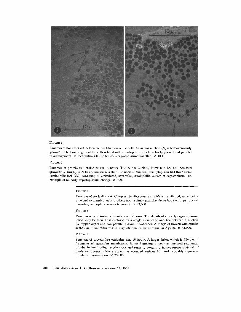
FIGURE 2
Pancreas of stock diet rat. A large acinus fills most of the field. An acinar nucleus (N) is homogeneously granular. The basal region of the cells is filled with ergastoplasm which is closely packed and parallel in arrangement . Mitochondr ia (M) lie between ergastoplasmic lamcllae. X 4000.
FIGURE 3
Pancreas of protein-free ethionine rat, 6 hours. The acinar nucleus, lower left, has an increased granulari ty and appears less homogeneous than the normal nucleus. The cytoplasm has three small osmiophilic loci (EL) consisting of vesiculatcd, agranular, osmiophilic masses of e rgas toplasm--an example of an early ergastoplasmic change. X 4000.
FIGURE
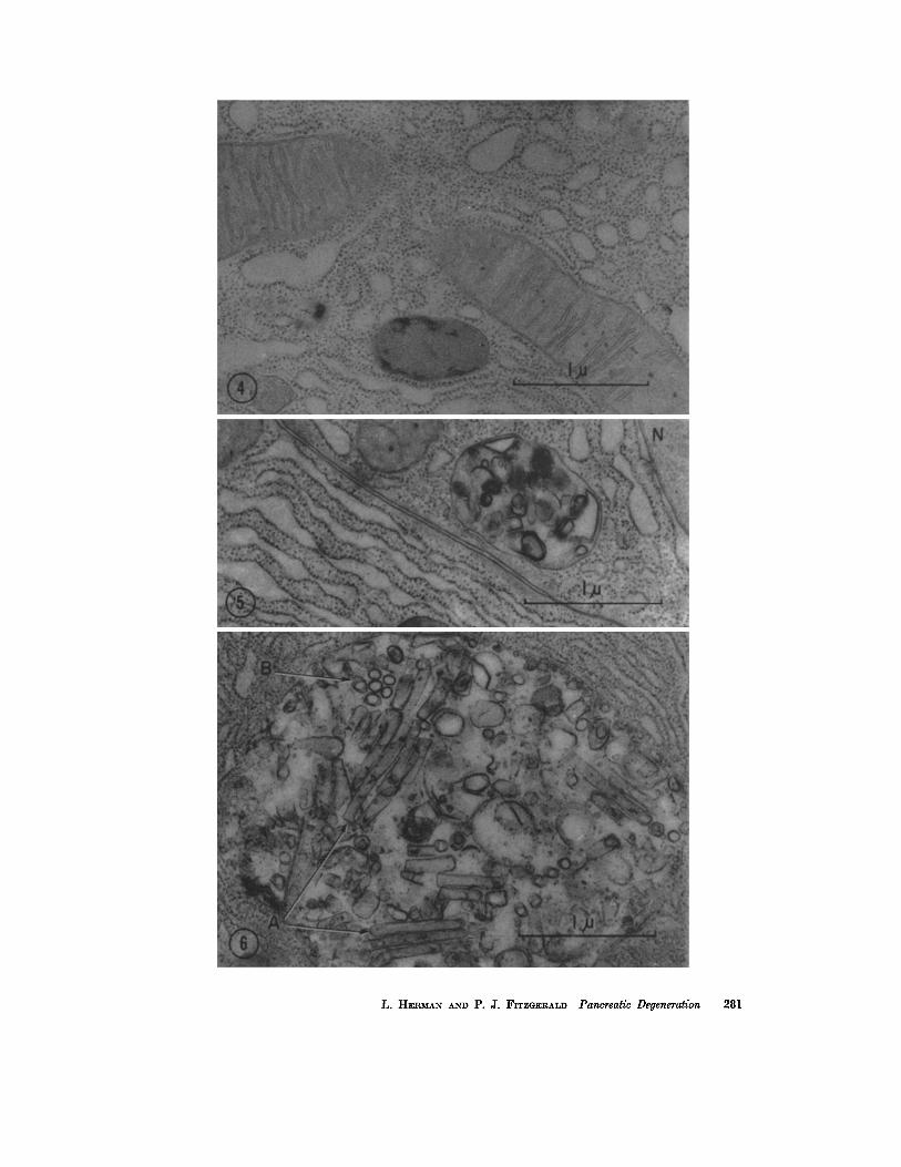
Pancrcas of stock diet rat. Cytoplasmic ribosomes are widely distributed, some being at tached to membranes and others not. A finely granular dense body with pcrlpheral , irregular, osmiophilic masses is present. X 33,000.
FIGURE 5
Pancreas of protein-free ethionine rat, 12 hours. The details of an early ergastoplasmic lesion may be seen. It is enclosed by a single membrane and lies between a nucleus (N, upper right) and two parallel plasma membranes. A tangle of broken osmiophilie agranular membranes within may encircle less dense vesicular regions. X 33,000.
FIGURE 6
Pancreas of protein-free ethionine rat, 18 hours. A larger lesion which is filled with fragments of agranular membranes. Some fragments appear as enclosed segmental tubules in longitudinal section (A) and seem to contain a homogeneous material of moderate density. Others appear as rounded vesicles (B) and probably represent tubules in cross-section. X 33,000.
280 THE JOURNAL OF CELL BIOLOGY • VOLUME 12, 1962
L. HEI~MA~ AZ~'D P. J. FITZGERALD Pancreatic Deyeneration 281
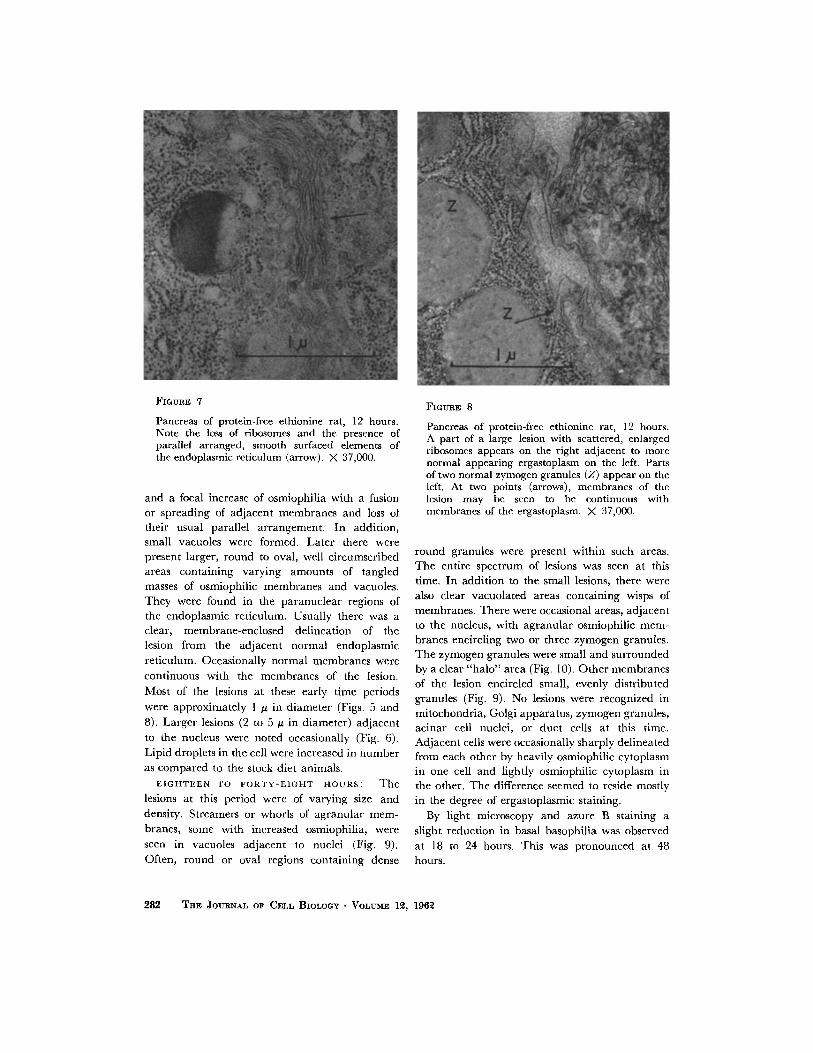
FIGURE 7
Pancreas of protein-free ethionine rat, 12 hours. Note the loss of ribosomes and the presence of parallel arranged, smooth surfaced elements of the endoplasmic reticulum (arrow). ;K 37,000.
and a focal increase of osmiophilia with a fusion or spreading of adjacent membranes and loss ot their usual parallel arrangement. In addition, small vacuoles were formed. Later there were present larger, round to oval, well circumscribed areas containing varying amounts of tangled masses of osmiophilic membranes and vacuoles. They were found in the paranuclear regions of the endoplasmic reticulum. Usually there was a clear, membrane-enclosed delineation of the lesion from the adjacent normal endoplasmic reticulum. Occasionally normal membranes were continuous with the membranes of the lesion.
Most of the lesions at these early time periods
were approximately 1 ~ in diameter (Figs. 5 and 8). Larger lesions (2 to 5 # in diameter) adjacent to the nucleus were noted occasionally (Fig. 6). Lipid droplets in the cell were increased in number as compared to the stock diet animals.
E I G H T E E N TO F O R T Y - E I G H T HOURS: The lesions at this period were of varying size and density. Streamers or whorls of agranular mem- branes, some with increased osmiophilia, were seen in vacuoles adjacent to nuclei (Fig. 9). Often, round or oval regions containing dense
FIGURE 8
Pancreas of protein-free ethionine rat, 12 hours. A part of a large lesion with scattered, enlarged ribosomes appears on the right adjacent to more normal appearing ergastoplasm on the left. Parts of two normal zymogen granules (Z) appear on the left. At two points (arrows), membranes of the lesion may be seen to be continuous with membranes of the ergastoplasm. X 37,000.
round granules were present within such areas. The entire spectrum of lesions was seen at this time. In addition to the small lesions, there were also clear vacuolated areas containing wisps of membranes. There were occasional areas, adjacent to the nucleus, with agranular osmiophilic mem- branes encircling two or three zymogen granules. The zymogen granules were small and surrounded by a clear "ha lo" area (Fig. I0). Other membranes of the lesion encircled small, evenly distributed granules (Fig. 9). No lesions were recognized in mitochondria, Golgi apparatus, zymogen granules, acinar cell nuclei, or duct cells at this time. Adjacent cells were occasionally sharply delineated from each other by heavily osmiophilic cytoplasm in one cell and lightly osmiophilic cytoplasm in the other. The difference seemed to reside mostly in the degree of ergastoplasmic staining.
By light microscopy and azure B staining a slight reduction in basal basophilia was observed at 18 to 24 hours. This was pronounced at 48 hours.
282 THE JOURNAL OF CELL BIOLOGY • VOLUME 12, 1962
B. LATER DEGENERATIVE CHANGES--DAYs T w o TO EmHT
Acinar and lobular patterns were retained. Acinar cells were swollen and there was decreased basophilia (azure B staining). Acinar cell nuclei had lost the diffuse fine granularity of the normal nucleus and frequently showed coarse clumps of
areas. The nucleoli appeared more osmiophilic than in the stock or protein-free groups, and oc- casionally showed vacuoles in their centers (Fig. 12).
The zymogen granules appeared relatively nor- mal except that they were no longer closely packed but were more widely dispersed throughout the
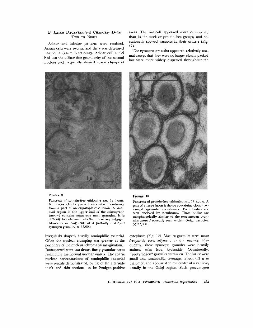
FIGURE 9
Pancreas of protein-free ethionine rat, 18 hours. Numerous closely packed agranular membranes from a part of an ergastoplasmic lesion. A small oval region in the upper half of the micrograph (arrow) contains numerous small granules. It is difficult to determine whether these are enlarged ribosomes or fragments of a partially destroyed zymogen granule. X 37,000,
irregularly shaped, heavily osmiophilic material, Often the nuclear clumping was greater at the periphery of the nucleus (chromatin margination). Interspersed were less dense, finely granular areas resembling the normal nuclear matrix. The coarse nuclear concentrations of osmiophilic material were readily demonstrated, by use of the alternate thick and thin sections, to be Feulgen-positive
FIGURE 10
Pancreas of protein-free ethionine rat, 18 hours. A part of a large lesion is shown containing closely ar- ranged agranular membranes. Four bodies are seen enclosed by membranes. These bodies are morphologically similar to the prozymogen gran- ules more frequently seen within Golgi vacuoles. X 37,000.
cytoplasm (Fig. 12). Mature granules were more frequently seen adjacent to the nucleus. Fre- quently, these zymogen granules were heavily stained with lead hydroxide. Occasionally, "prozymogen" granules were seen. The latter were small and osmiophilic, averaged about 0.3 # in diameter, and appeared in the center of a vacuole, usually in the Golgi region. Such prozymogen
L. HERMAN AND P. J. FITZGERALD Pancreatic Degeneration 283
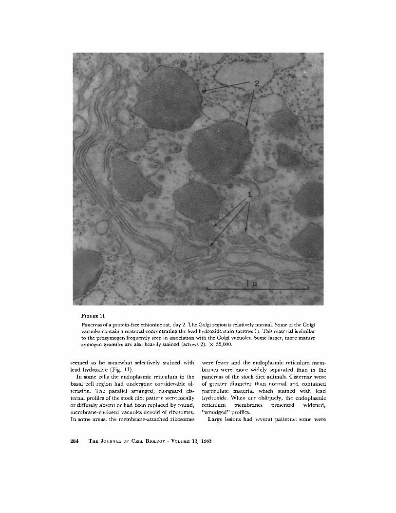
FIGURE 11
Pancreas of a protein-free ethionine rat, day 2. The Golgi region is relatively normal. Some of the Golgi vacuoles contain a material concentrating thc lead hydroxide stain (arrows 1 ). This material is similar to the prozymogen frequently seen in association with the Golgi vacuoles. Some larger, morc mature zymogcn granules are also heavily stained (arrows 2). X 55,000.
seemed to be somewhat selectively stained with lead hydroxide (Fig. 11).
In some cells the endoplasmic re t iculum in the basal cell region had undergone considerable al- teration. The parallel arranged, elongated cis- ternal profiles of the stock diet pa t t e rn were focally or diffusely absent or had been replaced by round, membrane-enclosed vacuoles devoid of ribosomes. In some areas, the membrane -a t t ached ribosomes
were fewer and the endoplasmic re t iculum mem- branes were more widely separated than in the pancreas of the stock diet animals. Cisternae were of greater d iameter t han normal and conta ined part iculate mater ia l which stained with lead hydroxide. W h e n cut obliquely, the endoplasmic re t iculum membranes presented widened, " smudged" profiles.
Large lesions had several pat terns: some were
284 ThE JOURNAL OF CELL BIOLOGY " VOLUME 12, 196~2
FiouRs 12
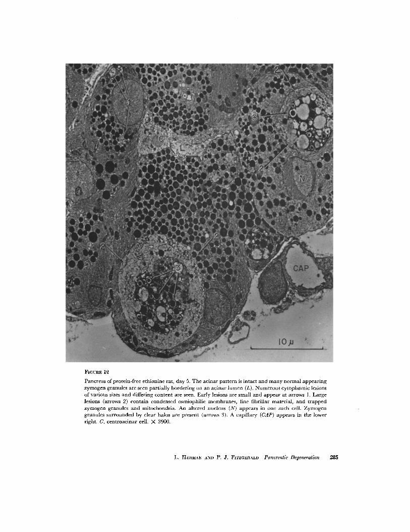
Pancreas of protcin-frec cthionlnc rat, day 5. The acinar pat tern is intact and many normal appear ing zymogen granules are seen partially bordering on an acinar lumen (L). Numerous cytoplasmic lesions of various sizes and differing content arc seen. Early lesions arc small and appear at arrows 1. Large lesions (arrows 2) contain condensed osmiophilic membranes , fine fibrillar material, and t rapped zymogen granules and mitochondria. An altered nucleus (N) appcars in one such cell. Zymogen granules surrounded by clear halos are present (arrows 3). A capillary (CAP) appear s in the lower right. C, centroacinar cell. X 3900.
L. HERMAN AND P. J. FITZGERALD Pancreatic Degeneration 285
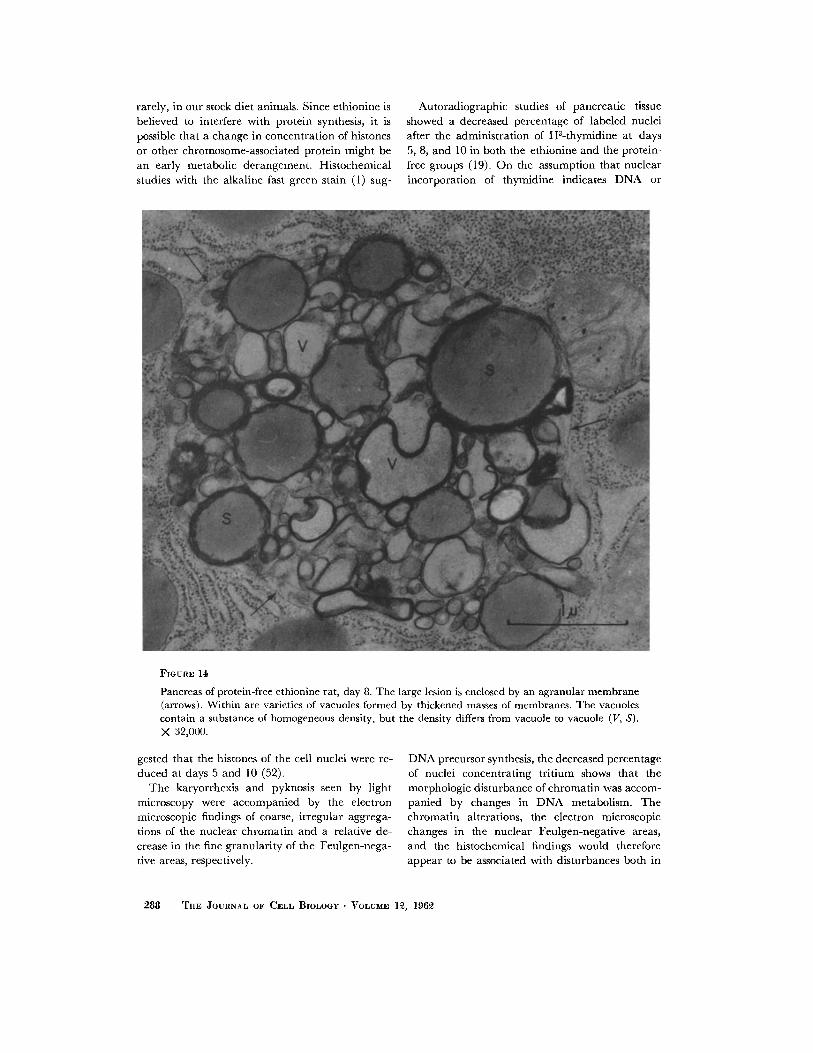
vacuolated areas with only wisps of membranes within (Fig. 13); some contained mitochondria or zymogen granules (Fig. 13); and others contained the parallel arranged, granule-free membranes described earlier. Some cells contained round nuclei and cytoplasm with whorled membranous structures (Fig. 14). Occasionally a whorled osmio- philic body was present. These had closely packed, concentric membranes which were heavily osmio- philic and occasionally were accompanied by slightly swollen ribosomes regularly spaced along the membrane, q-he interior of such bodies fre- quently contained a less dense, homogeneous granule-free area (Figs. 15 and 15 A).
The membranes, vacuoles, and vesicles of the Golgi apparatus appeared relatively normal early in the ethionine-injected animals.
Lipid droplets were prominent in most of the cells and were greatly increased in number and size as compared with those in the pancreas of the stock diet animals.
Most mitochondria appeared to be of normal size and appearance. A few altered mitochondria were seen. These were swollen, with disrupted, short cristae attached to the inner margins of the outer limiting double membrane.
C. MAXIMUM DEGENERATIVE C H A N G E s - - D A Y s EIGHT TO T E N
Maximum ethionine changes occurred in most animals at day 10. Acinar tissue in some areas was greatly reduced and the pancreas consisted mainly of ducts, capillaries, collagen, and inflammatory cells. The lobular and acinar outlines were gen- erally absent or greatly distorted. Areas where acinar cells persisted showed dilated acinar lumina and the cells were considerably reduced in size. In some areas, acinar patterns were recognizable even though considerable damage to many cells had occurred. Frequently, strands and clumps of heavily osmiophilic, coalesced endoplasmic reticu- lum (osmiophilic plaques) could be seen. Nuclei, where intact, contained areas of coarse, dense granularity. Nucleoli were decreased in number or,
where present, consisted of a small mass of in- creased osmiophilia with small vacuoles (Fig. 16).
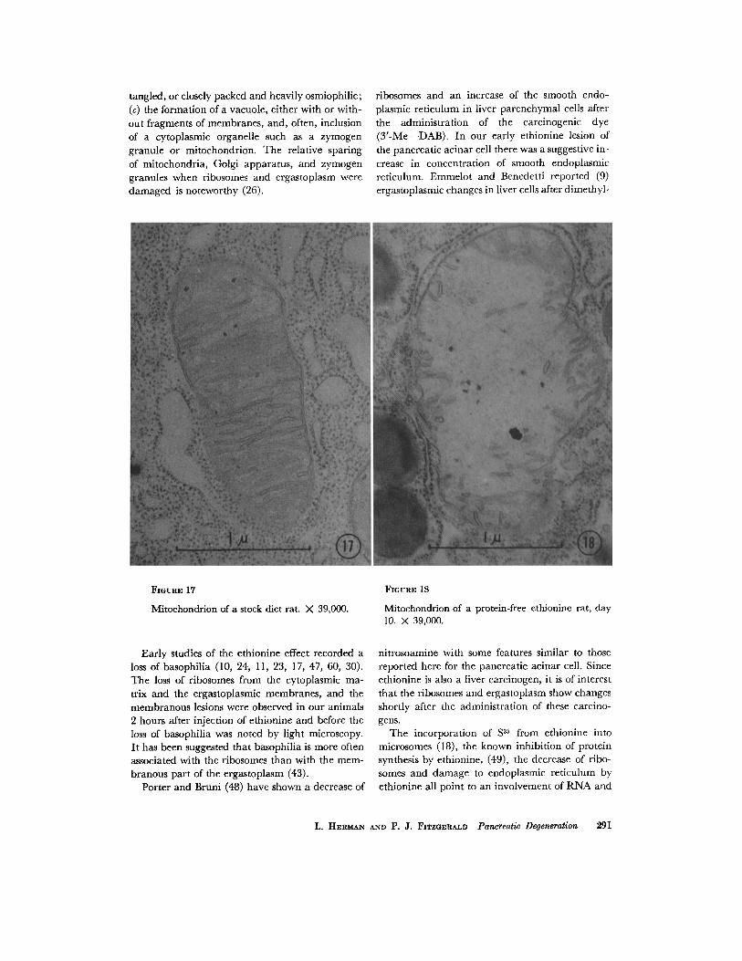
The Golgi apparatus was preserved but atrophic. Mitochondria were swollen, were less dense, and contained short, broken cristae (com- pare Figs. 17 and 18).
With the atrophy and disappearance of paren- chymal cells at day 10 the ducts became promi- nent. The smallest ductules contained three to five cells encircling the lumen, which had a thin basement membrane. The cells were cuboidal and their free luminal edge contained microvilli which were of variable length but less frequent, shorter, and straighter than those seen on the free border of the acinar cell. The duct cell membranes, un- like those of acinar cells, were very conspicuous and laterally interdigitated with adjacent cells. Duct cell nuclei were indented, and the nucleo- plasm was a mixture of coarse and fine granules of irregular pattern. Duct cell nucleoli were small. The cytoplasm was sparse and the endoplasmic reticulum membranes were generally absent, al- though vesicles bounded by rough surfaced mem- branes were seen occasionally. A few ribosomes were scattered freely in the cytoplasmic matrix. Occasionally, duct and ductule cells were abnor- mal and displayed the characteristic lesion found in the degenerating aeinar cell. Numerous "lipid- like" droplets were also present within the abnor- mal cell. Its Golgi apparatus was intact and promi- nent (Fig. 19). Mitochondria were unchanged.
DISCUSSION
Nuclear Lesions
A coarse clumping of DNA, quite distinct from that of the usual fine granularity of the normal acinar cell nucleus, and the alignment of chroma- tin along the inner circumference of the nuclear membrane ("chromatin margination") were also noted in our protein-free animals, in cells infected with virus (38), in the parenchymal liver cell nuclei of rats following fasting-refeeding (in refer- ence 13, compare Figs. 23 and 24 with 31), and,
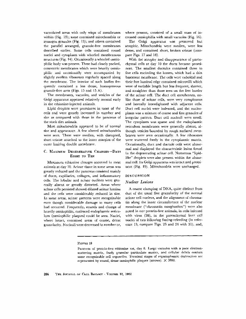
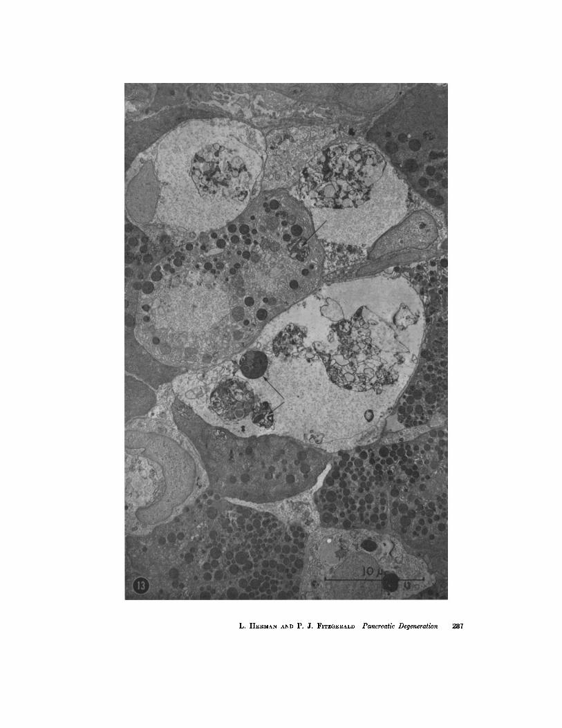
FIGURE 13
Pancreas of protein-free ethionine rat, day 8. Large vacuoles with a poor electron- scattering matrix, finely granular particulate matter, and cellular debris contain some recognizable cell organelles. Terminal stages of ergastoplasmic destruction are represented by round, dense osmiophilic plaques (arrows). >( 3800.
286 ThE JOURNAL OF CELL BIOLOGY • VOLUME ]~, 196~
L. HERMAN AND P. Z. FITZGERALD Pancreatic Degeneration 287
rarely, in our stock diet animals. Since ethionine is believed to interfere with protein synthesis, it is possible that a change in concentration of histones or other chromosome-associated protein might be an early metabolic derangement. Histochemical studies with the alkaline fast green stain (1) sug-
Autoradiographic studies of pancreatic tissue showed a decreased percentage of labeled nuclei after the administration of H3-thymidine at days 5, 8, and 10 in both the ethionine and the protein- free groups (19). On the assumption that nuclear incorporation of thymidine indicates D N A or
FmuaE 140
Pancreas of protein-free ethionine rat, day 8. The large lesion is enclosed by an agranular membrane (arrows). Within are varieties of vacuoles formed by thickened masses of membranes. The vacuoles contain a substance of homogeneous density, but the density differs from vacuole to vacuole (V, S). X 32,000.
gested that the histones of the cell nuclei were re- duced at days 5 and 10 (52).
The karyorrhexis and pyknosis seen by light microscopy were accompanied by the electron microscopic findings of coarse, irregular aggrega- tions of the nuclear chromatin and a relative de- crease in the fine granularity of the Feulgen-nega- tive areas, respectively.
DNA precursor synthesis, the decreased percentage of nuclei concentrating tritium shows that the morphologic disturbance of chromatin was accom- panied by changes in DNA metabolism. The chromatin alterations, the electron microscopic changes in the nuclear Feulgen-negative areas, and the histochemical findings would therefore appear to be associated with disturbances both in
288 T h E JOURNAL OF CELL BIOLOGY • VOLUME 1~, 196~2
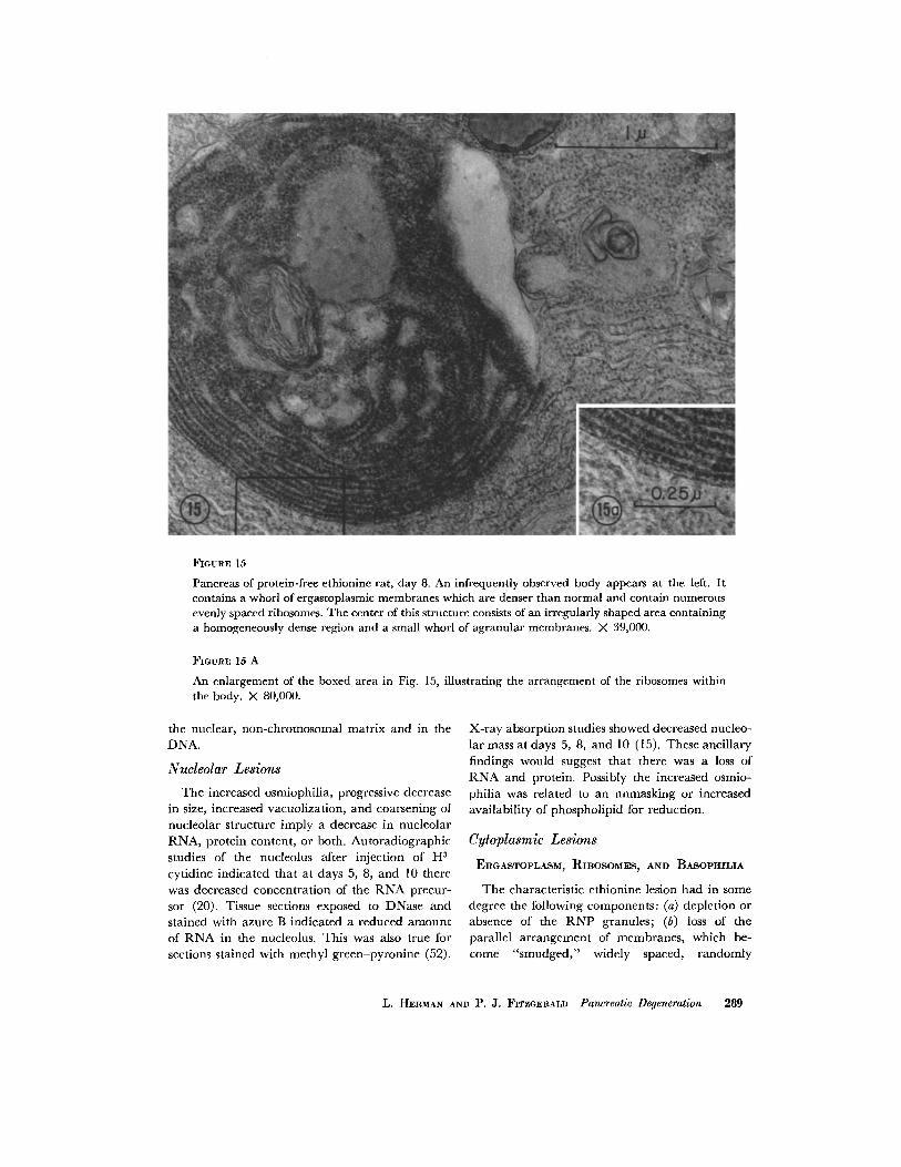
FIOURE 15
Pancreas of protein-free ethioninc rat, day 8. An infrequently observed body appears at the left. It contains a whorl of ergastoplasmic membranes which are denser than normal and contain numerous evenly spaced ribosomes. The center of this structure consists of an irregularly shaped area containing a homogeneously dense region and a small whorl of agranular membranes. X 39,000.
FmuuE 15 A
An enlargement of the boxed area in Fig. 15, illustrating the arrangement of the ribosomes within the body. X 80,000.
the nuclear, non-chromosomal matrix and in the DNA.
Nucleolar Lesions
The increased osmiophilia, progressive decrease in size, increased vacuolization, and coarsening of nucleolar structure imply a decrease in nucleolar RNA, protein content, or both. Autoradiographic studies of the nucleolus after injection of H 3- cytidine indicated that at days 5, 8, and 10 there was decreased concentration of the R N A precur- sor (20). Tissue sections exposed to DNase and stained with azure B indicated a reduced amount of R N A in the nucleolus. This was also true for sections stained with methyl green-pyronine (52).
X-ray absorption studies showed decreased nucleo- lar mass at days 5, 8, and 10 (15). These ancillary findings would suggest that there was a loss of R N A and protein. Possibly the increased osmio- philia was related to an unmasking or increased availability of phospholipid for reduction.
Cytoplasmic Lesions
ERGASTOPLASM~ RIBOSOMES~ AND BASOPHILIA
The characteristic ethionine lesion had in some degree the following components: (a) depletion or absence of the R N P granules; (b) loss of the parallel arrangement of membranes, which be- come "smudged," widely spaced, randomly
L. HERMAN AND 1 ~, J. FITZGERALD Pancreatic Degeneration 289
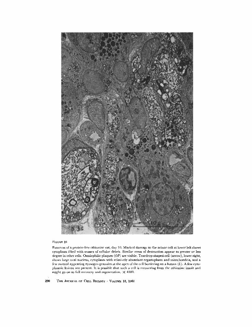
FIGURE 16
Pancreas of a protein-free e thionine rat, day 10. Marked d a m a g e to the acinar cell at lower left shows cytoplasm filled with masses of cellular debris. Similar areas of destruct ion appear to greater or less degree in other cells. Osmiophi l ic plaques (OP) are visible. Tea rd rop - shaped cell (arrow), lower right, shows large oval nucleus, cy toplasm with relatively a b u n d a n t ergas toplasm and mitochondr ia , and a few no rma l appear ing zymogen granules at the apex of the cell border ing on a l umen (L). A few cyto- plasmic lesions are present. I t is possible tha t such a cell is recovering f rom the ethionine insult and migh t go on to full recovery and regenerat ion. X 4300.
290 THE JOURNAL OF CELL BIOLOGY • VOLUME 1~, 196~2
tangled, or closely packed and heavily osmiophilic; (c) the formation of a vacuole, either with or with- out fragments of membranes, and, often, inclusion of a cytoplasmic organelle such as a zymogen granule or mitochondrion. The relative sparing of mitochondria, Golgi apparatus, and zymogen granules when ribosomes and ergastoplasm were damaged is noteworthy (26).
ribosomes and an increase of the smooth endo- plasmic reticulum in liver parenchymal cells after the administration of the carcinogenic dye (3'-Me--DAB). In our early ethionine lesion of the pancreatic acinar cell there was a suggestive in- crease in concentration of smooth endoplasmic reticulum. Emmelot and Benedetti reported (9) ergastoplasmic changes in liver cells after dimethyl-
FXGtmE 17
Mitochondrion of a stock diet rat. X 39,000.
Early studies of the ethionine effect recorded a loss of basophilia (10, 24, 11, 23, 17, 47, 60, 30). The loss of ribosomes from the cytoplasmic ma- trix and the ergastoplasmic membranes, and the membranous lesions were observed in our animals 2 hours after injection of ethionine and before the loss of basophilia was noted by light microscopy. It has been suggested that basophilia is more often associated with the ribosomes than with the mem- branous part of the ergastoplasm (43).
Porter and Bruni (48) have shown a decrease of
FIGURE 18
Mitochondrion of a protein-free ethionine rat, day 10. X 39,000.
nitrosoamine with some features similar to those reported here for the pancreatic acinar cell. Since ethionine is also a liver carcinogen, it is of interest that the ribosomes and ergastoplasm show changes shortly after the administration of these carcino- gens.
The incorporation of S 35 from ethionine into microsomes (18), the known inhibition of protein synthesis by ethionine, (49), the decrease of ribo- somes and damage to endoplasmic reticulum by ethionine all point to an involvement of RNA and
L. HEa~tAN AND P. J. FITZGERALD Pancreatic Degeneration 291
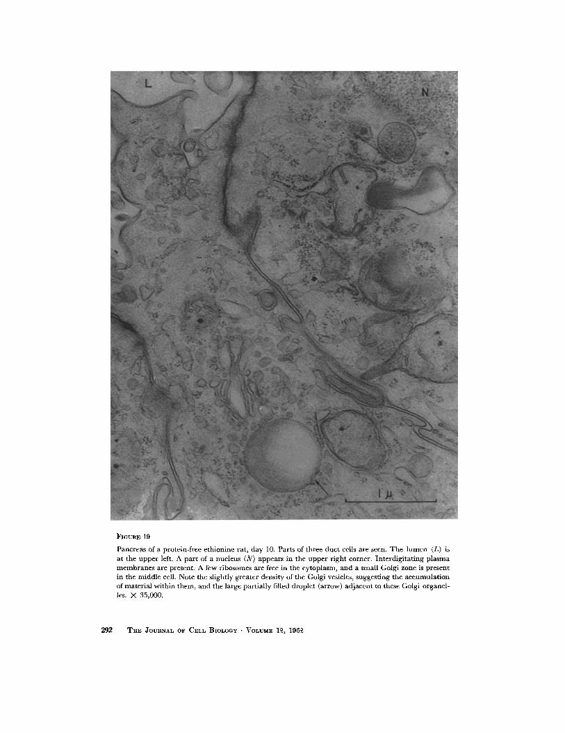
FIGURE 19
Pancreas of a protein-free c thionine rat, day 10. Parts of three duc t cells are sccn. T h e l u m c n (L) is at the uppe r left. A par t of a nucleus (N) appears in the uppe r r ight corner. In terd ig i ta t ing p l a sma m e m b r a n e s are prcsent. A few ribosomes arc frec in the cytoplasm, and a small Golgi zone is prescnt in the middle cell. Note the slightly greater density of the Golgi vesicles, suggest ing the accumula t ion of mater ia l within them, and the large partial ly filled droplet (arrow) ad jacent to these Golgi organcl- les. X 35,000.
292 THE JOURnaL OF CELL BIOLOGy • VOLUME 1~, 196~
protein metabolism. Farber and Magee's recent suggestion (10)that RNA metabolism maybe the primary biochemical locus of ethionine action would be consistent with our findings in that ribo- somes and the ergastoplasmic membranes were the first cell organelles to show conspicuous dam- age. Presumably, under such circumstances, pro- tein metabolism alterations would be secondary to the initial insult to RNA metabolism.
~YMOGEN GRANULES~ MITOCttONDRIA~ AND GOLGI APPARATUS
Most authors have reported a decrease or ab- sence of zymogcn granules early in the ethioninc effect. Histochcmical studies (52) showed a de- crease at days 5 to 8. Nevertheless, there were a few intact cells at days l0 to 12 which contained zymogen granules of normal appearance and num- ber.
Consistent with the morphologic findings of relatively little damage to zymogen granules at days 2 to 8 were the pancreatic lipasc and amylase activities found in this laboratory which showed no marked decrease until day 8 (35), although these enzymatic activity levels arc not conclusive evi- dence of the presence of zymogen granules or of lack of damage to zymogen granules (27). At days l0 and 12 the marked depletion of zymogen gran- ules was associated with a considerable decrease in the enzymatic activities (35).
Although the above findings might suggest that zymogen granule formation represents a late stage in a long process of some days' duration, most studies indicate a much shorter period of hours to days in the animal on a stock diet (18, 45, 56). A complicating but possibly explanatory factor in our studies was the markedly decreased food in- take of the animals on the protein-free ethionine regimen as compared with the protein-free con- trois. In the latter, zymogen granules were de- creased at day 5 and absent at days 8 to 10 (64). These results suggest the possibility that feeding stimulated the release of zymogen granules in the protein-free group and the absence of dietary pro- tein caused a decrease in synthesis of zymogen granules. The very low intake of food, or the ethionine toxicity, may have decreased the stimu- lus for discharge of zymogen granules in the ethionine-injected animals. The suggestion has been made that zymogen granule formation and discharge may be controlled by separate mecha- nisms (28).
Although some mitochondria showed changes in cells that were severely damaged at days 5 and 8, most mitochondria were relatively unaffected until day 10, when they resembled (Fig. 18) those seen in liver parenchymal cells following acute hypoxia and carbon tetrachloride poisoning (36, 7) or refeeding following starvation (54).
The sparing of the Golgi apparatus until days 8 and 10, when there was a reduction in the num- bers of membranes, vacuoles, and vesicles, and an absence of prozymogen granules within the Golgi vacuoles, suggests that the membranes of the Golgi apparatus are different from the ergasto- plasmic membranes.
VACUOLIZATION~ ~CLouDY SWELLING~ ~
HYDROPIC DEGENERATION~ AND FATTY DEGENERATION
By the use of alternate thick and thin sections it was shown that some of the vacuoles seen by light microscopy at days 5 to 8 corresponded to the ethionine vacuolar lesion seen by electron micros- copy. Many of these vacuoles contained mem- branes and "trapped" cellular organelles as well as a "fluid matrix" (Fig. 13). Some of the vacuoles seen by light microscopy stained with the fat stain oil red O (52). Others did not. It is believed that those vacuoles staining for fat also showed lipid droplets in the electron micrographs. Concomi- tantly there was a progressive increase of organ fluid, as determined by chemical analysis, which reached a peak at day 10, when the dry weight was only 40 per cent of the control value (35). The vacuoles not staining for fat or glycogen would ful- fill the criteria for hydropic vacuolization.
It has been suggested by Morgan et al. (38) that reversible changes in mitochondria produced by viral infection were part of the pattern of "cloudy swelling" (22). Since in our material mitochondria were uninvolved during the height of "cloudy swelling," mitochondrial change, morphologically speaking, is not obligatory for the presence of the lesion. Christie and Judah (7) suggested, as a re- sult of their studies with carbon tetrachloride on liver cells, that underlying the phenomenon of hydropic degeneration is damage to mitochondria. In our study, the non-involvement of mitochondria when hydropic degeneration was advanced indi- cates that morphologic changes in the mito- chondrion are not obligatory for this lesion. It may be germane to point out that cloudy swelling and hydropic degeneration have not been too
L. ~ I E R ~ N AND P. J. FITZGERALD Pancreatic Degeneration 293
clearly defined by the light microscopist. A study by the finer resolution of electron microscopy might lead to a better understanding of these entities.
Cytoplasmic lipid was demonstrated in many cells by light microscopy and fat stains (52) as well as by electron microscopy. Its basal distribu- tion in acinar ceils during the early degenerative phase may be related to the higher basal concen- tration of substances entering a cell from the adjacent capillaries, or to metabolic changes asso- ciated with the endoplasmic lesion. Fatty deposi- tion subsequent to decreased synthesis of proteins has been noted as an ethionine effect in liver cells (11), in tissue culture of starved "L" cells (29), in liver cell damage after carbon tetrachloride (41, 4, 53) and a liver carcinogen (48), and in the regenera- tion of liver after partial hepatectomy. The occurrence of more marked lipid droplet ac- cumulations in our protein-free animals (64) than in the ethionine animals might have been related to the different feeding habits of the two groups, or to the greater depletion of the fat depots in the ethionine group. Whether the fat was of endogenous or exogenous origin (3, 29) has not been determined.
Debatable Bodies
OsMIoPHILIC WHoRLs
At days 8 and l0 a distinctive lesion was seen. It was a whorled collection of ergastoplasm with increased osmiophilic membranes, yet the mem- branes were concentric, closely packed, and had heavily staining ribosomes (Figs. 15, 15 A). The deep osmiophilia was more commonly associated with degenerative lesions, but the oriented, con- ccntric membranes suggested an orderly process. There was a slight resemblance in the whorled pattern of this complex to the classical nebenkern pattern of ergastoplasm, which was conspicuous at day 12, but the membranes were more closely packed and more osmiophilic than the nebenkern of regenerating pancreas (25). It is possible that such a body represented regenerated ergasto- plasm which was recovering from the ethionine effect.
LYSOSOMES~ CYTOSOMES~ MICROBODIES
In an attempt to visualize the lysosome of de Duve we have studied many normal and ab- normal pancreases. Some oval or round homoge- neous bodies (lysosomes) (40), cytosomes (48), or
microbodies (48, 53) were seen occasionally in the pancreases of our stock diet (Fig. 4), ethionine- injected, and protein-free groups. Because of their rarity it was not possible to speak with assurance of any slight change in number. However, no marked increase in number was apparent.
Lesions Reported by Other Investigators
Some published electron micrographs indicate the presence of changes which are similar to but not identical with the ergastoplasmic lesions re- ported here. Weiss (66) injected neutral red into mice and reported alterations in the ergasto- plasm, zymogen granules, mitochondria, and Golgi granules of the acinar cells. Robertson (51) infected adult mice with pleurodynia virus and was able to produce an experimental form of pan- creatitis. He noted a "laminated body" consisting of whorled membranes which bore a resemblance to the ergastoplasmic lesions seen in this study. Recently, Seifert and Gieseking (55) have de- scribed in detail similar lesions with ethionine.
Speci~city of Ethionine Lesion
The individual components of the ethionine lesion--loss of basophilia, cloudy swelling, hy- dropic and fatty vacuolization, pyknosis, karyor- rhexis, and dissolution--are common, in varying degree, to many pathologic changes seen by light microscopy. The electron microscopic changes in the pancreas are similar to some of the changes seen in liver cells secondary to the liver carcinogens (9, 48). I t is possible, though doubtful, that the ethionine-produced lesions described here are spe- cific. They are quite different from the pancreatic changes produced by cobalt (31) or glucagon (32). Certainly the constellation of ethionine-pro- duced morphologic changes in the pancreas, liver, and kidney as seen by light microscopy is char- acteristic. Whether the initial lesions seen by electron microscopy--primarily ribosomal and membranous--are specific or characteristic of such compounds as ethionine and the carcinogens, or are merely common pathways in the degeneration of the ultrastructural components of many lesions, is a matter for future studies to determine.
This investigation was supported by United States Public Health Service grants nos. C3300 and C3301.
The authors gratefully acknowledge the assistance of Dr. Bernard Weisblum, Mr. Alan Lieberman, and Mr. Charles Harman. Received for publication, July 2, 1961.
294 THE JOVR~AL OF CELL BIOLOGY • VOLVME 1~, 196~
B I B L I O G R A P H Y
1. ALFERT, M., and GESCHWIND, I. I., Proc. Nat. Acad. Sc., 1953, 39, 991.
2. ALVIZOURI, M., and WARREN, S., Arch. Path., 1954, 57, 130.
3. BENSCH, K. G., KING, D. W., and SOCOLOW, F. L., J. Biophysic. and Biochem. Cytol., 1961, 9, 135.
4. BERNHARD, W., and ROUILLER, C., J. Biophysic. and Biochem. Cytol., 1956, 2, No. 4, suppl., 73.
5. BORYSKO, E., d. Biophysic. and Biochem. Cytol., 1956, 2, No. 4, suppl., 3.
6. CAULFIELD, J. B., J. aiophysie, and Biochem. Cytol., 1957, 3, 827.
7. CHRISTIE, G. S., and JUDAH, J. D., Proc. Roy. Soc. London, series B, 1954, 142, 241.
8. EKHOLM, R., and EDLUND, Y., J. Ultrastruct. Research, 1959, 2,453.
9. EMMELOT, P., and BENEDETTI, E. L., J. Biophysic. and Biochem. Cytol., 1960, 7, 393.
10. FARBER, E., and MAGEE, P. N., Biochem. J., 1960, 76, 58.
11. FARBER, E., and POPPER, H., Proc. Soc. Exp. Biol. and ivied., 1950, 74, 838.
12. FARQUHAR, M. G., and WELLINGS, S. R., J. Biophysic. and Biochem. Cytol., 1957, 3, 319.
13. FAWCETT, D. W., J. Nat. Cancer Inst., 1955, 15, 1475.
14. FERREIRA, D., J. Ultrastruct. Research, 1957, 1, 14. 15. FITZGERALD, P. J., in X-ray Microscopy and
Microradiography, (V. E. Cosslett, A. Eng- str6m, and H. H. Pattee, editors), New York, Academic Press, Inc., 1957, 507.
16. FITZGERALD, P. J . , Lab. Invest., 1960, 9, 67. 17. FITZGERALD, P. J., and ALWZOURI, M., Nature,
1952, 170, 929. 18. FITZGERALD, P. J., and HELLMAN, L., Lab.
Invest., 1961, 10, 2. 19. FITZGERALD, P. J. , and VINIJCHAIKUL, K., Lab.
Invest., 1959, 8, 319. 20. FITZGERALD, P. J. , and VINIJCHAIKUL, K., in
preparation. 21. FLAX, M. n. , and HIMES, M. H., Physiol. Zool.,
1952, 25, 297. 22. GANSLER, H., and ROUILLER, C., Schweiz. Z.
allg. Path. u. Bakt., 1956, 19, 217. 23. GOLDBERG, R. C., and CHAIKOFF, I. L., Arch.
Path., 1951, 52,230. 24. GOLDEERG, R. C., CHAmOFF, I. L., and DODGE,
A. H., Proc. Soc. Exp. Biol. and Med., 1950, 74, 369.
25. HERMAN, L., and FITZGERALD, P. J., J. Cell Biol., 1962, 12, 297.
26. HERMAN, L., FITZGERALD, P. J. , WEISS, M., and POLEVOY, I. S., Proc., 4th Internat. Conf.
Electron Micr. (Berlin, 1958), Berlin, Springer- Verlag, 1960, 2, 372.
27. HOKIN, L. E., Biochim. et Biophysica Acta, 1955, 18, 379.
28. JUNQUEmA, L. C. U., and HIRSCrt, G. C., Internat. Rev. Cytol., 1956, 5,323.
29. KING, D. W., SOCOLOW, E. L., and BENSGH, K. A., 3". Biophysic. and Biochem. Cytol., 1959, 5, 421.
30. KINNEY, T. D., KAUFMAN, H., and KLAVINS, J. V., Arch. Path., 1955, 60, 639.
31. LACY, P. E., and CARDEZA, A. F., Fed. Proc., abstract 1743, 1958, 444.
32. LACY, P. E., and CARDEZA, A. F., Diabetes, 1958, 7,368.
33. LUFT, J. H., J. Biophysic. and Biochem. Cytol., 1961, 9,409.
34. LUFT, J. H., and WooD, R. L., personal com- munication, July 20, 1959.
35. MARSH, W., and FITZGERALD, P. J., in prepara- tion.
36. MeI.BERT, E., and GUERRITORE, D., Beitr. path. anat., 1957, 117, 33.
37. MOORE, D. H., and GRIMLEY, P. H., J. Biophysic. and Biochem. Cytol., 1957, 3, 255.
38. MORGAN, C., ROSE, H. M., and MORE, D. M., Ann. New York Acad. Sc., 1957, 68, 302.
39. MUNGER, B. L., Am. J. Anat., 1958, 103, 1. 40. NOVIKOFF, A. B., BEAUFAY, H., and DE DUVE,
C., J. Biophysic. and Biochem. Cytol., 1956, 2, No. 4, suppl., 179.
41. OBERLING, C., and ROUILLER, C., Ann. anat. path., 1957, 1,401.
42. PALADE, G. E., J. Exp. Med., 1952, 95, 285. 43. PALADE, G. E., or. Biopt~ysic. and Biochem. Cytol.,
1955, 1, 59. 44. PALADE, G. E., J. Biophysic. and Biochem. Cytol.,
1956, 2,417. 45. PALADE, G. E., and SmKEVITZ, P., J. Biophysic.
and Biochem. Cytol., 1956, 2, 671. 46. PALAY, S. L., in Frontiers in Cytology, (S. L.
Palay, editor), New Haven, Yale University Press, 1958, 305.
47. POPPER, H., DE LA HUERTA, J., and KOCH- WESER, D., Am. J. Path., 1952, 28, 518.
48. PORTER, K. R., and BRUNI, C., Cancer Research, 1959, 19,997.
49. RABINOWITZ, M., OLSEN, M. C., and GREEN- BERG, D. M., J. Biol. Chem., 1957, 227,217.
50. ROBERTSON, J. S., Australian J. Exp. Biol. and Med. Sc., 1954, 32, 229.
51. ROBERTSON, J. S., Australian J. Exp. Biol. and Med. Sc., 1954, 32, 393.
52. ROQUE, A., in preparation.
L. HERMAN AND P. J. FITZGERALD Pancreatic Degeneration 295
53. ROUILLER, C., and BERNHARD, W., J. Biophysic. and Biochem. Cytol., 1956, 2, No. 4, suppl., 355.
54. ROUILLER, C., and GANSLER, H., in Symposium on the Fine Structure of Cells, 8th Congress of Cell Biology (Leiden, 1954), Groningen, The Netherlands, P. Noordhoff Ltd., 1955, 82.
55. SEIFERT, G., and GIESEKING, R., Beitr. path. Anat. u. allg. Path., 1961, 124, 81.
56. SIEKEVlTZ, P., and PALADE, G. E., J. Biophysic. and Biochem. Cytol., 1958, 4, 203.
57. SJiSSTRAND, F. S., and HANZON, V., Exp. Cell Research, 1954, 7, 393.
58. SJ/SSTRAND, F. S., and HANZON, V., Exp. Cell Research, 1954, 7,415.
59. SJ/SSTRAND, F. S., AND HANZO~, V., Experientia, 1954, 10, 367.
60. WACHSTEIN, M., and MEISEL, E., Lab. Invest., 1953, 2,253.
61. WATANABE, Y., J. Electronmicr., 1955, 3, 43. 62. WATSON, M. L., Biochim. et Biophysica Acta,
1954, 15, 475. 63. WATSON, M. L., J. Biophysic. and Biochem. Cytol.,
1958, 4, 727. 64. WEISELUM, B., HERMAN, L., and FITZGERALD,
P. J., J. Cell Biol., 1962, 12, 313. 65. WEISS, J. M., J. Exp. Mecl., 1953, 93, 607. 66. WEaSS, J. M., J. Exp, Mecl., 1955, 101,213. 67. WILEIAMSON, J. R., and LACY, P. E., Arch. Path.,
1959, 67, 102.
296 TSE JOURNAL OF CELL BIOLOOX" • VOLUME 1~, 196£