Embed Size (px)
Citation preview

CtrA controls cell division and outer membranecomposition of the pathogen Brucella abortus
Nayla Francis,1 Katy Poncin,1
Antonella Fioravanti,2† Victoria Vassen,1
Kevin Willemart,1 Thi Anh Phuong Ong,1
Luca Rappez,1‡ Jean-Jacques Letesson,1
Emanuele G. Biondi2,3 and Xavier De Bolle1*1Microorganisms Biology Research Unit (URBM),
Narilis, University of Namur, Namur, Belgium.2Unit�e de Glycobiologie Structurale et Fonctionnelle,
UMR 8576 CNRS – Universit�e de Lille, 50 Avenue
Halley, Villeneuve d’Ascq, France.3Laboratoire de Chimie Bact�erienne, Institut de
Microbiologie de la M�editerran�ee, Aix-Marseille
Universit�e, CNRS, UMR 7283, Marseille, France.
Summary
Brucella abortus is a pathogen infecting cattle, able
to survive, traffic, and proliferate inside host cells. It
belongs to the Alphaproteobacteria, a phylogenetic
group comprising bacteria with free living, symbiotic,
and pathogenic lifestyles. An essential regulator of
cell cycle progression named CtrA was described in
the model bacterium Caulobacter crescentus. This
regulator is conserved in many alphaproteobacteria,
but the evolution of its regulon remains elusive. Here
we identified promoters that are CtrA targets using
ChIP-seq and we found that CtrA binds to promoters
of genes involved in cell cycle progression, in addi-
tion to numerous genes encoding outer membrane
components involved in export of membrane pro-
teins and synthesis of lipopolysaccharide. Analysis
of a conditional B. abortus ctrA loss of function
mutant confirmed that CtrA controls cell division.
Impairment of cell division generates elongated and
branched morphologies, that are also detectable
inside HeLa cells. Surprisingly, abnormal bacteria are
able to traffic to the endoplasmic reticulum, the usual
replication niche of B. abortus in host cells. We also
found that CtrA depletion affected outer membrane
composition, in particular the abundance and spatial
distribution of Omp25. Control of the B. abortus
envelope composition by CtrA indicates the plasticity
of the CtrA regulon along evolution.
Introduction
Brucella abortus is a facultative intracellular pathogen
(Moreno and Moriyon, 2006) preferentially infecting cat-
tle, although humans can be accidental hosts. Infection
by B. abortus causes a disease called brucellosis, a
worldwide zoonosis. B. abortus can infect both epithelial
cells (such as HeLa and Vero cells) (Detilleux et al.,
1990) and professional phagocytes (macrophages and
dendritic cells) (Archambaud et al., 2010). Once inside
host cells, B. abortus resides in a membrane-bound
compartment called BCV for Brucella containing
vacuole. B. abortus intracellular trafficking is biphasic; in
a first a non-proliferative phase the BCV interacts with
early and then late endosomes (Pizarro-Cerda et al.,
1998a; Chaves-Olarte et al., 2002; Starr et al., 2008),
as shown by the acquisition of Lamp1, a marker of late
endosomes and lysosomes. Then, in most cell types
(Salcedo et al., 2013), the second phase is character-
ized by bacterial proliferation in a compartment harbor-
ing endoplasmic reticulum (ER) markers (Pizarro-Cerda
et al., 1998a; Celli et al., 2003; 2005). After this prolifer-
ation step, BCVs can acquire autophagic markers and
bacteria spread to neighboring cells (Starr et al., 2012).
Recently, new evidence showed that cell cycle and
virulence of B. abortus are coordinated (De Bolle et al.,
2015; Deghelt et al., 2014). B. abortus cell cycle starts
with cell division, that generates two unequal daughter
cells (Van der Henst et al., 2013). Each daughter cell
has a period in which chromosome replication is not ini-
tiated, they are proposed to be at the so-called G1
stage. When the chromosomal replication has started,
the bacteria are at the S (DNA synthesis) phase. The
stage between the end of chromosomal replication and
cell division is G2. We recently developed tools to iden-
tify B. abortus at the G1 stage, at the single cell level
(Deghelt et al., 2014). Bacteria in the G1 stage of their
Accepted 23 November, 2016. *For correspondence. E-mail [email protected]; Tel. (132) 81 72 44 38; Fax (132) 81 72 42 97.Present addresses: †Structural Biology Research Center, VIB, VrijeUniversiteit Brussels, Brussels, Belgium ‡European Molecular Biol-ogy Laboratory (EMBL), Structural and Computational BiologyDepartment, Heidelberg, Germany
VC 2016 John Wiley & Sons Ltd
Molecular Microbiology (2017) 103(5), 780–797 � doi:10.1111/mmi.13589First published online 10 January 2017

cell cycle are more infectious than their counterparts in
S or G2 phases (Deghelt et al., 2014). Furthermore,
during the early non-proliferative phase of the infection,
bacteria remained in G1 phase for up to 6 h and were
arrested for their growth (Deghelt et al., 2014). B. abor-
tus is thus able to block its cell cycle while trafficking
through the endocytic pathway. Around 8 h post-
infection (PI) in HeLa cells, bacteria resumed chromo-
some replication and growth while still residing in
Lamp11 compartments. However, the newly generated
daughter cells were delivered into Lamp1- BCVs
(Deghelt et al., 2014).
Brucella abortus is a member of the Alphaproteobac-
teria class, and many key regulators controlling the cell
cycle progression of the model organism Caulobacter
crescentus are conserved in B. abortus (Hallez et al.,
2004; Brilli et al., 2010). In particular the response regu-
lator and transcription factor CtrA is exclusively present
in Alphaproteobacteria and well conserved among them
(Brilli et al., 2010). In C. crescentus this transcription
factor controls the expression of genes involved in polar
morphogenesis, division, DNA methylation and chemo-
taxis (Quon et al., 1998; Reisenauer et al., 1999; Laub
et al., 2002). CtrA also binds the replication origin of C.
crescentus chromosome, thereby preventing the initia-
tion of its replication (Quon et al., 1998). CtrA regulates
similar processes in Sinorhizobium meliloti, a symbiont
of legume plants (Pini et al., 2015). In these two micro-
organisms, CtrA amount oscillates during cell cycle
thanks to regulations occurring at multiple levels
(Domian et al., 1997; Holtzendorff et al., 2004; Pini
et al., 2015). In B. abortus, DNase I footprinting assays
suggested that CtrA is also involved in cell cycle regula-
tion as it is able to bind the promoter of ccrM coding for
an essential DNA methyltransferase (Robertson et al.,
2000), and promoters of ftsE and minC genes, that are
involved in division (Bellefontaine et al., 2002). CtrA also
binds its own promoter (Bellefontaine et al., 2002). In C.
crescentus CtrA recognizes two consensus sequences,
the “TTAA(N7)TTAAC” 9-mer box (Quon et al., 1998)
and the “TTAACCAT” 8-mer box (Laub et al., 2002),
which are also found in predicted CtrA target promoters
in B. abortus (Bellefontaine et al., 2002; Hallez et al.,
2004).
At the post-translational level, phosphorylation and
proteolysis of CtrA are controlled by a complex network
(Curtis and Brun, 2010) that is predicted to be con-
served in many Alphaproteobacteria (Brilli et al., 2010).
The phosphorylation cascade controlling CtrA activity in
B. abortus is conserved and functional (Willett et al.,
2015). Alteration of CtrA control generates defects in
intracellular survival and a shift in the abundance in
ccrM transcripts (Willett et al., 2015). However, the regu-
lon of CtrA in B. abortus was poorly explored until now.
Here, we investigated the regulon of B. abortus CtrA
by performing a chromatin immunoprecipitation followed
by deep sequencing (ChIP-seq) analysis. A detailed
analysis of CtrA binding sites on B. abortus genome not
only revealed that CtrA binds to the promoters of genes
involved in cell cycle control and progression, but it also
binds to the promoters of genes involved in biogenesis
of the outer membrane. We show that CtrA is dispensa-
ble for elongation but is essential for division, CtrA
absence generating large branched morphologies both
in culture and inside host cells. Moreover, CtrA is
involved in the control of outer membrane composition.
Finally, we show that the activity of two CtrA-bound pro-
moters change according to bacterial cell size, suggest-
ing that CtrA is indeed a cell cycle regulator.
Results
Investigating the CtrA regulon by ChIP-seq
Since CtrA was recently proposed to control B. abortus
cell cycle (Bellefontaine et al., 2002; Willett et al., 2015),
a ChIP-seq analysis was performed to map CtrA binding
sites on B. abortus 544 genome, when this bacterium is
grown in rich medium until the mid-exponential phase.
The ChIP-seq data and the annotated Genbank files are
available as Supplemental material (Chr1.gb, ChIP-
seq_CtrA_chr1.txt, Chr2.gb and ChIP-seq_CtrA_chr2.txt
files). From this analysis, 109 CtrA binding regions were
selected (Supporting Information Table S1). CtrA binding
sites are scattered on the two chromosomes (Support-
ing Information Fig. S1). Of these regions, 71% had a
predicted 9-mer or 8-mer consensus binding site with 0,
1, or 2 mismatches, and 97% mapped to intergenic
regions. Among the CtrA-bound sequences with no pre-
dicted 9-mer or 8-mer box, 57% had at least one
“TTAA(C)” half site. CtrA binding pattern to DNA showed
a single peak coinciding with a predicted binding site
upstream of cpdR (BAB2_0042) and ccrM (BAB1_0516)
(Fig. 1). CtrA binding upstream of its own promoter
showed two peaks of equal size overlapping multiple
consensus sequences (Fig 1). These peaks corre-
sponded to the regions protected from DNase I diges-
tion by purified phosphorylated CtrA in an in vitro assay
(Bellefontaine et al., 2002). It should be noted that this
intergenic region bound by CtrA could also serve to reg-
ulate the expression of another gene (BAB1_1615),
which has an opposite orientation to ctrA (BAB1_1614).
Similarly, CtrA binding between BAB2_1162 and repA
(BAB2_1163, a gene putatively involved in the segrega-
tion of chromosome II replication origins) had a double
peaks pattern, but the peaks were of unequal size, and
the apparently stronger binding site contains a half site
CtrA regulon of the pathogen Brucella abortus 781
VC 2016 John Wiley & Sons Ltd, Molecular Microbiology, 103, 780–797

TTAAC (Fig. 1). Some other CtrA binding patterns to
DNA were less expected. For instance, CtrA bound a
region upstream of divK (BAB2_0628, coding for a regu-
lator of cell cycle (Hallez et al., 2007b; Mignolet et al.,
2010)) at the level of a TTAAC half site despite the pres-
ence of a 9-mer box around 300 base pairs upstream
the actual binding site (Fig. 1). CtrA also bound a region
inside the ddl open reading frame (BAB1_1447), which
is in operon with ftsQ, ftsA, and ftsZ. Ddl is a D-Ala-D-
Ala ligase while FtsQ, FtsA, and FtsZ are cell division
proteins. Interestingly, this binding site overlaps three
TTAAC half sites. A similar binding profile was observed
in C. crescentus, where CtrA also bound a sequence
within the ddl gene upstream of the ftsQA operon (Laub
et al., 2002).
The direct binding of CtrA to promoters identified in
ChIP-seq was confirmed by an electrophoretic mobility shift
assay (EMSA), using minC, dnaA, ftsQ, bamA, omp25,
and tolQ promoters as probes (Supporting Information Fig.
S2), suggesting that –at least in the case of these target
genes– CtrA alone is able to directly bind these promoters.
The genome-wide analysis of the functional classes of
CtrA-targeted genes revealed an enrichment of genes
involved in cell cycle (cell division, replication, DNA methyl-
ation, and cell cycle control) as expected, but also numer-
ous genes involved in envelope biogenesis/homeostasis.
Indeed, genes predicted to be involved in envelope com-
position and cell cycle are significantly enriched among
CtrA targets, as they constitute 33.3% and 11.5% of CtrA
regulon respectively compared with 3.3% and 2.6% of the
whole genome of B. abortus (p< 0.001 in a v2 analysis).
CtrA is predicted to directly control many genes
involved in cell division (Supporting Information Table
S1). These include the minCDE operon coding for the
Min system (Meinhardt and de Boer, 2001), whose func-
tion is to control the mid-cell placement of the Z ring.
This role in Z ring placement is in agreement with the
MinD oscillation reported in B. abortus (Hallez et al.,
2007a). The promoters of the genes coding for proteins
involved in Z ring formation and subsequent constriction
(ftsQAZ, ftsB, ftsEX) are also directly bound by CtrA.
The genes (pal and tolQRAB) coding for proteins
involved in the invagination of the outer membrane dur-
ing cell division (Gerding et al., 2007) are also direct tar-
gets of CtrA, suggesting that CtrA potentially controls
the whole cell division process. CtrA is also binding the
promoters of genes or operons involved in dNTP syn-
thesis (nrdHIEF), the initiation of chromosome I replica-
tion (dnaA), the partition of chromosome II origins
(repAB) (Deghelt et al., 2014), and the segregation of
chromosomes at the termination of replication (ftsK)
(Stouf et al., 2013).
Fig. 1. In vivo CtrA binding sites detected by ChIP-seq. The number of reads per nucleotide is plotted for six promoter regions enriched byCtrA pull-down. Red bars surrounded by red rectangles represent predicted 8-mer and 9-mer binding sites. Green bars surrounded by greenrectangles represent TTAA(C) half binding sites. Arrows under gene names represents the start of the coding sequences.
782 N. Francis et al. �
VC 2016 John Wiley & Sons Ltd, Molecular Microbiology, 103, 780–797

Fig. 2. CtrA depletion generates cell division defects strain in B. abortus.A. Phase contrast (“Phase”) and fluorescence (“TexasRed”) microscopy images of a CtrA depletion strain labeled with TRSE and grown withIPTG (“1IPTG”) show that bacteria have a normal morphology. Upon IPTG removal (“2IPTG”), bacteria elongate (3 h), form chains andbranch (7 and 15 h). TRSE allows covalent binding of amine groups present at the bacterial surface to Texas Red. Growth occurring afterTRSE labeling results in the incorporation of unlabeled envelope material. The scale bar corresponds to 2 mm.B. CtrA detection by Western blot shows a quick decrease in CtrA amount and apparent clearance 120 min post-IPTG removal.
CtrA regulon of the pathogen Brucella abortus 783
VC 2016 John Wiley & Sons Ltd, Molecular Microbiology, 103, 780–797
CtrA is binding many B. abortus promoters involved in
envelope composition (Supporting Information Table S1).
Indeed, it is targeting genes involved in LPS biosynthesis
(lpxD-fabZ-lpxAB and lpxE), LPS export to the OM (lptAB
and lptFGD), OM proteins composition (omp2b, omp25,
ropB, omp19, BAB1_0045, BAB1_0075, BAB1_1701, and
BAB2_0314) and the incorporation of proteins into the OM
(bamA). Moreover, CtrA binds to the promoter of six
genes coding for L, D-transpeptidases homologs
(BAB1_0047, BAB1_0138, BAB1_0589, BAB1_0978,
BAB1_1159, BAB1_1867), enzymes that link m-Dap resi-
dues within the peptidoglycan mesh (Magnet et al., 2008).
The function of these L, D-transpeptidases is unexplored in
B. abortus, but one of these L, D-transpeptidases (homolo-
gous to BAB1_0589) was found to be localized at the
growth pole in Agrobacterium tumefaciens (Grangeon
et al., 2015). PopZ is also localized at the growth pole in
B. abortus (Deghelt et al., 2014) and A. tumefaciens
(Grangeon et al., 2015), and its gene is also a direct target
of CtrA in B. abortus. These observations suggest that
polar differentiation could be controlled by CtrA in B.
abortus.
One striking feature of the CtrA regulon in B. abor-
tus is the high proportion of genes encoding proteins
involved in the control of CtrA. As depicted in Support-
ing Information Fig. S3, the divJ, divK, divL, chpT,
cpdR, rcdA, sciP, and ccrM genes are proposed to
control CtrA, but our data also suggest that these
genes are direct targets of CtrA, highlighting the
potential circular topology of this regulation network,
consistent with cell cycle control. CtrA was reported to
control ccrM transcripts levels in B. abortus (Willett
et al., 2015), which is consistent with its binding to the
ccrM promoter in vitro (Bellefontaine et al., 2002) and
in vivo (Supporting Information Table S1). It is note-
worthy that enrichment of reads at the dnaA promoter
is weak, suggesting either CtrA binding is infrequent or
it happens only in a small fraction of the bacterial
population.
It is also worth mentioning that only few genes pro-
posed to encode virulence factors are directly bound by
CtrA. These include a manganese transporter gene
mntH (Anderson et al., 2009) and a periplasmic super-
oxide dismutase gene sodC (Gee et al., 2005). CtrA
proposed direct targets also comprise the main tran-
scriptional regulator of flagellar genes (ftcR) (Leonard
et al., 2007) and several putative DNA repair genes
(uvrC, addBA, mutM, and tagA).
We decided to further investigate the role of CtrA in
regulating cell division and envelope composition by
constructing a CtrA depletion strain and analysing its
phenotype in culture and in the context of a cellular
infection.
CtrA is crucial for B. abortus cell division
In C. crescentus, CtrA is the master regulator controlling
many important genes required for cell cycle progres-
sion. Here we investigated the B. abortus CtrA function
in vivo by generating a ctrA depletion strain, as this
gene was suggested to be essential (Bellefontaine
et al., 2002). First, a wild type (WT) copy of ctrA was
cloned on a replicative plasmid as a fusion with an
IPTG-inducible promoter; then the chromosomal ctrA
deletion was obtained by allelic replacement in the pres-
ence of IPTG. When the growth medium was supple-
mented with IPTG, the DctrA plac-ctrA strain harbored a
WT morphology (Fig. 2A). Upon IPTG removal, CtrA
was cleared within 2 h from the cells (Fig. 2B). Abnor-
mal morphologies appeared from 3 h post IPTG removal
and consisted of elongated cells and cells with
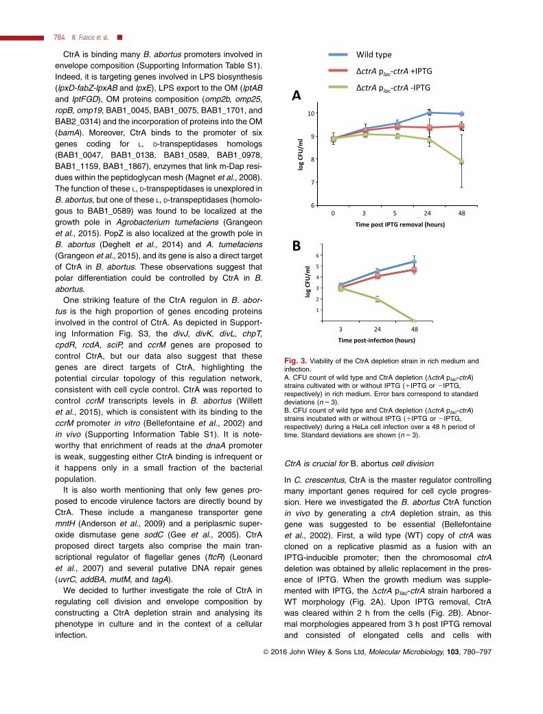
Fig. 3. Viability of the CtrA depletion strain in rich medium andinfection.A. CFU count of wild type and CtrA depletion (DctrA plac-ctrA)strains cultivated with or without IPTG (1IPTG or 2IPTG,respectively) in rich medium. Error bars correspond to standarddeviations (n 5 3).B. CFU count of wild type and CtrA depletion (DctrA plac-ctrA)strains incubated with or without IPTG (1IPTG or 2IPTG,respectively) during a HeLa cell infection over a 48 h period oftime. Standard deviations are shown (n 5 3).
784 N. Francis et al. �
VC 2016 John Wiley & Sons Ltd, Molecular Microbiology, 103, 780–797

mislocalized constrictions (Fig. 2A). At 3 h post-IPTG
removal, a fraction of CtrA-depleted bacteria (10.9%)
were longer than 2.75 mm while only 1.6% of WT bacte-
ria and 1.3% of the depletion strain grown with IPTG
exceeded this size (p< 0.05). A highly significant pro-
portion (p< 0.01) of CtrA-depleted bacteria (6.3%) had
a mislocalized constriction, i.e. detectable septa located
very close to one pole (Fig. 2A; white arrow heads),
compared with the WT strain (0.88%) and to the CtrA
depletion strain grown with IPTG (1.33%) (Fig. 2A).
Seven hours after IPTG was removed from the culture,
we observed bacteria that grew to form multiple
branches while others generated small chains, inter-
preted as filamentation with aborted divisions segment-
ing the bacteria (Fig. 2A). If the incubation in a
CtrA-depleted state is prolonged (15 h), bacteria kept on
branching. These results suggest that in the absence of
CtrA, bacterial elongation is maintained but division is
highly perturbed, since it is either abolished (there are
almost no visible constriction sites in branching bacteria)
or division is initiated at various positions but it is often
not completed since bacteria form chains.
We next characterized the viability of the CtrA deple-
tion strain. The viability of the CtrA depletion strain in
rich culture medium was assessed by counting the num-
ber of colony forming units (CFU) (Fig. 3A). In these
assays, colonies were grown on plates in the presence
of IPTG, since this strain does not grow on plates with-
out IPTG, consistent with the essentiality of the ctrA
gene. When the CtrA depletion strain was cultivated in
the presence of IPTG in liquid medium, a stable number
of CFU was reached earlier than the WT control, and
the plateau was lower (Fig. 3A). In the absence of
IPTG, the number of CFU did not increase, and
remained constant for 24 h before decreasing (Fig. 3A).
The high variability of CFU numbers after 48 h of deple-
tion (Fig. 3A) could be due to a high variability in the
capacity of branched bacteria to divide and release via-
ble bacteria. These data suggest that CtrA is essential
for B. abortus growth and long-term survival in rich
medium.
To test the reversibility of the CtrA depletion on cell
division, the CtrA depletion strain was grown overnight
without IPTG, labeled with Texas Red Succinimidyl
Ester (TRSE) and inoculated in fresh medium supple-
mented with IPTG. The newly incorporated envelope
appears unlabeled with TRSE after growth and thus
new division sites appear as unlabeled rings (Brown
et al., 2012). After 3 h of repletion, we observed a reac-
cumulation of CtrA (Supporting Information Fig. S4A)
and several unlabeled constriction sites were visible on
the large bacteria (Supporting Information Fig. S4B). Six
hours after IPTG was added to the medium, several
division events were completed as shown by the release
of unlabeled or partially labeled bacteria (Supporting
Information Fig. S4B). Those bacteria were of different
size and shapes, demonstrating that the septa were
formed at ectopic sites. These results further confirm
that CtrA is essential for division in B. abortus, and that
CtrA depletion effect is reversible for the generation of
cell division events, but not for their correct positioning
in the cell. It also indicates that large branching bacteria
generated in the absence of IPTG are not dead, at least
after an overnight depletion of CtrA.
In the ChIP-seq study reported above, only one condi-
tion was tested, and it is likely that CtrA could be able to
bind other targets in different conditions. Moreover is it
also likely that CtrA could be a crucial regulator in a
fraction of its targets, but only an accessory regulator
for other promoters. The availability of a depletion strain
for CtrA allowed us to test some CtrA targets promoters
for their dependence on CtrA. Using reverse transcrip-
tion followed by quantitative PCR (RT-qPCR), we found
that the abundance of ccrM transcript is strongly
dependent on CtrA since after a CtrA depletion condi-
tion (without IPTG) of 6 h, there is a significant
(p<0.01) 17.1 (60.8) fold decrease of ccrM mRNA
abundance compared with the control condition (with
IPTG). RT-qPCR analysis of other predicted CtrA targets
(omp25 and ftsEX) revealed statistically relevant
changes in mRNA abundance, but of very low amplitude
(typically <40%), highlighting the complexity of the regu-
lation network involving CtrA.
The activity of CtrA target promoters varies in function
of bacterial cell size
Since CtrA is a cell cycle regulator in C. crescentus
(Quon et al., 1998; Reisenauer et al., 1999; Laub et al.,
2002) and S. meliloti (Pini et al., 2015), we wondered if
CtrA is also able to regulate its targets according to cell
cycle. Because B. abortus is not synchronizable as of
yet, we monitored the activity of CtrA target promoters
at the single cell level, and we then sorted bacteria
according to their size to reconstruct their cell cycle. A
reporter system was designed to monitor the activity of
ccrM (pccrM), repAB (prepAB), ctrA (pctrA), and pleC
(ppleC) promoters by fusing each of them to a gene cod-
ing for an unstable GFP (GFP-ASV) (Andersen et al.,
1998) on a medium-copy replicative vector (Terwagne
et al., 2013). The ccrM and ctrA transcription follows a
tightly regulated profile throughout C. crescentus cell
cycle while PleC protein amount remains stable
(Zweiger et al., 1994; Quon et al., 1996; Wheeler and
Shapiro, 1999). The repAB, ctrA, and ccrM promoters
are bound by CtrA in the ChIP-seq (Supporting
CtrA regulon of the pathogen Brucella abortus 785
VC 2016 John Wiley & Sons Ltd, Molecular Microbiology, 103, 780–797

Information Table S1), while the pleC promoter is not, at
least in the conditions tested here.
Currently, unlike C. crescentus cell cycle, the B. abor-
tus cell cycle is not synchronizable. To test if ctrA, ccrM,
repAB, and pleC promoters are controlled along cell
cycle, fluorescence intensity of the B. abortus reporter
strains was measured in three independent experiments
and mean fluorescence intensity was plotted against
bacterial cell size (Fig. 4). The pctrA and prepAB activities
changed according to cell length, and they display oppo-
site profiles as maximal fluorescence intensity was
measured in intermediate bacteria for prepAB and in
small and large bacteria for pctrA reporters. These data
suggest that pctrA activity is maximal in large dividing
bacteria, and this activity decreases after division (Fig.
4). The maximal activity of pctrA in large bacteria is con-
sistent with cell division defect in the CtrA depletion
strain (Fig. 2). On the contrary, prepAB seems to be
turned on early in the cell cycle, leading to an accumula-
tion of GFP-ASV in intermediate bacteria (Fig. 4). These
data correlate with the initiation of replication of chromo-
some II at about half of the cell cycle of B. abortus
(Deghelt et al., 2014). The pccrM activity profile is similar
to pctrA (Fig. 4); differences between bacterial length
classes are however not significant, probably due to the
high variability of fluorescence intensity between experi-
ments, while variations of prepAB and pctrA activities
according to cell length were significant (Supporting
Fig. 4. Activity profile of repAB, ctrA, ccrM and pleC promoters according to cell length.Phase and fluorescence microscopy images of B. abortus reporter strains were analyzed with the MicrobeTracker program. Bacteria wereordered according to their cell length and the mean cell length and mean fluorescence intensity was calculated for a sliding window (fromsmallest to largest bacteria) of 300 bacteria. The mean fluorescence intensities were normalized to the average fluorescence intensity of thewhole population of a given experiment, allowing the representation of results from three independent experiments on the same plot. Eachexperiment is shown with a different color. The number of bacteria analyzed for prepAB are 1632, 1402, and 2377; for pctrA 1456, 1487, and1467; for pccrM 1446, 753, and 1678; for ppleC 1888, 1297, and 1953.
786 N. Francis et al. �
VC 2016 John Wiley & Sons Ltd, Molecular Microbiology, 103, 780–797

Information Fig. S5). The ppleC did not show any signifi-
cant variation in its activity according to bacterial cell
size (Fig. 4 and Supporting Information Fig. S5). Taken
together, these data suggest that two promoters bound
by CtrA in vivo are differentially regulated during B.
abortus cell cycle, supporting the role of CtrA as a cell
cycle regulator.
The ccrM and repAB promoters contain two and one
proposed CtrA binding sites respectively (Supporting
Information Fig. S6). Using the pccrM-gfpasv and prepAB-
gfpasv fusions cited above, we created mutants in the
CtrA binding boxes (Supporting Information Fig. S6) and
we were able to show that mutagenesis of CtrA binding
box 2 for pccrM and mutagenesis of the CtrA binding box
in prepAB abolished activity of these promoters, strongly
suggesting that CtrA is crucial to activate them. Of
course, we cannot exclude that these mutations also
impair the binding of other crucial factors required for
the activity of repAB and ccrM promoters. The mutation
of CtrA binding box 1 in pccrM did not yield a significant
effect, suggesting that its role is either subtle or
restricted to a condition that was not present in our
experiments.
CtrA is required for B. abortus division and survival in
HeLa cells
To assess the role of CtrA during infection, the depletion
strain was used to infect HeLa cells. Bacteria were incu-
bated with HeLa cells for 1 h with IPTG. Cells were then
washed and gentamicin was added to kill extracellular
bacteria. The CtrA depletion strain was able to infect
HeLa cells and to replicate intracellularly almost to the
same extent as the WT when IPTG was kept in the
medium (Fig. 3B). When IPTG was removed after the
initial hour of internalization, a similar number of CFU
was recovered 3 h PI, and then the CFU counts
dropped dramatically and went below the detection limit
at 48 h PI. We verified that the presence of Triton X-
100, used for the extraction of bacteria from host cells,
did not decrease the CFU counts for the CtrA depletion
strain in the absence of IPTG (Supporting Information
Fig. S7). Altogether, these data suggest that CtrA is cru-
cial for B. abortus viability during HeLa cells infection.
Similarly to the rich medium condition, we analyzed
the morphology of the CtrA depletion strain during infec-
tion. As expected, this strain had WT morphology when
IPTG was kept in the medium (Fig. 5A). When the infec-
tion was performed in the absence of IPTG, bacteria
with aberrant morphologies appeared from 10h PI, but
their proportion was variable from one infection to the
other. The intracellular branched morphologies are simi-
lar to those observed after a long depletion in culture
(Fig. 2). If bacteria are labeled with TRSE prior to infec-
tion, they also display a Texas Red fluorescence at the
base of the branched morphology (Fig. 5B; white arrow
head). The emergence of abnormal morphologies late in
the trafficking is consistent with the previously reported
biphasic trafficking of B. abortus in HeLa cells (Deghelt
et al., 2014). Indeed, B. abortus intracellular growth is
detected between 6 and 8 h PI in HeLa cells, suggest-
ing a growth arrest of at least 6 h (Deghelt et al., 2014).
The CtrA depletion generates elongated morphologies
at 10 h PI, suggesting that growth was also arrested for
several hours before, otherwise these abnormal mor-
phologies would have appeared around 3 h PI. This
suggests that CtrA is not crucial to control the timing of
the intracellular growth recovery.
Fig. 5. Morphology of the CtrA depletion strain during infection.A. Immunofluorescence microscopy of HeLa cells infected for 15 h with the depletion strain in presence or in absence of IPTG. Phasecontrast images were merged with anti-Brucella staining (cyan) to detect intracellular bacteria. The scale bars correspond to 5 mm. Theabsence of IPTG, bacteria with normal and abnormal morphologies can be found in variable proportions from one infection to the other.B. Representative image of an abnormal morphology generated by the CtrA depletion strain 15 h post-infection in HeLa cells, with bacterialabeled with TRSE before infection. The TRSE-labeled part corresponds to the old pole of the initial bacterium that invaded the host cell(white arrow head). DAPI (staining the nucleus in blue), anti-Brucella (green) and Texas Red are merged.
CtrA regulon of the pathogen Brucella abortus 787
VC 2016 John Wiley & Sons Ltd, Molecular Microbiology, 103, 780–797
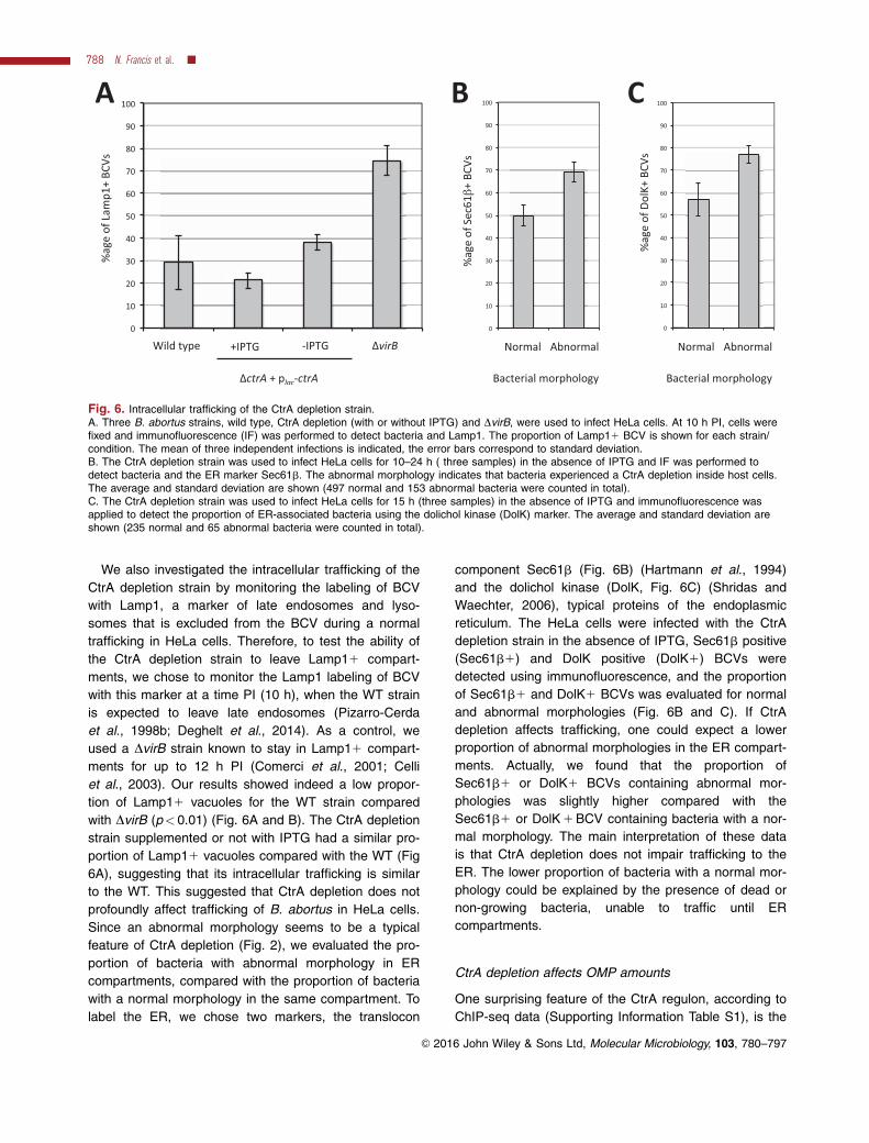
We also investigated the intracellular trafficking of the
CtrA depletion strain by monitoring the labeling of BCV
with Lamp1, a marker of late endosomes and lyso-
somes that is excluded from the BCV during a normal
trafficking in HeLa cells. Therefore, to test the ability of
the CtrA depletion strain to leave Lamp11 compart-
ments, we chose to monitor the Lamp1 labeling of BCV
with this marker at a time PI (10 h), when the WT strain
is expected to leave late endosomes (Pizarro-Cerda
et al., 1998b; Deghelt et al., 2014). As a control, we
used a DvirB strain known to stay in Lamp11 compart-
ments for up to 12 h PI (Comerci et al., 2001; Celli
et al., 2003). Our results showed indeed a low propor-
tion of Lamp11 vacuoles for the WT strain compared
with DvirB (p< 0.01) (Fig. 6A and B). The CtrA depletion
strain supplemented or not with IPTG had a similar pro-
portion of Lamp11 vacuoles compared with the WT (Fig
6A), suggesting that its intracellular trafficking is similar
to the WT. This suggested that CtrA depletion does not
profoundly affect trafficking of B. abortus in HeLa cells.
Since an abnormal morphology seems to be a typical
feature of CtrA depletion (Fig. 2), we evaluated the pro-
portion of bacteria with abnormal morphology in ER
compartments, compared with the proportion of bacteria
with a normal morphology in the same compartment. To
label the ER, we chose two markers, the translocon
component Sec61b (Fig. 6B) (Hartmann et al., 1994)
and the dolichol kinase (DolK, Fig. 6C) (Shridas and
Waechter, 2006), typical proteins of the endoplasmic
reticulum. The HeLa cells were infected with the CtrA
depletion strain in the absence of IPTG, Sec61b positive
(Sec61b1) and DolK positive (DolK1) BCVs were
detected using immunofluorescence, and the proportion
of Sec61b1 and DolK1 BCVs was evaluated for normal
and abnormal morphologies (Fig. 6B and C). If CtrA
depletion affects trafficking, one could expect a lower
proportion of abnormal morphologies in the ER compart-
ments. Actually, we found that the proportion of
Sec61b1 or DolK1 BCVs containing abnormal mor-
phologies was slightly higher compared with the
Sec61b1 or DolK 1 BCV containing bacteria with a nor-
mal morphology. The main interpretation of these data
is that CtrA depletion does not impair trafficking to the
ER. The lower proportion of bacteria with a normal mor-
phology could be explained by the presence of dead or
non-growing bacteria, unable to traffic until ER
compartments.
CtrA depletion affects OMP amounts
One surprising feature of the CtrA regulon, according to
ChIP-seq data (Supporting Information Table S1), is the
Fig. 6. Intracellular trafficking of the CtrA depletion strain.A. Three B. abortus strains, wild type, CtrA depletion (with or without IPTG) and DvirB, were used to infect HeLa cells. At 10 h PI, cells werefixed and immunofluorescence (IF) was performed to detect bacteria and Lamp1. The proportion of Lamp11 BCV is shown for each strain/condition. The mean of three independent infections is indicated, the error bars correspond to standard deviation.B. The CtrA depletion strain was used to infect HeLa cells for 10–24 h ( three samples) in the absence of IPTG and IF was performed todetect bacteria and the ER marker Sec61b. The abnormal morphology indicates that bacteria experienced a CtrA depletion inside host cells.The average and standard deviation are shown (497 normal and 153 abnormal bacteria were counted in total).C. The CtrA depletion strain was used to infect HeLa cells for 15 h (three samples) in the absence of IPTG and immunofluorescence wasapplied to detect the proportion of ER-associated bacteria using the dolichol kinase (DolK) marker. The average and standard deviation areshown (235 normal and 65 abnormal bacteria were counted in total).
788 N. Francis et al. �
VC 2016 John Wiley & Sons Ltd, Molecular Microbiology, 103, 780–797

high proportion of direct targets corresponding to genes
encoding outer membrane components, particularly
outer membrane proteins (OMPs). To reveal a possible
impact of CtrA depletion on the abundance of some of
these OMPs, three integral OMPs (Omp2b, Omp25, and
BamA) and two proposed OM lipoproteins (Omp16 and
Omp19) were detected by Western blot on a B. abortus
wild type strain, on the CtrA depletion strain cultivated
with IPTG and on the same strain depleted in CtrA over-
night. While the amount of Omp16 and Omp19 seems
to remain unchanged in the absence of CtrA, a slight
decrease in the amount of Omp2b and BamA was
observed (Fig. 7A). Omp25 abundance was lower in the
absence of CtrA, and strongly decreased at longer
depletion times (Fig. 7B). Given that OMPs of groups 2
and 3 (Omp2b and Omp25) are the major OMPs in Bru-
cella envelope (Dubray and Charriaut, 1983), their
reduced abundance in the absence of CtrA could lead
to the perturbation of the envelope, which could have
dramatic consequences for the bacterium when it is
inside host cells (Fig. 3C).
We also labeled Omp25 present at the surface of B.
abortus by immunofluorescence. The localization of
Omp25 on the CtrA-depletion strain grown overnight
without IPTG was heterogeneous. This was observed
with a monoclonal antibody directed against Omp25
(Supporting Information Fig. S8). The labeling was often
partial and concentrated on the tip of the branches (Fig.
8A). For the wild type strain, 86.6% of the bacteria were
either completely unlabeled or displayed an homogene-
ous labeling (Fig. 8B). The DctrA plac-ctrA depletion
strain cultivated in the presence of IPTG displayed a
similar proportion of unlabeled or homogeneously
labeled bacteria (82.2%) (Supporting Information Fig.
S8). Partially labeled bacteria were counted in three
independent experiments, and their proportion is
reported in Fig. 8C. The proportion of partially labeled
bacteria was significantly higher for the depletion strain
compared with the wild type strain (p<2.1 1025 in a
Scheff�e statistical analysis). These data suggest that in
the absence of CtrA, Omp25 localization on the surface
of B. abortus is perturbed. Curiously, the Omp25 label-
ing pattern was symmetric, meaning that if the tip of one
branch is labeled, the tip of the other branches is also
labeled (Fig. 8A). One plausible explanation is that
Omp25 is incorporated in the outer membrane in a simi-
lar manner in all parts of the branched cells that are
generated at the same time. In this model, Omp25 pro-
teins remain immobile in the outer membrane, probably
because they are directly or indirectly bound to peptido-
glycan (Cloeckaert et al., 1992).
Discussion
The essential transcription factor CtrA is known to be at
the heart of a complex network regulating the cell cycle
progression of the model organism C. crescentus. CtrA
is essential and its regulon is also partially conserved in
S. meliloti (Pini et al., 2015). However, in phylogeneti-
cally more distant organisms such as Rhodospirillales
(Greene et al., 2012) or Rhodobacterales, CtrA is not
essential and has evolved to regulate cell cycle-
independent processes (Lang and Beatty, 2000; Cheng
et al., 2011; Mercer et al., 2012; Francez-Charlot et al.,
2015). Analysis of the expression of CtrA targets in B.
abortus suggests that CtrA is involved in cell cycle-
dependent regulation (Fig. 4).
Fig. 7. Effect of the CtrA depletion on outer membrane proteinsabundance.A. Western blots on B. abortus lysates of the wild type (WT) strainand the CtrA depletion strain grown with or without IPTG for onenight, using monoclonal antibodies recognizing OMPs whose geneswere identified by ChIP-seq as being potentially regulated by CtrA.B. Western blots on lysates of the CtrA depletion strain grownwithout IPTG for 0, 7, 15, and 24 h, using a monoclonal anti-Omp25 antibody (A68/4B10/F05). Omp10 was detected byWestern blot as a loading control.
CtrA regulon of the pathogen Brucella abortus 789
VC 2016 John Wiley & Sons Ltd, Molecular Microbiology, 103, 780–797

Here, we show that CtrA regulon in B. abortus is par-
tially conserved in comparison with C. crescentus and
S. meliloti CtrA regulons. They indeed share genes
involved in cell cycle regulation such as DNA methyla-
tion, chromosome replication, and segregation and divi-
sion (Laub et al., 2002; Pini et al., 2015). However, as
previously suggested by the identification of a limited
number of CtrA targets in B. abortus (Bellefontaine
et al., 2002), a similar process can be regulated through
different target genes. For example, CtrA is binding to
the mipZ promoter in C. crescentus (Fumeaux et al.,
2014), allowing the control of Z ring positioning, while
this control is proposed to be mediated by the binding to
the minCDE promoter in B. abortus (Bellefontaine et al.,
2002), as also recently elucidated in S. meliloti (Pini
et al., 2015). These comparisons underline the plasticity
of the CtrA regulon along evolution.
Depletion of CtrA leads to a severe (Fig. 2) and
reversible (Supporting Information Fig. S4) cell division
defect in B. abortus. However, among the direct targets
of CtrA found in the conditions tested here, cell cycle-
related genes are not restricted to cell division. Indeed,
genes involved in replication, the DivK-CtrA regulation
network and the recruitment of proteins to the poles
(popZ) have also their promoter enriched by ChIP-seq.
Besides cell cycle-related genes, one obvious conclu-
sion of the ChIP-seq experiment reported here is the
high proportion of genes involved in envelope biogene-
sis or homeostasis. Indeed, CtrA predicted regulon is
enriched in genes coding for LPS and outer membrane
proteins biosynthesis and export. The presence of CtrA
is crucial for the production of normal amounts of
Omp25 (Fig. 7) and for an homogeneous distribution of
Omp25 on the surface of the bacterium (Fig. 8). We
Fig. 8. Effect of CtrA depletion on Omp25 localization.A. Phase contrast microscopy and associated fluorescent anti-Omp25 signal of the CtrA depletion strain cultivated in the absence of IPTG. Inthese bacteria, the Omp25 signal is localized at the tip of the branches, revealing the heterogeneity of the outer membrane composition inthese bacteria. These localization patterns also suggest that Omp25 diffusion in the outer membrane is slow.B. Homogeneous localization of anti-Omp25 signal on the wild type strain. The scale bars correspond to 2 lm.C. Proportion of partially labeled bacteria generated with the anti-Omp25 antibody, for the wild type strain or the depletion strain cultivated inthe presence or in absence of IPTG. Standard deviations are shown (n 5 3).
790 N. Francis et al. �
VC 2016 John Wiley & Sons Ltd, Molecular Microbiology, 103, 780–797

cannot exclude that alteration of Omp25 localization on
the surface is an indirect effect of the inhibition of cell
division. Indeed it is likely that the generation of elon-
gated and branched morphologies takes more time than
the generation of normal shaped bacteria, thus partially
labeled bacteria could reveal the oscillating nature of
Omp25 incorporation in the outer membrane in these
cells. A B. abortus deletion strain for omp25 was found
to be attenuated in cattle (Edmonds et al., 2001), but
more recent data suggested that this attenuation could
be explained by a higher internalization and a higher
intracellular killing of the omp25 mutant compared with
the wild type strain (Manterola et al., 2007). The molec-
ular functions of the highly abundant Omp25 are still
unknown; it was shown to inhibit TNFa production in
human macrophages (Jubier-Maurin et al., 2001) and it
could be involved in defining the properties of the outer
membrane by interacting with the LPS (Manterola et al.,
2005). Interestingly, in S. meliloti, CtrA binds to the pro-
moter of ropB gene (Pini et al., 2015), encoding an
Omp25 homolog involved in outer membrane stability in
Rhizobium leguminosarum (Vanderlinde and Yost,
2012). It is thus possible that in Rhizobiales, CtrA con-
trols factors involved in outer membrane biogenesis or
homeostasis. Their control by CtrA would have been
acquired after the divergence from the common ances-
tor with C. crescentus, since CtrA regulon of C. cres-
centus is not particularly enriched in genes involved in
outer membrane biogenesis or homeostasis (Laub
et al., 2002; Fumeaux et al., 2014).
Depletion of CtrA results in a strong inhibition of cell
division (Fig. 2A), possibly explaining why the ctrA gene
is essential in B. abortus as suggested in previous stud-
ies (Bellefontaine et al., 2002; Willett et al., 2015). The
inhibition of cell division results in branched morphology
or formation of small chains of cells, the latter being
probably produced by incomplete septation. It is note-
worthy that the CtrA-loss of function phenotype is
reversible, as induction of ctrA expression after depletion
resulted in the reactivation of cell division (Supporting
Information Fig. S4). Perturbation of division in the CtrA-
depleted condition is likely explained by the presence of
numerous genes and operons involved in division in the
CtrA predicted regulon. Indeed, the minCDE operon is
involved in Z ring placement, and many genes and oper-
ons proposed to be involved in the cell division process,
like ftsQAZ, ftsEX, ftsK, and the pal(omp16)-tolQRAB
locus are detected as possible direct targets of CtrA in
B. abortus (Supporting Information Table S1). The
deregulation of some of these genes is probably suffi-
cient to block the whole cell division process.
Depletion of CtrA also resulted in altered morphology
of bacteria infecting host cells (Fig. 5B). During infection
elongated and branched morphologies can only be
generated by growth, suggesting that these bacteria are
able to uptake nutrients during cellular infection, after an
initial stage of growth arrest that could be due to starva-
tion (see below). In host cells, the timing of the appear-
ance of the altered morphologies is different from the
timing of their formation in bacteriological medium.
Indeed, in HeLa cells abnormal morphologies of the
CtrA depletion strain appeared around or after 10 h PI
(Fig. 5B), mainly consisting of elongated cells (Fig. 5A)
resembling bacteria recovered 3 h after IPTG removal in
rich culture medium (Fig. 2A). At 15 h PI, branched bac-
teria were observed (Fig. 5A), similarly to the 7 h deple-
tion in culture (Fig. 2A). Interestingly, B. abortus was
shown to resume its intracellular growth around 8 h PI
in HeLa cells (Deghelt et al., 2014). The observation of
elongated bacteria at or after 10 h PI for the CtrA-
depleted bacteria thus suggests that the absence of
CtrA does not drastically change the timing of growth
arrest/resumption in HeLa cells. If CtrA is not required
for the control of the intracellular growth arrest/resump-
tion, it is likely that other regulation networks, like those
triggered by starvation (Dozot et al., 2006), could be
involved. Previous work also showed that B. abortus
growth is resumed in Lamp11 compartments around
8 h PI and that daughter cells are found almost exclu-
sively in Lamp1- compartment from 10 h PI (Deghelt
et al., 2014). While CtrA depleted bacteria with a normal
morphology could be dead or non-growing bacteria
unable to traffic until the ER, bacteria that have grown
to generate branching morphology are able to traffic to
the ER, demonstrating that they are able to perform this
crucial step of intracellular trafficking. This suggests that
despite their morphological alterations, these bacteria
are probably still able to produce a functional VirB sys-
tem, which is required for this step of their intracellular
trafficking (Comerci et al., 2001; Delrue et al., 2001).
Thus CtrA-dependent cell cycle control and intracellular
trafficking seem to be relatively independent processes
in B. abortus. The dramatic drop of the CFU counts at
48 h PI (Fig. 3B) suggests that the branched bacteria
are unable to survive for long periods in host cells.
In conclusion, our data support the idea that along
evolution of the CtrA regulon, CtrA has kept the control
of cell division in many Alphaproteobacteria. However, in
B. abortus and possibly in Rhizobiales it also acquired
new functions, including the control of envelope compo-
sition. It is interesting to realize that CtrA has to be
cleared from S. meliloti cells to allow them to differenti-
ate into nitrogen-fixing bacteroids inside host plants
(Pini et al., 2013), illustrating that fundamental proc-
esses of bacterial cell cycle have been adapted to the
lifestyle of pathogens and symbionts within the
Alphaproteobacteria.
CtrA regulon of the pathogen Brucella abortus 791
VC 2016 John Wiley & Sons Ltd, Molecular Microbiology, 103, 780–797
Experimental procedures
Bacterial strains and media
E. coli strains DH10B, BL21 (DE3) and DB3.1 were grown
in Luria-Bertani (LB) medium at 378C. Derivatives of the B.
abortus 544 NalR strain were cultivated in 2YT rich medium(1% yeast extract, 1.6% peptone, 0.5% NaCl) at 378C. Anti-
biotic concentrations are the following: ampicillin, 100 mg/
ml; kanamycin, 20 or 50 mg/ml; chloramphenicol, 20 mg/ml;nalidixic acid, 25 mg/ml; rifampicin, 20 mg/ml; gentamicin, 50
mg/ml. B. abortus strains were constructed as previously
described (Deghelt et al., 2014). Plasmids are listed in Sup-
porting Information Table S2.
Cloning of the pBBR-MCS1-placI-lacI-plac-ctrA
The placI-lacI-plac sequence was amplified from the pSRK-
Kan vector (Khan et al., 2008) using Phusion High-Fidelity
DNA Polymerase (New England BioLabs) and SacI-Kan3’
and plac-R1 primers (see Supporting Information Table S3).The ctrA coding sequence was amplified from B. abortus
544 purified genomic DNA using ctrA-F2 and KpnI-ctrA-R2
primers. The PCR product was fused to the placI-lacI-plac
sequence by joining PCR. The placI-lacI-plac-ctrA insert was
then cloned in the pBBRMCS1 using SacI and KpnI restric-
tion enzymes. By using these enzymes, the insert was
cloned in the opposite orientation to the plac promoter ofthe pBBRMCS1.
Cloning of a ctrA deletion for allelic exchange
The ctrA gene was deleted from B. abortus 544 chromo-
some by allelic replacement. A 750 base pair (bp)-region
upstream and another one downstream of ctrA were ampli-fied by PCR using PstI-Up-ctrA-F/Up-ctrA-R and Down-
ctrA-F/SalI-Down-ctrA-R pairs of primers respectively and
both PCR products were fused together by joining PCR.
The PCR product was cloned in the pNPTS138 vector (M.R. K. Alley, Imperial College of Science, London, UK) carry-
ing a kanamycin resistance cassette and a sucrose sensi-
tivity cassette.
Cloning of reporter vectors
Promoter regions were amplified from B. abortus 544 puri-fied genomic DNA using Phusion High-Fidelity DNA Poly-
merase and fused by joining PCR to gfp(ASV). The pairs of
primers used to amplify the promoter regions are XbaI-pctrA-F1/pctrA-R1, XbaI-prepAB-F1/prepAB-R1, XbaI-pccrM-F1/
pccrM-R1 and XbaI-ppleC-F1/ppleC-R1. The pair of primers
used to amplify the gfp(ASV) gene is gfP(ASV)-F2/XhoI-
gfp(ASV)-R2. XbaI and XhoI restriction sites were added tothe upstream and downstream primers. The fusion was first
cloned in pGEMT digested by EcoRV, generating blunt
ends, and sequenced. A XbaI-XhoI restriction allowed thetransfer of the insert to a pBBRMCS1 vector, in the oppo-
site direction to the lac promoter of the vector.Mutagenesis of the CtrA boxes was generated in the
same way, but using the constructs with a wild type
promoter as DNA template. The XbaI-pgene-F1 and XhoI-
gfp(ASV)-R2 were kept as they were but their correspond-
ing pair of primer (R1 and F2) were modified to include the
mutated CtrA-binding boxes. In the case of pccrM-mut1&2-
gfp(ASV), boxes were mutated sequentially (CtrA-binding
box 1, followed by CtrA-binding box 2). The wild type and
mutated promoters are shown in Supporting Information Fig
S5.
Chromatin immunoprecipitation with anti-CtrA antibodies
An 80 ml culture of B. abortus 544 at an OD600 of 0.8 was
centrifuged to harvest the bacteria. Protein-DNA crosslink-
ing was performed in 10 mM sodium phosphate buffer (pH
7.6) and 1% formaldehyde for 10 min at RT and 30 min on
ice. Bacteria were harvested by centrifugation at 8500 rpm
for 5 min at 48C, washed twice in cold PBS and resus-
pended in lysis buffer (10 mM Tris-HCl pH 7.5, 1 mM
EDTA, 100 mM NaCl, 2.2 mg/ml lysozyme, 20 ml protease
inhibitor solution). Zirconia/Silica beads (Biospec Products)
of 0.1 and 0.5 mm diameter were added. Bacteria were
lysed in the cell Disruptor Genie from Scientific Industries
at maximal amplitude (2800) for 25 min at 48C. ChIP buffer
was added (1.1% TritonX-100, 1.2 mM EDTA, 16.7 mM
Tris-HCl pH 8.0, 167 mM NaCl, protease inhibitors) and
bacteria were incubated at 378C for 10 min for further lysis.
The lysate was sonicated on ice (Branson Sonifier Digital
cell disruptor S-450D 400W) by applying 15 bursts of 20 s
(50% duty) at 30% amplitude to cut the DNA to fragments
of about 300 base pairs and centrifuged at 14,000 rpm for
3 min to pellet the debris. The supernatant was normalized
by protein content by measuring the absorbance at
280 nm. Around 7.5 mg of protein was diluted in 1 ml of
ChIP buffer supplemented with 0.01% SDS and pre-cleared
in 80 ml of protein A-agarose beads (Roche) and 100 mg
BSA. Polyclonal anti-CtrA antibodies (Bellefontaine et al.,
2002) were added to the supernatant (dilution 1:1000) and
incubated for one night at 48C to form immune complexes
which were then incubated with 80 ml of protein A-agarose
beads pre-saturated with BSA for 2 h at 48C. Beads were
then washed once with low salt buffer (0.1% SDS, 1%
TritonX-100, 2 mM EDTA, 20 mM Tris-HCl pH 8.1, 150 mM
NaCl), high salt buffer (0.1% SDS, 1% TritonX-100, 2 mM
EDTA, 20 mM Tris-HCl pH 8.1, 500 mM NaCl) and LiCl
buffer (0.25 M LiCl, 1% NP-40, 1% sodum deoxycholate,
1 mM EDTA, 10 mM Tris-HCl pH 8.1) and twice with TE
buffer (10 mM Tris-HCl pH 8.1 and 1 mM EDTA). Protein-
DNA complexes were eluted with 500 ml of elution buffer
(1% SDS and 0.1M NaHCO3). Reverse crosslinking was
performed in presence of 300 mM of NaCl O/N at 658C.
Samples were treated with 2 mg of Proteinase K for 2h at
458C in 40 mM EDTA and 40 mM Tris-HCl pH 6.5. DNA
was extracted using QIAgen minelute kit and resuspended
in 30 ml of Elution Buffer. ChIP DNA was sequenced using
Illumina MySeq.
Analysis of the sequencing data
Sequencing data consisted of a number of reads per nucle-
otide. Computing of average and variance in a window of 1
792 N. Francis et al. �
VC 2016 John Wiley & Sons Ltd, Molecular Microbiology, 103, 780–797

million base pairs allowed the calculation of Z score pour
each base pair (i.e., the number of standard deviation from
the average). Genomic regions with reads numbers above
the threshold (Z>4) were kept and considered to be bound
by CtrA. These regions were mapped to the genome of B.
abortus 2308, a close relative to the B. abortus 544 strain.
The mapping is available in the Supplemental files ChIP-
seq_CtrA_chr1.txt and ChIP-seq_CtrA_chr2.txt, that can be
analyzed using the Chr1.gb and Chr2.gb genomic sequen-
ces respectively, with the Artemis program (freely available
at the following website http://www.sanger.ac.uk/science/
tools/artemis).
Electrophoretic mobility shift assay
DNA probes of 50–70 pb were prepared by amplifying pro-
moter regions from B. abortus 544 purified genomic DNA
using Phusion High Fidelity DNA Polymerase (see Support-
ing Information Table S3 for list of primers, named as
EMSA-F/R-promoter). Each PCR product was concentrated
and purified after migration on agarose gel electrophoresis,
using NucleoSpinVR Gel and PCR Clean-up (Macherey-
Nagel) according to manufacturer’s instructions. These puri-
fied PCR products were then used as template for a new
amplification with a cyanine-5(Cy5)-labeled primer (Inte-
grated DNA Technologies) (Supporting Information Table
S3). The labeled amplicons were purified after gel electro-
phoresis and quantified using a QubitVR 3.0 Fluorometer
(Thermo Fisher Scientific).His6-CtrA was purified and phosphorylated as previously
described (Bellefontaine et al., 2002). Binding reactions
were prepared in a final volume of 20 ml with 0–340 mg of
CtrA, 3 ng of labeled probes and when necessary with 400
ng of competing probe (non-labeled PCR product) in bind-
ing buffer (10 mM Tris pH 7.4, 10 mM MgCl2, 50 mM NaCl,
10% glycerol). Samples were incubated for 30–45 min at
378C and 10 ml were loaded onto a 13.3% polyacrylamide
gel (3.9 ml of ddH2O, 700 ml of 5X TBE buffer, 2.3 ml of
40% acrylamide stock 19:1, 70 ml of 10% APS and 7 ml of
TEMED). TBE is 89 mM Tris, 89 mM boric acid and 2 mM
EDTA, pH 8.3. Samples were run at 100V for 3h20 in 0.5 3
TBE buffer at 48C. Samples were revealed using an Amer-
sham Imager 600 (GE Healthcare Life Sciences) with the
Cy5 fluorescence channel.
Reverse transcription followed by quantitative PCR
The B. abortus strains were grown in rich medium (2YT) until
exponential phase, washed and growth was restart in rich
medium, with or without IPTG for 6 h. Then bacteria were
washed in PBS, collected by centrifugation and immediately
frozen and stored at 2808C until processing. RNA was then
extracted with TriPure isolation reagent (Roche) according to
the instructions of the manufacturer. DNA contamination was
eliminated by incubation with DNase I (Fermentas). RNA was
reverse transcribed with specific primers, using the High
capacity cDNA Reverse Transcription kit (Applied Biosys-
tems). Specific cDNAs were amplified using FastStart Univer-
sal SYBR Green Master (Roche) with a LightCycler 96
Instrument (Roche). The specificity of the PCR was assessed
by melting-point analysis and gel electrophoresis. Results
were normalized using the housekeeping groEL gene as a
reference. Primer sequences (with a name starting by RT-
qPCR) are available in Supporting Information Table S3.
TRSE labeling
Bacteria were harvested by centrifugation at 7000 rpm for 2
min. They were then washed thrice with phosphate-
buffered saline (PBS) and incubated with Texas Red succi-
nimidyl ester (TRSE) from Invitrogen diluted at 1 mg/ml in
PBS for 15 min at room temperature (RT) in the dark. Bac-
teria were then washed once with PBS and twice with the
appropriate medium, 2YT for growth assays and Dulbecco’s
Modified Eagle’s Medium (DMEM) for HeLa cells infections.
Microscopy and analysis of GFP fluorescence in the
reporter systems using MicrobeTracker
Brucella abortus strains labeled with TRSE were analyzed
by fluorescence microscopy as previously reported (Deghelt
et al., 2014). The pairwise comparisons of the proportions
of morphotypes such as elongated cells or bacteria with
mislocalized constriction sites were made using a Scheffe
analysis. B. abortus strains expressing a promoter-gfp
fusion were observed using a Nikon 80i (objective phase
contrast 3100, plan Apo) connected to a Hamamatsu
ORCA-ER camera. Cell meshes were obtained using the
Matlab-based MicrobeTracker software (Sliusarenko et al.,
2011), determining the cell length and quantifying the aver-
age amount of fluorescence per bacterium. Data were then
transferred to Excel files using the “XLStotmeansteparea.m”
script. Data were sorted according to bacterial cell length,
and the mean cell length and mean fluorescence intensity
were calculated using a sliding window of 300 bacteria.
HeLa cells culture and infection
HeLa cells (from the Centre d’Immunologie de Marseille-
Luminy, Marseille, France) were cultivated at 378C and in a
5% CO2 atmosphere in DMEM (Invitrogen) supplemented
with 10% fetal bovine serum (Gibco), 0.1 g/l non-essential
amino acids and 0.1 g/l sodium pyruvate (Invitrogen). For the
infection, HeLa cells were seeded in 24-well plates (on cover-
slips for immunolabeling) at a concentration of 4.104 cells/ml.
On the day of the infection, an O/N culture of B. abortus was
diluted in DMEM to reach an MOI (multiplicity of infection) of
300. Bacteria were added to HeLa cells and the 24-well
plates were centrifuged at 1200 rpm for 10 min at 48C. Cells
were then incubated at 378C in a 5% CO2 atmosphere for
1 h. Cells were washed twice in PBS and fresh medium sup-
plemented with 50 mg/ml gentamicin was added.
Immunolabeling of infected HeLa cells
Cells were fixed in PBS 2% paraformaldehyde (Prolabo) for
20 min at RT then permeabilized in PBS 0.1% Triton X-100
for 10 min. Cells were incubated for 45 min with primary
CtrA regulon of the pathogen Brucella abortus 793
VC 2016 John Wiley & Sons Ltd, Molecular Microbiology, 103, 780–797
and secondary antibodies supplemented with 0.1% Triton
X-100 and 3% bovine serum albumin (BSA, Sigma Aldrich).
Brucella were detected with the A76-12G12 monoclonal
antibody (non-diluted hybridoma culture supernatant) fol-
lowed by a secondary anti-mouse antibodies coupled to
Alexa-488 diluted 500 times (Sigma Aldrich). Coverslips
were washed thrice with PBS and once with ddH2O and
mounted with Mowiol (Sigma). Antibodies are listed in Sup-
porting Information Table S4.For Lamp1 labeling, cells were fixed in methanol-acetone
(80%-20%) for 20 min at RT. Bacteria and Lamp1 were
labeled with a rabbit anti-Brucella serum diluted 2000 times
and mouse anti-Lamp1 antibodies diluted 200 times in PBS
2% BSA. Secondary anti-rabbit antibodies coupled to
Pacific Blue and anti-mouse antibodies coupled to Alexa-
488 (Sigma Aldrich) were diluted 500 times in PBS 2%
BSA. Coverslips were washed thrice in PBS 2% BSA and
mounted with Mowiol. For each strain in each condition
(6 IPTG), the number of BCVs analyzed was as follows:
66–77 for the wild type strain, 62–100 for the depletion
strain with IPTG, 48–70 for the depletion strain without
IPTG, and 36–71 for the DvirB strain.For endoplasmic reticulum (ER) labeling, cells were fixed
in PBS 2% paraformaldehyde for 20 min at RT, then
washed twice with PBS before to be permeabilized with
PBS 0.2% saponin (Sigma Aldrich) for 20 min at RT. Cells
were then blocked for 30 min with 0.2% saponin, 3% BSA,
50 mM NH4Cl in 0.1% dPBS-Tween20. ER was labeled
either with primary anti-DOLK rabbit antibody (Abcam,
ab93609) diluted 200 times or with anti-Sec61b rabbit anti-
body (B. Dobberstein, Universit€at Heidelberg, Heidelberg,
Germany) diluted 100 times, both in 3% BSA, 0.2% sapo-
nin and 0.1% dPBS-Tween20. Secondary anti-rabbit
coupled to Alexa-488 (Sigma Aldrich) diluted 500 times
were used for staining after washing thrice with PBS. B.
abortus were labeled using non-diluted A76-12G12 primary
antibody obtained from homemade hybridoma culture
supernatant in 0.2% saponin and 3% BSA, followed by a
secondary anti-mouse antibody coupled to TxRed (Sigma
Aldrich) diluted 500 times in 3% BSA, 0.2% saponin and
0.1% dPBS-Tween20. Coverslips were washed thrice in
PBS and once in ddH2O before to be mounted on Mowiol.
The infections were repeated three times independently,
and 167–317 BCVs were analyzed in each infection.
Growth curve and CFU counts
Growth curves were performed by using Bioscreen C from
Oy Growth curves. O/N cultures were diluted to an OD of
0.1 and the OD was measured every 30 min during 70 h.
For CFU counts in culture, wild type (WT) B. abortus 544
and the CtrA depletion strain supplemented with IPTG were
diluted to 1026 or 1027 in 2YT and 100 ml were plated on
2YT, supplemented with chloramphenicol and 1 mM IPTG
for the depletion strain. The depletion strain grown without
IPTG was diluted to 1024 or 1025. For CFU counts after
infection, HeLa cells were lysed with 0.01% TritonX-100
PBS for 10 min at RT. Several dilutions were plated on 2YT
supplemented with chloramphenicol and IPTG if needed.
Plates were incubated for 3–4 days at 378C.
Western blot analysis
One milliliter of a B. abortus culture was concentrated to an
OD600 of 10 in PBS. Bacteria were inactivated for 1 h at
808C and loading buffer was added. Fifteen ml of bacterial
lysate was loaded in each well. After migration, proteins
were transferred onto a nitrocellulose membrane which was
blocked in PBS supplemented with 0.05% Tween and 5%
milk for at least 1 h. The membrane was incubated for 1 h
with the appropriate serum (diluted 10, 100, or 10003
depending on the serum) and secondary antibodies
coupled to HRP (diluted 50003) (Dako Denmark) diluted in
PBS 0.05% Tween 1% milk. The membrane was washed
for 3 times for 5 min. The Clarity Western ECL Substrate
(Biorad) and Image Quant LAS 4000 (General Electric)
were used to reveal the bands.
Immunodetection of Omp25 on bacteria
Bacteria grown overnight in rich culture medium were
washed twice in PBS by centrifugation at 4000 rpm for 2.5
min and resuspension. Washed bacteria were resuspended
in non-diluted hybridoma culture supernatant containing
monoclonal anti-Omp25 antibodies and secondary anti-
mouse antibodies coupled to Alexa-488 diluted 500 times in
PBS, and were incubated for 40 min at RT on a wheel. Bac-
teria were washed twice in PBS and 2 ml were dropped on
an agarose pad (1% PBS agarose) for microscopy.
Acknowledgements
We thank V�eronique Dhennin from the UMR8199 sequencing
service LIGAN-PM Equipex (Lille Integrated Genomics
Advanced Network for personalized medicine) and Aur�elie
Mayard for assistance in protein purification and Western blot-
ting. This research has been funded by the Interuniversity
Attraction Poles Programme initiated by the Belgian Science
Policy Office (https://www.belspo.be/) to J.-J. Letesson and by
grants from Fonds de la Recherche Scientifique-Fonds
National de la Recherche Scientifique (FRS-FNRS, http://
www.fnrs.be) (PDR T.0053.13 and PDR Brucell-cycle
T.0060.15, CDR J.0091.14 and FRFC 2.4.541.08 F) to X. De
Bolle. We thank UNamur (https://www.unamur.be/) for finan-
cial and logistic supports. This work was supported by the
French Agence Nationale de Recherche (ANR-JCJC-2011-
Castacc) (http://www.agence-nationale-recherche.fr/) and the
Region Pas-De-Calais (http://www.nordpasdecalais.fr) CPER
to A. Fioravanti and Emanuele G. Biondi. N. Francis held an
Aspirant fellowship from FRS-FNRS. K. Poncin and V. Vassen
are supported by a Ph.D. grant from FRIA (FRS-FNRS). The
authors declare no conflict of interest.
Author contributions
NF, KP, AV, VV, KW, TAPO, and LR acquired and ana-
lyzed the data; NF, JJL, EB, and XDB designed the
study; NF, JJL, EB, and XDB wrote the manuscript.
794 N. Francis et al. �
VC 2016 John Wiley & Sons Ltd, Molecular Microbiology, 103, 780–797

References
Andersen, J.B., Sternberg, C., Poulsen, L.K., Bjorn, S.P.,
Givskov, M., and Molin, S. (1998) New unstable variants
of green fluorescent protein for studies of transient gene
expression in bacteria. Appl Environ Microbiol 64: 2240–
2246.
Anderson, E.S., Paulley, J.T., Gaines, J.M., Valderas, M.W.,
Martin, D.W., Menscher, E., Brown, et al. (2009) The
manganese transporter MntH is a critical virulence deter-
minant for Brucella abortus 2308 in experimentally
infected mice. Infect Immun 77: 3466–3474.Archambaud, C., Salcedo, S.P., Lelouard, H., Devilard, E.,
de Bovis, B., Van Rooijen, N., et al. (2010) Contrasting
roles of macrophages and dendritic cells in controlling ini-
tial pulmonary Brucella infection. Eur J Immunol 40:
3458–3471.Bellefontaine, A.F., Pierreux, C.E., Mertens, P.,
Vandenhaute, J., Letesson, J.J., and De Bolle, X. (2002)
Plasticity of a transcriptional regulation network among
alpha-proteobacteria is supported by the identification of
CtrA targets in Brucella abortus. Mol Microbiol 43: 945–
960.Brilli, M., Fondi, M., Fani, R., Mengoni, A., Ferri, L.,
Bazzicalupo, M., and Biondi, E.G. (2010) The diversity
and evolution of cell cycle regulation in alpha-proteobac-
teria: a comparative genomic analysis. BMC Syst Biol 4:
52.Brown, P.J., de Pedro, M.A., Kysela, D.T., Van der Henst,
C., Kim, J., De Bolle, X., et al. (2012) Polar growth in the
Alphaproteobacterial order Rhizobiales. Proc Natl Acad
Sci USA 109: 1697–1701.Celli, J., de Chastellier, C., Franchini, D.M., Pizarro-Cerda,
J., Moreno, E., and Gorvel, J.P. (2003) Brucella evades
macrophage killing via VirB-dependent sustained interac-
tions with the endoplasmic reticulum. J Exp Med 198:
545–556.Celli, J., Salcedo, S.P., and Gorvel, J.P. (2005) Brucella
coopts the small GTPase Sar1 for intracellular replication.
Proc Natl Acad Sci USA 102: 1673–1678.
Chaves-Olarte, E., Guzman-Verri, C., Meresse, S.,
Desjardins, M., Pizarro-Cerda, J., Badilla, J., et al. (2002)
Activation of Rho and Rab GTPases dissociates Brucella
abortus internalization from intracellular trafficking. Cell
Microbiol 4: 663–676.Cheng, Z., Miura, K., Popov, V.L., Kumagai, Y., and Rikihisa,
Y. (2011) Insights into the CtrA regulon in development of
stress resistance in obligatory intracellular pathogen Ehrli-
chia chaffeensis. Mol Microbiol 82: 1217–1234.
Cloeckaert, A., Zygmunt, M.S., de Wergifosse, P., Dubray,
G., and Limet, J.N. (1992) Demonstration of
peptidoglycan-associated Brucella outer-membrane pro-
teins by use of monoclonal antibodies. J Gen Microbiol
138: 1543–1550.Comerci, D.J., Martinez-Lorenzo, M.J., Sieira, R., Gorvel,
J.P., and Ugalde, R.A. (2001) Essential role of the VirB
machinery in the maturation of the Brucella abortus-con-
taining vacuole. Cell Microbiol 3: 159–168.Curtis, P.D., and Brun, Y.V. (2010) Getting in the loop: regu-
lation of development in Caulobacter crescentus. Micro-
biol Mol Biol Rev 74: 13–41.
De Bolle, X., Crosson, S., Matroule, J.Y., and Letesson, J.J.
(2015) Brucella abortus cell cycle and infection are coor-
dinated. Trends Microbiol 23: 812–821.Deghelt, M., Mullier, C., Sternon, J.F., Francis, N., Laloux,
G., Dotreppe, D., et al. (2014) G1-arrested newborn cells
are the predominant infectious form of the pathogen Bru-
cella abortus. Nat Commun 5: 4366.
Delrue, R.M., Martinez-Lorenzo, M., Lestrate, P., Danese,
I., Bielarz, V., Mertens, P., et al. (2001) Identification of
Brucella spp. genes involved in intracellular trafficking.
Cell Microbiol 3: 487–497.Detilleux, P.G., Deyoe, B.L., and Cheville, N.F. (1990) Pene-
tration and intracellular growth of Brucella abortus in non-
phagocytic cells in vitro. Infect Immun 58: 2320–2328.
Domian, I.J., Quon, K.C., and Shapiro, L. (1997) Cell
type-specific phosphorylation and proteolysis of a tran-
scriptional regulator controls the G1-to-S transition in a
bacterial cell cycle. Cell 90: 415–424.Dozot, M., Boigegrain, R.A., Delrue, R.M., Hallez, R.,
Ouahrani-Bettache, S., Danese, I., et al. (2006) The stringent
response mediator Rsh is required for Brucella melitensis
and Brucella suis virulence, and for expression of the type IV
secretion system virB. Cell Microbiol 8: 1791–1802.Dubray, G., and Charriaut, C. (1983) Evidence of three
major polypeptide species and two major polysaccharide
species in the Brucella outer membrane. Ann Vet Res
14: 311–318.Edmonds, M.D., Cloeckaert, A., Booth, N.J., Fulton, W.T.,
Hagius, S.D., Walker, J.V., and Elzer, P.H. (2001) Attenu-
ation of a Brucella abortus mutant lacking a major 25
kDa outer membrane protein in cattle. Am J Vet Res 62:
1461–1466.
Francez-Charlot, A., Kaczmarczyk, A., and Vorholt, J.A.
(2015) The branched CcsA/CckA-ChpT-CtrA phosphore-
lay of Sphingomonas melonis controls motility and biofilm
formation. Mol Microbiol 97: 47–63.Fumeaux, C., Radhakrishnan, S.K., Ardissone, S.,
Theraulaz, L., Frandi, A., Martins, D., et al. (2014) Cell
cycle transition from S-phase to G1 in Caulobacter is
mediated by ancestral virulence regulators. Nat Commun
5: 4081.Gee, J.M., Valderas, M.W., Kovach, M.E., Grippe, V.K.,
Robertson, G.T., Ng, W.L., et al. (2005) The Brucella
abortus Cu, Zn superoxide dismutase is required for opti-
mal resistance to oxidative killing by murine macrophages
and wild-type virulence in experimentally infected mice.
Infect Immun 73: 2873–2880.Gerding, M.A., Ogata, Y., Pecora, N.D., Niki, H., and de
Boer, P.A. (2007) The trans-envelope Tol-Pal complex is
part of the cell division machinery and required for proper
outer-membrane invagination during cell constriction in E.
coli. Mol Microbiol 63: 1008–1025.Grangeon, R., Zupan, J.R., Anderson-Furgeson, J., and
Zambryski, P.C. (2015) PopZ identifies the new pole, and
PodJ identifies the old pole during polar growth in Agro-
bacterium tumefaciens. Proc Natl Acad Sci USA 112:
11666–11671.Greene, S.E., Brilli, M., Biondi, E.G., and Komeili, A. (2012)
Analysis of the CtrA pathway in Magnetospirillum reveals
an ancestral role in motility in alphaproteobacteria.
J Bacteriol 194: 2973–2986.
CtrA regulon of the pathogen Brucella abortus 795
VC 2016 John Wiley & Sons Ltd, Molecular Microbiology, 103, 780–797
Hallez, R., Bellefontaine, A.F., Letesson, J.J., and De Bolle,
X. (2004) Morphological and functional asymmetry in
alpha-proteobacteria. Trends Microbiol 12: 361–365.Hallez, R., Letesson, J.J., Vandenhaute, J., and Bolle, X.D.
(2007a) Gateway-based destination vectors for functional anal-
yses of bacterial ORFeomes: application to the Min system in
Brucella abortus. Appl Environ Microbiol 73: 1375–1379.
Hallez, R., Mignolet, J., Van Mullem, V., Wery, M.,
Vandenhaute, J., Letesson, J.J., et al. (2007b) The asym-
metric distribution of the essential histidine kinase PdhS
indicates a differentiation event in Brucella abortus.
EMBO J 26: 1444–1455.
Hartmann, E., Sommer, T., Prehn, S., Gorlich, D., Jentsch,
S., and Rapoport, T.A. (1994) Evolutionary conservation
of components of the protein translocation complex.
Nature 367: 654–657.Holtzendorff, J., Hung, D., Brende, P., Reisenauer, A.,
Viollier, P.H., McAdams, H.H., and Shapiro, L. (2004)
Oscillating global regulators control the genetic circuit
driving a bacterial cell cycle. Science 304: 983–987.Jubier-Maurin, V., Boigegrain, R.A., Cloeckaert, A., Gross,
A., Alvarez-Martinez, M.T., Terraza, A., et al. (2001)
Major outer membrane protein Omp25 of Brucella suis is
involved in inhibition of tumor necrosis factor alpha pro-
duction during infection of human macrophages. Infect
Immun 69: 4823–4830.Khan, S.R., Gaines, J., Roop, R.M., 2nd, and Farrand, S.K.
(2008) Broad-host-range expression vectors with tightly
regulated promoters and their use to examine the influ-
ence of TraR and TraM expression on Ti plasmid quorum
sensing. Appl Environ Microbiol 74: 5053–5062.Lang, A.S., and Beatty, J.T. (2000) Genetic analysis of a
bacterial genetic exchange element: the gene transfer
agent of Rhodobacter capsulatus. Proc Natl Acad Sci
USA 97: 859–864.
Laub, M.T., Chen, S.L., Shapiro, L., and McAdams, H.H.
(2002) Genes directly controlled by CtrA, a master regu-
lator of the Caulobacter cell cycle. Proc Natl Acad Sci
USA 99: 4632–4637.Leonard, S., Ferooz, J., Haine, V., Danese, I., Fretin, D.,
Tibor, A., et al. (2007) FtcR is a new master regulator of
the flagellar system of Brucella melitensis 16M with
homologs in Rhizobiaceae. J Bacteriol 189: 131–141.Magnet, S., Dubost, L., Marie, A., Arthur, M., and Gutmann,
L. (2008) Identification of the l, d-transpeptidases for pepti-
doglycan cross-linking in Escherichia coli. J Bacteriol 190:
4782–4785.Manterola, L., Guzman-Verri, C., Chaves-Olarte, E.,
Barquero-Calvo, E., de Miguel, M.J., Moriyon, I., et al.
(2007) BvrR/BvrS-controlled outer membrane proteins
Omp3a and Omp3b are not essential for Brucella abortus
virulence. Infect Immun 75: 4867–4874.Manterola, L., Moriyon, I., Moreno, E., Sola-Landa, A.,
Weiss, D.S., Koch, M.H., et al. (2005) The lipopolysac-
charide of Brucella abortus BvrS/BvrR mutants contains
lipid A modifications and has higher affinity for bacteri-
cidal cationic peptides. J Bacteriol 187: 5631–5639.Meinhardt, H., and de Boer, P.A. (2001) Pattern formation
in Escherichia coli: a model for the pole-to-pole oscilla-
tions of Min proteins and the localization of the division
site. Proc Natl Acad Sci USA 98: 14202–14207.
Mercer, R.G., Quinlan, M., Rose, A.R., Noll, S., Beatty, J.T.,
and Lang, A.S. (2012) Regulatory systems controlling
motility and gene transfer agent production and release
in Rhodobacter capsulatus. FEMS Microbiol Lett 331:
53–62.Mignolet, J., Van der Henst, C., Nicolas, C., Deghelt, M.,
Dotreppe, D., Letesson, J.J., and De Bolle, X. (2010)
PdhS, an old-pole-localized histidine kinase, recruits the
fumarase FumC in Brucella abortus. J Bacteriol 192:
3235–3239.Moreno, E., and Moriyon, I. (2006) The genus Brucella.
Prokaryotes 5: 315–456.Pini, F., De Nisco, N.J., Ferri, L., Penterman, J., Fioravanti,
A., Brilli, M., et al. (2015) Cell Cycle Control by the Mas-
ter Regulator CtrA in Sinorhizobium meliloti. PLoS Genet
11: e1005232.
Pini, F., Frage, B., Ferri, L., De Nisco, N.J., Mohapatra,
S.S., Taddei, L., et al. (2013) The DivJ, CbrA and PleC
system controls DivK phosphorylation and symbiosis in
Sinorhizobium meliloti. Mol Microbiol 90: 54–71.Pizarro-Cerda, J., Meresse, S., Parton, R.G., van der Goot,
G., Sola-Landa, A., Lopez-Goni, I., et al. (1998a) Brucella
abortus transits through the autophagic pathway and rep-
licates in the endoplasmic reticulum of nonprofessional
phagocytes. Infect Immun 66: 5711–5724.
Pizarro-Cerda, J., Moreno, E., Sanguedolce, V., Mege, J.L.,
and Gorvel, J.P. (1998b) Virulent Brucella abortus pre-
vents lysosome fusion and is distributed within
autophagosome-like compartments. Infect Immun 66:
2387–2392.Quon, K.C., Marczynski, G.T., and Shapiro, L. (1996) Cell
cycle control by an essential bacterial two-component
signal transduction protein. Cell 84: 83–93.
Quon, K.C., Yang, B., Domian, I.J., Shapiro, L., and
Marczynski, G.T. (1998) Negative control of bacterial
DNA replication by a cell cycle regulatory protein that
binds at the chromosome origin. Proc Natl Acad Sci USA
95: 120–125.Reisenauer, A., Quon, K., and Shapiro, L. (1999) The CtrA
response regulator mediates temporal control of gene
expression during the Caulobacter cell cycle. J Bacteriol
181: 2430–2439.Robertson, G.T., Reisenauer, A., Wright, R., Jensen, R.B.,
Jensen, A., Shapiro, L., and Roop, R.M., 2nd (2000) The
Brucella abortus CcrM DNA methyltransferase is essen-
tial for viability, and its overexpression attenuates intracel-
lular replication in murine macrophages. J Bacteriol 182:
3482–3489.
Salcedo, S.P., Chevrier, N., Lacerda, T.L., Ben Amara, A.,
Gerart, S., Gorvel, V.A., et al. (2013) Pathogenic Brucella repli-
cate in human trophoblasts. J Infect Dis 207: 1075–1083.Shridas, P., and Waechter, C.J. (2006) Human dolichol
kinase, a polytopic endoplasmic reticulum membrane pro-
tein with a cytoplasmically oriented CTP-binding site.
J Biol Chem 281: 31696–31704.Sliusarenko, O., Heinritz, J., Emonet, T., and Jacobs-
Wagner, C. (2011) High-throughput, subpixel precision
analysis of bacterial morphogenesis and intracellular
spatio-temporal dynamics. Mol Microbiol 80: 612–627.Starr, T., Child, R., Wehrly, T.D., Hansen, B., Hwang, S.,
Lopez-Otin, C., et al. (2012) Selective subversion of
796 N. Francis et al. �
VC 2016 John Wiley & Sons Ltd, Molecular Microbiology, 103, 780–797

autophagy complexes facilitates completion of the Bru-
cella intracellular cycle. Cell Host Microbe 11: 33–45.
Starr, T., Ng, T.W., Wehrly, T.D., Knodler, L.A., and Celli, J.
(2008) Brucella intracellular replication requires trafficking
through the late endosomal/lysosomal compartment. Traf-
fic 9: 678–694.Stouf, M., Meile, J.C., and Cornet, F. (2013) FtsK actively
segregates sister chromosomes in Escherichia coli. Proc
Natl Acad Sci USA 110: 11157–11162.Terwagne, M., Mirabella, A., Lemaire, J., Deschamps, C.,
De Bolle, X., and Letesson, J.J. (2013) Quorum sensing
and self-quorum quenching in the intracellular pathogen
Brucella melitensis. PLoS One 8: e82514.Van der Henst, C., de Barsy, M., Zorreguieta, A.,
Letesson, J.J., and De Bolle, X. (2013) The Brucella
pathogens are polarized bacteria. Microbes Infect 15:
998–1004.Vanderlinde, E.M., and Yost, C.K. (2012) Mutation of the
sensor kinase chvG in Rhizobium leguminosarum nega-
tively impacts cellular metabolism, outer membrane sta-bility, and symbiosis. J Bacteriol 194: 768–777.
Wheeler, R.T., and Shapiro, L. (1999) Differential localiza-tion of two histidine kinases controlling bacterial cell dif-ferentiation. Mol Cell 4: 683–694.
Willett, J.W., Herrou, J., Briegel, A., Rotskoff, G., andCrosson, S. (2015) Structural asymmetry in a conservedsignaling system that regulates division, replication, andvirulence of an intracellular pathogen. Proc Natl Acad SciUSA 112: E3709–E3718.
Zweiger, G., Marczynski, G., and Shapiro, L. (1994) A Cau-lobacter DNA methyltransferase that functions only in thepredivisional cell. J Mol Biol 235: 472–485.
Supporting information
Additional supporting information may be found in theonline version of this article at the publisher’s web-site.
CtrA regulon of the pathogen Brucella abortus 797
VC 2016 John Wiley & Sons Ltd, Molecular Microbiology, 103, 780–797