Embed Size (px)
Citation preview

Cryo-electron microscopy
Methods in Molecular Biophysics, Spring 2009
Sample preparationSingle-particle reconstruction
Image manipulationExamples
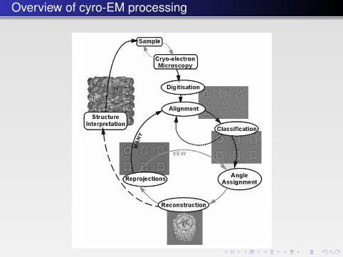
Overview of cyro-EM processing
van Heel et al, Q. Rev. Biophys. 33, 307 (2000)

Particle picking
Particle detection is the first thing to do in the single particle reconstruction as soon asthe Cryo-EM images are digitized and/or CTF-corrected. The goal of this step is tolocate all particles. Since achieving high-resolution reconstruction often requires overhundreds of thousands of particles, it is important to design a fast and automaticalgorithm for particle detection. We have developed two methods for this task. One isbased on data clustering and the other is based on Voronoi diagram and distancetransform. See http://cvcweb.ices.utexas.edu/cvc/projects/angstrom/bm/index.php

Automated particle picking

Projecting the image
The CT/MRI reconstruction is based on a theorem called Fourier Slice Theorem,which relates the Fourier transform of any projection of the original structure to theFourier transform of the original structure. Fig.2(a) shows an example of a 2Dstructure reconstruction from its 1D projections. With enough number of projections,we can compute their Fourier transforms, each of which is "embedded" in the Fourierspace and then an interpolation technique is used to "reconstruct" the Fouriertransform of the original structure f(x, y) in the entire Fourier space. After we obtainF(u, v), the inverse Fourier transform of F(u, v) gives the original structure f(x,y). Fig.2(b) illustrates the overall pipeline of reconstruction. It is straightforward to extend thisprocedure into 3D structure reconstruction.

More on projections

Image enchancement
The images obtained from the cryo-electron microscopy are usually verynoisy and have very low contrast. It is necessary to smooth the noise as wellas enhance the contrast. This also happens right after the 3D electrondensity maps are reconstructed. The following pictures show an example ofour approaches on the reconstructed electron density map of Rice DwarfVirus (RDV). The Cryo-EM images or the reconstructed maps may also beimproved by Contrast Transfer Function (CTF) correction.

Image enchancement

Moving towards atomic resolution
Although atomic details are not detectable in reconstructed 3D cryo-EM maps, giventheir low feature resolution, it is sometimes feasible to locate secondary structures(alpha helices and beta sheets). An approach for detecting alpha helices in 3D mapsis where the alpha helix is modelled with a cylinder (length and thickness) and thecylinder is correlated with the segmented protein map. Since the best solution isachieved by exhaustively searching in translation space (3D) and orientation space(2D), this method is computationally expensive. A related exhaustive searchapproach, designed for beta sheet detection uses a disk (planar) model for betasheets.