Embed Size (px)
Citation preview
1
Course : PGPathshala-Biophysics
Paper 10 : Techniques used in Molecular Biophysics II
Module 09 : DNA Replication Models, Mechanisms
Content Writer: Dr. Jayanth Kumar, AIIMS, NEW DELHI
DNA is the genetic material of eukaryotes and some prokaryotes including bacteria. Bacterial
DNA is double stranded and usually free of other proteins. On the contrary, eukaryotic DNA
is wound into complicated secondary and tertiary structures. It is also wound around histone
proteins which restrict or allow access to the DNA for transcription adding another layer of
complexity to gene expression regulation.
DNA structure:
The presence of nucleic acids in the nuclei of pus cells was demonstrated by Friedrich
Miescher in 1869. He named the acidic substance as 'nuclein' which referred to a mixture of
nucleic acids and protein. DNA was established as the genetic material by the Hershey-Chase
and the Mcleod-Avery-Macarty experiments. Subsequently, the first x-ray diffraction
experiments of DNA were carried out by Rosalind Franklin and Maurice Wilkins (1950)
following which the double helical structure of DNA was elucidated by James Watson and
Francis Crick (1953).
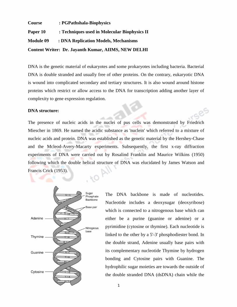
The DNA backbone is made of nucleotides.
Nucleotide includes a deoxysugar (deoxyribose)
which is connected to a nitrogenous base which can
either be a purine (guanine or adenine) or a
pyrimidine (cytosine or thymine). Each nucleotide is
linked to the other by a 5'-3' phosphodiester bond. In
the double strand, Adenine usually base pairs with
its complementary nucleotide Thymine by hydrogen
bonding and Cytosine pairs with Guanine. The
hydrophilic sugar moieties are towards the outside of
the double stranded DNA (dsDNA) chain while the
2
hydrophobic bases are stacked in the centre. This is called base stacking interactions. DNA
has an absorption maximum at 260 nm. Due to the base stacking interactions, the absorbance
of dsDNA is much lower than that of the corresponding single stranded DNA (ssDNA). This
is called the hypochromic effect.
DNA Replication:
DNA replication is the biological process of producing two identical copies of DNA from one
original molecule. Replication is the basis for biological inheritance. Since DNA is made up
of a double helix of two complementary strands during replication, these strands are
separated. Each strand of the original DNA molecule then serves as a template for the
production of a daughter strand.
Models of DNA Replication:
The Watson and Crick DNA model implies an elegant mechanism for replication:
a. Unwind the DNA molecule.
b. Separate the two strands.
c. Make a complementary copy for each strand.
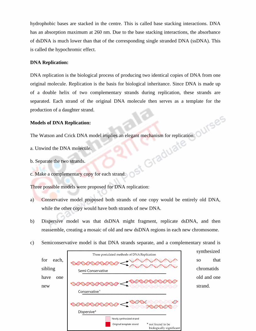
Three possible models were proposed for DNA replication:
a) Conservative model proposed both strands of one copy would be entirely old DNA,
while the other copy would have both strands of new DNA.
b) Dispersive model was that dsDNA might fragment, replicate dsDNA, and then
reassemble, creating a mosaic of old and new dsDNA regions in each new chromosome.
c) Semiconservative model is that DNA strands separate, and a complementary strand is
synthesized
for each, so that
sibling chromatids
have one old and one
new strand.

3
The correct model of DNA replication was established by the Messelson and Stahl
experiment. The experiment made use of heavy Nitrogen (15N) and light Nitrogen (14N) to
label DNA. E.Coli were grown in 15N containing media for several generations and then
shifted to 14N containing media. When DNA is extracted from these cells and centrifuged on a
salt density gradient, the DNA separates out at the point at which its density equals that of the
salt solution. In the first generation, DNA contained a mixture of 15N and 14N which proved
that DNA replication is semiconservative.
DNA replication in prokaryotes

4
In general, DNA is replicated by: a) uncoiling of the helix b) strand separation by breaking of
the hydrogen bonds between the complementary strands and c) synthesis of two new strands
by complementary base pairing. Replication begins at a specific site in the DNA called the
origin of replication (ori).
DNA replication is bidirectional from the origin of replication. DNA replication occurs in
both directions from the origin of replication. To begin DNA replication, unwinding enzymes
called DNA helicases cause the two parent DNA strands to unwind and separate from one
another at the origin of replication to form two "Y"-shaped replication forks. Helix
destabilizing proteins (Single strand binding proteins: SSBs) bind to the single-stranded
regions so the two strands do not rejoin.
Coiling of the dsDNA during the unwinding of the strands at the replication fork leads to
formation of supercoils. Enzymes called topoisomerases produce breaks in the DNA and then
rejoin them in order to relieve the stress produced by supercoiling. There are two type of
topoisomerases: Type I which create a nick in a single strand of DNA and relieve stress, Type
II when a double strand break is created, stress is released followed by resealing of the nick.
As the strands continue to unwind in both directions around the entire DNA molecule, new
complementary strands are produced by the hydrogen bonding of free DNA nucleotides with
those on each parent strand. As the new nucleotides line up opposite each parent strand by

5
hydrogen bonding, enzymes called DNA polymerases join the nucleotides by way of
phosphodiester bonds. The nucleotides lining up by complementary base pairing are
deoxynucleoside triphosphates (dNTPs). As the phosphodiester bond forms between the 5'
phosphate group of the new nucleotide and the 3' OH of the last nucleotide in the DNA strand,
two of the phosphates are removed providing energy for bonding.
DNA polymerase
DNA polymerases have the property that they can only add nucleotides at the free 3' end.
Hence DNA polymerases can extend the daughter strands in the 5' to 3' direction. The
template strand is therefore read in the 3' to 5' direction. So the two strands of DNA cannot be
replicated the same way. The two strands are antiparallel: one parent strand - the one running
3' to 5' is called the leading strand can be copied directly down its entire length and the other
parent strand - the one running 5' to 3' is called the lagging strand must be copied
discontinuously in short fragments – Okazaki fragments of around 100-1000 nucleotides each
as the DNA unwinds.
DNA polymerase enzymes cannot begin a new DNA chain from scratch. It can only attach
new nucleotides onto 3' OH group of a nucleotide in a preexisting strand. To start the
synthesis of the leading strand and each DNA fragment of the lagging strand, an RNA
polymerase complex called a primosome or primase is required. The primase is capable of
joining RNA nucleotides without requiring a preexisting strand of nucleic acid - this forms an
RNA primer with a free 3' OH group where DNA polymerase can add nucleotides further.
After a few nucleotides are added, primase is replaced by DNA polymerase. DNA polymerase
can now add nucleotides to the 3' end of the short RNA primer. The primer is later degraded
and filled in with DNA.
There are three DNA polymerases found in bacteria: I, II and III. All of them have 5' to 3'
DNA polymerase activity. DNA polymerase I also has 5'-3' exonuclease activity as well as 3'-
5' exonuclease activity. DNA polymerase I is involved in removal of RNA primers after
replication as well as in proofreading of DNA.
DNA polymerase II is involved in repair of damaged DNA. It has 3' → 5' exonuclease
activity. However there are multiple proofs that this is not the main polymerase involved in
DNA Replication.
6
A. E.Coli Strains lacking the gene show no defect in growth or replication.
B. Synthesis of Pol II is induced during the stationary phase of cell growth - a phase in which
little growth and DNA synthesis occurs.
C. Pol II has a low error rate but it is much too slow to be of any use in normal DNA
synthesis.
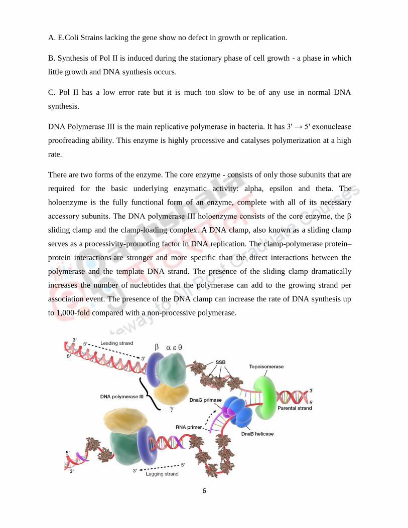
DNA Polymerase III is the main replicative polymerase in bacteria. It has 3' → 5' exonuclease
proofreading ability. This enzyme is highly processive and catalyses polymerization at a high
rate.
There are two forms of the enzyme. The core enzyme - consists of only those subunits that are
required for the basic underlying enzymatic activity: alpha, epsilon and theta. The
holoenzyme is the fully functional form of an enzyme, complete with all of its necessary
accessory subunits. The DNA polymerase III holoenzyme consists of the core enzyme, the β
sliding clamp and the clamp-loading complex. A DNA clamp, also known as a sliding clamp
serves as a processivity-promoting factor in DNA replication. The clamp-polymerase protein–
protein interactions are stronger and more specific than the direct interactions between the
polymerase and the template DNA strand. The presence of the sliding clamp dramatically
increases the number of nucleotides that the polymerase can add to the growing strand per
association event. The presence of the DNA clamp can increase the rate of DNA synthesis up
to 1,000-fold compared with a non-processive polymerase.

7
Initiation of replication
Replication starts when DNA at the origin of replication denatures to expose the bases,
creating a replication fork. Replication is usually bidirectional from the origin. E. coli has one
origin, oriC, which has:
A minimal sequence of about 245 bp required for initiation.
Three copies of a 13-bp AT-rich sequence.
Four copies of a 9-bp sequence.
The following sequence of events occur in E. coli initiating DNA synthesis: Initiator proteins
attach. E. coli’s initiator protein is DnaA (from the dnaA gene). DNA helicase (from dnaB)
binds initiator proteins on the DNA, and denatures the AT-rich region using ATP as an energy
source. DNA primase (from dnaG) binds helicase to form a primosome, which synthesizes a
short (5–10nt) RNA primer. Following the formation of the RNA primer, DNA polymerase III
takes over to polymerize the leading and the lagging strands. At the end of replication, the
RNA primer is replaced by DNA by the activity of DNA polymerase I.
Termination of Replication takes place when the replication fork encounters certain
termination sequences which trap the replication fork from moving back. The two forks
moving from the opposite ends meet and replication is complete. The concatenated DNA
strands are separated by DNA topoisomerase IV, which is a Type II topoisomerase.

8
Fidelity of DNA replication
DNA replication exhibits a high degree of fidelity. Mistakes during the process are extremely
rare. DNA pol III makes only one mistake per 108 bases made. There are several reasons why
fidelity is high:
Instability of mismatched pairs
Configuration of the DNA polymerase active site
Proofreading function of DNA polymerase
Instability of mismatched pairs: Complementary base pairs have much higher stability than
mismatched pairs. This feature only accounts for part of the fidelity: It has an error rate of 1
per 1,000 nucleotides
Configuration of the DNA polymerase active site: DNA polymerase is unlikely to catalyze
bond formation between mismatched pairs. This induced-fit phenomenon decreases the error
rate to a range of 1 in 100,000 to 1 million
Proofreading function of DNA polymerase: DNA polymerases can identify a mismatched
nucleotide and remove it from the daughter strand. The enzyme uses its 3’ to 5’ exonuclease
activity to remove the incorrect nucleotide. It then changes direction and resumes DNA
synthesis in the 5’ to 3’ direction

9
DNA Replication in Eukaryotes:
The basic principle behind DNA replication in eukaryotes is similar to that of prokaryotes.
However there are some key differences. There are multiple origins of replication unlike
prokaryotes which have only one origin of replication. For example, the human genome has
around 30000 origins of replication. Eukaryotes have around 15 DNA polymerases. DNA
polymerase α functions as the primase creating the RNA primer required to initiate
replication. DNA polymerase β is involved in proofreading and DNA repair. DNA
polymerase δ is the main polymerase involved in replication of the leading and the lagging
strands. Eukaryotes have mitochondria which carries its own genetic material. DNA
polymerase γ carries out replication and repair of mitochondrial DNA. Other polymerases are
not well characterized. Not all the DNA polymerases are exonucleases in eukaryotes.
DNA replication in Eukaryotes is synchronized with the progression of the cell cycle. During
the S phase of the cell cycle, DNA synthesis begins. The function of helicase is taken over by
the MCM (Minichromosome maintenance) group of proteins. Proteins like Cdc6 and Cdt
prevent replication from progressing until the cyclin dependent kinases become active and
phosphorylate them thus linking cell cycle changes to DNA replication.

10
In prokaryotes replication proceeds at about 1000 nucleotides per second, and thus is done in
no more than 40 minutes. In Eukaryotes replication takes proceeds at 50 nucleotides per
second, and is completed in 60 minutes.
Telomerase
The RNA primer of the last Okazaki fragment cannot be replaced by DNA because of the
absence of a free 3'-OH group to add nucleotides. This results in an overhang at the ends of
chromosomes. Overhangs are good substrates for exonucleases. To prevent this the cell
utilizes the enzyme telomerase, also called terminal transferase. This is a ribonucleoprotein
that adds a species-dependent telomere repeat sequence to the 3' end of telomeres. A telomere
is a region of repetitive sequences at each end of eukaryotic chromosomes. Telomeres protect
the end of the chromosome from DNA damage or from fusion with neighbouring
chromosomes.
Telomerase is a reverse transcriptase enzyme that carries its own RNA molecule (e.g., with
the sequence "CCCAAUCCC" in vertebrates) which is used as a template when it elongates
telomeres. As more and more repeats are added, DNA polymerase III starts adding more
Okazaki fragments on the complementary strand. Thus the ends of the chromosome are
prevented from damage and loss of genetic material.
Telomerase is highly active in normal stem cells and most cancer cells. It is normally absent
from, or at very low levels in, most somatic cells. This explains the finite lifespan of somatic
cells since they keep losing genetic material from the ends of chromosomes and are not able
to replace them.

11
Summary:
DNA
replication is
the biological
process of
producing two
identical copies
of DNA from one
original
molecule.
DNA
replication is
semiconservative
It is carried out by multiple proteins including DNA polymerase III in prokaryotes
Only one origin of replication is present in prokaryotes and multiple are present in
eukaryotes
DNA replication is possible only in the 5' to 3' direction: Hence two different types of
replication take place in the two strands: Leading strand and lagging strand synthesis
The short fragments formed in the lagging strand: Okazaki fragments
Telomerase is a reverse transcriptase which helps to protect the ends of chromosomes.
DNA replication is highly synchronized to the cell cycle in eukaryotes

12
The β clamp helps in increasing the processivity of DNA polymerase
Termination sequences and DNA topoisomerase help in the termination of replication.