Embed Size (px)
Citation preview

World Journal of Cardiovascular Diseases, 2014, 4, 35-38 WJCD http://dx.doi.org/10.4236/wjcd.2014.42006 Published Online February 2014 (http://www.scirp.org/journal/wjcd/)
OPEN ACCESS
Coronary sinus reentrant tachycardia after atrial fibrillation ablation: From bad to worse*
Pietro Turco
Electrofisiology Laboratory, Multimedica Hospital, Milano, Italia Email: [email protected] Received 29 October 2013; revised 8 December 2013; accepted 17 December 2013 Copyright © 2014 Pietro Turco. This is an open access article distributed under the Creative Commons Attribution License, which permits unrestricted use, distribution, and reproduction in any medium, provided the original work is properly cited. In accordance of the Creative Commons Attribution License all Copyrights © 2014 are reserved for SCIRP and the owner of the intellectual property Pietro Turco. All Copyright © 2014 are guarded by law and by SCIRP as a guardian.
ABSTRACT Herein we present a case of atrial tachycardia as a sequel of AF ablations. A 42-year-old man was ad-mitted to our department because of a very sympto-matic tachycardia. The patient, because of paroxys-mal AF and typical atrial flutter, had been already submitted (three times) to ablation procedures in both left (pulmonary vein insulation) and right atria (cavo-tricuspidal isthmus). During the electrophysio-logical study, a huge and very fast atrial tachycardia was induced: 230 ms cycle length, 1/1 atrio-ventricu- lar conduction with the ventricular rate of 260 bpm, complete left bundle branch block, and clinically recognized by the patient. Four minutes later, a 2/1 AV conduction without branch block permitted map-ping and ablation. A high-density mapping around isthmus and coronary sinus (CS) was performed. The analysis of the chronological activation clearly showed a circuit propagation around the CS ostium with a very slow conduction in the anterior zone enlightened by the tight color progression, and counterclockwise activation of the right atrium lateral wall. In anterior zone of CS ostium diastolic fragmented electrograms were detected. After preventing his position localiza-tion, radiofrequency delivering (35 W) was effective
to interrupt the arrhythmia in 3 seconds. Energy de-livering was continued to anchor the lesion to the in-ferior vena cava. Confirmation of successful ablation was determined by unsuccessful attempts at reinduc-tion of the arrhythmia, in basal state and during infu-sion of isoproterenol. KEYWORDS Atrial Flutter; Atrial Fibrillation; Transcatheter Ablation; Three-Dimensional Mapping
1. INTRODUCTION The increased number of transcatheter ablations in the atria for treatment of atrial fibrillation (AF) contributes to an increased incidence of organized macro reentrant tachycardia [1-4].
Herein we present a case of atrial tachycardia as a se-quel of AF ablations.
2. CASE REPORT A 42-year-old man was admitted to our department be-cause of a very symptomatic tachycardia. The patient, because of paroxysmal AF and typical atrial flutter, had been already submitted (three times) to ablation proce-dures in both left (pulmonary vein insulation) and right atria (cavo-tricuspidal isthmus).
In spite of anti-arrhythmic therapy, the patient, four days before his admission, experienced a new very symp-tomatic (sweating, hypotension and dizziness) tachycar-dia for which was submitted to ablation procedure again.
At the procedure the patient, on pharmacological wash- out, presented a stable sinus rhythm.
By coronary sinus (CS) stimulation, with extra stimu-lus technique, a huge very fast atrial tachycardia was induced: 230 ms cycle length, 1/1 atrio-ventricular con-
*Core Tip. The increased number of transcatheter ablations in the atria for treatment of atrial fibrillation is contributing to an increased inci-dence of organized macro reentrant tachycardia.
Herein we present a case of atrial tachycardia as a sequel of atrial fi-brillation ablations. During EP study a huge very fast, 260 bpm, atrial tachycardia was induced. A high-density mapping (Carto) indicated a reentry circuit around CS. In anterior edge of CS diastolic fragmented electrograms were localized. Radiofrequency delivering was effective to interrupt the arrhythmia. Energy delivering was continued to anchor the lesion to the inferior vena cava. In conclusion, extensive liner le-sions in right and left atria and normal anatomical barriers (tricuspidal annulus, Eustachian ridge and crista terminalis, scars) could contribute to the onset of conduction corridors (man-made maze) that could sus-tain more aggressive arrhythmias.

P. Turco / World Journal of Cardiovascular Diseases 4 (2014) 35-38
Copyright © 2014 SciRes. OPEN ACCESS
36
duction with ventricular rate of 260 bpm, complete left bundle branch block and clinically recognized by the patient (Figure 1). Four minutes later a 2/1 AV conduc-tion without branch block permitted mapping and abla-tion (Figure 2).
A 7.5 F 3.5 mm irrigated-tip roving catheter (Navistar Thermocool, Diamond Bar, CA, USA) inserted by right femoral vein for electroanatomical mapping and ablation was used.
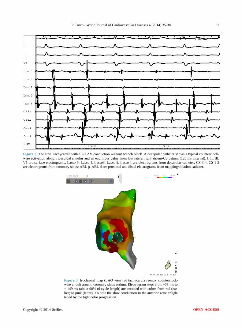
Electroanatomical mapping (CARTO 3, Diamond Bar, CA, USA) commenced in the right atrium because of the CS proximal-distal activation sequence. In order to re-construct the chronology of the entire macroreentry cir-cuit and to identify the mid-diastolic isthmus (MDI), a specific setting of the window of interest was used, as previously described [1]. Chronological activation analy-sis on the entire cycle (230 ms) indicated the MDI (the interface between the red and the purple colors) of typi-cal atrial flutter, but more anterior. A decapolar catheter showed a typical counterclockwise activation along tri-cuspidal annulus and an enormous delay from low lateral right atrium-CS ostium, more than would be expected in case of a slow conduction (low lateral-proximal CS in-terval of 120 ms.). A high-density mapping around isth-mus and CS was performed (Figure 3). The re-analysis of the chronological activation clearly showed a circuit propagation around the CS ostium with a very slow
conduction in the anterior zone enlightened by the tight color progression, and counterclockwise activation of the right atrium lateral wall (Figure 3). In anterior zone of CS ostium diastolic fragmented electrograms were de-tected. Entrainment that could have confirmed that sus-pected path was truly in the circuit, was not performed to avoid atrial fibrillation degeneration. After preventive His position localization, radiofrequency delivering (35 W) was effective to interrupt the arrhythmia in 3 sec. Energy delivering was continued to anchor the lesion to the inferior vena cava. Confirmation of successful abla-tion was determined by unsuccessful attempts at reinduc-tion of the arrhythmia, in basal state and during infusion of isoproterenol.
3. DISCUSSION In the era of transcatheter ablation procedures in the right and left atria to treat AF, incidence of organized ma-cro-reentrant tachycardia, as a sequel of extensive linear ablation, is 5% - 30% [2-4]. In Chugh et al. series on atrial flutters that arose after left atrial circumferential ablation of AF, in approximately 25% of cases they used the musculature of the CS [2]. Furthermore, Chae et al. reported a series of 21 micro-reentrant atrial tachycardias that occurred after circumferential pulmonary vein abla-tion. They indicated that the most common sites of origin
Figure 1. Induction of the huge very fast atrial tachycardia, 230 ms cycle length with 1/1 atrio-ventricular conduction (ventricular rate of 260 bpm ) and complete left bundle branch block. I, II, III, aVR, aVL, aVF, V1,V2, V3, V4, V5, V6 are surface electrograms.
P. Turco / World Journal of Cardiovascular Diseases 4 (2014) 35-38
Copyright © 2014 SciRes. OPEN ACCESS
37
Figure 2. The atrial tachycardia with a 2/1 AV conduction without branch block. A decapolar catheter shows a typical counterclock-wise activation along tricuspidal annulus and an enormous delay from low lateral right atrium-CS ostium (120 ms interval). I, II, III, V1 are surface electrograms; Lasso 5, Lasso 4, Lasso3, Lasso 2, Lasso 1 are electrograms from decapolar catheter; CS 3-4, CS 1-2 are electrograms from coronary sinus; ABL p, ABL d are proximal and distal electrograms from mapping/ablation catheter.
Figure 3. Isochronal map (LAO view) of tachycardia reentry counterclock-wise circuit around coronary sinus ostium. Electrogram steps from −53 ms to + 149 ms (about 90% of cycle length) are encoded with colors from red (ear-lier) to pink (latter). To note the slow conduction in the anterior zone enligh-tened by the tight color progression.
P. Turco / World Journal of Cardiovascular Diseases 4 (2014) 35-38
Copyright © 2014 SciRes. OPEN ACCESS
38
were the CS, the antrium of a pulmonary vein, and the anterior wall of the left atrium or base of the left atrial appendage [3].
Morady et al. reported that 75% of atrial tachycardias occurred after wide-area radiofrequency ablation to iso-late the pulmonary veins is caused by macroreentry also around CS [4].
Extensive liner lesions in right (isthmus, intercaval, superior vena cava) and left atria (circumferential pul-monary vein isolation, roof, anterior and posterior left isthmus) and normal anatomical barriers (tricuspidal an-nulus, Eustachian ridge and crista terminalis, scars) could contribute to the onset of conduction corridors: a man- made maze. In our case the stimulus goes slowly around CS ostium, especially in anterior zone, and spreads trough the right atrium corridors with a fast repetitive sequence with a counterclockwise activation of the lateral wall.
The result of extensive linear ablations is the onset of more organized tachyarrhythmias, often with rapid ven-tricular rates, persistent and difficult to suppress with rhythm control medications: from bad to worse.
4. CONCLUSIONS In conclusion, our experience suggests that in case of extensive ablation lines for persistent atrial fibrillation or redo of paroxysmal atrial fibrillation, the highest thera-peutic effectiveness is obtained by mapping and ablating all remaining circuits. At the last step, for the remaining circuit underlying a tachycardia with a more or less con-stant cycle length, electroanatomical mapping and abla-tion are mandatory.
This behaviour could prevent the arising of new tach-yarrhythmias (man-made) which in the majority of cases is more symptomatic than atrial fibrillation.
REFERENCES [1] De Ponti, R., Verlato, R., Bertaglia, E., Del Greco, M.,
Fusco, A., Bottoni, N., Drago, F., Sciarra, L., Ometto, R., Mantovan, R. and Salerno-Uriarte, J.A. (2007) Treatment of macro-re-entrant atrial tachycardia based on electroa-natomic mapping: Identification and ablation of the mid- diastolic isthmus. Europace, 9, 449-457. http://dx.doi.org/10.1093/europace/eum055
[2] Chugh, A., Oral, H., Good, E., Han, J., Tamirisa, K., Lemola, K., Elmouchi, D., Tschopp, D., Reich, S., Igic, P., Bogun, F., Pelosi Jr., F. and Morady, F. (2005) Cathe-ter ablation of atypical atrial flutter and atrial tachycardia within the coronary sinus after left atrial ablation for atri-al fibrillation. Journal of the American College of Cardi-ology, 46, 83-91. http://dx.doi.org/10.1016/j.jacc.2005.03.053
[3] Chae, S., Oral, H., Good, E., Dey, S., Wimmer, A., Crawford, T., Wells, D., Sarrazin, J.F., Chalfoun, N., Ku- hne, M., Fortino, J., Huether, E., Lemerand, T., Pelosi, F., Bogun, F., Morady, F., Chugh, A., et al. (2007) Atrial ta- chycardia after circumferential pulmonary vein ablation of atrial fibrillation: Mechanistic insights, results of ca-theter ablation, and risk factors for recurrence. Journal of the American College of Cardiology, 50, 1781-1787. http://dx.doi.org/10.1016/j.jacc.2007.07.044
[4] Morady, F., Oral H. and Chugh, A. (2009) Diagnosis and ablation of atypical atrial tachycardia and flutter compli-cating atrial fibrillation ablation. Heart Rhythm, 6, S29- S32. http://dx.doi.org/10.1016/j.hrthm.2009.02.011


![Sinus tachycardia: Evaluation and management...reentrant tachycardia is a reentrant arrhythmia that is paroxysmal with a discrete onset and offset (unlike sinus tachycardia) [1]. Distinguishing](https://img.dokumen.tips/doc/110x75/5e522e5c6d98f111335a4f1d/sinus-tachycardia-evaluation-and-management-reentrant-tachycardia-is-a-reentrant.jpg)