Embed Size (px)
Citation preview

Conversion of Vitamin B6 Compoundsto Active Forms in the Red Blood Cell
BARBARAB. ANDERSON,CATHERINEE. FULFORD-JONES, J. ANTHoNYCHILD,MICHAEL E. J. BEARD, and CHRISTOPHERJ. T. BATEMANFrom the Department of Haematology, St. Bartholomew's Hospital,London, England
A B S T R A C T In studies with pyridoxine and other B6compounds in blood, the active forms pyridoxal and py-ridoxal phosphate were measured by differential assaysusing Lactobacillus casei. Red cell uptake of tritiatedpyridoxine was also measured. A new metabolic pathwayfor conversion of pyridoxine to active forms was dem-onstrated in red cells.
In vivo studies in normal subjects suggested that pyri-doxine was taken up by red cells where it was convertedto pyridoxal phosphate and then pyridoxal, followed bygradual release of a proportion of pyridoxal into plasma.In vitro incubation of pyridoxine with blood confirmedthis observation.
Increasing amounts of pyridoxine were taken up andconverted as the amount added to blood was increased,and only very small numbers of red cells were needed toconvert appreciable amounts. Conversion was markedlyinhibited at temperatures lower than 370C, and stoppedaltogether at - 20'C.
Release of pyridoxal into plasma was always directlyproportional to the amount of pyridoxal formed and tothe volume of plasma present. That pyridoxal phosphatewas not released into plasma was demonstrated in storedblood, for pyridoxine was converted mainly only as faras pyridoxal phosphate, probably due to inactivation ofthe phosphatase. Pyridoxal phosphate remained in thered cells.
Pyridoxine was converted when incubated with washedred cells in saline or phosphate buffer suspension(0.08 M). In saline suspension, pyridoxal formed butwas not released in the absence of plasma. In phosphatebuffer suspension, pyridoxal phosphate was formed butwas not changed to pyridoxal, probably due to inac-tivation of phosphatase by excess phosphate.
This work was presented in part at the 8th InternationalCongress of Haematology, Munich 1970.
Received for publication 30 November 1970 and in revisedform 25 February 1971.
Pyridoxamine was converted to active forms in redcells less efficiently. Pyridoxal entered red cells rapidly,equilibrating between plasma and cells within 1 min inthe same ratio as pyridoxal formed inside red cells.Pyridoxal phosphate did not enter red cells in wholeblood but did so readily in washed cells in saline.
INTRODUCTIONThere are three forms of vitamin B6; pyridoxine (thealcohol), pyridoxamine (the amine), and pyridoxal (thealdehyde). These can also exist in the phosphorylatedform. Pyridoxal phosphate is known to be the mainactive coenzyme form and it acts as such in many en-zyme systems in the body.
Pyridoxamine and pyridoxal predominate in meat andother animal products, but in fruit, vegetables, andcereals pyridoxine is the main form (1-4). It is ap-parent that pyridoxine must be an important form of Befor many individuals and it has also been most com-monly used for therapy. The mode and site of conversionof pyridoxine to forms active in the body is thereforeof considerable biological and clinical interest.
Exactly how and where the conversion of pyridoxineand other B6 compounds takes place in the body has notbeen fully established. The pathway usually postulatedfor pyridoxine is by conversion first to pyridoxine phos-phate and then to pyridoxal phosphate, the conversionsbeing activated by the appropriate enzymes, a kinase andoxidase respectively. This reaction has been demonstratedin vitro in liver homogenates (5), but whether this isthe essential or sole site of conversion is doubtful.
We measured the appearance of pyridoxal and pyri-doxal phosphate in blood at varying time intervals af-ter oral administration of pyridoxine. The microbiologi-cal assay method with Lactobacillus casei was used (6).We found that there was a rapid conversion of pyri-doxine to these forms, and this led us to carry out in
The Journal of Clinical Investigation Volume 50 1971 1901
vitro studies in which pyridoxine and other forms ofB. were incubated with blood under varying conditions.The red cell was found to play an important role.
METHODS
All blood samples were taken from healthy members of thestaff and were heparinized (125 IU lithium heparin per 10ml). Blood and Be compounds were protected from lightthroughout (6).
The unlabeled Be compounds were obtained from SigmaChemical Co. (98-99% crystalline) except pyridoxine-5'-phosphate which was obtained from Mann Research Labs,Inc. Tritium-labeled pyridoxine (Pyridoxine-T(G) Hydro-chloride) was obtained from the Radiochemical Centre, Am-ersham, England.
Experimental procedureIn vivo studies. A 3-ml blood sample was taken from a
subject who had fasted overnight. An oral dose of 50 mgof pyridoxine HCO was then given with water and nofood was taken for 3 hr. 3-ml blood samples taken at in-tervals from 10 min up to 24 hr afterwards, were plungedimmediately into ice and prepared for storage as describedbelow.
In vitro studies. In the form of pyridoxine HCl, therequired amount of pyridoxine (50-2000 ng) in 0.01 mlof 0.15 M NaCl was added per ml of blood. The same con-ditions applied to other forms of B&. Tritium-labeled pyri-doxine (pyridoxine-8H) was added for measurement of redcell uptake and the specific activity was always adjustedso that 0.005 ACi was added per ml of blood.
Whole blood (or red cell suspension), and apparatuswere equilibrated to 370C in a warm room. In every ex-periment a base line sample was kept for assay. B. wasadded to the required amount of blood within 2 hr of beingdrawn, then 3-ml portions were put in stoppered conicalflasks and were incubated in a shaking waterbath at 370Cat 70 strokes per minute. The portions were removed atintervals up to 3 hr, plunged immediately into ice, and pre-pared for storage as described below.
Subsequent treatment and storage of bloodTo stop enzyme activity at the end of each time interval,
about 0.3 ml of whole blood (or red cell suspension) wasimmediately deep frozen to - 200C. Plasma (or suspendingfluid) was separated immediately from the remaining bloodat 4'C and deep frozen to - 200C.
For assay of red cells, plasma from 1 ml of blood wasremoved immediately at 4VC, cells were washed three timesin cold 0.15 M NaCI, the volume was made up to 1 ml withcold 0.15 M NaCl, and cells were deep frozen to - 20'C.
Measurement of B6 compoundsMeasurement of pyridoxal and pyridoxal phosphate. The
assay organism Lactobacillus casei NCIB 8010 (ATCC7469) was used as described for the assay of serum byAnderson, Peart, Fulford-Jones (6). L. casei measuresonly pyridoxal directly and responds to 0.01 ng per ml. Tomeasure pyridoxal phosphate it must first be hydrolyzed topyridoxal. Neither pyridoxine nor any of the other knownforms of B. can be measured at the dilutions used, whetherhydrolyzed or not.
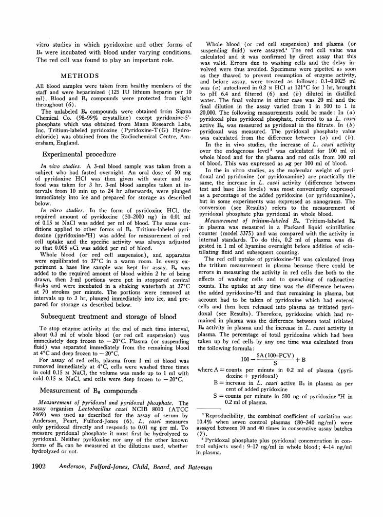
Whole blood (or red cell suspension) and plasma (orsuspending fluid) were assayed.' The red cell value wascalculated and it was confirmed by direct assay that thiswas valid. Errors due to washing cells and the delay in-volved were thus avoided. Specimens were pipetted as soonas they thawed to prevent resumption of enzyme activity,and before assay, were treated as follows: 0.1-0.0025 mlwas (a) autoclaved in 0.2 N HCl at 121'C for 1 hr, broughtto pH 6.4 and filtered (6) and (b) diluted in distilledwater. The final volume in either case was 20 ml and thefinal dilution in the assay varied from 1 in 500 to 1 in20,000. The following measurements could be made: In (a)pyridoxal plus pyridoxal phosphate, referred to as L. caseiactive B6, was measured as pyridoxal in the filtrate. In (b)pyridoxal was measured. The pyridoxal phosphate valuewas calculated from the difference between (a) and (b).
In the in vivo studies, the increase of L. casei activityover the endogenous level 2 was calculated for 100 ml ofwhole blood and for the plasma and red cells from 100 mlof blood. This was expressed as /Ag per 100 ml of blood.
In the in vitro studies, as the molecular weight of pyri-doxal and pyridoxine (or pyridoxamine) are practically thesame, the increase in L. casei activity (difference betweentest and base line levels) was most conveniently expressedas a percentage of the added pyridoxine (or pyridoxamine),but in some experiments was expressed as nanograms. Theconversion (see Results) refers to the measurement ofpyridoxal phosphate plus pyridoxal in whole blood.
Measurement of tritium-labeled B,. Tritium-labeled B.in plasma was measured in a Packard liquid scintillationcounter (model 3375) and was compared with the activity ininternal standards. To do this, 0.2 ml of plasma was di-gested in 1 ml of hyamine overnight before addition of scin-tillating fluid and subsequent counting.
The red cell uptake of pyridoxine-2H was calculated fromthe tritium measurement in plasma because there could beerrors in measuring the activity in red cells due both to theeffects of washing cells and to quenching of radioactivecounts. The uptake at any time was the difference betweenthe added pyridoxine-2H and that remaining in plasma, butaccount had to be taken of pyridoxine which had enteredcells and then been released into plasma as tritiated pyri-doxal (see Results). Therefore, pyridoxine which had re-mained in plasma was the difference between total tritiatedB. activity in plasma and the increase in L. casei activity inplasma. The percentage of total pyridoxine which had beentaken up by red cells by any one time was calculated fromthe following formula:
100 _ 5A(100-PCV) + B
where A = counts per minute in 0.2 ml of plasma (pyri-doxine + pyridoxal)
B = increase in L. casei active Be in plasma as percent of added pyridoxine
S = counts per minute in 500 ng of pyridoxine-'H in0.2 ml of plasma.
'Reproducibility, the combined coefficient of variation was10.4% when seven control plasmas (80-340 ng/ml) wereassayed between 10 and 40 times in consecutive assay batches(7).
2 Pyridoxal phosphate plus pyridoxal concentration in con-trol subjects used: 9-17 ng/ml in whole blood; 4-14 ng/mlin plasma.
1902 Anderson, Fulford-Jones, Child, Beard, and Bateman
RESULTSB
In vivo studiesORALINGESTION OF 50 MGOF PYRIDOXINE HCLAppearance of L. casei active B6 in blood. Increased
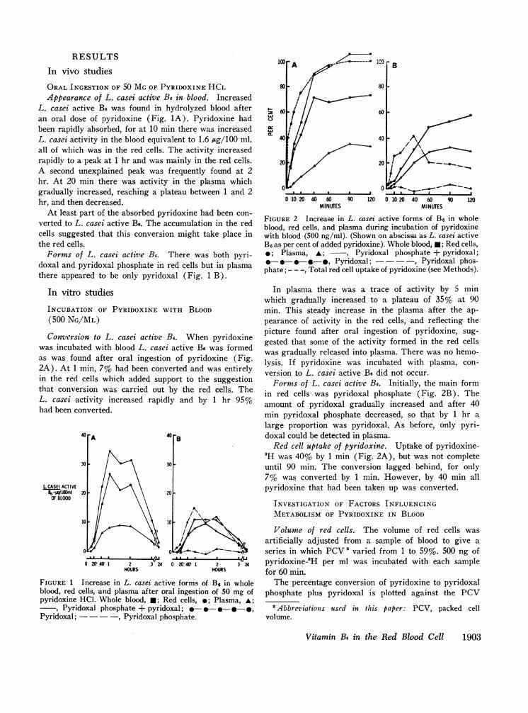
L. casei active B8 was found in hydrolyzed blood afteran oral dose of pyridoxine (Fig. 1A). Pyridoxine hadbeen rapidly absorbed, for at 10 min there was increasedL. casei activity in the blood equivalent to 1.6 Ag/100 ml,all of which was in the red cells. The activity increasedrapidly to a peak at 1 hr and was mainly in the red cells.A second unexplained peak was frequently found at 2hr. At 20 min there was activity in the plasma whichgradually increased, reaching a plateau between 1 and 2hr, and then decreased.
At least part of the absorbed pyridoxine had been con-verted to L. casei active Be. The accumulation in the redcells suggested that this conversion might take place inthe red cells.
Forms of L. casei active B6. There was both pyri-doxal and pyridoxal phosphate in red cells but in plasmathere appeared to be only pyridoxal (Fig. 1 B).
In vitro studies
INCUBATION OF PYRIDOXINE WITH BLOOD(500 NG/ML)Conversion to L. casei active B6. When pyridoxine
was incubated with blood L. casei active Be was formedas was found after oral ingestion of pyridoxine (Fig.2A). At 1 min, 7% had been converted and was entirelyin the red cells which added support to the suggestionthat conversion was carried out by the red cells. TheL. casei activity increased rapidly and by 1 hr 95%had been converted.
40 -A 40 B
30-30
LCASEI ACTIVEB6-j#1i 'mI 20 - 20 -
OF BLOOD
10 10
0 0
0 20 401 2 3 24 0 20' 40' 1 2 3 24HOURS HOURS
FIGURE 1 Increase in L. casei active forms of B6 in wholeblood, red cells, and plasma after oral ingestion of 50 mg ofpyridoxine HCL. Whole blood, *; Red cells, *; Plasma, A;
, Pyridoxal phosphate + pyridoxal; - -9 -,Pyridoxal; -, Pyridoxal phosphate.
~60 i 60
40' 40 -
20 20 -"
FIuE MNTS10 00 1010201001 04 0 9 2
FIUE2Increase in L. casei active forms of B3, in wholeblood, red cells, and plasma during incubation of pyridoxinewith blood (500 ng/ml). (Shown on abscissa as L. casei activeB, as per cent of added pyridoxine). Whole blood, 0; Red cells,
0;Plasma, A; ~, Pyridoxal phosphate + pyridoxal;0-0-e-.-., Pyridoxal; - ,Pyridoxal phos-
phate; - - -, Total red cell uptake of pyridoxine (see Methods).
In plasma there was a trace of activity by 5 minwhich gradually increased to a plateau of 35% at 90min. This steady increase in the plasma after the ap-pearance of activity in the red cells, and reflecting thepicture found after oral ingestion of pyridoxine, sug-gested that some of the activity formed in the red cellswas gradually released into plasma. There was no hemo-lysis. If pyridoxine was incubated with plasma, con-version to L. casei active Be did not occur.
Forms of L. casei active B6. Initially, the main formin red cells was pyridoxal phosphate (Fig. 2B). Theamount of pyridoxal gradually increased and after 40min pyridoxal phosphate decreased, so that by 1 hr a
large proportion was pyridoxal. As before, only pyri-doxal could be detected in plasma.
Red cell uptake of pyridoxine. Uptake of pyridoxine-'H was 40% by 1 min (Fig. 2A), but was not completeuntil 90 min. The conversion lagged behind, for only7% was converted by 1 min. However, by 40 min allpyridoxine that had been taken up was converted.
INVESTIGATION OF FACTORS INFLUENCINGMETABOLISM OF PYRIDOXINE IN BLOOD
Volume of red cells. The volume of red cells was
artificially adjusted from a sample of blood to give aseries in which PCV3 varied from 1 to 59%. 500 ng ofpyridoxine-3H per ml was incubated with each samplefor 60 min.
The percentage conversion of pyridoxine to pyridoxalphosphate plus pyridoxal is plotted against the PCV
' Abbreviationis used in this paper: PCV, packed cellvolume.
Vitamin B6 in the Red Blood Cell 1903

100
80
LU
f.)LAJa_
0 10 20 30 40 50 60PACKEDCELL VOLUME-PERCENT
FIGURE 3 Conversion to L. casei active B6 (pyridoxal phos-phate plus pyridoxal) after incubation of pyridoxine (500ng/ml) for 60 min with samples of the same blood adjusted tocontain increasing volumes of red cells. (Shown on abscissa astotal L. casei active B6 as per cent of added pyridoxine).
(Fig. 3). There was a significant conversion of 6%when the PCV was only 1% and as much as 37%(175 ng/ml) when the PCV was 7%. The conversionincreased up to a PCV of 43% but for a PCV of 59%there was a slight inhibition. The red cell uptake did notexceed the conversion and appeared to be the limitingfactor.
Amount of pyridoxine added. Amounts of 50-2000ng of pyridoxine-3H were incubated per ml of blood fortimes varying from 5 min to 2 hr, and the red celluptake of pyridoxine and conversion to pyridoxal phos-phate plus pyridoxal were measured (Fig. 4).
UPTAKE: As the amount of added pyridoxine was in-creased larger amounts were taken up by the cells. Theproportion in relation to the added amounts only de-
iamr 5 MINUTES
1400
1200
1000
B6ngfml
OF BLOOD800I.
6001
4001
200
zX6
c a
0 250500 1000
20 MINUIES
//'{/s/
. .
/
/
creased slightly. A large proportion entered the cellswithin the first 5 min, but a slower uptake followed.
CONVERSION: At first, the conversion lagged behindthe uptake as shown previously, but by 2 hr even themain part of that taken up from 2000 ng was converted.
Pyridoxal formation and plasma volume. The ap-pearance of pyridoxal in plasma was related to at leasttwo factors:
1. AMOUNTOF PYRIDOXAL FORMED IN BLOOD: In-creasing amounts of pyridoxine (50-2000 ng/ml) wereincubated with samples of the same blood for timesvarying from 20 min to 2 hr. The amount of pyri-doxal released into plasma bears a direct linear relation-ship to the amount of pyridoxal formed in blood (Fig.5), and not to the total conversion to pyridoxal phos-phate plus pyridoxal. The mean percentage of pyridoxalwhich appeared in the plasma was 33% of the totalpyridoxal formed in blood.
2. THE AMOUNT OF PLASMA PRESENT: Increasingamounts of plasma removed from samples of the sameblood were replaced by equal volumes of 0.15 M NaCl.Thus, the amount of red cells and total volume wereconstant while the amount of plasma present in eachsample decreased. 500 ng of pyridoxine per ml was incu-bated with each sample and approximately the sameamount was converted to pyridoxal in each. There isa direct linear relationship between the amount of py-ridoxal appearing in the plasma and the amount ofplasma present (Fig. 6).
In addition, plasma was removed from whole bloodafter incubation of 500 ng of pyridoxine per ml for 90min, in which circumstances there was maximum releaseof pyridoxal into plasma (see Fig. 2A). Fresh plasma
60MI NUTES 2OMINUFES
~~ ~~~~~~~~~~~~~~~~~~~~~~~~~~~~~~~~~~~~~I
fI
I7
FIGURE 4 Red cell uptake of different amounts of pyridoxine-3Hincubated with blood for varying lengths of time, and the conversionto L. casei active B6. ---, Total red cell uptake of pyridoxine;
-*A, Pyridoxal phosphate plus pyridoxal in whole blood.
1904 Anderson, Fulford-Jones, Child, Beard, and Bateman
2000 250 500 1000 2000 25050 1000 2000 -ADDED PYRIDOXINE ng/ml OF BLOOD

60
40PLASMA
PYRIDOXALng/ml
BLOOD
1501
TOTALPLASMA
PYRIDOXAL(rig)
50
0 400 800 1200TOTAL CONVERTEDPYRIDOXALng/ml OF BLOOD
FIGURE 5 Relationship of amount of pyridoxal passing intoplasma with amount of pyridoxal formed after incubation ofdifferent amounts of pyridoxine (50, 100, 250, 500, 1000, and2000 ng/ml) with blood for varying lengths of time. Incubatedfor 20 min A; 60 min A; 120 min .
was added and after a further incubation for 60 minan amount of pyridoxal relative to that in the red cellswas again released into the plasma. On the other hand,when pyridoxine was incubated with washed cells sus-pended in 0.15 M NaCl there was practically no releaseof pyridoxal into saline, in spite of the fact that a majorproportion was converted to pyridoxal.
Temperature. 500 ng of pyridoxine-3H per ml ofblood was incubated at temperatures of 37, 22, and 4°Cfor times up to 60 min. The uptake of pyridoxine intocells, the conversion to pyridoxal phosphate plus pridoxaland the release of pyridoxal into plasma were all de-creased by lowering the temperature of incubation. At4°C there was very marked inhibition with practicallyno release into plasma.
However, conversion of pyridoxine continued slowlyat 4°C and 73% had been converted in 24 hr. Therewas no conversion at - 200 C.
Storage of blood. Portions of blood were kept forperiods up to 7 days, some on the bench at room tem-perature (23°C) and others at 4°C, and conversion topyridoxal phosphate and pyridoxal was measured whenthe stored blood was incubated with pyridoxine (500ng/ml) for 60 min at 37°C (Fig. 7).
CONVERSION: The total conversion to L. casei activeB, remained normal in blood which had been kept at40C, and up to 48 hr at 230C. The longer that bloodhad been stored up to 48 hr the greater was the pro-portion of pyridoxal phosphate in the cells after incuba-tion with pyridoxine, with a consequent reduction inpyridoxal. This was very marked in blood stored at230C.
RELEASE: The amount of pyridoxal appearing inplasma after conversion of pyridoxine decreased in bloodthat had been kept longer than 5 hr. The decrease wasrapid in blood stored at 23°C and generally was in-
A
0 10 20 30 40 50 60 70AMOUNTOF PLASMAPRESENT
AS %OF TOTALVOLUME
FIGURE 6 Relationship of amount of pyridoxal passing intoplasma with amount of plasma present, after incubation ofpyridoxine (500 ng/ml) for 60 min with samples of the sameblood adjusted to contain increasing amounts of plasma. (Redcell volume constant and total volume kept constant with0.15 M NaCl).
directly proportional to the accumulation of pyridoxalphosphate in the red cells and paralleled the decreasingamounts of pyridoxal. This observation suggests thatthe phosphate form of pyridoxal does not pass out of thecell.
Washed red cells, intact and hemolyzed. 500 ng ofpyridoxine per ml was incubated for 60 min with (a)whole blood and (b) twice washed red cells suspendedeither in 0.15 M NaCl or in phosphate buffer in 0.15 MNaCl (pH 7.4) of varying molarity (0.01 M to 0.08 M),the concentration of cells being the same as that ofwhole blood. Conversion to L. casei active forms is illus-trated in Fig. 8. The total conversion was approxi-mately the same throughout, but practically no pyri-doxal passed into the suspending fluid. The proportion of
1001 STOREDAT 23C
80
z 60
0:. 40
20
100 r
80
60
40
20
STOREDAT 4°C
-I0 1 2 3 4 5 6 7 0 1 2 3 4 5 6 7
LENGTHOF STORAGE(DAYS)
FIGURE 7 Effect of storage of blood before incubation withpyridoxine (500 ng/ml) on conversion to L. casei active formsof B6 and release into plasma after incubation for 60 min.(Shown on abscissa as L. casei active B6 as per cent of addedpyridoxine). Whole blood, *; Red cells, *; Plasma, A; I
Pyridoxal phosphate + pyridoxal; --* , Py-ridoxal; - Pyridoxal phosphate.
Vitamin B6 in the Red Blood Cell 1905

100
za:)
0-
FIGURE 8 Comparison of conversion of pyridoxine (500ng/ml) to L. casei active forms of B6 after incubation for 60min with (a) whole blood, and (b) washed red cells suspendedin either 0.15 MNaCl or increasing concentrations of phosphatebuffer in 0.15 M NaCl. (Shown on abscissa as L. casei activeB6 as per cent of added pyridoxine). LR Pyridoxal phosphate+ pyridoxal; shaded area represents pyridoxal phosphate.
pyridoxal phosphate was 3% in whole blood, 19% inthe saline suspension, and increased progressively in thephosphate buffer suspensions to 100% at 0.08 M.
500 ng of pyridoxine-3H per ml was incubated forvarying times with (a) whole blood (b) twice washedred cells in 0.15 M NaCl, either intact or hemolyzed ineach case. Cells were hemolyzed by ultrasonic disinte-gration for 3 min. The conversion to pyridoxal phos-phate plus pyridoxal and in addition the uptake of pyri-doxine by intact cells were compared (Fig. 9), andboth were approximately the same in saline suspensionas in whole blood.
In hemolyzed cells conversion was lower than inintact cells, being markedly lower in hemolyzed wholeblood. In the latter, the form was entirely pyridoxalphosphate, and there was a large proportion in the hemo-lyzed washed cells.
INCUBATION OF DIFFERENT FORMSOF Be WITH BLOOD
Pyridoxamine. The actual uptake of pyridoxamineinto the cells could not be studied by our methods. When500 ng was incubated per ml of blood for varying times,an increase in pyridoxal phosphate and pyridoxal wasmeasured in the red cells, but only 50% had been con-verted at 2 hr.
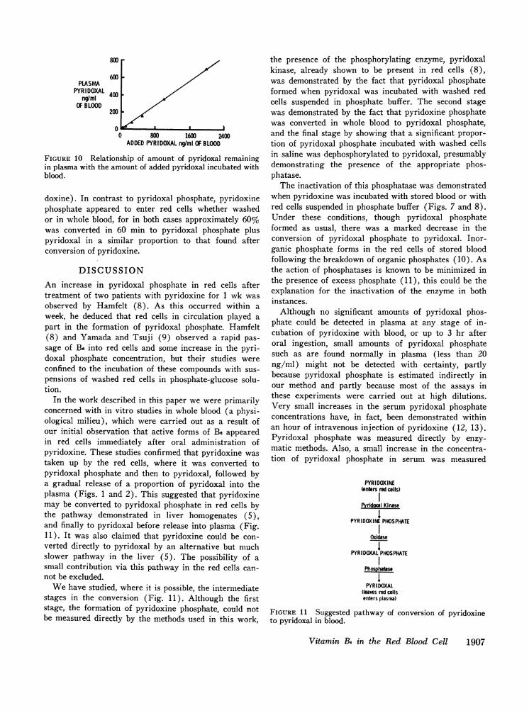
Pyridoxal. Amounts of 50-2000 ng of pyridoxalwere incubated per ml of blood for 60 min. The formwas unchanged, and the distribution between plasmaand red cells was approximately the same (mean 32.8%in plasma, 62.5% in cells) (Fig. 10), being also similarto that of pyridoxal converted from pyridoxine by red
cells (Fig. 5). Further, when 500 ng of pyridoxal wasincubated with blood for 1 min, there was equilibrationto the same distribution which remained the same upto the 2 hr studied (mean 31.5% in plasma, 67.4% incells).
On the other hand, when varying amounts of pyri-doxal were incubated with washed cells in 0.15 M NaClmost of the pyridoxal entered the cells within 1 min,leaving only a trace in the saline.
Pyridoxal phosphate. Amounts of 200-2000 ng ofpyridoxal phosphate (in terms of pyridoxal) were in-cubated per ml of (a) whole blood and (b) washedcells in 0.15 M NaCl for 60 min. In whole blood pyri-doxal phosphate remained in plasma with no significantamounts entering the red cells. In contrast, when pyri-doxal phosphate was incubated with washed cells in 0.15M NaCl, pyridoxal phosphate entered the washed redcells, only 20% remaining in the plasma even when2000 ng was added.
Interconversion of pyridoxal and pyridoxal phosphate.When either pyridoxal or pyridoxal phosphate was in-cubated with whole blood, the form remained un-changed. When either form was incubated with washedcells suspended in phosphate buffer (0.08 M) almosthalf the added pyridoxal was phosphorylated to formpyridoxal phosphate, but pyridoxal phosphate, thoughentering the cells, did not change. However, if pyridoxalphosphate was incubated with washed cells suspendedin 0.15 M NaCl, at least half was dephosphorylated toform pyridoxal.
Pyridoxine phosphate. Pyridoxine phosphate was in-cubated with (a) whole blood and (b) washed cellssuspended in 0.15 M NaCl (500 ng/ml in terms of pyri-
I--zLJ
c.
0 5 20 40 60MINUTES
FIGURE 9 Comparison of red cell uptake of pyridoxine-3H(500 ng/ml) incubated for different lengths of time with (a)whole blood and (b) washed red cells in 0.15 M NaCl, andcomparison of subsequent conversion to L. casei active B6(pyridoxal phosphate plus pyridoxal) in intact and hemolyzedred cells. (Shown on abscissa as B6 as per cent of added pyri-doxine). Whole blood, *; red cells, a; washed red cell suspen-sion, 0; washed red cells, 0; , intact cells;hemolyzed cells; - - -, total red cell uptake of pyridoxine.
1906 Anderson, Fulford-Jones, Child, Beard, and Bateman
PLASMAPYRIDOXAL
ng/mlOF BLOOD
800
600
400
200
800 1600 2400ADDEDPYRIDOXALng/ml OF BLOOD
FIGURE 10 Relationship of amount of pyridoxal remainingin plasma with the amount of added pyridoxal incubated withblood.
doxine). In contrast to pyridoxal phosphate, pyridoxinephosphate appeared to enter red cells whether washedor in whole blood, for in both cases approximately 60%was converted in 60 min to pyridoxal phosphate pluspyridoxal in a similar proportion to that found afterconversion of pyridoxine.
DISCUSSIONAn increase in pyridoxal phosphate in red cells aftertreatment of two patients with pyridoxine for 1 wk wasobserved by Hamfelt (8). As this occurred within aweek, he deduced that red cells in circulation played apart in the formation of pyridoxal phosphate. Hamfelt(8) and Yamada and Tsuji (9) observed a rapid pas-sage of B3 into red cells and some increase in the pyri-doxal phosphate concentration, but their studies wereconfined to the incubation of these compounds with sus-pensions of washed red cells in phosphate-glucose solu-tion.
In the work described in this paper we were primarilyconcerned with in vitro studies in whole blood (a physi-ological milieu), which were carried out as a result ofour initial observation that active forms of B6 appearedin red cells immediately after oral administration ofpyridoxine. These studies confirmed that pyridoxine wastaken up by the red cells, where it was converted topyridoxal phosphate and then to pyridoxal, followed bya gradual release of a proportion of pyridoxal into theplasma (Figs. 1 and 2). This suggested that pyridoxinemay be converted to pyridoxal phosphate in red cells bythe pathway demonstrated in liver homogenates (5),and finally to pyridoxal before release into plasma (Fig.11). It was also claimed that pyridoxine could be con-verted directly to pyridoxal by an alternative but muchslower pathway in the liver (5). The possibility of asmall contribution via this pathway in the red cells can-not be excluded.
Wehave studied, where it is possible, the intermediatestages in the conversion (Fig. 11). Although the firststage, the formation of pyridoxine phosphate, could notbe measured directly by the methods used in this work,
the presence of the phosphorylating enzyme, pyridoxalkinase, already shown to be present in red cells (8),was demonstrated by the fact that pyridoxal phosphateformed when pyridoxal was incubated with washed redcells suspended in phosphate buffer. The second stagewas demonstrated by the fact that pyridoxine phosphatewas converted in whole blood to pyridoxal phosphate,and the final stage by showing that a significant propor-tion of pyridoxal phosphate incubated with washed cellsin saline was dephosphorylated to pyridoxal, presumablydemonstrating the presence of the appropriate phos-phatase.
The inactivation of this phosphatase was demonstratedwhen pyridoxine was incubated with stored blood or withred cells suspended in phosphate buffer (Figs. 7 and 8).Under these conditions, though pyridoxal phosphateformed as usual, there was a marked decrease in theconversion of pyridoxal phosphate to pyridoxal. Inor-ganic phosphate forms in the red cells of stored bloodfollowing the breakdown of organic phosphates (10). Asthe action of phosphatases is known to be minimized inthe presence of excess phosphate (11), this could be theexplanation for the inactivation of the enzyme in bothinstances.
Although no significant amounts of pyridoxal phos-phate could be detected in plasma at any stage of in-cubation of pyridoxine with blood, or up to 3 hr afteroral ingestion, small amounts of pyridoxal phosphatesuch as are found normally in plasma (less than 20ng/ml) might not be detected with certainty, partlybecause pyridoxal phosphate is estimated indirectly inour method and partly because most of the assays inthese experiments were carried out at high dilutions.Very small increases in the serum pyridoxal phosphateconcentrations have, in fact, been demonstrated withinan hour of intravenous injection of pyridoxine (12, 13).Pyridoxal phosphate was measured directly by enzy-matic methods. Also, a small increase in the concentra-tion of pyridoxal phosphate in serum was measured
PYRIDOXINE(enters red cells)
Pyridocal Kinase
PYRIDOXINE PHOSPHATE
Oxidase
PYRIDOXALPHOSPHATE
Phosphatase
IPYRIDOXAL
(leaves red cellsenters plasma)
FIGURE 11 Suggested pathway of conversion of pyridoxineto pyridoxal in blood.
Vitamin B6 in the Red Blood Cell 1907

after oral ingestion of 2 or 25 mg reaching a small peakat 3 hr (13), unlike the large peak of pyridoxal mea-sured by us much earlier, i.e. about 1 hr after ingestion(Fig. 1).
The distribution of pyridoxal between plasma andred cells was constant during incubation after red cellconversion of pyridoxine (Fig. 5) and after addition ofpyridoxal to blood (Fig. 10), irrespective of the amountsadded. The fact that the proportion of pyridoxal inplasma was also dependent on the amount of plasmapresent (Fig. 6) suggests that a factor in plasma, per-haps a specific protein, controls this. If pyridoxal isbound to a protein, the binding is loose, for L. casei isable to utilize all the pyridoxal after incubation of bloodwith pyridoxine or pyridoxal without previous extraction.
The instant equilibration of pyridoxal between plasmaand red cells was in contrast to the slower patternof uptake of pyridoxine (Fig. 2), which is probablypartly controlled by the comparative delay in conversionto active forms. The uptake of pyridoxine was unchangedin washed cells in saline (Fig. 9), suggesting a control-ling receptor inside the cell for pyridoxine. On the otherhand, almost all pyridoxal instantly entered the cellsunder these conditions, which corroborates the findingthat converted pyridoxal inside the cell is not releasedin the absence of plasma. Pyridoxal phosphate added towhole blood remained in the plasma and did not enterthe cells, although it readily entered washed cells in sa-line. This suggested that a firm binding to a plasmaprotein might prevent its entry into cells, which was notthe case with pyridoxal or pyridoxine. The contrastingbehavior of the different B6 forms is marked. There isevidence that they vary in their affinity for protein, andthis has initiated investigations into the protein bindingof B6.
Not only was pyridoxal phosphate not able to enter thered cell, but it appears that if it formed intracellularlyand persisted, as demonstrated in stored blood, it didnot leave the cell (Fig. 7). But there is also evidencethat in stored blood the factor controlling pyridoxallevels in plasma deteriorates, for when pyridoxal wasincubated with stored blood, although pyridoxal phos-phate did not form, the distribution was changed withless pyridoxal remaining in the plasma. This could not,however, explain the sharp decrease of pyridoxal foundin plasma when pyridoxine was incubated with storedblood, and most probably the explanation is the inabilityof pyridoxal phosphate to leave the cell; hence its con-version to pyridoxal under normal circumstances forrelease into plasma.
The findings of this work suggest that all forms ofB6 incubated with and entering the red cells are con-verted mainly via pyridoxal phosphate ultimately topyridoxal, a proportion of which is then gradually re-
leased into plasma. It is not known whether this conver-sion affects the red cell function but it is interesting thatpyridoxal phosphate, like 2,3-diphosphoglycerate andATP causes a shift to the right in the oxygen dissocia-tion curve (14-16).
It is also not certain whether pyridoxal released intoplasma is utilized elsewhere. It is possible that pyridoxalis the main transport form and that following its easypassage into cells of other tissues of the body, for ex-ample marrow cells, pyridoxal phosphate, the coenzyme,forms inside the cell. This could explain the significanceof the wide distribution of pyridoxal kinase in differenttissues (17). However, pyridoxal phosphate could onlyform and remain as such provided that there was not aphosphatase also present capable of breaking down pyri-doxal phosphate. Alternatively, this pyridoxal might bemainly excreted after conversion to pyridoxic acid in theliver.
Homogenates of liver and brain have been shown toconvert pyridoxine either to pyridoxal phosphate or topyridoxal (5). It should be taken into account that itis likely that these homogenates contained some red cells,and we have shown that a very small number of red cellscan convert a significant amount of pyridoxine (Fig. 3).Wehave carried out preliminary studies with white bloodcells and marrow cells but have not been able to demon-strate that these cells can convert any significant amountsof pyridoxine.
It is interesting that where other vitamins are con-cerned, there have been various reports of uptake intored cells and the consequent appearance of related formsin the cells (18-23). However, it is not known whetherthe red cell converts these other vitamins to active formswhich are also subsequently released into plasma.
Investigations are in progress into the conversion ofpyridoxine to active forms in blood in various conditions.A condition of particular interest is the alcohol-inducedsideroblastic anemia. Recently, evidence of an impairedconversion of pyridoxine to pyridoxal phosphate in thebody has been demonstrated in this condition (24). Ifthe conversion in blood is of importance in the body, thedemonstration of a defect at any stage might have somebearing on this, and on the pyridoxine-responsive sidero-blastic anemias.
ACKNOWLEDGMENTS
We are very grateful to Professor D. L. Mollin for hishelp and advice in the preparation of this paper and toDr. A. M. Dawson for his interest and encouragement. Wewould also like to thank Drs. R. Green, A. H. Waters,P. A. Newmark, B. Brozovic, and A. M. Musso for helpfuldiscussions during the course of this work and Mrs. MaryRawlins for help with some of the assays.
This work was supported by grants to Professor D. L.Mollin from the Wellcome Trust and Medical ResearchCouncil.
1908 Anderson, Fulford-Jones, Child, Beard, and Bateman

REFERENCES1. Rabinowitz, J. C., and E. E. Snell. 1948. The vitamin B6
group XIV. Distribution of pyridoxal, pyridoxamine andpyridoxine in some natural products. J. Biol. Chem. 176:1157.
2. Toepfer, E. W., and J. Lehmann. 1961. Procedure forchromatographic separation and microbiological assay ofpyridoxine, pyridoxal and pyridoxamine in food extracts.J. Ass. Oflic. Agr. Chem. 44: 426.
3. Polansky, M. M., and E. W. Murphy. 1966. Vitamin B6components in fruits and nuts. J. Amer. Diet. Ass. 48:109.
4. Polansky, M. M. 1969. Vitamin B6 components in freshand dried vegetables. J. Amer. Diet. Ass. 54: 118.
5. Wada, H., T. Morisue, Y. Nishimura, Y. Morino, Y.Sakamoto, and K. Ichihara. 1959. Enzymatic studies onpyridoxine metabolism. Proc. Jap. Acad. 35: 299.
6. Anderson, B. B., M. B. Peart, and C. E. Fulford-Jones.1970. The measurement of serum pyridoxal by a micro-biological assay using Lactobacillus casei. J. Clin. Pathol.(London). 23: 232.
7. Hill, A. B. 1966. The variability of observations. InPrinciples of Medical Statistics. The Lancet Ltd., Lon-don. 8th edition. 85.
8. Hamfelt, A. 1967. Pyridoxal kinase activity in bloodcells. Clin. Chim. Acta. 16: 7.
9. Yamada, K., and M. Tsuji. 1968. Transport of vitaminB6 in human erythrocytes. J. Vitaminol. (Kyoto). 14:282.
10. Aylward, F. X., B. R. S. Mainwaring, and J. F. Wilkin-son. 1940. Effects of some preservatives on stored blood.Lancet. 1: 685.
11. Snell, E. E. 1961. Some enzymatic transformations ofvitamin B6 and their inhibition. Proc. Int. Congr. Bio-chem. 5th Moscow. 1961. 24: 268.
12. Wada, H., T. Morisue, Y. Sakamoto, and K. Ichihara.1957. Quantitative determination of pyridoxal-phosphateby apotryptophanase of Escherichia coli. J. Vitaminol.(Kyoto). 3: 183.
13. Hines, J. D., and D. S. Love. 1969. Determination ofserum and blood pyridoxal phosphate concentrations withpurified rabbit skeletal muscle apophosphorylase b. J.Lab. Clin. Med. 73: 343.
14. Chanutin, A., and R. R. Curnish. 1967. Effect of organicand inorganic phosphates on the oxygen equilibrium ofhuman erythrocytes. Arch. Biochem. Biophys. 121: 96.
15. Benesch, R. E., R. Benesch, and C. I. Yu. 1969. Theeffect of pyridoxal phosphate on the oxygenation ofhemoglobin. Fed. Proc. 28: 604.
16. Paniker, N. W., and E. Beutler. 1970. Effect of normalmetabolites on the oxygen-hemoglobin equilibrium. Proc.Soc. Exp. Biol. Med. 135: 389.
17. McCormick, D. B., M. E. Gregory, and E. E. Snell.1961. Pyridoxal phosphokinases. I. Assay, distribution,purification and properties. J. Biol. Chem. 236: 2076.
18. Axelrod, A. E., T. D. Spies, and C. A. Elvehjem. 1941.The effect of a nicotinic acid deficiency upon the coen-zyme I content of the human erythrocyte and muscle.J. Biol. Chem. 138: 667.
19. Leifer, E., J. R. Hogness, L. J. Roth, and W. H. Lang-ham. 1948. Utilization of nicotinic acid and its amide bythe human erythrocyte in vitro. J. Amer. Chem. Soc. 70:2908.
20. Preiss, J., and P. Handler. 1958. Biosynthesis of diphos-phopyridine nucleotide. I. Identification of intermediates.J. Biol. Chem. 233: 488.
21. Lee, K. W., D. M. Abelson, and Y. 0. Kwon. 1968.Nicotinic acid-6-'4C metabolism in man. Amer. J. Clint.Nutr. 21: 223.
22. Beutler, E. 1969. Effect of flavin compounds on gluta-thione reductase activity: in vivo and in vitro studies.J. Clin. Invest. 48: 1957.
23. Baker, H., 0. Frank, A. D. Thomson, and S. Feingold.1969. Vitamin distribution in red blood cells, plasmaand other body fluids. Amer. J. Clin. Nutr. 22: 1469.
24. Hines, J. D., and D. H. Cowan. 1970. Studies on thepathogenesis of alcohol-induced sideroblastic bone-mar-row abnormalities. N. Engl. J. Med. 283: 441.
Vitamin B6 in the Red Blood Cell 1909