Embed Size (px)
Citation preview

CONTROL OF BIOLUMINESCENCE
OPERATING THE LIGHT SWITCH IN PHOTOPHORES FROM MARINE ANIMALS
AKADEMISK AVHANDLING
för filosofie doktorsexamen i zoofysiologi som enligt naturvetenskapliga fakultetens beslut kommer att försvaras offentligt fredagen den 20 februari 2009, kl. 10.00 i föreläsningssalen,
Zoologiska institutionen, Medicinaregatan 18, Göteborg
av
JENNY KRÖNSTRÖM
Department of Zoology/Zoophysiology
2009


DISSERTATION ABSTRACT
Jenny Krönström (2009) Control of bioluminescence - operating the light switch in photophores from marine animals Department of Zoology/Zoophysiology, University of Gothenburg, Box 463, 405 30 Göteborg
Physiologically controlled photocytes, capable of producing bioluminescence, are a common feature in the ocean among animals ranging from cnidarians to fish. The aim of this thesis was to study and compare the nature of this control, in both distantly and closely related species from the groups Teleostei Crustacea, and Cnidaria.
This was done using histochemistry and electron microscopy to reveal the internal morphology of the different photophores and to identify the location of nerves and signalling substances inside these organs. Moreover, luminescence responses of isolated photocytes, photophores or live animals, exposed to drugs with effects on adrenergic, noradrenergic, 5-hydroxytryptaminergic and nitric oxide signalling mechanisms, were measured.
Nitric oxide donors had modulating, primarily quenching, effects on the luminescence from the fish species Argyropelecus hemigymnus and Porichthys notatus and the krill Meganyctiphanes norvegica. However, a few of the A. hemigymnus photophores, and a part of the P. notatus response were potentiated when using nitric oxide donors. The variety in nitric oxide responses was reflected by the presence of nitric oxide synthase-like material in different cell types, including neurons, photocytes and lens/filter cells, in the photophores from the studied fish species and Meganyctiphanes norvegica.
Capillary sphincter cells and capillary endothelia contained nitric oxide synthase-like material in Meganyctiophanes norvegica photophores. Moreover, varicose nerve fibres, containing 5-hydroxytryptamine, followed the capillaries and reached the sphincter cells, suggesting that nitric oxide and 5-hydroxytryptamine may interact and control the resistance for haemolymph flow in the photophores, but other mechanisms are also discussed in the thesis. Contractile properties of the sphincter structures, and possibly the endothelial cells, were supported by the presence of muscle-like filaments in the sphincter structures and filamentous actin in both sphincter and endothelial cells. Relaxation of sphincters and capillaries may increase the flow of oxygenated haemolymph to the light-producing cells, thus stimulating or facilitating luminescence. Further indications for this scenario were a stimulation of luminescence by muscle relaxing substances and a quenching of the 5-hydroxytryptamine stimulated luminescence by a muscle contracting substance. Attempts to study an adrenergic mechanism in cnidarians failed for unknown reasons.
In conclusion, it was shown that a nitric oxide signalling system is present in the photophores from several luminescent species. The variety of nitric oxide responses, as well as the variety of morphological arrangements and patterns of innervation in the studied photophores emphasise the biodiversity of bioluminescence.


Till Johan och Freja


LIST OF PAPERS
This thesis is based on following papers and manuscripts, which are referred to in the text by their Roman numerals: I Krönström J, Holmgren S, Baguet F, Salpietro L and Mallefet J (2005) Nitric
oxide in control of luminescence from hatchetfish (Argyropelecus hemigymnus) photophores. J. Exp. Biol., 208, 2951-2961
II Krönström J and Mallefet J (2009) Is NO commonly involved in control of
photogenesis in bioluminescent fish? Manuscript III Krönström J, Dupont S, Mallefet J, Thorndyke M, Holmgren S (2007)
Serotonin and nitric oxide interaction in the control of bioluminescence in Northern krill, Meganyctiphanes norvegica (M. Sars). J. Exp. Biol., 209, 3179-3187
IV Krönström J, Karlsson W, Johansson BR, Holmgren S (2009) Involvement of
contractile elements in control of bioluminescence in Northern krill, Meganyctiphanes norvegica (M. Sars) Submitted


LIST OF CONTENTS
ABBREVIATIONS.......................................................................................... 1 INTRODUCTION..........................................................................................3 Signalling substances ..................................................................................4 Catecholamines.............................................................................................................. 4 5-hydroxytryptamine (5-HT)......................................................................................... 4 Nitric oxide (NO) ........................................................................................................ 5 Evolution of bioluminescence.....................................................................6 Light-producing systems (luciferins and luciferases) ................................6 Photophores.................................................................................................8 Control of luminescence.............................................................................. 10 AIMS ............................................................................................................ 13 METHODOLOGICAL ASPECTS ................................................................. 14 Animals........................................................................................................ 14 Microscopy studies...................................................................................... 14 Fixation and embedding of tissue ................................................................................... 15 Histochemistry ......................................................................................................... 15 Electron microscopy .................................................................................................. 15 Antibodies and probes ................................................................................................... 15 Nerve markers......................................................................................................... 16 NOS antibodies....................................................................................................... 16 Actin and myosin detection....................................................................................... 16 Secondary antibodies ................................................................................................ 17 Pharmacology.............................................................................................. 17 Experimental setup ....................................................................................................... 17 Fish......................................................................................................................... 17 Meganyctiphanes norvegica........................................................................................ 18 Cnidaria.................................................................................................................. 19 Drugs ........................................................................................................... 19 NO donors ................................................................................................................... 20 NOS inhibitors............................................................................................................. 20 Muscle effecting drugs..................................................................................................... 20 Data analysis ...............................................................................................21 RESULTS AND DISCUSSION ......................................................................22

Morphology of photophores........................................................................22 Variation in the morphology of fish photophores ............................................................. 22 Krill photophores contain capillary sphincters .................................................................. 23 Nitric oxide mechanisms ............................................................................24 NOS-like proteins are contained in photophores from several species................................ 24 NO has various physiological effects on luminescence reactions ......................................... 25 Effects of NO on luminescence are independent of cGMP ............................................... 26 Putative effects of NO on photophore respiration and oxygen availability......................... 27 Connections between muscle relaxation, flow of haemolymph and luminescence in krill ............................................................................28 Adrenaline has inconstant effects on sea-pen luminescence .....................29 SUMMARY AND CONCLUSIONS...............................................................30 ACKNOWLEDGEMENTS.............................................................................35 REFERENCES ...............................................................................................37

1
ABBREVIATIONS
AcT acetylated tubulin BH4 (6R)-5,6,7,8-tetrahydrobiopterin 8BrcGMP 8-Bromoguanosine 3’,5’-cyclic monophosphate sodium salt monohydrate cAMP cyclic adenosine monophosphate cGMP cyclic guanosine monophosphate ED50 effective dose for 50% of response eNOS endothelial nitric oxide synthase FAD flavin adenine dinucleotide GFP green fluorescent protein 5-HT 5-hydroxytryptamine iNOS inducible nitric oxide synthase Lmax maximal light production Lmax fast maximal light production of Porichthys notatus fast light response Lmax slow maximal light production of Porichthys notatus slow light response Ltot total light production L-NAME NG-nitro-L-arginine methyl ester NADPH nicotinamide adenine dinucleotide phosphate NADPHd nicotinamide adenine dinucleotide phosphate diaphorase NO nitric oxide NOS nitric oxide synthase nNOS neuronal nitric oxide synthase O.C.T. optimal cutting temperature q quanta RPLmax maximum rate of light production SNAP S-nitroso-N-acetylpencillamine SNP sodium nitroprusside TL1/2 time to half extinction of light response Tip time to inflection point TLmax time to maximal light production TLmax slow time to maximal light production of Porichthys notatus slow light response TLmax fast time to maximal light production of Porichthys notatus fast light response Tstart time to start of light response ODQ [1,2,4] oxadizolo [4,3-a]quinoxalin-1-one KCl potassium cloride KCN potassium cyanide

2

3
INTRODUCTION
Bioluminescence is light produced by live organisms. The ability to produce light is a common feature in the animal kingdom, even though most people are not aware of the widespread occurrence of this phenomenon. Bioluminescent species are so far found in ca one third of the animal phyla, and the majority of these dwell in the marine habitat (Harvey 1916, Herring 1987, Haddock and Case 1994).
Bioluminescence is produced by a chemical reaction between a substrate, luciferin, and an enzyme, luciferase. Luciferin, the light emitting molecule, is excited in the luminescence reaction and emits light as the molecule relaxes to the normal state (fig. 1). This reaction requires oxygen in most cases (Hastings 1983, Shimomura 2006). Luciferin and luciferase are produced in endogenous light-producing cells so called photocytes (this is by far the most common, Herring 1987) or in bacteria in symbiotic relation with a multicellular organism such as a fish or squid. The light-producing reaction can take place inside or outside the cells (Hastings and Morin 1991). Symbiotic luminous bacteria are usually contained in specialised pockets in the skin or gut of the host animal, while photocytes are gathered to form either luminous areas on the body or in specific light organs (photophores). Photophores contain accessory structures such as reflector, filter or lens to direct and modify the light emitted by the photocytes to fit the ambient situation (Denton, et al. 1985). There are several ecological benefits to bioluminescence including camouflage (counter-illumination), interspecies signalling, scaring predators, luring prey and illumination, depending on species (Widder 1999).
Fig. 1 General bioluminescence reaction. The light emitter luciferin reacts with oxygen and the enzyme luciferase to form an exited form of luciferin, indicated with *. Light (hυ) is emitted when the molecule relaxes to normal state. Modified from Wilson and Hastings 1998
Animals possessing bacterial bioluminescent symbionts can only control the light by covering or uncovering the bacteria, which are constantly glowing (animals with bacterial symbionts will not be discussed further in this thesis). However, in species with endogenous photocytes the production and intensity of light is precisely regulated (Hastings and Morin 1991). For example, fireflies are communicating between sexes with flashes of specific length and frequency and are capable of producing long and short flashes like a “Morse code” for this purpose (Herring 2000a). In hatchetfish (Argyropelecus spp.) and many other fish species living in the mesopelagic (200-1000 m depth) part of the ocean, the intensity of light is closely adjusted to the illumination from the surface and used as a camouflage (Denton, et al. 1972, Widder 1999).
In contrast to the large interest in the chemical nature of the bioluminescent reactants, luciferins and luciferases, and their application possibilities in modern molecular biology, the physiological control of light production has been extensively studied only in a few species. However, bioluminescence is of great interest in an evolutionary and functional perspective, since
luciferin + O2 + luciferase luciferin* luciferin + hυluciferin + O2 + luciferase luciferin* luciferin + hυ

4
light-producing capabilities have evolved separately several times and appears in distantly related animal groups (Herring 1987).
In this thesis, the control mechanisms for endogenous light production in marine fish, krill
and sea-pens have been investigated. Most focus has been on the hatchetfish (Argyropelecus hemigymnus), the Northern krill (Meganyctiphanes norvegica) and the signalling substance nitric oxide (NO), but mechanisms involving 5-hydroxytryptamine (5-HT) and adrenaline/noradrenaline have also been investigated to some extent.
Signalling substances
Catecholamines The catecholamines noradrenaline and adrenaline are synthsised in neurons and/or chromaffin tissue of all vertebrate groups and are involved in the autonomic control system, regulating blood pressure, heart performance, metabolism and numerous other involuntary events. Noradrenaline is synthesized from L-tyrosine via a chain of events, involving the enzymes tyrosin hydroxlase, aromatic L-amine decarboxylase and dopamine �-hydroxylase. Adrenaline is derived from noradrenaline through the action of the enzyme phenylethanolamine-N-methyl transferase (Nilsson 1983).
Adrenaline and noradrenaline are less common in invertebrates, where dopamine and octopamine are the major catecholamines expressed, but nevertheless the presence of adrenaline or noradrenaline has been reported in species from several invertebrate groups including molluscs, cnidarians, echinoderms, crustaceans and insects (Carlberg and Rosengren 1985, Delcomyn and Prosser, Walker and Holden-Dye 1991). Vertebrate catecholamine receptors are α- or β-receptors with several subtypes, of which homologues to the mammalian β2 receptor are most frequently found in invertebrates (Palacios, et al. 1989). Adrenaline stimulates luminescence in the sea-pen Renilla koellikeri, probably via β-like receptors (Anctil and Boulay 1982, Awad and Anctil 1993). Further effects of noradrenaline and adrenaline on luminescent systems are summarised in the section “Control of luminescence” below.
5-Hydroxytryptamine (5-HT) 5-HT, often called serotonin, is a monoamine that functions both as a neurotransmitter and a hormone, and is synthesized from L-tryptophan by the enzymes tryptophan hydroxylase and aromatic L-aminoacid decarboxylase (Nilsson 1983). It is present in the nervous system of vertebrates and invertebrates (Walker and Holden-Dye 1991, Beltz 1999, Moreau, et al. 2002) and stored in endocrine tissue, such as endocrine cells in fish intestine and crustacean pericardial organ (Anderson 1983, Beltz and Kravitz 1986, Anderson and Campbell 1988).
Seven different 5-HT receptor families, with numerous subtypes, have been identified in mammals. The intracellular effects of 5-HT stimulation may be an increase or decrease of intracellular cAMP, stimulation of phospholipase C or opening of sodium-potassium channels in the plasma membrane, depending on the receptor type stimulated (Saudou and Hen 1994, Tierney 2001). In this thesis, 5-HT mechanisms of action have been studied in krill. The 5-HT

5
receptor functions appear to be similar in invertebrates as in mammals (Blenau and Baumann 2001) and e.g. a receptor with 5-HT2-like properties has been identified physiologically in the crayfish Procambarus clarkii (Tabor and Cooper 2002). Furthermore, a 5-HT1crust receptor closly related to the mammalian 5HT1 receptor, but with unknown mechanism of action has been cloned from prawn (Macrobrachium rosenbergii), spiny lobster (Panulirus interruptus) and the crayfish (Procambarus clarkii) (Sosa, et al. 2004).
5-HT has numerous effects on the heart, muscle and behaviour of crustaceans, but most interestingly, it stimulates krill luminescence (Kay 1962, Kuramoto and Ebara 1984, Wilkens 1987, Delcomyn and Prosser 1991, Fregin and Wiese 2002).
Nitric oxide (NO) NO is an important messenger molecule in many biological systems ranging from plants to mammals (Torilles 2001). It is a gaseous molecule which is both water- and lipid-soluble and diffuses easily through membranes into a target cell, or other neighbouring cells. In biological systems, NO is produced from L-arginine by the enzyme nitric oxide synthase (NOS), resulting in citrulline as a byproduct. This reaction requires the cofactors NADPH, FAD, heme, calcium calmoduline and BH4 (Bredt and Snyder 1994). NOS is present in three isoforms in mammalians: endothelial NOS (eNOS), inducible NOS (iNOS) and neuronal NOS (nNOS). In general, eNOS and nNOS are considered to be constitutive, while iNOS is a player in the immune system and induced by, for example, bacterial lipopolysaccarides (Bredt and Snyder 1994, Laing, et al. 1999). However, some studies indicate that nNOS and eNOS can also be induced (Alderton, et al. 2001).
In nonmammalian animals it appears that all these isoforms do not always exist, but in fish and sea urchin, there are indications for at least two NOS isoforms (Bicker 2001, Cox, et al. 2001). NOS usually shows more sequence similarity between species than between isoforms, and the parts binding the cofactors required for the function of the enzyme are highly conserved regions of the NOS molecule (Cox, Mariano, Heck, Laskin and Stegeman 2001).
The first record of NO as a physiologically active molecule described a relaxing substance produced by endothelial cells that acted on rabbit vascular smooth muscle (Palmer, et al. 1987). This muscle relaxing effect has been recorded in several animal groups including echinoderms (Elphick and Melarange 1998, Elphick and Melarange 2001) and fish (Agnisola 2005). NO has also been shown to be important in both the autonomic and the central nervous system, acting as a retrograde transmitter and causing, for example, long term potentiation and memory consolidation in mammals and insects (Lincoln, et al. 1997, Bicker 2001). NO is unusual as a neurotransmitter in the sense that it is not stored in vesicles in the nerve terminals and lack receptors on the cell surface (Bredt and Snyder 1994).
The most commonly reported mechanism of NO action is through stimulation of soluble guanylyl cyclase, that leads to production of cGMP, which in turn has several mechanisms of action depending on cell type (Koesling, et al. 2004). Several other mechanisms of NO action have also been suggested, such as affecting excitability of neurons/cells via direct effects on ion-channels and causing inhibition of cellular respiration by binding to oxygen sites in the mitochondrial electron transport chain (Brown 1995, Erxleben and Hermann 2001, Hermann and Erxleben 2001). In bioluminescent systems, NO is so far not well explored, but it is

6
established that firefly bioluminescence is controlled by NO (as described in the section “Control of luminescence” below, Trimmer, et al. 2001).
Evolution of bioluminescence
Bioluminescent species are believed to have evolved independently about 30 times over time (Herring 1987). The following citation of Harvey (1952) illustrates the apparent randomness of the emergence of bioluminescent species:
“…, as if a handful of damp sand has been cast over the names of various groups written on a blackboard, with luminous species appearing wherever a mass of sand struck. ...It is an extraordinary fact that one species in a genus may be luminous and another closely allied species contains no trace of luminosity.”
Hence, there is no common ancestor for all bioluminescent species and the luminescent
systems have different origins, resulting in diverse morphological and physiological solutions for producing and emitting light. On the basis of the unpredictable appearance of luminescent species throughout evolution, and the necessity for oxygen in most luminescence reactions, it has been speculated that bioluminescence might have evolved to eliminate oxygen or reactive oxygen species from the tissue: functions necessary for all animals (Rees, et al. 1998, Timmins, et al. 2001a). For example, in cell culture, the luciferin coelenterazine has been shown to reduce death of human fibroblasts exposed to oxidative stress (Rees, et al. 1998). Moreover, coelenterazine is detected not only in light organs, but is also frequently found in the digestive tract and hepatopancreas of both luminous and non-luminous fish, cephalopods and decapods (Mallefet and Shimomura 1995, Thomson, et al. 1997, Rees, et al. 1998).
Light-producing systems (luciferins and luciferases)
In bioluminescence systems, the luciferins are the light-emitting molecules. At least nine different luciferins have been isolated and some of them are found in several phyla, implicating that the molecule might be passed on in the food chain (fig. 2, Shimomura 2006). Experiments with the jellyfish Aquorea victoria and the midshipman fish, Porichthys notatus, show a requirement for a luciferin in the diet for the ability to produce light (Barnes, et al. 1973, Thompson and Tsuji 1989, Haddock, et al. 2001). In contrast, the enzymes, luciferases, are endogenous, produced by the animal itself, irrespective of whether luciferin is internally produced or received via the food (Shimomura 2006).
Bioluminescence can be of different colours, but the majority of the primary bioluminescence reactions between luciferin and luciferase produce blue light, and blue is also the most common colour of light emitted from the photophores in marine animals (Widder, et al. 1983). This is of advantage since blue wavelengths travels furthest in seawater (Kampa 1970). Other colours, for example green, can be obtained with fluorescent proteins like the well known

7
green fluorescent protein (GFP). Professors O. Shimomura, M. Schalfie and R. Y. Tsien were awarded with the Nobel Prize 2008 for the discovery of GFP and the development of widely used applications in molecular biology for this protein. The energy from the luciferin-luciferase reaction is transferred to GFP and emitted as green light (Shimomura 2006). The structure of the luciferase is also of importance for the colour of light emitted, as well as the presence of pigmented filters covering the photocytes (Seliger and McElroy 1964, Denton, et al. 1970).
The luciferin coelenterazine is found in coelenterates (i.e. Cnidaria and Ctenophora) and was first isolated from the jellyfish A. victoria (Shimomura and Johnson 1972), but it has also been found in many other animals possessing intracellular or extracellular bioluminescence, such as sea-pens, squid, shrimp and fish (Shimomura and Johnson 1975, Rees, et al. 1992, Mallefet and Shimomura 1995, Shimomura 2006). In A. Victoria, the coelenterazine is a part of a photoprotein complex (aequorin) also comprising oxygen and luciferase. Addition of calcium to aequorin stimulates luminescence without requiring exogenous oxygen (Shimomura 2006). Like in A. victoria, calcium is the single stimulus controlling the luminescence reaction in the sea-pen Renilla spp. However, in Renilla spp. and other anthozoans, for example Veritillium cynomorium, coelenterazine is stored bound to a luciferine-binding protein together with luciferase in intracellular compartments (luminosomes) (Henry 1975, Shimomura 2006). Binding of calcium to the luciferin-binding protein results in the release of coelenterazine, which then is free to react with available oxygen and luciferase resulting in a luminescence reaction (Anderson and Cormier 1973, Cormier 1978). Moreover, A. victoria, Renilla spp. and many other cnidarians emit green light with the assistance of GFP (Anderson and Cormier 1973).
Coelenterazine has also been found in sufficiently high concentrations in the photophores of the fish species A. hemigymnus, Cyclothone braueri, Myctophum punctatum and Vinciguerria attenuata, to suggest that this is the functional luciferin in these systems (Rees, et al. 1990, Mallefet and Shimomura 1995). A luminescence reaction is obtained when V. attenuata tissue extracts are mixed with coelenterazine, further supporting that coelenterazine is the luciferin in this fish (Rees, et al. 1990). One report suggests that A. hemigymnus and several lanternfish species (Myctophidae spp.) possess bacterial luminescence, based on the detection of the bacterial luciferase genes lux A and B (Vibrio fisheri), in tissue samples from these species (Foran 1991). However, the same experiments repeated by another group gave contradictory results arguing against bacterial luminescence in these fish (Haygood, et al. 1994). In contrast, it is clearly established that the P. notatus, living in more shallow waters, uses the luciferin from a small ostracod (Cypridina hilgendorfii) that is common in their habitats (12 Harper 1999, 22 Mensinger and Case 1991). The ostracod luciferin-luciferase light reaction requires oxygen and calcium (Shimomura, et al. 1961).
The krill luciferin-luciferase reaction is also dependent on oxygen and is very pH sensitive (Shimomura and Johnson 1967, Shimomura 1995). The pH sensitivity is shared by the dinoflagellate light reaction and, moreover, the structure of the dinoflagellate luciferin molecule is very similar to the krill luciferin (fig. 1), possibly suggesting that the krill might receive the luciferin when feeding on dinoflagellates (Schmidt and Atkinson 2006).

8
Fig. 2 Molecular structure of the luciferins which are used for light production by species discussed in this thesis. References: White, et al. 1961, Bode and Hastings 1963, White, et al. 1963, Kishi, et al. 1966, Hori, et al. 1977, Inoue, et al. 1977, Nakamura, et al. 1988 Photophores
Photophores are morphologically advanced organs containing light-producing cells, photocytes, and accessory structures such as reflector, filter or lens to direct and modify the light emitted by the photocytes. Different types of photophores are common among bioluminescent animals, but some groups, like sea-pens or brittlestars, lack photophores and have the photocytes gathered in luminous tissue areas, without accessory structures (Anctil, et al. 1984b, Hastings and Morin 1991, Deheyn, et al. 1996). For pictures of some of the photophores and luminous tissues studied, see fig. 3.
All the fish species in this study possess ventrally located photophores, probably used for counter-illumination of the ventral surface of the fish, to avoid forming a dark silhouette when viewed from below. However, both counter-illumination (Harper and Case 1999) and courtship displays of the photophores have been reported in P. notatus (Crane 1965), and many of the lanternfish species possess additional caudal photophores for interspecies communication (Barnes and Case 1974, Paxton, et al. 1984). Similarly to the fish species, krill have their photophores positioned ventrally, which suggests a counter-illumination purpose (Widder 1999), but interspecies communication has been observed (Mauchline 1960) and some krill species even have a sexual dimorphism in the pattern or size of the light organs (Herring 1978). However, in M. norvegica, studied in papers III and IV, the number and distribution of photophores is the same in both sexes.
O
NHNH2N
H
NH
HN
Cypridina-luciferinHO
COOHS SN N
Firefly-luciferin
OHO
N NNH
Coelenterazine
COONa
COONa
O
OH
NHHN
NH HN
Dinoflagellate-luciferin
Me
Me
MeCH2H
Me
COONa
O
OOH
NHHN
NH HN
Me
Me
COONaMe
Me
CH2OHKrill-luciferin
O
NHNH2N
H
NH
HN
Cypridina-luciferin
O
NHNH2N
H
NH
HN
O
NHNH2N
H
NH
HN
Cypridina-luciferinHO
COOHS SN N
Firefly-luciferinHO
COOHS SN N
Firefly-luciferin
OHO
N NNH
Coelenterazine
OHO
N NNH
OHO
N NNH
CoelenterazineCoelenterazine
COONa
COONa
O
OH
NHHN
NH HN
Dinoflagellate-luciferin
Me
Me
MeCH2H
Me
COONa
COONa
O
OH
NHHN
NH HN
Dinoflagellate-luciferin
Me
Me
MeCH2H
Me
COONa
O
OOH
NHHN
NH HN
Me
Me
COONaMe
Me
CH2OHKrill-luciferin
COONa
O
OOH
NHHN
NH HN
Me
Me
COONaMe
Me
CH2OHKrill-luciferin

9
The development of photophores differs among species in both succession of photophore formation and tissue origin. Photophores are either developed simultaneously (e.g C. braueri and A. hemigymnus) or in succession (e.g. V. attenuata and M. norvegica); however, photophores developed late may still become functional at the same time as earlier developed light organs (C. braueri and V. attenuata) (Petersson 1968, Ahlstrom 1973). Photophores are usually superficial structures and, for example, formed from the ectoderm in krill and from the ectoderm and mesoderm in P. notatus (Petersson 1968, Anctil and Case 1977).
Adult photophores are morphologically very different structures among species and animal groups, but photocytes and the accessory structures reflector, lens and/or filter cells are common features of most types (Herring and Morin 1978). With accessory structures it is possible to narrow the spectrum of wavelengths, changing colour of the emitted light, and direct and reflect the primary light produced by the photocytes. For example, the counter-illuminating hatchetfish (Argyropelecus spp.) use reflectors to scatter the light from the photocytes according to a specific pattern, optimal for blending with the weak illumination from the surface (Denton, et al. 1969, Denton, Gilpin-Brown and Wright 1970, Denton, Gilpin-Brown and Wright 1972). Cells containing a filtering pigment cover the exit for the light and narrow the spectrum of wavelengths, emitted from the hatchetfish photocytes. The spectrum of the emitted light, after filtering, very closely matches the ambient light spectrum at 500 m depth in clear oceanic water (Denton and Herring 1978, Denton, et al. 1985). The intensity of light is physiologically controlled to match the light from the surface, and studies with P. notatus and the lanternfish Symbolophorus californiensis show that the fish adjust their luminescence to an experimentally set background illumination (Case, et al. 1977, Harper and Case 1999)
Fig 3. Schematic pictures of (A) A. hemigymnus, (B) M. norvegica and (C) P. phosphorea and photographs of their photophores and luminescent tissue (a, b and c respectively). The hatchetfish, Argyropelecus hemigymnus, has ventrally located photophores (a, arrows), seen as pink dots in the photograph. (B) The krill, M. norvegica, has two ocular photophores (upper arrow, b1) and eight ventral (i.e. lower arrow, b2) photophores distributed between four pairs of pleopods and on the ventral part of the cephalothorax. (C) The luminescent autozooid polyps of the sea-pen colony Pennalula phosphorea are situated on the edge of the colony leafs (for example in boxed area). (c) The area containing photocytes is fluoresceing in green, indicating presence of GFP. Bars are a: ca 1 cm, b1, b2 and c: ca 100 μm Picture a is shown with kind permission from J. Mallefet.
A
B C
a
b1
b2
c
A
B C
a
b1
b2
c
10
Control of luminescence
Physiological and morphological investigations of isolated photophores, or live luminescent animals, indicate that the control mechanisms for luminescence onset, intensity and duration are, at least partly, neuronal in most species studied, possessing endogenous photocytes. Implications for nervous control are e.g. the responsiveness of photophores to electrical stimuli and the presence of nerves detected with electron microscopy or light microscopy in the photophores of fish, krill and sea-pens (Nicol 1954, Petersson 1968, Barnes and Case 1974, Baguet 1975, Anctil and Case 1977). The photocytes are directly innervated in many species, but the krill photocytes are unusual in lacking direct innervation, and nerves instead reach other cells near the photocytes (discussed further below, paper IV, Herring and Locket 1978).
Electrical stimulation of the spinal cord or isolated photophores induces light reactions in several species of lanternfish (Myctophum punctatum, Benthosema glaciale, Stenobrachius leucopsarsus, Lampanyctus ritteri, Triphoturus mexicanus) with varying kinetics (mainly rapid flashes) depending on type of luminous tissue (ventral or caudal photophores) and stimulus strength and duration (Anctil 1971, Barnes and Case 1974, Christophe and Baguet 1982). The photophores or luminescent tissue of A. hemigymnus, Maurolicus muelleri, and P. notatus, M. norvegica and the sea-pen Pennatula phosphorea are also stimulated to flash by electricity (Nicol 1957, Nicol 1958, Anderson and Case 1975, Baguet 1975, Fregin and Wiese 2002).
Adrenaline is the most extensively investigated stimulus for marine bioluminescence and has been found to induce light production in several fish and cnidarian species. Whole colonies or single polyps from the sea-pen R. koellikeri respond with luminescence to adrenergic stimuli (Anctil and Boulay 1982). Uptake of adrenalin in tissue containing photocytes has also been demonstrated in R. koellikeri (Anctil, et al. 1984b). Injection of adrenaline or noradrenaline in live fish, or when applied to isolated photophores, stimulates luminescence in A. hemigymnus, M. muelleri and P. notatus and these catecholamines are also present in extracts of photophores from A. hemigymnus, M. muelleri, V. attenuata and M. punctatum (Salpietro, et al. 1998). However, attempts to stimulate luminescence in different lanternfish including M. punctatum with adrenaline, noradrenaline or acetylcholine have failed, possibly due to problems with maintaining these fish live and wealthy after capture (Anctil 1971, Barnes and Case 1974).
Isolated photophores from A. hemigymnus respond to stimulation with both adrenaline and noradrenaline with a long lasting glow. However, the photophores are much more sensitive to adrenaline (200 times lower ED50). Responses start already at exposure to 10-8 M adrenaline, while concentrations of between 10-5 and 10-4 M noradrenaline are required for a proper response. Photophores deriving from separate individuals are either weakly or strongly responding to adrenaline (fig. 4). The maximal light emission of the strongly responding photophores is approximately seven times higher than the maximal response of weakly responding organs (Baguet and Marechal 1977). The physiological reason for the differences in response is unknown, but probably not related to the physiological state of the light organs, since there is no difference in oxygen consumption between strongly responding and weakly (or non) responding photophores, indicating that both types are in the same physiological state (Mallefet and Baguet 1985).

11
Isolated photophores from P. notatus respond to noradrenaline with two phases of luminescence, a fast and short response followed by a slow, longer response (fig. 4, Christophe and Baguet 1983, Christophe and Baguet 1985, J. Krönström, C. Vanderlinden and J. Mallefet, previously unpublished data). Results from experiments with different adrenergic agonists and antagonists, specifically directed towards α- or β-adrenergic receptors, suggest that the fast response is mediated via α-receptors and the slow via β-receptors. However, the results are not completely clear (Christophe and Baguet 1983, Christophe and Baguet 1985).
Fig. 4 Original recordings of luminescence from (a) Porichthys notatus and (b) Argyropelecus hemigymnus after stimulation, at time 0, with noradrenaline (0.1 mM) and adrenaline (0.5 mM) respectively. The P. notatus recording is from a single photophore showing the characteristic fast and slow light responses in succession. The A. hemigymnus traces are from photophores from separate individuals, one showing the weak (solid line) and the other the strong (hatched line) adrenaline response. The A. hemigymnus recording is made by J. Mallefet and shown with his kind permission.
5-HT has an inhibitory effect on electrically stimulated luminescence in P. notatus, but the
response stimulated with noradrenaline is not affected (Baguet 1975). Moreover, immunoreactivity against 5-HT has been found in the photocytes of P. notatus and they also sequester radioactively labelled 5-HT in the photocytes. This suggests a role for 5-HT in the control of luminescence from P. notatus photophores (Anctil, et al. 1984b, Mallefet and Anctil 1992a). In contrast to the inhibitory effects on luminescence on P. notatus photophores, 5-HT stimulates bioluminescence in live krill (Kay 1962, Kay 1965, Fregin and Wiese 2002), but the mechanism for this is not known. Another often reported stimulus for luminescence in krill is light, either from artificial light sources like camera flashes or from other krill (Mauchline 1960, Kay 1965).
Fireflies also answer to light signals with light production on there own. Octopamine, a catecholamine commonly expressed in invertebrate nervous systems, has been known to stimulate luminescence in fireflies, but the mechanisms for octopamine action were not understood until after the discovery of the importance and universal occurrence of the signalling substance NO. Only then, NO was identified as the mediator, produced in response to octopamine stimulation of a cell type adjacent to the photocytes in the firefly photophore. NO, in turn, triggers luminescence (Trimmer, et al. 2001).
Rel
ativ
e lig
ht in
tens
ity×1
06
1
10 20 30 40 20 30 40100
65432
87
00
Porichthys notatus Argyropelecus hemigymnus
slow resp.
Time (min)
fast resp.
weak resp.
strong resp.a b
Rel
ativ
e lig
ht in
tens
ity×1
06
1
10 20 30 40 20 30 40100
65432
87
00
Porichthys notatus Argyropelecus hemigymnus
slow resp.
Time (min)
fast resp.
weak resp.
strong resp.a b

12
As can be understood from the previous sections, the understanding of reaction mechanisms controlling bioluminescence, with few exceptions, is fragmentary. Extensive investigations of light-controlling mechanisms have only been performed on a few species or groups including the cnidarians Renilla spp. and A. victoria, fireflies, brittlestars and a few fish species. Studies of the physiological control of luminescence in brittlestars show that the knowledge of the control mechanisms can not be automatically extrapolated even between closely related species. For example, the brittlestar species Amphiura filiformis and Amphipholis squamata share a cholinergic light control, while Ophiopsila californica responds to tryptamine and Ophiopsila aranea responds to none of the substances tested on the other species (Dewael and Mallefet 2002, Vanderlinden and Mallefet 2004).

13
AIMS
In this thesis, I have investigated the control mechanisms for bioluminescence in various species. The aims were to extend the number of luminescent species investigated and to add new knowledge to partly understood systems, such as the krill photophores. Species from the groups Teleostei, Crustacea and Cnidaria were chosen in order to compare the nature of the physiological control of luminescence among distantly related groups.
More specific aims were to:
Identify and describe the presence and pattern of nitric oxide synthase containing structures in photophores from different species.
Investigate the role of nitric oxide in the physiological control of luminescence these species.
Elucidate the nature and physiological significance for luminescence control of capillary sphincter structures in photophores of the krill Meganyctiphanes norvegica.

14
METHODOLOGICAL ASPECTS
Animals
Marine bioluminescent animals with endogenous photocytes belonging to the groups Teleosts (papers I and II), Crustacea (papers III and IV) and Cnidaria (previously unpublished data) were studied. For evolutionary relationships, see table 1 after the “Results and Discussion” section.
The cnidarian sea-pens Pennatula rubra, Pteroides spinosum and Veritillium cynomorium were hand-collected by divers from mud bottoms in the Mediterranean outside Banyuls, France. Pennatula phosphorea was collected by hand or with Agassiz-scraper from the mud bottoms of the Gullmar fjord, Sweden. The sea-pens were kept in tanks provided with running seawater (at ambient temperature) at the Oceanology Observatory of Banyuls, France or at the Sven Lovén Centre for Marine Sciences, Fiskebäckskil, Sweden. The animals were not fed, but presumably consumed small organisms found in the tank seawater, supplied directly from the Mediterranean or the Gullmar fjord.
Krill, Meganyctiphanes norvegica, were collected with an Isaacs-Kidd mid-water trawl in the Gullmar fjord, Sweden. They were kept in running seawater (6ºC) at the Sven Lovén Centre for Marine Sciences or in aquaria filled with water collected in the Gullmar fjord at the Department of Zoophysiology, Göteborg, Sweden. The water was changed approximately every third day and the animals were fed mainly with Artemia salina larvae. The krill were used within two weeks after collection. Within this time frame, the time in captivity did not affect the ability to respond to light-producing stimuli.
Mesopelagic bioluminescent fish species Argyropelecus hemigymnus, Maurolicus mulleri, Vinciguerria attenuata, Cyclothone braueri and the lanternfish species Electrona risso, Hygophum benoiti and Myctophum punctatum were captured with hand net from a small boat in the Strait of Messina, Sicily, Italy, or collected at dawn from the beaches of the Strait. This was possible as strong currents and winds in the Messina area during the winter and early spring force fresh specimens of mesopelagic fish to shallow waters and make some of them wash ashore on the beaches (Baguet 1975). The fish were kept in seawater in the fridge and used the same day.
Shallow-water midshipman fish, Porichthys notatus, were collected by divers in Santa Barbara (California, USA) and shipped by air to Brussels (Belgium) and transported to the Laboratory of Marine Biology, University of Louvain, Louvain-la-Neuve (Belgium) where they were kept in aquaria (15-17 ºC) until use. The fish were fed with frozen, thawed fish.
Microscopy studies
Different histochemical techniques, including immunohistochemistry and NADPH diaphorase (NADPHd) staining, were performed on sections of photophores from all fish species and M. norvegica in order to detect the presence and location of elements involved in the control of light

15
production. Electron microscopy was used to study in detail certain cell types, central for the process of light production, in the M. norvegica photophores.
Fixation and embedding of tissue Histochemistry Details regarding the fixation and further treatment of the histochemical samples are given in the individual papers. In general, for immunohistochemistry, tissue was dissected out and fixed in Zamboni’s fixative, rinsed and dehydrated in a graded alcohol series for subsequent treatment with xylene, rehydrated to phosphate-buffered saline, cryoprotected in phosphate-buffered saline with sucrose, embedded in either O.C.T or agar and quickly frozen in isopentane chilled with liquid nitrogen. Agar was used for increased stability when sectioning samples with large differences in tissue density, for example whole fish with bones or krill with a dense exoskeleton see Holmberg, et al. 2001.
Preparations destined for treatments involving the actin probe, rhodamin phalloidin, were fixed in 4% formaldehyde. My conception is that this rendered a slightly poorer morphological resolution than Zamboni’s fixative, especially in the M. norvegica preparations (paper IV).
Electron microscopy Electron microscopy was used to study the sphincter structures in M. norvegica photophores (paper IV). Photophores were fixed in glutaraldehyde (2.5%) with formaldehyde (2%) or in glutaraldehyde (2.5%). Both fixations gave equally good morphological resolution in the sections. The protocol for preparation of electron microscopy samples is described in detail in paper IV.
Antibodies and probes Indirect immunohistochemistry was used to detect NOS-containing structures inside, or associated to, the photophores of fish (papers I and II) and krill (paper III) and to visualize nerve fibres innervating these organs. In addition NADPHd staining was used to detect NOS in P. notatus (paper II). Antibodies against 5-HT, myosin and actin and an actin probe (rhodamin phalloidin) were used in search for 5-HT, myosin and actin in M. norvegica photophores (papers III and IV).
Commercially available monoclonal and polyclonal antibodies raised against, primarily, mammalian antigens were used. Detailed information on all the antibodies is given in tables in the respective papers. The staining with antibodies directed against proteins is referred to as, for example, NOS-like in this thesis as the non-mammalian species may have other substances that the antibody will bind to, possibly with equal or higher affinity. Even though controls have been performed it is not possible to be completely certain of the identity of the non-mammalian proteins detected, hence the use of i.e NOS-like. Furthermore, the amino acid sequence and tertiary structure may vary somewhat among species, and the affinity of antibodies raised against mammalian substances may not be as strong in non-mammalian species. 5-HT has a common structure throughout the animal kingdom and therefore the binding of antibodies raised against 5-HT is less likely to be affected by the species origin of the antigen.
16
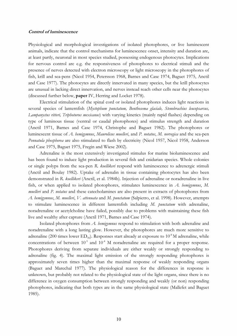
Nerve markers To detect nerve fibres in fish (papers I and II), an antibody raised against acetylated tubulin (AcT) was used. AcT is restricted to nerve cells in the Atlantic cod (Gadhus morhua) and has the antibody has successfully labelled nerve fibres in several other fish species including zebrafish (Danio rerio), Atlantic cod (G. morhua) and rainbow trout (Oncorhynchus mykiss) (Rutberg, et al. 1995, Johnsson, et al. 2001, Holmberg, et al. 2003). In preparations from all fish species in papers I and II, the morphological patterns of the AcT labelling indicate that nerve fibres are marked. However, the binding of the AcT antibody depends on the level of acetylation of tubulin, which is a dynamic process. Therefore it can not be certain that all nerves are detected with this antibody (Takemura, et al. 1992).
The AcT antibody did not stain M. norvegica neurons and could therefore not be used as a neuronal marker in this species, even though it has been used in studies of other crustacean and insect nervous systems and successfully labels nerve fibres in the developing nervous system of the caridean shrimp (Palaemonetas argentinus) and the horse shoe crab (Limulus polyhemus), as well as mature nerve fibres in the brain of adult crickets (Grillus bimaculatus) (Harzsch, et al. 1997, Harzsch, et al. 1999, Mittman and Scholtz 2003, Mashaly, et al. 2008). A neuronal marker developed for fruit fly (Drosophila melanogaster 22C10, Developmental Studies Hybridoma Bank, IA, USA) was also unsuccessful in labelling nerves in M. norvegica.
An antibody raised against synapsin (Drosophila anti-synapsin, antiSYNORF1, 3C11, Developmental Studies Hybridoma Bank), a protein binding vesicles to the cytoskeleton in synaptic parts of the neuron, did successfully label structures inside and outside M. norvegica photophores (fig. 5). This antibody has recently become widely used on crustacean nervous systems. It labels developing nervous systems in brine shrimp and crayfish (Artemia salina, Cherax destructor and Marmorcrebs) and synaptic-dense areas in the adult stomatogastric ganglion of crab, crayfish and lobster (Cancer pagurus, Homarus americanus and Cherax destructor) (Skiebe and Ganeshina 2000, Vilpoux, et al. 2006). However, a marker for axons in krill is still lacking.
NOS antibodies NOS sequences are not known for the specific species used in this study. Therefore, sequence analyses were performed to compare the antigen amino acid sequences of the NOS antibodies used, with known NOS sequences of as closely related species as possible, giving a theoretical indication of the ability of the antibodies to detect NOS in the fish and krill species used. The antibody nNOSsc1025 showed the strongest NOS-like immunoreactivity in fish samples and also proved to have the largest sequence identity, among the antibodies used, between the original antigen and a section of fish nNOS (84%, Oryzias lanipes and 79% Danio rerio, accession numbers AB163430 and NM_131660, respectively) (papers I and II).
For krill, the uNOS PA1039 antigen had the best compatibility with another crustacean NOS (100%, Geocarcinus lateralis, accession number AY552549), and the antibody gave good labelling. Furthermore, a similar labelling was received with the antibody nNOS B220, which also showed considerable alignment between its antigen and the crustacean NOS (paper III).
The antibodies uNOS PA1039, nNOS 31030 and nNOS B220 (for details see paper I, page 2953) were tested on Amphiura filiformis preparations (previously unpublished data).
Actin and myosin detection To confirm the muscular nature of M. norvegica sphincter structures (see paper IV) attempts were made to detect actin and myosin filaments in these structures. This was

17
done using antibodies against actin and myosin and a probe for filamentous actin (Rhodamin phalloidin, see paper IV, page 6 for a table of actin and myosin antibodies and probes). Rhodamin phalloidin is a peptide toxin, isolated from the mushroom Amanita phalloides (Wieland 1986), binding specifically to filamentous actin in many species e.g. lobster (Homarus americanus) (Wilkens, et al. 2008).
Different antibodies raised against myosin heavy and light chains in smooth and skeletal muscle were used in attempts to confirm the muscular nature of the sphincter structures in krill photophores. Only the antibody M4276, raised against myosin in fast skeletal muscle from rabbit, showed weak staining in tail muscle from M. norvegica used as control, and none detected myosin inside the light organs. M4276 is the only antibody in the test which has been previously reported to label myosin in a crustacean species (Procambarus clarcii, LaFramboise, et al. 2000).
Secondary antibodies The secondary antibodies used were commercially available, conjugated to either biotin or a fluorophore. For visualization of biotin-conjugated antibodies, subsequent binding with an avidin-peroxidase complex was used, producing a brown precipitate when the substrate was added. Naturally fluorescent material inside photocytes or, occasionally, other cell types in photophores is common. This is useful for detecting the photocytes in the fluorescence microscope, but limits the use of flourophore colours that can be used. Blue fluorescing material was found inside photocytes from A. hemigymnus, P. notatus, V. attenuata and M. muelleri while filter/lens cells from V. attenuata fluoresced in pink and red/orange fluorescence was detected in the photophores from C. braueri. Fluorescent structures were lacking in M. norvegica photophores, while luminous tissue from the sea-pen P. phosphorea was brightly fluorescing in green, indicating the presence of a green fluorescent protein (GFP, described in the introduction) in these photocytes.
Pharmacology
Experimental setup Pharmacological experiments were carried out to investigate the effect of different bioactive compounds (putative transmitters or other signalling molecules) on the light production of the photophores of A. hemigymnus (paper I), P. notatus (fig. 6), M. norvegica (papers III and IV), the echinoderm Amphiura filiformis and cnidarian sea-pens (previously unpublished data). The experiments were performed on live animals, tissue preparations or isolated photocytes. A Berthold FB12 luminometer, measuring light with wavelengths between 370 and 630 nm and calibrated with a standard light source (470 nm, Betalight, SRB technologies, Winston-Salem, NC, USA) was used to record the light reaction in all experiments. The studied species emit light with peaks of emission at wavelengths between 470 and 500 nm (Widder, et al. 1983). A neutral density filter was positioned between the luminescent object and the detector when light responses exceeded the detection limit for the luminometer detector.
Fish The ventral photophores from A. hemigymnus were dissected from the fish as described in paper I and the tissue piece divided in two halves, containing anterior and posterior

18
photophores, respectively. The preparations were placed in a holder containing saline for deep-sea fish (Hanks solution, paper I). The holder was placed in the luminometer, with photophores facing the detector, and test substances were added to the saline. Anterior and posterior photophores showed identical reactions to the concentration of adrenaline used (0.1 mM) and one part was used as control and the other for treatment (paper I). These photophore preparations are viable for at least 13 h, indicated by a stable oxygen consumption (Mallefet and Baguet 1985), but to avoid any possible effects due to origin of preparation and time after dissection, the preparations were used alternatingly as control and treatment.
P. notataus were anesthetized in quinaldine (3.7% in seawater for 1 min), and skin strips containing 4-6 photophores were dissected out. Separate photophores were freed from surrounding tissue and placed in a holder with the photophores facing the detector of the luminometer. Application of test substances were made as described above.
Meganyctiphanes norvegica Live M. norvegica were used, as isolated photophores from krill do not respond reliably to known bioluminescence stimuli (Herring and Locket 1978, personal observations). Different set-ups and experimental protocols were used in papers III and IV.
In paper III the specimens were freely swimming in a test tube containing 10 ml of seawater. The animals were “standing” in the test tube and could swim a small distance (ca 1 cm) up from the detector of the luminometer. Prior to the experiments, the specimens were exposed to 1h of daylight in order to standardize the pre-experimental light exposure of each specimen. Krill show “light-induced light reaction” (Mauchline 1960, Kay 1965, paper III) and this reaction was allowed inside the luminometer before the animals were taken out and injected with test substance in the abdominal muscle.
The protocol described above was improved in paper IV. 1h light exposure was omitted from the protocol and all experiments and handling of krill were performed in red light, which the krill can not detect (Widder and Frank 2001). This was done in order to avoid the light induced light reaction and to avoid exposing the specimens to more “unnatural” light than necessary. The test tube was changed for a small, aerated glass aquarium, fitting into the luminometer. A metal holder was glued to the dorsal side of the cephalothorax of the krill and then attached to the aquarium and directed in such a way that the ventrally positioned photophores were facing the detector of the luminometer. The animals were still able to move the photophores, but this way they were fixed at the same distance from the detector throughout the experiment and the chances of the photophores pointing towards the detector were essentially improved, compared to the “standing” position in the test tube in paper III. In paper IV, test substances were added to the water around the krill (not injected as in paper III), hence the specimens were not further disturbed after attaching them in the aquarium.
Different kinetics of the 5-HT stimulated light reaction was received when injecting the substance compared to dissolving it in the water. When 5-HT is injected the luminescence reaction is finished after 20-30 min (paper III), while when dissolved in the water luminescence slowly reaches a steady level and does not fade for hours (paper IV). The concentration of drug reaching the receptor is not known, but is likely to be higher when the test substance is injected. However, the exposure time is probably longer when the substance is dissolved in the water. For instance, in paper III, 5 μl of 1 mM 5-HT was injected, resulting in a theoretical maximal

19
concentration of 0.03 mM in the haemolymph, while in paper IV the drug in the water had the concentration of 0.01 mM, and has to penetrate the body surface, gills or exoskeleton. In both instances, a fraction of 5-HT will be broken down while circulated in the haemolymph, before reaching the receptors in the photophores. However, both methods gave a robust and repeatable response to 5-HT.
Cnidaria Pharmacological experiments were performed on photocytes in situ in tissue preparations from four species of cnidarian sea-pens, or on aliquots of isolated photocytes from two different species. Sea-pens are soft, feather-shaped, octocorallian colonies inhabited by polyps with different specialisations. Large so called autozooids are feeding polyps, while small siphonozooids keep the water tension and circulation in the colony (Anctil, et al. 1984a). Both autozooids and siphonozooids have tissue compartments containing photocytes. The location of the autozooid photocytes is shown in fig. 3)
The protocol for experiments with cnidarian tissue was changed several times since many preparations did not show reliable responses to KCl, which was used as positive control substance (Case and Morin 1966, De Bremaeker, et al. 1996). The general protocol is described here, with the limits for the variations in concentrations and times that were tested. Individual autozooids, containing luminescent tissue, was dissected from anaesthetized sea-pen colonies. Magnesium chloride (3.5%) or propylen phenoxitol (0.03-0.05%) was used as anaesthesia and the exposure times were 15-60 min, depending on the sensitivity of the specific specimens to the anaesthetics. The preparations were then rinsed in clean, aerated seawater for 0.5 h-24 h prior to the experiments, placed in separate test tubes containing a small volume of seawater, and inserted into the luminometer. Test substances were injected in the tube to dilute in the ambient medium. Moreover, whole sea-pen colonies were immersed in test substances and the luminescence responses viewed with the dark adapted naked eye.
Separate photocytes from the species V. cynomorium and P. rubra were isolated from autozooid tissue using a protocol developed for echinoderm tissue, slightly modified (De Bremaeker, et al. 2000). Finely cut autozooids were digested in pronase (0.5% in artificial seawater) for 30 min (30 ºC), and after sedimentation and centrifugation the fraction containing cells was added to a percoll solution (63% in artificial seawater, 1050 mM osmolarity) and separated with ultracentrifugation (30 min, 19000 rpm). The fraction containing photocytes was retrieved, further separated and subsequently kept in artificial seawater.
Drugs
The concentrations of drugs used in the different studies are based on other published studies on fish and invertebrates, when available. When possible, the test substances were dissolved in saline or seawater. When ethanol or dimethyl sulfoxide were used as a solvent, the concentration used never had an effect on the luminescence responses.
20
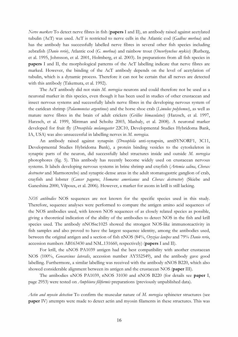
NO donors The NO donors sodium nitroprusside (SNP), S-nitroso-N-acetylpencillamine (SNAP) and hydroxylamine were used to examine the effect of NO on the adrenaline stimulated light response of A. hemigymnus (paper I) and the noradrenalin stimulated response of P. notatus (previously unpublished data). SNP and SNAP were also used to investigate the involvement of NO in the control of M. norvegica luminescence (paper III). SNP and SNAP both release NO in solution (Ioannidis, et al. 1996, Ohta, et al. 1997), as opposed to hydroxylamine that is broken down intracellularly by catalase to yield NO (Ohta, et al. 1997). Hydroxylamine had a stronger effect on A. hemigymnus and P. notatus stimulated luminescence compared to SNP and SNAP. Possibly the localized release of NO, received with the intracellular breakdown of hydroxylamine, could contribute to a higher concentration of NO in the target cell and hence have a larger effect on the luminescence than SNP and SNAP.
NOS inhibitors To block endogenous production of NO, various NOS inhibitors can be used. In paper I, the NOS inhibitor L-thiocitrulline was used on isolated light organs from hatchetfish and L-NAME was used on midshipman fish photophores as well as on live M. norvegica. L-thiocitrulline and L-NAME are S- and N-substituted analogues of L-arginine, respectively (Granik and Grigorév 2002). They both inhibit all NOS isoforms, but L-thiocitrulline is more efficient in inhibiting iNOS compared to the eNOS and nNOS isoforms in mammalian tissue (Joly, et al. 1995), while L-NAME shows moderate selectivity for eNOS and nNOS (Alderton, et al. 2001).
Muscle-effecting drugs To study the putative muscle character and the involvement of sphincter structures in control of krill luminescence, different drugs affecting muscle contraction and relaxation were used in paper III. Substances with different actions were chosen in order to ensure that the effects observed were derived from muscular contractions or relaxations and not from possible effects on other cell types involved in light production.
Two muscle-relaxing drugs, verapamil and papaverine, and two muscle-contracting drugs, thapsigargin and Bay K8544 were used. Both relaxing substances had a stimulatory effect, on the M. norvegica luminescence, even though they have partly different physiological mechanisms. Verapamil inhibits contraction in muscle cells through inhibition of L-type calcium channels in muscle cell membranes (Nwoga, et al. 1996), while papaverine can have several mechanisms of action, which are more or less prominent depending on type of tissue. The effects include muscle relaxation through elevation of cAMP levels by blocking phosphodiesterase that normally metabolizes cAMP, inhibition of mitochondrial respiration and blockade of calcium channels (possibly by an indirect effect of cAMP increase) (du Moulin and Schulz 1975, Kaneda, et al. 1998). Papaverine has primarily been used on mammalian smooth muscle, but has effects on cAMP-levels in crustacean neurons (Araki, et al. 2005).
Both muscle-contracting drugs elevate the calcium concentration in the muscle cell cytoplasm, with different mechanisms. Thapsigargin very specifically inhibits calcium-ATPases from restoring endoplasmatic and sarcoplasmatic calcium stores, thus over time emptying these stores, elevating the calcium concentration in the cytosol and creating contraction of the muscle

21
cell (Treiman, et al. 1998, Wallace, et al. 1999). Bay K8544 also elevates the calcium concentration in muscle cell cytoplasm, although this is achieved through stimulation of L-type calcium channels in the plasma membrane, a separate mechanism from thapsigargin (Monterrubio, et al. 2000, Erxleben and Hermann 2001). Neither of the muscle-contracting drugs stimulated the M. norvegica luminescence, but they had separate effects on the 5-HT stimulated light production (paper IV). Thapsigargin had a quenching effect on the luminescence, in concert with a contraction of the muscular sphincters in the krill photophores, while Bay K8544 surprisingly potentiated the 5-HT stimulated luminescence (paper IV). More experiments are needed to evaluate the causes of these diverging responses.
Data analysis
Different parameters were used to characterize the luminescence response in the pharmacological studies. For the A. hemigymnus (paper I) three parameters were used to characterize the light emission. The light maximum in quanta per second (Lmax, q/s), time to light maximum in seconds (TLmax, s) and time to half extinction of the light response also expressed in seconds (TL1/2). Similar parameters, except TL1/2, were used in P. notatus and sea-pens. Due to the double phased light response of P.notatus one Lmax and one TLmax was decided for each response (Lmax fast, Tlmax fast, Lmax slow and TLmax slow), moreover the ratio between Lmax fast and Lmax slow was used when searching for effects of the treatments on the luminescence from this fish.
Recordings from M. norvegica luminescence did not describe a smooth curve, due to photophores movements during the measurements. Therefore the parameters described above were difficult to determine and not applied. A new set of parameters were used in paper III and IV. In paper III, the primary luminescence data were accumulated over time and the curve received was fitted to a Gompertz growth curve (paper III, page 3182). From the curve, the total light emitted during the experimental period (Ltot, q/s), the maximal rate of light production (RLPmax, q/s2) and the time from experimental start to RLPmax (time to inflection point, Tip, s) were calculated and used in the analysis (paper III, page 3182). As described above, dissolving test substances in the ambient water gave a different kinetics of the light response from injecting the substances. The Gomperzt growth curve did not fit the data obtained in paper IV and therefore an Lmax (q/s) was used, calculated as an average of the light response at the maximal level during 10 min. The kinetic parameters Tstart and TLmax were also used. However, especially TLmax is a rather rough measurement due to the fluctuating primary light curve and it is given in minutes rather than seconds as in A. hemigymnus and P. notatus.

22
RESULTS AND DISCUSSION
The studies in this thesis were conducted to investigate the control mechanisms for bioluminescence in different species with endogenous photocytes. The most important results from papers I-IV are summarized and discussed here, together with some results from previously unpublished studies. For further details, the reader is referred to the individual papers.
Morphology of photophores
The photophore morphology of the investigated species has been described in detail (papers I-IV), and in some instances this adds new observations to already described species. These new observations are summarized below. Notably, photophores from the krill M. norvegica and all fish species in the study were found to be innervated.
Variation in the morphology of fish photophores The photophores of V. attenuata and C. braueri are described with figures for the first time (paper II). Schematic pictures of these organs were constructed from light microscopy observations and literature descriptions (Bassot 1966, Herring and Locket 1978). The photophores of the two species have a similar external appearance: small, black and bead shaped. However, they belong to different classes of photophores, according to the classification of Bassot 1966, based on the arrangement and type of photocytes. V. attenuata has intracellular luminescence whereas C. braueri photocytes secrete components for the luminous reaction to channels between the photocytes. These products are collected in a common duct, possibly where the luminescence reaction occurs (Bassot 1966, Herring and Locket 1978, paper II). This was also the most intensely innervated area in C. braueri photophores (paper II). Nerve fibres were also detected among the photocytes in V. attenuata, but most fibres were present among the outer lens/filter cells, which has not been described before in the literature (paper II). The outer lens/filter was encircled by a collar structure very similar to a structure with light guiding properties found in krill, the laminar ring (Herring and Locket 1978, paper II).
The observations of the photophore morphology in the other fish species studied (A. hemigymnus, P. notatus, M. muelleri and the lanternfish E. risso, H. benoiti and M. punctatum) agrees with earlier observations of light organs from the same, or closely related, species. The luminescence of these fish species is probably intracellular since they lack channels or ducts for luminous products, secreted by the light-producing cells (Bassot 1966, O´Day 1972, paper II). A. hemigymnus and M. muelleri have a similar photophore structure, with the photocytes gathered in a common chamber with several openings for the exit of light (Bassot 1966, Cavallaro, et al. 2004), while the lanternfish species have shallow, cup-shaped, photophores with flattened photocytes connected as a syncytium (O´Day 1972, Anctil and Case 1977). P. notatus have deeper cup-shaped photophores with the photocytes positioned in the dorsal part of the organ. The most prominent structure of the P. notatus photophores is a large cone-shaped lens (Nicol 1957, Mallefet and

23
Anctil 1992b). In conclusion, the morphology of fish photophores is very different among species in general and in the specific species presented here, even though all organs described above probably are used for the same purpose, counter-illumination.
Krill photophores contain capillary sphincters The morphological observations, using electron microscopy, on M. norvegica photophores in paper IV are compatible with observations in two other krill species Euphaucia pacifica and Euphaucia gibboides (Harvey 1977, Herring and Locket 1978), indicating that the general structure of krill photophores is similar between species. The light-producing cells are positioned in the centre of the light organ, forming a striated structure called the lantern. The lantern functions as a sinus, into which oxygenated haemolymph entering the photophore is drained via numerous capillaries (Herring and Locket 1978, paper IV). The opening of each capillary is guarded by a cell type containing filamentous material (sphincter cell).
The general structure of the sphincter cells has been described in previous publications (Petersson 1968, Harvey 1977, Herring and Locket 1978), but in paper IV these cells were studied in detail by using electron microscopy and histochemistry in M. norvegica. The filamentous material consists of thick and thin muscular filaments arranged in blocks oriented along or around the capillary as a sphincter structure. Both capillary epithelium and sphincter structures contain filamentous actin, labelled with the actin probe rhodamin phalloidin. The sphincter structures are seen as thickenings at the outflow of the capillaries, indicating a concentration of filamentous actin, probably identical to the thin filaments observed with electron microscopy (paper IV). (For an even more detailed description of M. norvegica photophores and schematic pictures, see paper III and IV). The light-producing cells are not innervated (paper IV). Instead, varicose nerve fibres, containing 5-HT, follow the capillaries and reach the sphincter cells, as shown with antibodies against 5-HT (paper IV). Varicose nerve fibres are normally an indication of terminals with transmitter release. This was recently further confirmed by synapsin immunohistochemistry (fig. 5) which labelled nerves along the capillaries, showing the presence of nerve terminals along the whole length of the capillaries throughout the photophore, not only at the sphincter structures. In double labelled preparations 100% coexistence was found between 5-HT and synapsin immunoreactivity. The 5-HT and synapsin-labelled fibres possibly affect the endothelial cells of the capillaries or an adjacent cell type (A-cells, paper III, page 3180), so far believed to have a supportive function (Herring and Locket 1978, Skiebe and Ganeshina 2000). Innervation of sphincter cells and whole capillaries suggested that these structures might be regulated to control the haemolymph flow, and hence the oxygen supply, to the light-producing cells. Several scientists have speculated that oxygen might be the single stimulus for luminescence in krill (Petersson 1968, Harvey 1977, Herring and Locket 1978).

24
Fig. 5 Detail from Meganyctiphanes norvegica photophore. (a) Numerous capillaries (c) containing filamentous actin (rhodamin phalloidin, red), ramify in the photophores and end with sphincter structures (sp) at the lantern (la, light-producing structure) in the centre of the organ. (b) Varicose nerves (synapsin, green), (c) containing 5-HT (blue), follow the capillaries and reach the sphincter structures. Bars are 25μm Nitric oxide mechanisms
Histochemical and/or pharmacological indications for the involvement of NO in the control of bioluminescence were obtained in surprisingly many of the species included in this thesis (table 1, page 33). However, the results suggest several different mechanisms rather than a common role for NO in the investigated species, also multiple functions within photophores of the same species.
NOS-like proteins are contained in photophores from several species Presence of NOS-like proteins, shown with immunohistochemistry, indicates a capacity for NO production in the photophores of krill and all the mesopelagic fish species studied. NOS-like immunoreactivity appeared in several different structures, e.g. photocytes, nerves and lens/filter cells, within the same photophore in lanternfish species, A. hemigymnus, M. muelleri and V. attenuata, suggesting multiple roles for NO within the same light organ.
The lantern fish photocytes as well as nerves reaching the photocytes contains NOS-like protein, a prerequisite for an involvement of NO on the luminescence reaction. In A. hemigymnus and M. muelleri the NOS-like substance is present in nerves or in structures, which could possibly be nerves or vessels (M. muelleri), among the photocytes, but also inside the lens or filter cells, implicating a possible role for NO both in the production and modulation of light. Modulations of light characteristics by NO, rather than a direct effect on the luminescence reaction, is also suggested by the location of NOS-like immunoreactivity in nerves among lens or filter cells, and in the cells building the collar structure (mentioned above) in V. attenuata photophores. A putative way of modulating the light emitted by the photocytes would be to change the light absorbing or diffracting properties of the lens or filter cells. Since NO is known to induce melanogenesis in human skin cells (Cals-Grierson and Ormerod 2004), it is tempting to suggest that NO could regulate the production of light absorbing pigment in the lens or filter cells by a similar mechanism.
In C. braueri and M. norvegica photophores, NOS-like immunoreactivity is associated to a single type of tissue structure, epithelial cells of the collecting duct for luminous products in C. braueri, and capillary epithelium and sphincter cells in M. norvegica. The epithelial cells in C. braueri
a cb
la
c
sp
a cb
la
cc
sp

25
are in close association to the photocytes and NO produced there has a good theoretical chance of reaching the photocytes, as it can spread tens of millimetres from the source (Edelman and Gally 1992). NO interferes with the exocytosis machinery in some systems and could possibly directly affect excretion of luminous products from C. braueri photocytes (Lowenstein 2007), but may as well have other functions. The presence of NOS-like immunoreactivity in capillary epithelium and sphincter cells in M. norvegica photophores implies a possible role for NO in controlling the supply of oxygen transported to the lantern with the haemolymph (further discussed below).
NOS-like immunoreactivity could not be detected in photophores from P. notatus (paper II) and luminescent tissue of an echinoderm, the brittlestar A. filiformis (J. Krönström, C. Vanderlinden and J. Mallefet, previously unpublished data). This could indicate that NOS is lacking in the luminescent system of these species, or that the tested antibodies did not fit a putative P. notatus or A. filiformis NOS. The intense staining of photocytes from P. notatus, using the NADPHd technique (paper II), could suggest a presence of a NOS protein in these cells but could possibly attributed to from some other NADPHd enzymes present in the photoytes. In mammals, different NADPH dehydrogenases, NADPH ferrihaemoprotein reductase, NADPH cytochrome c2 and NAD(P)+ transhydrogenase are all enzymes which can reduce NADPH and produce staining with the NADPHd technique (Blotter, et al. 1995).
NOS has been detected with immunohistochemistry in the nervous system of the echinoderm starfish Marthasterias glacialis, using antibodies against mammalian nNOS (Martinez, et al. 1994) and two different NOS isoforms have been sequenced in the sea urchin Arbacia punctulata (Cox, et al. 2001). This indicates a high probability for the existence of a brittlestar NOS, although this was not detected with the antibodies used in this study. On the other hand, the lack of effect with the NO donor SNAP on the luminescence of the brittlestars Ophiopsila aranea and Ophiopsila californica, and of SNAP and the NOS inhibitor L-NAME on A. filiformis luminescence, suggests that NO is not involved in the control of light production in this species (Dewael and Mallefet 2002, personal observations).
From the results of the histochemical studies it can be concluded that NOS-like material is present in several, closely and distantly related, species but is probably not commonly present in all luminescent systems. Clearly, histochemical studies are not enough to elucidate the effects and significance of NO in control of bioluminescence, and functional studies are needed for this purpose. Therefore, in this thesis, pharmacological experiments investigating the effect of NO donors and NOS inhibitors on light production have been performed on isolated photophores from A. hemigymnus and P. notatus, live M. norvegica and A. filiformis arm segments.
NO has various physiological effects on luminescence reactions NO modulates the luminescence reaction stimulated with adrenaline (A. hemigymnus, paper I), noradrenaline (P. notatus, fig. 6) or 5-HT (M. norvegica, paper III), but had no effect per se, in contrast to the mechanism in fireflies where NO stimulates light production (Trimmer, et al. 2001). The effects of NO donors on the adrenaline (A. hemigymnus) or 5-HT (M. norvegica) stimulated luminescence are mainly inhibitory, although a small portion of the A. hemigymnus samples instead, for unknown reasons, are potentiated by NO donors. The level of inhibition is correlated to the ability of the separate photophores to respond to adrenaline, strongly

26
responding organs are more inhibited by NO donors than weakly responding ones (paper I, page 2956). In concert with the inhibitory effect of the NO donors, the NOS inhibitors L-thiocitrulline and L-NAME potentiated the stimulated light reactions in A. hemigymnus and M. norvegica, respectively, again apart from some A. hemigymnus samples that showed the opposite reaction.
The different levels of response to NO donors and inhibitors between A. hemigymnus individuals are reflected in the density of NOS-like immunoreactive nerve fibres observed among the photocytes in the light organs from separate individuals, using immunohistochemistry (paper I). Experiments show that all NOS isoforms, not only iNOS, can be induced (Alderton, et al. 2001) and NOS expression in A. hemigymnus could possibly be regulated to meet the current need for NO in the photophores, regardless of the NOS isoform present. Furthermore, a large potentiating effect on the luminescence from photophores weakly responding to adrenaline, of the NOS inhibitor L-thiocitrulline, may indicate an inhibitory NO tonus in these preparations. An NO tonus is, for instance, present in vessels and intestine of fish and mammalians (Daniel, et al. 1994, Karila and Holmgren 1995, Mustafa, et al. 1997, Agnisola 2005).
Similarly to the dual effects of NO on A. hemigymnus light production, NO appears to potentiate the first phase of the P. notatus noradrenaline stimulated light response, but inhibit the second phase. This is illustrated by a higher Lmax fast/Lmax slow ratio in the presence of the NO donor hydroxylamine than with noradrenaline alone. In concert, this ratio was significantly lowered by L-NAME, mainly due to an inhibitory effect on the fast response. A possible explanation for this is an NO stimulated increased release of noradrenaline from nerve terminals among the photocytes, which has been shown in intestinal tissue from mammals (Yamamoto, et al. 1994, Sotirov and Papasova 2000). An increased release of noradrenaline would primarily stimulate α-receptors on the photocytes, believed to be responsible for the fast light response (Christophe and Baguet 1983, Christophe and Baguet 1985).
Fig. 6 Effects of the NO-donor hydroxylamine (OH-amine, 0.1 mM) and the NOS-inhibitor L-NAME (0.1 mM) on the ratio between the peak of the fast and slow light response from P. notatus, compared to the noradrenaline (NA, 0.1 mM) control. Statistical method was ANOVA with log-transformed values and Tukey’s post-hoc test. * indicates statistical significant difference between treatment and control (p<0.05). Error bars show standard error and number of replicates were: nNA=12, nOH-amine=9 and nL-NAME=6. Previously unpublished data (J. Krönström, C. Vanderlinden and J. Mallefet)
Effects of NO on luminescence are independent of cGMP Like the distribution of NOS-like immunoreactivity in the photophores, the effects of NO on the luminescence differ between the investigated species. What appears to be common though, is the independence of a cGMP effector pathway for NO in the photophores. Pharmacological experiments with A. hemigymnus (paper I) and M. norvegica (paper III) did not reveal effects of
NA L-NAMEOH-amine+NA+NA
L max
fast
/ Lm
axsl
ow
0.0
1.5
1.0
0.5
3.0
2.5
2.0
*
*
NA L-NAMEOH-amine+NA+NA
L max
fast
/ Lm
axsl
ow
0.0
1.5
1.0
0.5
3.0
2.5
2.0
*
*

27
either the cGMP analogues (8BrcGMP and dibutyryl cGMP) or the guanyl cyclase inhibitor ODQ on the adrenaline (A. hemigymnus) or the 5-HT (M. norvegica) stimulated light reactions (apart from a lower RPLmax in the cGMP+5-HT treatment compared to 5-HT controls). This is in contrast to the stimulation of cGMP production by NO found in many species in other signalling pathways and there are both functional and morphological evidence in the literature for such cGMP pathways in fish and crustaceans (Abebe, et al. 1995, Zhan, et al. 1999, Scholz 2001, Aonuma 2002). However, other effector pathways for NO have also been described, including modulation of potassium- and calcium-channels via a direct effect on the ion-channels in skeletal muscle cells from the isopod Idothea baltica, (Erxleben and Hermann 2001, Hermann and Erxleben 2001) and blockade of mitochondrial respiration by direct binding of NO to elements in the electron transport chain, for example in mammalian muscle cells and nerve terminals (Brown 1995).
Putative effects of NO on photophore respiration and oxygen availability The stimulatory effect of NO on luminescence in firefly (Photuris spp. and Photinus spp.) is mediated via blockade of mitochondrial respiration, leaving oxygen free to take part in the luminescence reaction (Timmins, et al. 2001b, Trimmer, et al. 2001). A similar blockade of photocyte respiration is a feasible mechanism for NO action in both A. hemigymnus and P. notatus. In isolated photophores from P. notatus, light production is induced when mitochondrial respiration is blocked with KCN (Mallefet and Baguet 1984). The relationship between light production and cellular respiration is not as clear in A. hemigymnus photophores. However, oxygen consumption is changing during luminescence, suggesting a connection between light production and cellular respiration in this species, though probably with a different reaction mechanism than that of P. notatus photophores (Mallefet and Baguet 1985).
NO inhibits the light production in M. norvegica, which is in contrast to the situation in firefly (Trimmer, et al. 2001). This agrees with the different morphological arrangements of NOS-containing structures in the two species. In fireflies, NO is released directly to the photocytes, while in M. norvegica the location of NOS-like material around capillaries and sphincter cells (described above) suggests that NO could be involved in the control of the flow of oxygenated haemolymph to the lantern (Greenfield 2001, Trimmer, et al. 2001) (paper III). In mammals NO is a vasodilatory substance (Bredt and Snyder 1994), but a putative vasodilatory function is not compatible with the inhibition of the light response caused by NO. According to earlier hypotheses, a dilation of capillaries and/or sphincter structures would allow more oxygenated haemolymph to enter the lantern and instead induce light emission (Petersson 1968, Harvey 1977, Herring and Locket 1978). On the contrary, it is feasible that a contraction of the filamentous material in the sphincter structure could be stimulated by an NO mediated opening of calcium channels in the membrane of the sphincter cells. An increased inward calcium current, caused by NO, has been shown in muscle cells from the crustacean Idothea Baltica (Erxleben and Hermann 2001). Contraction of the sphincter cells, and consequently a decreased flow of oxygenated haemolymph to the lantern, is in concert with the observed quenched luminescence at exposure to NO donors (paper III).
Moreover, NO could possibly be released to the haemolymph, transported to the lantern and have a direct effect on the photocytes, for example a hyperpolarization of these cells, making

28
them less reactive to other stimuli (Hermann and Erxleben 2001). It is feasible that NO is transported to the lantern bound to haemocyanin, since this crustacean blood pigment functions as an NO scavenger. In addition, a replacement of oxygen for NO in the haemocyanin molecule would result in less oxygen to the light-producing cells and hence less light production, regardless of the volume of haemolymph passing through the lantern (Jacklet and Koh 2001).
The pharmacological studies confirm the histochemical observations, that NO is involved in the control of diverse luminescence systems, ranging from krill to several fish species, possessing fundamentally different types of photophores. However, the effects of NO are not compatible between species. This agrees with the different evolutionary origin of the luminescence systems (Herring 1987).
Connections between muscle relaxation, flow of haemolymph and luminescence in krill
Morphological observations thus suggest that muscular sphincter structures are central in the control of luminescence in krill (paper IV, Herring and Locket 1978). In order to study the functional role of these structures, the effects of muscle relaxing and contracting drugs on the luminescence response were investigated (paper IV).
Stimulation of the light response by the muscle relaxing drugs, verapamil and papaverine, indicates that increased access to oxygen, through relaxation of the sphincter structures and increased flow of haemolymph, could be a single stimulus for luminescence in these photophores, as speculated by earlier investigators (above). In concert, the muscle contracting drug thapsigargin partly quenched the luminescence reaction stimulated with 5-HT.
The dense 5-HT innervation reaching the sphincter cells (paper III and fig. 5), which contain contractile elements (paper IV, page 9 and 10), suggests that 5-HT might be the substance relaxing the sphincter structures. Different effects of 5-HT on crustacean musculature, for example increasing heart rate and contraction force of the stomatopod, Squilla oratoria, but also relaxing arterial valves in the lobster Panilurus japonicus have been shown (Kuramoto and Ebara 1984, Beltz and Kravitz 1986, Ando and Kuwasawa 2004). Moreover, it is possible that the whole capillaries are capable of relaxing and contracting, since labelling for filamentous actin was present in the capillary walls throughout the capillaries inside the photophores (fig. 5). In lobster (Homarus americanus) some vessels have recently been shown to possess contractile properties and contain the contractile elements actin, myosin and tropomyosin, even though they are lacking organised muscle filaments in the vessel wall (Wilkens, et al. 2008). The importance of the capillaries in M. norvegica photophores is further supported by the labelling of synapsin (fig. 5), indicating synaptic formations, in the nerves along the whole capillaries in the photophores.
5-HT has a potentiating effect on the light response when combined with each of the relaxing drugs. The mechanisms behind this are unclear, but it could possibly indicate that 5-HT has other, or additional, effects on the luminescence system than the postulated relaxing effects on the sphincter structures. For example 5-HT could stimulate the release of a second, so far unidentified, transmitter. This effect of 5-HT has been shown in the crayfish neuromuscular junction (Eynart 1981). Even so, it is difficult to picture the mechanisms behind the strongly potentiating effect of 5-HT on the papaverine and verapamile responses. It may be speculated

29
that the muscle relaxing drugs and 5-HT act in synergy during signal transduction. The relaxing effect of papaverine is mediated partly by inhibition of cAMP breakdown (du Moulin and Schulz 1975, Kaneda, et al. 1998) and 5-HT may also contribute to an increase in intracellular cAMP levels, which has been demonstrated for example in neurons in the crayfish, Procambarus clarkii, tail (Araki, et al. 2005), and in the neuromuscular junction of the crayfish, Orconectes virilis (Eynart 1981). A relaxing effect from a possible 5-HT stimulated cAMP increase could be enhanced by both papaverine and verapamil through blocking the influx of calcium in muscle cells (Nwoga, et al. 1996, Kaneda, et al. 1998).
The results from the studies of the involvement of the sphincter structures in control of light production in M. norvegica (paper IV), combined with the results from the investigation of the effects of NO on the same system (paper III) highlights the complexity of this control system in which several components, including oxygen, may be involved.
Adrenaline has inconstant effects on sea-pen luminescence
Within the echinoderm group, even closely related species can have different chemical coding of the nervous control of the luminescence reaction (Dewael and Mallefet 2002, Vanderlinden and Mallefet 2004). In order to find out whether a similar situation exists in cnidarians, attempts were made to study several cnidarian sea-pen species (P. phosphorea (Gullmar fjord), P. rubra, P. spinosum and V. cynomorium (Mediterranean)) for comparisons with previous studies in the sea-pen R. koellikeri.
In Renilla spp., adrenaline, but not noradrenaline, stimulated luminescence in whole colonies and in isolated auto- and siphonozooids, probably via �-like adrenoceptors (Anctil and Boulay 1982, Awad and Anctil 1993). However, in our previously unpublished studies (J. Krönström, C. Vanderlinden and J. Mallefet) inconclusive results were obtained with adrenergic drugs. Preparations from a few individuals of P. phosphorea responded with clear trains of short light flashes to both adrenaline and noradrenaline (0.001 and 0.1 mM), while others were unresponsive to catecholamine stimulation or possibly showed weak responses with varied kinetics. A few preparations from the Mediterranean species P. spinosum and V. cynomorium were also weakly responsive to adrenaline and noradrenaline (0.01 μM-0.01 mM). They showed several types of responses, ranging from trains of flashes to single, occasional, flashes or longer feeble glows. This instability in responses to adrenergic drugs was also observed in isolated photocytes from P. rubra and V. cynomorium.
Since our preparations were only occasionally stimulated with adrenaline or noradrenaline, it is possible that they for some reason were unstable. When all attempts to obtain stable preparations, through changes in the experimental protocol, failed we could not move on to investigate the NO system in sea-pens. However, experiments with R. koellikeri indicate that NO is not involved in the control of luminescence in this sea-pen species (Anctil, et al. 2005).

30
SUMMARY AND CONCLUSIONS
The studies included in this thesis introduce the signalling substance NO, earlier only investigated in fireflies, as a modulator of luminescence in several bioluminescent systems. Moreover, they contribute to the detailed description of photophore morphology, mainly of the krill M. norvegica photophores but also in several fish species, and to the understanding of the role of capillary sphincters in control of M. norvegica luminescence.
The studied photophores from fish and krill were all found to be innervated and 5-HT, which stimulates luminescence in krill, was shown for the first time in nerve structures inside M. norvegica photophores . The distribution of 5-HT-containing nerves inside these photophores, suggests that control of luminescence might be mediated through an action on sphincter structures and/or capillaries. Evidence presented in this thesis indicate that these structures have contractile properties which are of importance for luminescence control, presumably through changes in haemolymph flow and oxygen supply to the photocytes. Contractility may not be a common feature of crustacean vessels, but it is not unknown. Moreover, NOS is present in the area of sphincter structures and capillaries, suggesting that NO also takes part in this control. This is supported by a quenching effect of NO-donors, and a potentiating effect of an NOS inhibitor, on the 5-HT stimulated luminescence in M. norvegica.
The involvement of NO in luminescence control could be rather widespread since evidence for NOS-like proteins was found in photophores from several fish species, belonging to five different fish families, and in a crustacean. Photophores used for different purposes, like signalling as the lantern fish caudal photophores and possibly the photophores of M. norvegica and counter-illumination as the A. hemigymnus photophores, all contain NOS-like material. Interestingly, the distribution of NOS-like substance varies between photophores from different species and similarly, the effects of NO differ between the various luminous systems. The effects are mainly quenching in M. norvegica and A. hemigymnus, potentiating on a part of the P. notatus light response and directly stimulating on the previously studied firefly. The signalling pathway for NO is still unknown for the marine species, but as in fireflies, the NO effects are not mediated via cGMP, which is the most commonly reported second messenger for NO.
In future studies, detailed studies of the mechanisms of action for NO in bioluminescent systems could reveal unknown NO signalling pathways. In order to understand the commonality of NO in luminescence control, the effects of NO on luminescence in even more species from different animal groups, need to be studied. Furthermore, the basic knowledge of photophore morphology and innervation is still poor in many species and thorough investigations of the internal organization of photophores are important, also for the understanding of the function. The krill photophores are well described, and here the next step towards understanding of the control of light-production would be further investigations of the photophore capillary structures on a cellular level, and possibly micromeasurements of hemolymph flow and oxygen contents in different situations.

31
In conclusion, the studies included in this thesis adds knowledge of even more variety in the wonderful world of bioluminescence that already has large variation on several levels, ecological, morphological and last but not least functional.

32

33
Table 1 Systematic relationship, physiological characteristics of luminescence control, luciferins and luminescence behaviour of the studied species. For comparison a few species (indicated with *) and pieces of data from other studies are included. Abbreviations are adrenaline (A), noradrenaline (NA), acetylcholine (Ach), nitric oxide (NO), octopamine (Oct), 5-hydroxytryptamine (5-HT), immunoreactivity (IR), nitric oxide synthase (NOS), coelenterazine (coelent.), luciferin (lucif.), predator (pred.), communication (com.), counter-illumination (counter-ill.). Substances or effects within brackets are secondary to or weaker than the ones without brackets. + indicates positive immunoreactivity against a specific substance, while - means that the substance could not be detected with so far used antibodies. References: papers I-V, previously unpublished results (J. Krönström, C. Vanderlinden and J. Mallefet), Mauchline 1960, Crane 1965, Barnes and Case 1974, Baguet 1975, Baguet and Marechal 1977, Anctil and Boulay 1982, Christophe and Baguet 1983, Christophe and Baguet 1985, Mallefet and Anctil 1992b, Harper and Case 1999, Widder 1999, Herring 2000b, Trimmer, et al. 2001, Dewael and Mallefet 2002, Dupont, et al. 2004, Shimomura 2006
Phyla Class Order Family Species Light stimuli
Light modulators
IR in photophores Luciferine Light behaviour
Cnidaria Anthozoa Pennatulacea Pennatulidae Pennatula phosphorea A/NA? coelent.? avert pred.?
Pennatula rubra A/NA? coelent.? avert pred.?
Pteroides spinosum A/NA? coelent.? avert pred.?
Veritillium cynomorium A/NA? coelent.? avert pred.?
Renilla Koellikeri* A coelent avert pred.?
Echinodermata Ophiuroidea Ophiurida Amphiuridae Amphiura filiformis Ach (A) NOS- coelent. avert pred.?
Arthropoda Insecta Coleoptera Lampyridae Photuris spp.* NO (Oct) NOS+ Firefly-lucif. com.
Malacostraca Euphauciacea Euphausidae Meganyctiphanes norvegica 5-HT NO- 5-HT+, NOS+ Krill-lucif. counter-ill./com.?
Chordata Actinopterygii Stomiiformes Sternoptychidae Argyropelecus hemigymnus A (NA) NO-(+) NOS+ coelent.? counter-ill.
Maurolicus mulleri A NOS+ coelent.? counter-ill.
Phosichthyidae Vinciguerria attenuata NOS+ coelent.? counter-ill.
Gonostomatidae Cyclothone braueri NOS+ coelent.? counter-ill.
Myctophiformes Myctophidae Electrona risso NOS+ coelent.? counter-ill./com.
Hygophum benoiti NOS+ coelent.? counter-ill./com.
Myctophum punctatum NOS+ coelent.? counter-ill./com.
Batrachoidiformes Batrachoididae Porichthys notatus NA (A) 5-HT-, NO+(-)
A+, NA+, 5-HT+, NOS- Cypridina-lucif. counter-ill./com.?

34

35
ACKNOWLEDGEMENTS
First of all I would like to thank my supervisors Susanne Holmgren, Jerome Mallefet and Mike Thorndyke for giving me the opportunity to work with the most beautiful and exciting biological phenomenon in the world! Thank you: Susanne, for giving me an excellent research education and for maintaining a high standard and professionalism in everything you do. Thank you for showing your soft and understanding side when necessary and for doing my job when things, like giving birth, get in the way of paper deadlines. Thank you for nice company during field-trips and conferences and for joining me to every sushi-restaurant in Yokohama, even though you do not eat sushi…. Thank you for your encouragement when I am nervous or fed up, your opinion means a lot to me. Jerome, for introducing me to the science of bioluminescence and for teaching me how to study it. Thank you for wonderful field-trips to Sicily and France, and for taking very good care of me in Louvain-la-Neuve. Thank you for showing me animals I would never had seen if I had not known you, for introducing me to a Nobel Prize winner and for letting me taste your excellent cooking. Mike, for sharing your expertise in molecular biology and for always making me feel welcome whenever I turn up at Kristineberg, announced or unannounced. Thank you for introducing me to interesting scientists from all over the world, and for your great enthusiasm when it comes to biological research. I have never seen a more cheerful dissection of a hagfish!
I would also like to thank my other coworkers and/or coauthors for great cooperation. I have learned a lot from you all. Special thanks to: Christine Vanderlinden, for getting me started with the luminometer measurements and for putting your own projects aside to help me. Thank you for fun times inside and outside the lab and for letting me stay at your house (all the best to your parents), the first time I visited Belgium. Waldemar Karlsson, min enda och bästa examensstudent. Tack för ditt vetenskapliga bidrag till min avhandling och för ett fantastiskt underhållande år. Tack för alla möjliga och omöjliga teorier om det mesta, din ärliga nyfikenhet på mitt turbulenta privatliv, och tack för god husmanskost. Låt mig aldrig dricka i kapp med dig igen.
Thank you all staff at the Department of Zoology and at the Sven Lovén Centre for Marine Sciences, you are making it fun and easy to go to work. Tack till min examinator, Thrandur Björnsson för kritisk läsning av min avhandling. Tack båtpersonalen på Kristineberg för hjälp med insamling av djur i alla väder och för trevliga stunder på seglande och ankrat fartyg. Tack alla doktorander på Zoologen och Kristineberg, för bra gemenskap, för hjälp med allt möjligt från undervisning till forskning, trevliga luncher och kul fester.
Tack nuvarande och tidigare medlemmar av GENI och COCA-grupperna, för att ni alltid satt er in i, och visat intresse för, mitt ämne fast det varken handlar om tarmar eller kärl. För er noggranna dissekering av min avhandling och för proffsig utfrågning på grillningarna, för mysiga måndagsmöten och god kaka. Tack:

36
Henrik Seth, för din välutvecklade ”ta reda på”-hjärna. Tack för att du alltid lyssnar noga på mina små och stora vetenskapliga problem. Tack för en perfekt balans mellan arbetsfokus och småprat på vårt arbetsrum. Tack för att du flyttade åt mig och för att allting på något vis verkar vara så enkelt i dina ögon. Du jobbar mer än någon jag känner, men med minst ansträngning. Christina Hagström, för proffsig hjälp och trevlig atmosfär på hist lab. Det är så roligt att du delar min danspassion! Michael Axelsson, för din hjälp Waldemars arbete, men även mycket annat. Tack för att du har rest dig för att hjälpa till, i princip innan man uttalat frågan och för att allt har en snabb, och oftast teknisk, lösning. Tack för intressanta diskussioner med udda vinklingar och en ström av aldrig sinande idéer. Catharina Olsson, för din stora kunnighet inom skrivande och laborativa tekniker och din vilja att dela med dig av den. Tack för din principfasthet som gör att om man frågat dig något, och fått svar, så vet man att det är korrekt. Tack för att du organiserat våra måndagsmöten på ett givande sätt och för att du kommit med många genomtänkta och bra synpunkter på min avhandling. Jag tror du är mjukare än du låter, oftast. Albin Gräns och Fredrik Albertsson för trevlig stämning, givande diskussioner, kaffe och bra lunchsällskap. Anna Holmgren, Ågot Johansson och Monica Sundqvist, medlemmar av GENI-gruppen när jag började doktorera. Tack för att ni fick mig att känna mig välkommen och lärde mig gruppens fika och lunch kutym.
Jag vill också tacka min familj och mina vänner som gör livet roligt, lätt och enkelt att leva och som tar hand om mig på bästa sätt utanför jobbet. Tack: Mina ”medmammor” Sandra och Anja med familjer, för att ni förgyllt min föräldraledighet. Anna, min äldsta barndomsvän, för att du alltid finns när du behövs och för att ingenting förändras oss emellan fastän tiden går ibland. Annelie, för att du är så omtänksam och för en minnesvärd tid av 50- och 5+. Selma, vårt äventyr på Ostindiefararen och i Brasilien är oslagbart, där delade vi allt, bland annat mitt häftigaste bioluminiscens-ögonblick. Tack för din värme och skön tango. Mamma, Pappa, Marie och Elin, ni har gjort mig till en trygg och självsäker person. Tack för ert stöd i alla lägen. Johan, min kärlek, för att du verkar stolt över det jag gör och för att du alltid vet hur jag mår, egentligen. Tack för att du lagt dina intressen och måsten åt sidan och agerat ensamstående pappa, medan jag avslutat min avhandling. Det är fantastiskt att det är mig du vill vara med! Freja, min underbara dotter för att du saknat mig, och väntat på mig, och inte velat sova förrän jag jobbat färdigt sent på kvällen. Tack för att du är så rolig och gör mig så lycklig och stolt. Du är alltid centrum, jag är din mamma. The work presented in this thesis was financially supported with grants from the Swedish Science Research Council to S. Holmgren and from the Knut and Alice Wallenberg Foundation, the Wilhelm and Martina Lundgren foundation, the Helge Ax:son Johnsson Foundation and Jubileumsfonden to J. Krönström.
37
REFERENCES
Abebe W, Hussain T, Olanrewaju H and Mustafa J (1995) Role of nitric oxide in adenosine
receptor-mediated relaxation of porcine coronary artery. Heart Circ. Physiol. 38:H1672-H1678
Agnisola C (2005) Role of nitric oxide in the control of coronary resistance in teleosts. Comp. Biochem. Physiol. 142:178-187
Ahlstrom EH (1973) The diverse patterns of metamorphosis in Gonastomatiid fishes-an aid to classification. International symposium of the early life history of fish, Oban, Scottland, pp 659-674
Alderton WK, Cooper CE and Knowles RG (2001) Nitric oxide synthases: structure, function and inhibition. Biochem. J. 357:593-615
Anctil M (1971) Stimulation of bioluminescence in lanternfishes (Myctophidae). II. Can. J. Zool. 50:233-237
Anctil M and Case JF (1977) The caudal luminous organs of lanternfishes: general innervation and ultrastructure. Am. J. Anat. 149:1-22
Anctil M and Boulay D (1982) Monoaminergic mechanisms associated with control of luminescence and contractile activities in the coelentrate, Renilla köllikeri. J. Exp. Zool. 223:11-24
Anctil M, Descarries L and Watkins CK (1984a) Distribution of [3H]noradrenaline and [3H]serotonin in photophores of Porichthys notatus. Cell Tissue Res. 235:129-136
Anctil M, Germain G and LaRiviére L (1984b) Catecholamines in the coelentrate Renilla köllikeri. Cell Tissue Res. 238:69-80
Anctil M, Poulain I and Pelletier C (2005) Nitric oxide modulates peristaltic muscle activity associated with fluid circulation in the sea pansy Renilla koellikeri. J. Exp. Biol. 208:2005-2017
Anderson C (1983) Evidence for 5-HT-containing intrinsic neurons in the teleost intestine. Cell Tissue Res. 230:377-386
Anderson C and Campbell G (1988) Immunohistochemical study of 5-HT-containing neurons in the teleost intetine: relationship to the presence of enterochromaffin cells. Cell Tissue Res. 254:553-559
Anderson JM and Cormier MJ (1973) Luminosomes, the cellular site of bioluminescence in coelaentrates. J. Biol. Chem. 248:2937-2943
Anderson PAV and Case JF (1975) Electrical activity associated with luminescence and other colonial behaviour in the pennatulid Renilla köllikeri. Biol. Bull. 149:80-95
Ando H and Kuwasawa K (2004) Neuronal and neurohormonal control of the heart in the stomatopod crustacean, Squilla oratoria. J. Exp. Biol. 207:4663-4677
Aonuma H (2002) Distribution of NO-induced cGMP-like immunoreactive neurones in the abdominal nervous system of the crayfish, Procambarus clarkii. Zool. Sci. 19:969-979
Araki M, Nagayama T and Sprayberry J (2005) Cyclic AMP mediates serotonin-induced synaptic enhancement of lateral giant interneuron of the crayfish. J. Neurophysiol. 94:2644-2652
Awad EW and Anctil M (1993) Identification of beta-like adrenoreceptors associated with bioluminescence in the sea pansy Renilla koellikeri. J. Exp. Biol. 177:181-200
Baguet F (1975) Excitation and control of isolated photophores of luminescent fishes. Prog. Neurobiol. 5:97-125
Baguet F and Marechal G (1977) The stimulation of isolated photophores (Argyropelecus) by epinephrine and norepinephrine. Comp. Biochem. Physiol. 60C:137-143

38
Barnes AT, Case JF and Tsuji FJ (1973) Induction of bioluminescence in a luciferin deficient form of the marine teleost, Porichthys, in response to exogenous luciferin. Comp. Biochem. Physiol. 46A:709-723
Barnes AT and Case JF (1974) The luminescence of the lanternfish (Myctophidae): spontanous activity and responses to mechanical, electrical and chemical stimulation. J. Exp. Mar. Biol. Ecol. 15:203-221
Bassot J-M (1966) On the comparative morphology of some luminous organs. In: Johnsson FH and Haneda Y (eds) Bioluminescence in progress. Princeton university press, Princeton
Beltz BS and Kravitz EA (1986) Aminergic and peptidergic neuromodulation in Crustacea. J. Exp. Biol. 124:115-141
Beltz BS (1999) Distribution and functional anatomy of amine-containing neurons in decapod crustaceans. Microsc. Res. Tech. 44:105-120
Bicker G (2001) Sources and targets of nitric oxide signalling in insect nervous systems. Cell Tissue Res. 303:137-146
Blenau W and Baumann A (2001) Molecular and pharmacological properties of insect biogenic amine receptors: lessons from Drosophila melanogaster and Apis mellifera. Arch. Insect. Biochem. Physiol. 48:13-38
Blotter D, Grozandovic Z and Gossrau R (1995) Histochemistry of nitric oxide synthase in the nervous system. Histochem. J. 27:758-811
Bode VC and Hastings JW (1963) The purifiation and properties of the bioluminescent system in Gonyaulax poledra. . Arch. Biochem. Biophys. 103:488-499
Bredt DS and Snyder SH (1994) Nitric oxide: a physiologic messenger molecule. Ann. Rev. Biochem. 63:175-195
Brown GC (1995) Nitric oxide regulates mitochondrial respiration and cell functions by inhibiting cytochrome oxidase. FEBS Lett. 369:136-139
Cals-Grierson M-M and Ormerod AD (2004) Nitric oxide function in the skin. Nitric Oxide 10:179-193
Carlberg M and Rosengren E (1985) Biochemical basis for adrenergic neurotransmission in coelentrates. J. Comp. Biochem. Physiol. B 155:251-255
Case JF and Morin JG (1966) Glutamate suppression of neuroeffector processes in a coelenterate. Am. Zool. 6:525
Case JF, Warner J, Barnes AT and Lowenstine M (1977) Bioluminescence of lanternfish (Myctophidae) in response to changes in light intensity. Nature 265:179-181
Cavallaro M, Mammola CL and Verdiglione R (2004) Structural and ultrastructural comparison of photophores of two species of deep-sea fishes: Argyropelecus hemigymnus and Maurolicus muelleri. J. Fish Biol. 64:1552-1567
Christophe B and Baguet F (1982) Luminescence of isolated photophores and supracaudal gland from Myctophum punctatum: electrical stimulation. Comp. Biochem. Physiol. 71A:131-136
Christophe B and Baguet F (1983) Luminescence of isolated photocytes from Porichthys: adrenergic stimulation. J. Exp. Biol. 104:183-192
Christophe B and Baguet F (1985) The adrenergic control of the photocyte luminescence of the Porichthys photophore. Comp. Biochem. Physiol. 81C:359-365
Cormier MJ (1978) Comparative biochemistry of animal systems. In: Herring PJ (ed) Bioluminscence in action. Academic press, London, pp 75-108
Cox RL, Mariano T, Heck DE, Laskin JD and Stegeman JJ (2001) Nitric oxide synthase sequences in the marine fish Stenotomus chrysops and the sea urchin Arbacia punctulata, and phylogenetic analysis of nitric oxide synthase calmodulin-binding domains. Comp. Biochem. Physiol. B Biochem. Mol. Biol. 130:479-491
Crane JM (1965) Bioluminescent courtship display in the teleost Porichthys notatus. Copeia 2:239-241

39
Daniel EE, Haugh C, Woskowska Z, Cipris S, Jury J and Fox-Threlkeld T (1994) Role of nitric oxide-related inhibition in intestinal function: relation to vasoactive intestinal polypeptide. Am. J. Physiol. 266:G31-G39
De Bremaeker N, Mallefet J and Baguet F (1996) Luminescence control in the brittlestar Amphipholis squamata: Effect of cholinergic drugs. Comp. Biochem. Physiol. 115C:75-82
De Bremaeker N, Baguet F and Mallefet J (2000) Effects of catecholamines and purines on luminescence in the brittlestar Amphipholis squamata (Echinodermata). J. Exp. Biol. 203:2015-2023
Deheyn D, Alva V and Jangoux M (1996) Fine structure of the photogenous areas in the bioluminescent ophiuorid Amphipholis squamata (Echinodermata, Ophiuridea). Zoomorph. 116:195-204
Delcomyn F and Prosser L (1991) Central nervous systems. In: C.L.P (ed) Neural and integrative animal physiology. Wiley, NewYork
Denton EJ, Gilpin-Brown JB and Roberts BL (1969) On the organization and function of the photophores of Argyropelecus. J. Physiol. 204:38P-39P
Denton EJ, Gilpin-Brown JB and Wright PG (1970) On the 'filters' in the photophores of mesopelagic fish and on a fish emitting red light and especially sensitive to red light. J. Physiol. 208:72P-73P
Denton EJ, Gilpin-Brown JB and Wright PG (1972) The angular distribution of the light produced by some mesopelagic fish in relation to their camouflage. Proc. R. Soc. Lond. B 182:145-158
Denton EJ and Herring PJ (1978) On the filters in the ventral photophores of mesopelagic animals [proceedings]. J. Physiol. 284:42P
Denton EJ, Herring PJ, Widder EA, Latz MI and Case JF (1985) The roles of filters in the photophores of oceanic animals and their relation to vision in the oceanic environment. Proc. R. Soc. Lond. B 225:63-97
Dewael Y and Mallefet J (2002) Luminescence in ophiuroids (Echinodermata) does not share a common nervous control in all species. J. Exp. Biol. 205:799-806
du Moulin A and Schulz J (1975) Effects of some phosphodiesterase inhibitors on two different preparations of adenosine 3',5'-monophosphate phosphodiesterase. Experentia 15:883-884
Dupont S, Mallefet J and Vanderlinden C (2004) Effect of beta-adrenergic antagonists on bioluminescence control in three species of brittlestars (Echinodermata: Ophiuroidea). Comp. Biochem. Physiol. C Toxicol. Pharmacol. 138:59-66
Edelman GM and Gally JA (1992) Nitric oxide: linking space and time in the brain. Proc. Natl. Acad. Sci. USA 89:651-652
Elphick MP and Melarange R (1998) Nitric oxide function in an Echinoderm. Biol. Bull. 194:260-266
Elphick MR and Melarange R (2001) Neural control of muscle relaxation in echinoderms. J. Exp. Biol. 204:875-885
Erxleben C and Hermann A (2001) Nitric oxide augments voltage-activated calcium currents of crustacea (Idotea baltica) skeletal muscle. Neurosci. Lett. 300:133-136
Eynart J (1981) Cyclic AMP, 5-HT, and the modulation of transmitter release at the crayfish neuromuscular junction. J. Neurobiol. 12:505-513
Foran D (1991) Evidence of luminous bacterial symbionts in the light organs of myctophid and stomiiform fishes. J. Exp. Zool. 259:1-8
Fregin T and Wiese K (2002) The photophores of Meganyctiphanes norvegica (M. Sars) (Euphausiacea): mode of operation. Helgol. Mar. Res. 56:112-124
Granik VG and Grigorév NB (2002) Nitric oxide synthase inhibitors: biology and chemistry. Russ. Chem. Bull. 51:1973-1995

40
Greenfield MD (2001) Missing link in firefly bioluminescence revealed: NO regulation of photocyte respiration. BioEssays 23:992-995
Haddock SH, Rivers TJ and Robison BH (2001) Can coelenterates make coelenterazine? Dietary requirement for luciferin in cnidarian bioluminescence. Proc. Natl. Acad. Sci. USA 98:11148-11151
Haddock SHD and Case JF (1994) A bioluminescent chaetognath. Nature 367:225-226 Harper RD and Case JF (1999) Disruptive counterillumination and its anti-predatory value in the
plainfish midshipman Porichthys notatus. Mar. Biol. 134:529-540 Harvey BJ (1977) Circulation and dioptric apparatus in the photophore of Euphasia pacifica: some
ultrastructural observations. Can. J. Zool. 55:884-889 Harvey EN (1916) The mechanism of light production in animals. Science XLIV:208 Harvey EN (1952) Bioluminescence. Academic press Inc., NewYork Harzsch S, Anger K and Dawirs RR (1997) Immunocytochemical detection of acetylated α-
tubulin and Drosophila synapsin in the embryonic crustacean nervous system. Int. J. Dev. Biol. 41:477-484
Harzsch S, Benton J, Dawirs RR and Beltz B (1999) A new look at embryonic development of the visual system in decapod crustaceans: neuropil formation, neurogenesis, and apoptotic cell death. J. Neurobiol. 39:294-306
Hastings JW (1983) Biological Diversity, Chemical Mechanisms, and the Evolutionary Origin of Bioluminescent Systems. J. Mol. Evol. 19:309-321
Hastings JW and Morin JG (1991) Bioluminescence. In: Prosser L (ed) Neural and integrative animal physiology. Wiley, New York, pp 131-170
Haygood MG, Edwards DB, Mowlds G and Rosenblatt RH (1994) Bioluminescence of myctophid and stomiiform fishes is not due to bacterial luciferase. J. Exp. Zool. 270:225-231
Henry JP (1975) Control of the Ca2+ dependent luminecence of lumisomes by monovalent cations. Biochem. Biophys. Res. Comm. 62:253-259
Hermann A and Erxleben C (2001) Nitric oxide activates voltage-dependent potassium currents of crustacean skeletal muscle. Nitric Oxide 5:361-369
Herring PJ (1978) Bioluminescence of invertebrates other than insects. In: Herring PJ (ed) Bioluminescence in action. Academic Press, London, pp 199-240
Herring PJ and Locket NA (1978) The Luminescence and Photophores of Euphausiid Crustaceans. J. Zool. Lond. 186:431-462
Herring PJ and Morin JG (1978) Bioluminescence in fishes. In: Herring PJ (ed) Bioluminescence in action. Academic Press, London, pp 273-329
Herring PJ (1987) Systematic distribution of bioluminescence in living organisms. J. Biolumin. Chemilumin. 1:147-163
Herring PJ (2000a) Bioluminescent signals and the role of reflectors. J. Opt. A. 2:R29-R38 Herring PJ (2000b) Species abundance, sexual encounter and bioluminescent signalling in the
deep sea. Philos. Trans. R. Soc. Lond. B. Biol. Sci. 355:1273-1276 Holmberg A, Hägg U, Fritsche R and Holmgren S (2001) Occurrence of neurotrophin receptors
and transmitters in the developing Xenopus gut. Cell Tissue Res. 306:35-47 Holmberg A, Schwerte T, Fritsche R, Pelster B and Holmgren S (2003) Ontogeny of intestinal
motility in correlation to neuronal development in zebrafish embryos and larvae. J. Fish Biol. 63:318-331
Hori K, Charbonnaeu H, Hart RC and Cormier MJ (1977) Structure of native Renilla reniformis luciferin. Proc. Natl. Acad. Sci. USA 74:4285-4287
Inoue S, Kakoi H, Murata M, Goto T and Shimomura O (1977) Complete structure of Renilla luciferin and luciferylsulfate. Tetrahedron lett. 2685-2688

41
Ioannidis I, Batz M, Paul T, Korth HG, Sustmann R and De Groot H (1996) Enhanced release of nitric oxide causes increased cytotoxicity of S-nitroso-N-acetyl-DL-penicillamine and sodium nitroprusside under hypoxic conditions. Biochem. J. 318(Pt 3):789-795
Jacklet JW and Koh HY (2001) Nitric Oxide as an orthograde cotransmitter at central synapses of Aplysia: responses of isolated neurons in culture. Am. Zool. 41:282-291
Johnsson M, Axelsson M and Holmgren S (2001) Large veins in the Atlantic cod (Gadus morhua) and the rainbow trout (Oncorhynchus mykiss) are innervated by neuropeptide-containing nerves Anat. Embryol. 204:109-115
Joly GA, Narayanan K, Griffith OW and Kilbourn RG (1995) Characterization of the effects of two new arginine/citrulline analogues on constitutive and inducible nitric oxide synthases in rat aorta. Br. J. Pharmacol. 115:491-497
Kampa EM (1970) Underwater daylight and moonlight measurements in the eastern North Atlantic. J. Mar. Biol. Ass. UK 50:397-420
Kaneda T, Shimizu K, Nakajyo S and Urakawa N (1998) The difference in mechanisms of papaverine on vascular and intestinal smooth muscle. Eur. J. Pharmacol. 355:149-157
Karila P and Holmgren S (1995) Enteric reflexes and nitric oxide in the fish intestine. J. Exp. Biol. 198:2405-2411
Kay KH (1962) Bioluminescence of the euphasiid crustacean Meganyctiphanes norvegica; the influence of 5-hydroxytryptamine. Proc. Physiol. Soc. 165:63-64
Kay KH (1965) Light-stimulated and light-inhibited bioluminescence of the euphasiid Meganyctiphanes norvegica (G. O. Sars). Proc. R. Soc. Lond. B 162:365-386
Kishi Y, Goto T, Hirata Y, Shimomura O and Johnson FH (1966) Cypridina luminescence. I Structure of Cypridina luciferin Tetrahedron lett. 3427-3436
Koesling D, Russwurm M, Mergia E, Mullershausen F and Friebe A (2004) Nitric oxide-sensitive guanylyl cyclase: structure and regulation. Neurochem. Int. 45:813-819
Kuramoto T and Ebara A (1984) Neurohormonal modulation of the cardiac outflow through the cardioarterial valve in the lobster. J. Exp. Biol. 111:123-130
LaFramboise WA, Griffis B, Bonner P, Warren W, Scalise D, Guthrie RD and Cooper RL (2000) Muscle type-specific myosin isoforms in crustacean muscles. J. Exp. Zool. 286:36-48
Laing KJ, Hardie LJ, Aartsen W, Grabowski PS and Secombes CJ (1999) Expression of an inducible nitric oxide synthase gene in rainbow trout Oncorhynchus mykiss. Dev. Comp. Immunol. 23:71-85
Lincoln J, Hoyle CHV and Burnstock G (1997) Nitric oxide in health and disease. Cambridge University press, Cambridge
Lowenstein CJ (2007) Nitric oxide regulation of protein trafficking in the cardiovascular system. Cardiovasc. Res. 75:240-246
Mallefet J and Baguet F (1984) Oxygen consumption and luminescence of Porichthys photophores stimulated by potassium cyanide. J. Exp. Biol. 109:341-352
Mallefet J and Baguet F (1985) Effects of adrenaline on the oxygen consumption and luminescence of the photophores of the mesopelagic fish Argyropelecus hemigymnus. J. Exp. Biol. 118:341-349
Mallefet J and Anctil M (1992a) Immunohistochemical detection of biogenic amines in the photophores of the midshipman fish Porichthys notatus. Can. J. Zool. 70:1968-1974
Mallefet J and Anctil M (1992b) Immunohistochemical detection of biogenic amines in the photophores of the midshipman fish Porichthys notatus. Can. J. Zool. 70:1968-1975
Mallefet J and Shimomura O (1995) Presence of coelenterazine in mesopelagic fishes from the strait of Messina. Mar. Biol. 124:381-385
Martinez A, Riveros-Moreno JM, M. PJ, Moncada S and Sesema P (1994) Nitric oxide (NO) synthase immunoreactivity in the starfish Marthasteras glacialis. Cell Tissue Res. 275:599-603

42
Mashaly A, Winkler M, Frambach I, Gras H and Schurmann F-W (2008) Sprouting interneurons in mushroom bodies of adult cricket brains. J. Comp. Neurol. 508:153-174
Mauchline J (1960) The biology of the euphasiid crustacean, Meganyctiphanes norvegica (M. Sars). Proc. R. Soc. Edinb. B LXVII:141-179
Mittman B and Scholtz G (2003) Development of the nervous system in the "head" of Limulus polyhemus (Chelicerata: Xiphosura) morphological evidence for a correspondence between the segments of the chelicerae and of the (first) antennae or mandibulata. Dev. Genes. Evol. 213:9-17
Monterrubio J, Lizardi L and Zuazaga C (2000) Silent calcium channels in skeletal muscle fibers of the crustacean Atya lanipes. J. Membr. Biol. 173:9-17
Moreau X, Benzid D, De Jong L, Barthelemy RM and Casanova JP (2002) Evidence for the presence of serotonin in Mysidacea (Crustacea, Peracarida) as revealed by fluorescence immunohistochemistry. Cell Tissue Res. 310:359-371
Mustafa T, Agnisola C and Hansen JK (1997) Evidence for NO-dependent vasodilation in the trout (Oncorhynchus mykiss) coronary system. J. Comp. Physiol. B 167:98-104
Nakamura H, Musicki B, Kishi Y and Shimomura O (1988) Structure of the light emitter in krill (Euphausia pacifica) bioluminescence. J. Am. Chem. Soc. 110:2683-2685
Nicol JAC (1954) The nervous control of luminescent responses in polynoid worms. J. Mar. Biol. Ass. UK 33:225-255
Nicol JAC (1957) Observations on photophores and luminescence in the teleost Porichthys. J. Cell Sci. 98:179-188
Nicol JAC (1958) Observations on the luminescence of Pennatula phosphorea, with a note on the luminescence. J. Mar. Biol. Ass. UK 37:551-563
Nilsson S (1983) Autonomic nerve function in the vertebrates. Springer-Verlag, Berlin, Heidelberg, NewYork
Nwoga JC, Sniffen JC, Pena-Rasgado C, Kimler VA and Rasgado-Flores H (1996) Effects of pentachlorophenol on calcium accumulation in barnacle muscle cells. J. Physiol. 491:13-20
O´Day WT (1972) The histology and fine structure of some bioluminescent organs in deep-sea fishes anatomy. University of southern California, Los Angeles
Ohta K, Rosner G and Graf R (1997) Nitric oxide generation from sodium nitroprusside and hydroxylamine in brain. Neuroreport 8:2229-2235
Palacios JM, O'Dowd BF, Catecchia S, Hnatovich M, Caron MG and Lefkowitz RJ (1989) Adrenergic receptors homologies in vertebrate and invertebrate species examined by DNA hybridization. Life Sci. 44:2057-2065
Palmer RM, Ferrige AG and Moncada S (1987) Nitric oxide release accounts for the biological activity of endothelium-derived relaxing factor. Nature 327:524-526
Paxton JR, Ahlstrom EH and Moser HG (1984) Myctophidae: relationships. In: Moser HG, Richards WJ, Cohen DM, Fahay MP, Kendall AWj and Richerdson SL (eds), vol 1. Onotogeny and systematics of fishes. Am. Soc. Ichthyol. Herpetol. Spec. Publ., pp 239-244
Petersson G (1968) Studies on photophores in the Euphasiacea. Sarsia 36:1-39 Rees JF, Thompson EM, Baguet F and Tsuji FJ (1990) Detection of coelenterazine and related
luciferase activity in the tissues of the luminous fish, Vinciguerria attenuata. Comp. Biochem. Physiol. 96A:425-430
Rees JF, Thompson EM, Baguet F and Tsuji FJ (1992) Evidence for the utilization of coelenterazine as the luminescent substrate in Argyropelecus photophores. Mol. Mar. Biol. Biotechnol. 1:219-225
Rees JF, de Wergifosse B, Noiset O, Dubuisson M, Janssens B and Thompson EM (1998) The origins of marine bioluminescence: turning oxygen defence mechanisms into deep-sea communication tools. J. Exp. Biol. 201:1211-1221

43
Rutberg M, Billger M, Modig C and Wallin M (1995) Distribution of acetylated tubulin in cultured cells and tissues from the atlantic cod (Gadus morhua). Role of acetylation in cold adaptation and drug stability. Cell Biol. Int. 19:749-758
Salpietro L, Donato A, Baguet F, Mallefet J, Gargano MA and Marino R (1998) Determination by HPLC of adrenaline (E) and of noradrenaline (NE) in certain species of mesopelagic fish in the Strait of Messina. J. Biolumin. Chemilumin. 13:311-314
Saudou F and Hen R (1994) 5-Hydroxytryptamine receptor subtypes in vertebrates and invertebrates. Neurochem. Int. 25:503-532
Schmidt K and Atkinson A (2006) Protozoans as food source for Antarctic krill, Euphausia superba: Complementary insights from stomach content, fatty acids, and stable isotops Limnol. Oceanogr. 51:2409-2427
Scholz NL (2001) NO/cGMP signalling and the flexible organisation of motor behaviour in crustaceans. Am. Zool. 41:292-303
Seliger HH and McElroy WD (1964) The colors of firefly bioluminescence: enzyme configuration and species specificity. Proc. Natl. Acad. Sci. USA 52:75-81
Shimomura O, Johnson FH and Saiga Y (1961) Purification and properties of Cypridina luciferase. J. Cell Comp. Physiol. 58:113-124
Shimomura O and Johnson FH (1967) Extraction, purification, and properties of the bioluminescence system of the euphausid shrimp Meganyctiphanes norvegica. Biochemistry 6:2293-2306
Shimomura O and Johnson FH (1972) Structure of the light-emitting moiety of aequorin. Biochemistry 11:1602-1608
Shimomura O and Johnson FH (1975) Chemical nature of bioluminescence systems in coelenterates. Proc. Natl. Acad. Sci. USA 72:1546-1549
Shimomura O (1995) The roles of the two highly unstable components F and P involved in the bioluminescence of euphausiid shrimps. J. Biolumin. Chemilumin. 10:91-101
Shimomura O (2006) Bioluminescence: chemical principles and methods. World Scientific Publishing Co., Singapore
Skiebe P and Ganeshina O (2000) Synaptic neuropil in nerves of the crustacean stomatogastric nervous system: an immunocytochemical and electron microscopical study. J. Comp. Neurol. 420:373-397
Sosa MA, Spitzer N, Edwards DH and Baro DJ (2004) A crustacean serotonin receptor: cloning and distribution in the thoracic ganglia of crayfish and freshwater prawn. J. Comp. Neurol. 473:526-537
Sotirov E and Papasova M (2000) Nitric oxide modulates release of noradrenaline in guinea-pig gastric fundus. Brain Res. Bull. 51:401-405
Tabor JN and Cooper RL (2002) Physiologically identified 5-HT2-like receptors at the crayfish neuromuscular junction. Brain Res. 932:91-98
Takemura R, Okabe S, Umeyama T, Kanai Y, Cowan N and Hirokawa N (1992) Increased microtubule stability and α-tubulin acetylation in cells transfected with microtubule-associated proteins MAP 1B, MAP2 or tau. J. Cell Sci. 103:953-964
Thompson EM and Tsuji FJ (1989) Two populations of the marine fish Porichthys notatus, one lacking in luciferin essential for bioluminescence. Mar. Biol. 102:161-165
Thomson CM, Herring PJ and Campbell AK (1997) The widespread occurrence and tissue distribution of the imidazolopyrazine luciferins. J. Biolumin. Chemilumin. 12:87-91
Tierney AJ (2001) Structure and function of invertebrate 5-HT receptors: a review. Comp. Biochem. Physiol. 128:791-804
Timmins GS, Jackson SK and Swartz HM (2001a) The evolution of bioluminescent oxygen consumption as an ancient oxygen detoxification mechanism. J. Mol. Evol. 52:321-332
Timmins GS, Robb FJ, Wilmot CM, Jackson SK and Swartz HM (2001b) Firefly flashing is controlled by gating oxygen to light-emitting cells. J. Exp. Biol. 204:2795-2801

44
Torilles J (2001) Nitric oxide: One of the more conserved and widespread signalling molecules. Front. Biosci. 6:1161-1172
Treiman M, Caspersen C and Christensen SB (1998) A tool coming of age: thapsigargin as an inhibitor of sarco-endoplasmic reticulum Ca(2+)-ATPases. Trends Pharmacol. Sci. 19:131-135
Trimmer BA, Aprille JR, Dudzinski DM, Lagace CJ, Lewis SM, Michel T, Qazi S and Zayas RM (2001) Nitric oxide and the control of firefly flashing. Science Washington DC 292:2486-2488
Walker RJ and Holden-Dye L (1991) Evolutionary aspects of transmitter molecules, their receptors and channels. Parasitology 102 Suppl:S7-29
Wallace P, Ayman S, McFadzean I and Gibson A (1999) Thapsigargin-induced tone and capacitative calcium influx in mouse anococcygeus smooth muscle cells. Arch. Pharmacol. 360:368-375
Vanderlinden C and Mallefet J (2004) Synergic effects of tryptamine and octopamine on ophiuroid luminescence (Echinodermata). J. Exp. Biol. 207:3749-3756
White EH, McCarpa F, Field G and McElroy WD (1961) The structure and synthesis of firefly luciferin. J. Am. Chem. Soc. 83:2402-2403
White EH, McCarpa F and Field GF (1963) The structure and synthesis of firefly luciferine. J. Am. Chem. Soc. 85:337-343
Widder EA, Latz MI and Case JF (1983) Marine bioluminescence spectra measured with an optical multichannel detection system. Biol. Bull. 165:791-810
Widder EA (1999) Bioluminescence. In: Archer S, Djamgos MB, Loew E, Partridge JC and Vallerga S (eds) Adaptive mechanisms in the ecology of vision. Kluwer Academic Publishers, Great Britain, pp 555-581
Widder EA and Frank TM (2001) The speed of an isolume: a shrimp's eye view. Mar. Biol. 138:669-677
Wieland T (1986) Phallotoxins. Springer-Verlag, NewYork Wilkens JL (1987) Cardiac and circulatory control in decapod Crustacea with comparisons to
molluscs. Experentia 43:990-994 Wilkens JL, Cavey MJ, Shovkivska I, Zhang ML and ter Keurs HE (2008) Elasticity, unexpected
contractility and the identification of actin and myosin in lobster arteries. J. Exp. Biol. 211:766-772
Vilpoux K, Sandeman R and Harzsch S (2006) Early embryonic development of the central nervous system in the Australian crayfish and the Marbled crayfish (Marmorkrebs). Dev. Genes Evol. 216:209-223
Wilson T and Hastings JW (1998) Bioluminescence. Ann. Rev. Cell Dev. Biol. 14:197-230 Yamamoto R, Wada A, Asada Y, Yuhi T, Yanagita T, Niina H and Sumiyoshi A (1994)
Functional relation between nitric oxide and noradrenaline for the modulation of vascular tone in rat mesenteric vasculature. Arch. Pharmacol. 349:362-366
Zhan X, Li D and Johns RA (1999) Immunohistochemical evidence for the NO cGMP signalling pathway in respiratory ciliated epithelia of rat. J. Histochem. Cytochem. 47:1369