Embed Size (px)
Citation preview

BIOLUMINESCENCE
W. D. McELROY AND B. L. STREHLERDepartment of Biology and McCollum-Pratt Institute, The Johns Hopkins University, Baltimore,Maryland, and Institute of Radiobiology and Biophysics, University of Chicago, Chicago, Illinois
CONTENTSIntroduction................................................................................... 177General thermodynamic and physical considerations............................................ 178General physiology and biochemistry of luminous bacteria...................................... 180Biochemistry of luminescence.................................................................. 182
Bacterial luminescence..................................................................... 182Extraction of the system................................................................ 182Components required.................................................................. 182Diaphorase properties.................................................................. 183Effect of various environmental factors and inhibitor .................................. 184
Firefly luminescence....................................................................... 185Cypridina luminescence.................................................................... 187
Chemistry of luciferin.......................................................................... 187Bacterial luciferin ................................................ 187Firefly luciferin............................................................................ 188Cypridina luciferin......................................................................... 189
Luminescence in other forms: comparative aspects.............................................. 189Control and mechanism of luminescence........................................................ 190Applications of bioluminescence to biological problems......................................... 191
INTODUCTION ceased to luminesce could be stimulated to emit
Numerous studies during the past fifty years light by adding the hot water extract. On theon luminous orgasms have established beyond basis of this type of experiment, Dubois proposeddoubt that the necessary energy for light pro- the theory that there was, in the hot waterduction is yielded by chemical reactions (1, 2). extract, a substance stable to heat which wasBioluminescence is a special form of chemi- destroyed during its luminescent oxidationluminescence. The nature of the biochemical through the action of a catalyst present in thereactions which result in an electronically cold water extract. He named the heat stableexcited state of some molecule which subse- material luciferine and the enzyme whichquently emits light is, therefore, the central catalyzed its oxidation luciferase. This earlyproblem in bioluminescence. The present review work of Dubois was greatly extended andwill be concerned primarily with the nature of clarified through the efforts of E. Newtonthese biochemical reactions and the structure of Harvey and his associates at Princeton (2). Thethe light emitting molecule. Certain aspects of luciferin-luciferase reaction has been demon-the physiology of luminous bacteria will also be strated in a number of luminous species while adiscussed. variety of physical and chemical studies has
Luminescence is distributed widely in the been performed on the luminous extracts of theanimal and plant kingdoms, and Harvey has ostracod crustacean, Cypridina hilgendojUi.recently summarized most of this information in Subsequent studies on the nature of the luciferinhis book (2). The first definitive experiment In other forms led, however, to the concept thatregarding the nature of the components necessary the first rate limiting factor in cold waterfor light production was reported by Dubois in extracts was not necessarily the oxidizable1885 (3). He found that the luminous organs of substrate. In 1947 McElroy (4) demonstratedPyrophors, a luminous beetle, would cease to that the loss of light emission in cold wateremit light if immiersed in hot water. He found, extracts of fireffies could be restored by thehowever, that a cold water extract which had addition of adenosine triphosphate. Later ex-
177
on May 1, 2019 by guest
http://mm
br.asm.org/
Dow
nloaded from
178 W. D. McELROY AND B. L. STREHLER [VOL. 18
tensive studies (5, 6, 7) to be discussed below that these latter reactions are fundamentally noestablished the presence of a second factor which different from those which require enzymes aslogically could be called firefly luciferin. Although catalysts.this factor is destroyed during luminescent A molecule may be raised from a low, stableoxidation, it does not become rate limiting in the electronic state to an excited, unstable configura-cold water extracts except for a few species of tion by a variety of physical procedures (1).fireflies. Recently Strehler (8) has shown that Thus, in addition to chemical reactions as athe first rate limiting component for light source of energy for the excited state of aemission in luminous bacteria extracts is reduced molecule, thermal excitation may occur as, fordiphosphopyridine nucleotide. Later studies on example, in a wood fire or in the sun; the moleculethis system have demonstrated that at least two may be excited through the absorption of aother factors are required for light emission quantum of light (fluorescence); it may be(9, 10, 11). It is clear, therefore, that the earlier brought into an unstable electronic configurationoperational definition of luciferin is concerned by the intervention of physical distortiononly with the first rate limiting factor for light particularly in crystals (triboluminescence); andemission in crude extracts. It is not necessarily a it may be excited by means of an electricaltest for the molecule actually concerned in the discharge.final light emitting step. By comparing the bio- Chemiluminescence, however, is unique in thatchemistry and physiology of the three luminous the excitation energy is derived from exergonicspecies discussed above, we believe that a chemical reactions and in that it proceeds at anclearer idea of the general problem of the appreciable rate in solution at room temperature.luciferin-luciferase reaction will emerge. In the Although it is impossible at present to describepresent review, however, the luminous bacteria the exact mechanism of chemiluminescence evenwill be discussed in greater detail since much of for one single molecular type, the informationthe information on the cell-free preparations has which is available on the chemiluminescence ofbeen obtained during the past year. 3-amino-phthalhydrazide is reasonably complete.
Chemiluminescent properties of this compoundGENERAL THERMODYNAMIC AND in the presence of peroxide, base and activator
PHYSICAL CONSIDERATIONS such as ferricyanide were discovered by AlbrechtSince the terminal reactions in bioluminescence (12). The most definitive information bearing on
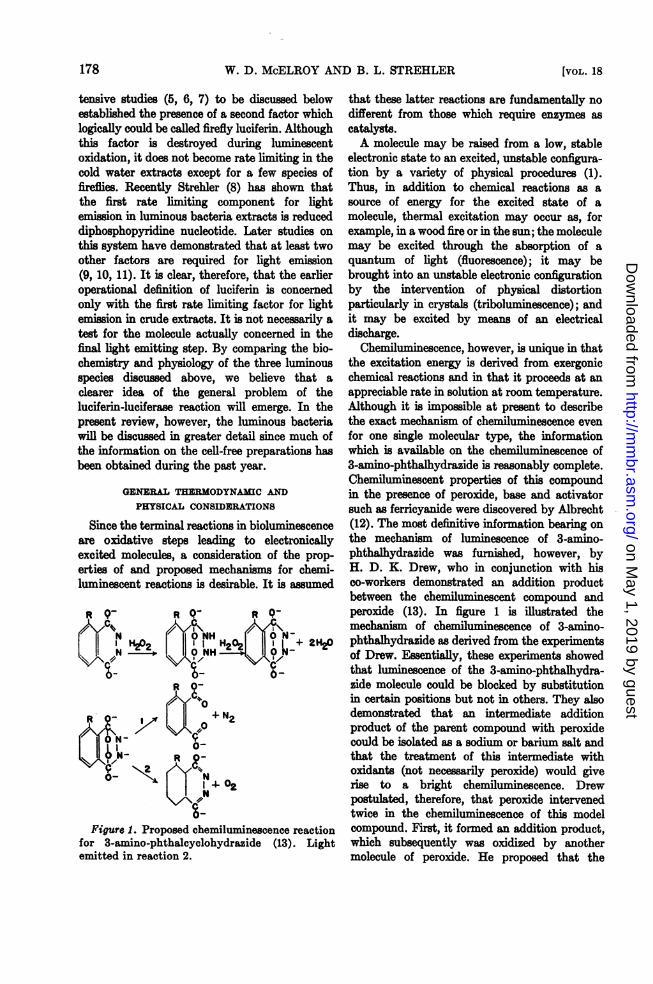
are oxidative steps leading to electronically the mechanism of luminescence of 3-amino-excited molecules, a consideration of the prop- phthalhydrazide was furnished, however, byerties of and proposed mechanisms for chemi- H. D. K. Drew, who in conjunction with hisluminescent reactions is desirable. It is assumed co-workers demonstrated an addition product
between the chemiluminescent compound andR 9- R R peroxide (13). In figure 1 is illustrated theNCC\\ , mechanism of chemiluminescence of 3-amino-1H02 | || H202 ||6+ 2V phthalhydrazide as derived from the experiments
X NH - of Drew. Esentially, these experiments showedthat luminescence of the 3-amino-phthalhydra-
R O- zide molecule could be blocked by substitution4-' 0 in certain positions but not in others. They also
RO- I | 11 +N2 demonstrated that an intermediate additionproduct of the parent compound with peroxide
N-|r 9- could be isolated as a sodium or barium salt ands0N-R a- that the treatment of this intermediate withg
t YAWN oxidants (not necessarily peroxide) would give{ T4- °2 rise to a bright chemiluminescence. Drew
postulated, therefore, that peroxide intervenedtwice in the chemiluminescence of this model
Figure 1. Proposed chemiluminescence reaction compound. First, it formed an addition product,for 3-amino-phthalcyclohydrazide (13). Light which subsequently was oxidized by anotheremitted in reaction 2. molecule of peroxide. He proposed that the
on May 1, 2019 by guest
http://mm
br.asm.org/
Dow
nloaded from

1954] BIOLUMINESCENCE 179
luminesent molecule acts as an intermediatecatalyst for the degradation of peroxide and thatthe energy liberated arises from this reactionrather than from the destruction of the lumi- wnescent molecule itself. Drew suggested that z3-amno-phthalhydrazide was the acceptor of /the energy from peroxide decomposition andacted as a mediator for energy liberation via light
Uemission. ,,The importance of organic hydroperoxides has cn
bee emphasized by a number of workers in thisfield. Recently Lhitz and Abrahmon (14) / _ _have studied light emision from the reaction of 4 600 700
zinc tetraphenylporphine (DH2) and tetralin MILLIMICRONSH Figure S. Spectal energy of light emitted from
I bacteria (left) and fireffies (right).hydroperoxide (>C-0-0-H). In this bi-molecular reaction they could demonstrate the blue violet to the red region of the spectrum.catalytic decomposition of the hydroperoxide by Thus, approximately 40-60 kcal perM of quantathe porphyrin dye. They propose the following Einstein) are required, and this, in the shortreaction mecanism: wavelength regions, corresponds to slightly moreH H energy than is available per two electrons at the
I I average reduction level of carbohydrate.C*O*O-H + DH2 CO-+ DH. + E60 Most bacterial luminescences and the lumi-
nescence of extracts of Achromobacter facheriH center in the blue-green at approximately 500C-O > nipm (2). The lunescece of Cypridina is a/C-Or + DH -. DH2 + brilliant blue with am mum at about 480 mp,
DH+* DII +light while the lumine ce of firefly extracts and ofDH,* the intact firefly shows a maximum at 565 mpThe isolation of the ketone and the slow destruc- and a subsidiary peak at 620 mA (15). Sometion of the dye during this reaction are evidence minor differences are apparent in the grossconsistent with the proposed m anism. In the eion spectra of different species of firefliescase of bioluminescence the requirement of (16, 17). Whether this is a true difference in theoxygen for light production may be used as an emission spectrum of the chemiluminescentargument for a reaction analogous to the compounds involved or of a filter action on theperoxididic cheniluminescence of synthetic part of the surrounding material is as yet notorganic compounds. The obvious difference completely settled. Inasmuch as the luciferinbetween the two processes is that an enzyme is obtained from the two species of fireflies canrequired for bioluminescence. The degree of cross-react and both seem to possess similaranalogy between in vitro cheluminescence and physical and enzymological properties, it seemsbioluminescence remains an open question until likely that the latter interpretation is correctthe intermediates in both processes have been (18). Macsire, in 1821 (19), showed that a heatedaccurately defined. Although all arguments based preparation of firefly tails emits a much redderon this information are tentative, nevertheless a light just prior to the extinction of the reactionconsideration of experimental findings concerning than is normally observed from the intactchemuluminescence may furnis certain clues animal. Harvey (20) has suggested that thefor the understanding of bioluminescence. The observed change is due to a difference in scat-possible participation of peroxides in the latter tering at higher temperatures because of a partialprocess will be discussed in a later section. denaturation of the surrounding structures. NoThe color of light emitted by two luminous such change in the color of the emitted light is
species is shown in figure 2. The wavelength observed when a mixture of the purified compo-distribution of light emitted extends from the nents is heated.
on May 1, 2019 by guest
http://mm
br.asm.org/
Dow
nloaded from
180 W. D. McELROY AND B. L. STREHLER [VOL. 18
While earlier analyses of the emission spectra Although Achromobacter ficheri will grow on aof luminous bacteria had indicated two funda- minimal medium consisting of inorganic saltsmental frequencies (21), more recent work by and glycerol as the carbon source, certainaminoSpruit and van der Burg (22), in which particular acids, notably methionine, histidine and lysine,care was taken to avoid scattering and self- increase the luminescence and decrease the lag inabsorption, has demonstrated no dichotomy of the growth. The nitrogen sources used mostfundamental frequencies but rather a single favorably by A. Jescheri for growth and lumi-emission maximum at around 500 mg. The wide nescence are the ammonium salts. The bacteriavariation in the spectral energy distribution of cannot use nitrate although they rapidly reducelight emitted by different luminous organisms is nitrate to nitrite. Serine, glutamic acid, andfurther support for the idea that different guanine will support growth, but luminescenceluciferins are involved in these reactions. A fails to develop. Both glutamine and asparaginecomparison of the various luciferins will be made will support growth and luminescence but are farin a later section. inferior to the ammonium salts. Under conditions
for optimum growth on ammonium salts FarghalyGENERALUPHYSIOOGYBANBCHERISTRYOF demonstrated that methionine plus histidine or
methionine plus lysine would increase theThere are a large number of different luminous luminescence 65 per cent without affecting
species of bacteria which are distributed among growth. In the presence of all three amino acidsvarious genera, including the Peudomonas and the luminescence was lower than in the foregoingVibrio, as well as the subgenus Photobacterium combinations. Methionine was the only amino(2). The two luminous species which have been acid which reduced the lag in the developmentused most extensively for physiological and bio- of luminescence. Doudoroff (26) had alsochemical studies are Photobacterium phosphoreum demonstrated that certain strains of luminousand Achromobacter fischeri (Bacterium pho8- bacteria require methionine for growth andphorescens indigenu8). Both are marine forms. light production.Doudoroff (23) was the first to make a careful In the basal medium described by Doudoroffstudy of the fermentative metabolism of several (23) andMcElroy and Farghaly (27), growth andspecies of luminous bacteria. He found that all light production were normal at 25 C or less, butof the facultatively anaerobic forms investigated both failed to occur above 27 C unless hydrolyzedshowed the same general "mixed acid" type of casein was added. Anderson (28) identified theanaerobic sugar dissimilation. Formic, acetic, amino acids in the hydrolyzed casein which werelactic and succinic acids, alcohol, CO, and required for growth at the higher temperature asacetylmethylcarbinol (acetoin) were among some methionine, glutamic, aspartic and arginine. Theof the products formed. Photobacterium pho8- elimination of any one of these amino acidsphoreum was capable of producing hydrogen. almost completely prevented growth and lightRecently Friedman (24) has demonstrated in production. Serine could replace aspartic andcell-free extracts enzymes of the Embden- arginine, provided lower concentrations ofMeyerhof system as well as those associated with methionine and glutamic were used. The criticalthe glucose monophosphate shunt pathway. requirement of amino acids for the developmentAmong the factors which have been found to of luminescence is illustrated by the mutational
influence bacterial luminescence are salt concen- studies on luminous bacteria by McElroy andtration, amino acids, carbon sources, and Farghaly (27). In a mutant requiring asparticmolecular oxygen. The work of Farghaly (25) acid for growth, they found that luminescenceand others has demonstrated that there is a failed to appear at a concentration of asparticrather critical optimum of salt concentration for acid which gave nearly maxium growth.bacterial luminescence and growth. The effects Higher concentrations of aspartic acid allowedon these two processes are not identical: the the rapid development of the luminescentprocess of growth is less inhibited by low salt system. With an aspartic acid concentrationconcentration than is luminescence. Analysis of which gave mum growth, it was found thatthe inhibition of luminescence development in a mixture of methionine, arginine, leucine andhypotonic media has shown that the effect is isoleucine would completely restore luminescence.mainly osmotic in nature. The elimination of any one of the four amino
on May 1, 2019 by guest
http://mm
br.asm.org/
Dow
nloaded from

1954] BIOLUMINESCENCE 181
acids from the mixture prevents the production A0of any significant amount of light, irrespectiveof the concentrations of the remaining 3 amino 80acids. The complexity of the relationship of 8 70Xamino acid requirement for growth and lumi- LC.i
nescence is illustrated by multiple mutants. , IMcElroy and Friedman (29) illustrated that by 0 50crossing two mutants which required single 4Z
I.-amino acids, growth could be restored to normal ¢ 40 40Iby adding the two growth factors. The lumi- Zo I3nescence, however, was only 10-15 per cent of /|normal. Friedman (24) has demonstrated that a
2 2
multiple mutant requiring arginine, proline,histidine, lysine, tyrosine and methionine for c 40 80 0l20growth did not luminesce when grown on the TIME (HOURS)minimal medium plus these amino acids. Lumi-
T
nescencedvd Figure S. Effect of aspartic acid on light pro-nescence developed normally, however, if the duction in a multiple mutant of Achromobactermedium which supported growth was supple- ficheri. Open circles refer to light intensity.mented with aspartic acid (figure 3). Graph on the right represents growth and light
Farghaly has studied a number of carbon production in the absence of aspartic while thesources for their ability to support growth and graph on the left represents the effect of supple-light production of A. fischeri (25). Of the 20 mentation with aspartic acid (24).compounds tested, only glycerol and glucose wereutilized by this organism for growth and lumi- respiration was the same in the bright and dimnescence. Friedman has since found that the strains as in dim strains plus riboflavin. Frombacteria can grow on fructose, provided small these observations Doudoroff concluded that aamounts of either glutamic acid, histidine, flavin was connected with one of the enzymesmethionine or cysteine are added to the medium involved in light production by the bacteria.(24). A certain amount of growth occurs in the As a result of work with inhibitors and otherpresence of low fructose concentrations, whereas indirect studies, a number of workers havehigher concentrations completely inhibit growth. suggested that the light emitting process ofThis amino acid requirement for fructose bacteria is coupled to the main respiratoryutilization remains unexplained. It is interesting pathway of these organisms (32, 33, 34, 35, 36).that Farghaly has found that these same four In Harvey's earlier studies (37) it was clear thatamino acids are effective in initiating growth in a the terminal oxidases were far more sensitive tolow COs atmosphere. Friedman found that the action of cyanide than was the luminescentextracts of cells grown on glucose rapidly reaction itself. Because of the suggestion thatphosphorylate fructose in the presence of ATP, cypridina luciferin might be a naphthoquinoneand that resting cell suspensions rapidly oxidize derivative (38, 39), Spruit and Schuiling (40)this compound. He has suggested that fructose studied the effect of a variety of these compoundsor compounds formed from fructose may inhibit on bacterial luescence. They found that thegrowth by reacting with free amino nitrogen in percentage inhibition of respiration was alwayscells. lower than that of luminescence. They explainedOther species of luminous bacteria have been this inhibition of luminescence as an oxidation
studied for their ability to grow on a variety of of luciferin whose redox potential was deduced tocarbon sources (see Harvey, 2). Johnson (30), in be -0.050 volts. Since addition of KCN relievedan extensive study, has shown that resting cells the inhibition of the naphthoquinones, Spruitof Photobtum phophorecene and A. ficheri and Schuiling believed the naphthoquinonesrapidly oxidize a variety of carbon sources. Most were being reoxidized by a cyanide sensitiveof these substrates do not support growth. catalyst. As will be discussed later, the partially
Doudoroff (31) in earlier studies found that purified luciferase preparation does indeed rapidlyadded riboflavin was essential for maximum reduce quinones and in so doing inhibits the lightluminescence during growth. Interestingly, the emitting reaction. McElroy and Kipnis (36)
on May 1, 2019 by guest
http://mm
br.asm.org/
Dow
nloaded from

182 W. D. McELROY AND B. L. STREHLER [VOL. 18
were unable, however, to confirm the cyanide bacteria. These pursuits were almost uniformlyeffect reported by Spruit and Schuiling. From unsuccessful, due perhaps to insufficient sensi-kinetic studies on the inhibition of light by tivity on the part of light measuring equipment2-methyl-1,4-naphthoquinone they concluded and the lack of many important biochemicalthat at least two pathways of electron transport intermediates. Nevertheless, Gerretsen (41) insupport luminescence. As a result of Friedman's 1920 succeeded in obtaining a weak "luciferin-studies (24) it seems possible that these two luciferase" reaction from extracts of Photo-pathways are the Embden-Meyerhof glycolytic bacterium Javanense. Unfortunately, Gerretsensystem and hexose monophosphate shunt. The did not pursue these studies, and other workerslatter pathway is apparently more sensitive to were unable to confirm his findings (2, 42).the quinones as far as supporting luminescence In 1951 Shoup and Strehler (unpublished)is concerned. In the presence of glucose and noted that acetonized powders from Achromo-4 X 10⁻⁶ molar 2-methyl-1 ,4-naphthoquinone bacterftscheri would luminesce brightly for severalluminescence is inhibited only 20 per cent. When, minutes after being suspended in water. Boiledhowever, the glucose disappears from the extracts of these same bacteria did not restore themedium, the light intensity rapidly drops to over luminescence after the cold water extracts had70 per cent inhibition. This is also true for the ceased to give light. Some dim luminescence didaction of dinitrophenol. Kinetic analysis of occur when hydrogen peroxide was added eitherluminescenceunder these two conditions indicates to a cold water or boiled extract, but this isquite clearly that only one of the possible rate probably due to the peroxidation of riboflavinlimiting pathways is supporting light emission. (43).All of these studies support the idea that the Components required. Following these observa-light emitting system in luminous bacteria tions, Strehler and Cormier (8, 10) undertookrepresents a by-pass of the cyanide sensitive examination of the luminescence of acetonizedportion of the cytochrome system. In addition, A. fischeri powders. They observed that thehowever, they suggest that the luminescent duration of luminescence depends on the concen-system is closely associated with the cytochrome tration of bacterial powders used and that asystem and, in fact, competes with the latter for luciferin-luciferase reaction was obtainable ifelectrons derived from the breakdown of food sufficiently concentrated extracts were employed.material. The factor which first became limiting for
luminescence in the crude extract was shown toBIOCHEMISTRY OF LUMINESCENCE be diphosphopyridine nucleotide (DPN+) or its
Bacterial Luminescence reduced homologue (DPNH). The relationship
Extraction of the system. Harvey's monograph between DPNH and light intensity is shown inBioluminesence (2) has reviewed early attemph figure 4. By the classical definition this com-gpound was bacterial lucifern just as ATP is
to extract the luminescent system from luminous firefly luciferin in the classical sense for mostspecies of fireflies. Initial attempts to demonstratea requirement for other diffusible factors were
t_- inconclusive, in that prolonged dialysis and( 2 acetone fractionation gave preparations still3 / capable of responding solely to added DPNH.z However, certain cofactors, such as flavin
mononucleotide, coenzyme A, and thiaminpyrophosphate, were effective in increasing theluminescence of crude extracts under variousconditions. In addition, a potent stimulation ofluminescence was obtainable from water extracts
0 0.5 1 1.5 of defatted hog kidney cortex and other tissues.DPNH-M x lo5 This material was called kidney cortex factor or
Figure 4. Relationship between light intensity KCF (10, 11).of bacterial extracts and DPNH concentration In contrast to the failure of Strehler and'(44). Cormier to demonstrate an absolute requirement
on May 1, 2019 by guest
http://mm
br.asm.org/
Dow
nloaded from

1964] BIOLUMINESCENCE 183
CoNc4"moles.104)4 12 20 28 36
I-~~~~~~~~~~~~~~o
Lu -z~~~L
Z 8 2
(201 .30
lCI) 4 o ..L-J ~~~~~~FMVNCONC.(T~0.5 10 6 2-R2 FMN
ADDED
1 2 6 26b~ soTIME-MINUTES OAI'P
Figure 5. Relationship between light intensity Figure 6. Relationship between light intensityof bacterial extract and riboflavin phosphate of bacterial extracts and palmitic aldehyde con-(FMN: flavin mononucleotide) concentration (9). centration (46).
for a factor other than DPNH for the lumi-nescence, McElroy et al. (9) were able to resolve lodistilled water autolysates of A. fischeri corm-pletely for FMN by a combination of acidprecipitation and ultraviolet irradiation. Their 8aepreparations would not emit light in the absence 2 / /of added BMN, thus indicating that the lumi- = 60/o onescent pathway consists of a DPN-flavin Lelectron transport system (figure 5). McElroy > F K/i/;et al. also observed a requirement for an ad- " 4ditional factor which they termed bacterial z Lluciferin. This factor was, in certain respects, 2analogous to the component in firefly lunu-nescence which McElroy and Strehler havecalled firefly luciferin (5, 6). It now appears that 0 2.5 5.0 7.5 0.0 12.5 5.0this component is similar to KCF since it has no OXYGEN TENSION (mm)observable effect in the presence of excess KCF. Figure 7. Effect of palmitic aldehyde on luni-Subsequently Cormier and Strehler (11, 45) nescence (triangles) and respiration (circles).identified the component in hog kidney cortex Open circles and triangles are the results withoutresponsible for the marked increase in lumig aldehyde (45).nescence as the long chain aldehyde, palmitalThis factor had been isolated in 1928 by Feulgen the bacterial enzyme and that the long chainet al. (46) as a breakdown product ofplasolgen, aldehydes are also required for the optimalthe palmitic acetal of glycerophosphocholine. In luminescence under these conditions (see fig-the presence of KCF, DPN, malate, and FMN, it ure 7).has been possible to obtain luminescence of cell Diaphorase properties. The partially purifiedand particle-free extracts of A. ficheri for 24 system acts as a typical diaphorase (44). Bothhours or more. The relationship between KCF methylene blue and 2-methyl 1,4-naphthoqui-and light intensity is shown in figure 6. Both the none are rapidly reduced by the system. Even inrate of luminescence and respiration at low the absence of the dyes, DPNH oxidation occursoxygen tensions are increased by KCF (see at a reasonable rate only if FMN is present.figure 7). Under normal oxygen tension the presence or
Strehler et al. (47) have recently demonstrated absence of the aldehyde does not affect thethat a bright luminescence will occur when DPNH oxidation in the partially purifiedreduced riboflavin or reduced FMN is added to preparations. It is possible, therefore, that much
on May 1, 2019 by guest
http://mm
br.asm.org/
Dow
nloaded from

184 W. D. McELROY AND B. L. STREHLER [VOL. 18
of the DPNH oxidation is through a pathway luminescence only after irradiating with ultra-which is not directly concerned with light violet light. It seems likely that the activeemission. Ferricyanide, likewise, leads to a very material is similar to the long chain aldehyde.rapid oxidation of DPNH. In fact, if sufficient The partially purified luciferase is rapidly in-ferricyanide is added to oxidize the DPNH, no activated by the irradiation which probablylight emission is observed from the reaction. On accounts for the decrease in light intensity afterthe other hand, with lower concentrations of prolonged ultraviolet treatment. The lumi-ferricyanide the light emission is completely nescent reaction is very sensitive to a variety ofdelayed until the ferricyanide is reduced, and chemical agents. Riboflavin and flavin adeninethen once this occurs, light appears. dinucleotide inhibit probably by competing
Effect of various environmental factors and with FMN at the site of its reduction. Theinhibitors. The general temperature dependencies inhibition by p-Cl-Hg-benzoate, reversed byof the bacterial luminescent reaction in vitro glutathione, and by copper and silver indicatesand in vivo are similar with an optimum at the necessity of a sulfhydryl group for enzymeapproximately 25 C (10, 48, 49, 51) (A. fischeri). activity. The inhibition by versene and cyanideIncreasing the salt concentration of the reaction indicates the possible involvement of a metal;mixture produces first an increase, then at however, in the partially purified preparationshigher concentrations, a decrease in the rate of consistent reactivation by metals after verseneluminescence. The pH dependence of the re- treatment has not been observed. The cyanideaction in crude extracts (10) exhibits a double may be acting by combining with the aldehyde.optimum (pH 6 and 8.5), while in more purified It is interesting that cyanide should inhibit thepreparations a single optimum is observed (44) isolated system, whereas it has very little effectat pH 7.0. even in higher concentrations on light emission
Gerretsen (41) observed an increase in lumi- in the intact bacterium.nescence after a short ultraviolet treatment, and A number of kinetic observations on the crudethis observation has been repeated by Harvey. and FMN resolved luciferase preparations haveSimilarly, irradiation of the crude enzyme with been made (50). Particularly, the time course ofultraviolet light (365 mMA) produces an initial luminescence has been followed when one of theincrease and then a decrease in luminescence necessary factors was added last, e.g., DPN+,(9, 10). This effect has been examined in some malate, DPNH, FMN, 02, KCF, FMNH2, anddetail byMcElroy and co-workers. By (NH4)2SO4 reduced riboflavin. These results are summarizedfractionation it is possible to separate from the in table 1. Assuming that the shortest half riselight emitting system a fraction which stimulates time would be characteristic of later steps in a
series of reactions, the following sequence hasTABLE 1 been postulated for bacterial extract lumi-
Time required for half-maximal luminescence in nescence.bacterial extracts when various essential
components are added last Malate + DPN+ -- DPNHDPNH + FMN -- DPN+ + FMNH2
Adde t Factors Present with Enzyme "1/2 RiseFMNH" +°2light
54 The effects of high hydrostatic pressures on theMalate DPN+, FMN, KCF, 02 108 luminescence of cell-free extracts have beenDPN+ Malate, FMN, KCF, 02 108 examined by Strehler and Johnson (51). TheirDPNH FMN, KCF, 02 2.7 results may be summarized as follows: WhenFMN DPNH, KCF, Os 2.7 pressure is applied, there is an initial rapidFMNH2 02 0.41 increase in luminescence followed by an expo-FMNH2 KCF, 02 0.075 nential decrease to some lower level. TheRFH2 02 0.27 magnitude of this effect depends upon theRFH2 KCF, 02 0.14 pressure, temperature and presence of KCF.KCF FMN, DPNH, K2 0.05 Typical results are shown in figure 8. These02 FM___ PNHK_______ results are explicable in terms of and consistentThe % rise time is the time required to reach with the kinetic evidence obtained earlier. The
M maximal light intensity. rise, when pressure is applied, is regarded as the
on May 1, 2019 by guest
http://mm
br.asm.org/
Dow
nloaded from

19541 BIOLUMINESCENCE 185
TO
i- 60 0.05 mg. CONC.
150 I I A. FISCHERI z48 e51lo26 C U ML-
140 40
830 o3
W120 0.0002o. ,nEXTRACTS2 000025
>_~~~ ~~~~~~~~~~~ 0NONE1150
LUW 2 4 6 S t0 52 54 56 58
500 OFF AT? CONG. MOLAR X 104
000OF4igure08.Effect of hydrostatic pressure on Figure 9. Relationship between light intensity
90 CEILLS of firefly extracts and adenosine triphosphate and
80scent oxidation of Gavi at high hydrostaticMg+ concentration (65).by amoimsulfate fractionation after elution
70 (7, 52, 53). These preparations emit no light inviewedaanteosthe absence of ATP. The luciferin has been puin-duetoannhbiio3 fled by extraction from the aqueous solution with
MINUTES acidified ethyl acetate and finally by columnFigure 8. Effect of hydrostatic pressure on the chromatography using "dowex 50o,fullers earth-
light intensity of bacterial cells and extracts. celite mixtures (52, 54) and celite,Hs0, chloro-Pressure was applied at 30 seconds and released at form-butanol columns. The properties of the75 and 195 seconds (51). luciferin will be discussed in a later section. In
other species of fireflies such as Photinus mel~anotisresult of an increased efficiency of the lumi- or Diphotu montanis the crude aqueous extractsnescent oxidation of flavin at high hydrostatic do not respond to ATP (18). To obtain light frompressures. The later exponential decline can be these preparations it is necessary to add theviewed as a depression in the pool size of FMNH2 purified luciferin obtainable from Photinusdue to an inhibition of the DPNH-FMN reaction. pyralis, as well as ATPThe effects are generally similar to those observed The response of the purified system, obtainedin the intact bacterium. The tentative identifica- from Photinuapyralis to varying concentrationstion of the slow reaction as a readjustment of of ATP and tis shown in figure 9. Thepool size was made by determining the prise variation of the light intensity with these twotime when FMN, DPNH, enzyme and KCF were components suggests that a complex betweenmixed. This value was compared with the time ATP and Mg++ is the effective substrate (55).constant exhibited by the system in moving to a This is particularly noticeable with low Mg++new steady state after the pressure had been concentrations where it is possible to demonstratealtered. At 5 C the times were approximately an inhibition with high ATP concentration. For15 sec. The luminescent oxidation of substrate the intermediate range of concentrations theamounts of reduced flavin was accelerated by mxalinitial light response is obtained whenhigh pressures. the molar ratio of ATP to Mg++ is one. In the
FireflyLuminescencepurified enzyme preparation light eissio cannotFisbeenprifedflymLrumiescquen sextrs be elicited by a variety of other phosphorylated
The production of light by extracts from compounds which have been tested. The initiationfireflies has been shown to depend upon the of light emission in the crude extracts by ADPpresence of adenosine triphosphate, a highly is due to the presence of an effective myokinasefluorescent compound called luciferin, luciferase, (7, 56). Inosine triphosphate, uridine tri-oxygen and magnesium ions (4, 5). The luciferase phosphate, acetyl phosphate, creatine phosphate,has been purified from crude aqueous extracts by inorganic pyrophosphate, and a variety of otherabsorption on calcium phosphate gels followed phosphorylated and nonphosphorylated cofactors
on May 1, 2019 by guest
http://mm
br.asm.org/
Dow
nloaded from

186 W. D. McELROY AND B. L. STREHLER (VOL. 18
fail to initiate light in the purified preparations. of oxygen, to an excited state which subsequentlyThe reaction is apparently specific for adenosine emits light. Recently it has been shown thattriphosphate. It is cyanide and azide insensitive under anaerobic conditions formation of the in-but can be inhibited by p-Cl-Hg-benzoate. The active complex still occurs (57). When oxygen islatter inhibition is reversed by glutathione. Light readmitted to a previously anaerobic system,emission is extremely sensitive to a variety of there is a flash of light which decreases within aamines, particularly hydroxylamine, while second to the low baseline level of luminescence.various benzimidazole and benzothiazole deriva- This flash can be interpreted in a number oftives inhibit by competing with the luciferin ways. It may be that under aerobic conditions(McElroy, unpublished). the consumption of the active intermediate is soWhen the purified firefly components are rapid as to prevent a true equilibrium between the
mixed, there is an initial high light intensity active and inactive complexes. Under anaerobicwhich rapidly declines to an intermediate conditions true equilibrium can be established,steady state level of luminescence of several and the momentary flash where oxygen is re-hours duration. Once the light intensity has admitted is due to the rapid utilization of anreached the low baseline level, it is possible to accumulated active intermediate. The formationshow by direct chemical analysis that most of and accumulation under anaerobic conditions ofthe ATP is still present. It is also available for a further product from the active intermediatethe hexokinase reaction (7). If additional indicated in figure 10 are possible. Consistentluciferase is added to the reaction mixture, with this latter interpretation are the followinghowever, a second flash similar to the original is earlier observations: Certain dried preparationsobtained. The results indicate that all of the low of firefly lanterns will luminesce when moistenedmolecular weight components necessary for light but fail to respond to ATP addition; moreover,production are present and available for maxi- powders of desiccated firefly lanterns boiled inmum light production. butyl alcohol have been observed to exhibit theThe decline of luminescence after its initiation same phenomena. Such an interpretation does
with ATP to the low steady state level is believed not necessitate the postulation of a slow equi-to be due to the reversible formation of an librium reaction between the inactive and activeinactive complex from an active intermediate. intermediates, as compared to the luminescentThe latter intermediate is presumably composed reaction which utilized this active intermediate.of four components as indicated in figure 10. Although the formation of the inactive complexProbably through a series of reactions the active does not require oxygen, it is dependent upon aintermediate is finally converted, in the presence second protein. The removal of inorganic pyro-
phosphatase from the luciferase increases theL,Hp+ E + Mg + ATP steady-state level of luminescence, while addition
of purified firefly or yeast pyrophosphatase'1t depresses it. The results of such an experiment
LH2*E*Mg*ATP are shown in figure 11. The fact that inorganic(ACTIVE INTERMEDIATE) -- +o,-w LIGHT pyrophosphatase is an essential protein in the1jMg L A Pi + MgPr formation of the inactive complex is important
PrSt ~ ~LHeE POPM Mg-Pr in explaining the effect of various pyrophosphatesLH2fE*Mg ATP-Mg*Pr + w-o-p or and polyphosphates on light emission. The ad-(INACTIVE COMPLEX) * LH2-ELMgSPOP. ATP.Mg-Pr dition of a variety of these compounds to the
NHoIIT OR luminescent reaction temporarily restores theCONTROL
~ ~ ~ LCOMPLLXj
MECHANISM llight to a high intensity. This action of inorganicCETYLCHOLINE pyrophosphate and triphosphate in stimulating
l AC4ATP1- PO11P+Ac-CoA +Ad.
light production, after its initiation by ATP, isCHOLINE + AC 9coA POP + Ac * Co A + Ad. attributed to the rapid breakdown of the inactive
Figure 10. Proposed scheme for firefly lumi- complex by these agents. In the case of pyro-nescence. phosphate the evidence shows that it competes
POP: pyrophosphate with ATP in formation of the active intermediate.Pr: protein Thus the addition of pyrophosphate before ATPPi: inorganic phosphate strongly inhibits light emission. On the other
on May 1, 2019 by guest
http://mm
br.asm.org/
Dow
nloaded from

1954] BIOLUMINESCENCE 187
hand, the delayed addition of pyrophosphate 100leads to an initial stimulation followed often bya decline and then a rise to a secondary peak 90which rapidly decreases to the baseline level as 80the pyrophosphate is hydrolyzed by the action of > lpyrophosphatase. Such results are to be expected t 70if pyrophosphate splits the inactive complex to Cn
form initially some active intermediate. The WIamount of active intermediate formed depends z soupon both the nature of the inactiV'e complex(s)and the mode of the splitting by pyrophosphate. 4 AThe simplest inactive complex presumably o 30 Bformed consists of luciferin-luciferase-Mg-ATP- DiMg-pyrophosphatase. Pyrophosphate could split 20such a complex to give rise to either an active E0(luciferin-luciferase-Mg-ATP) or an inactiveouciferin-luciferase-Mg-pyrophosphate) inter- c
10 2 30 4 5 6
mediate containing luciferase (see figure 10). The l0 20 30 40 50 60initial light intensity obtained with the addition TIMEEf- SECONpDhSof pyrophosphate would then be a measure of the Figure 11. Effect of inorganic pyrophosphataseactive intermediate formed. The secondary peak on the light intensity in firefly extracts. Curve Aof luminescence following the addition of pyro- represents the reaction with purified luciferase.phosphate would represent the slow release of Curves B, C, D, and E represent the effect ofluciferase from an inhibitory complex with pyro adding increasing amounts of inorganic pyro-phosphate. The effect of various pyrophosphatase phosphatase (55).inhibitors, such as Mn++, Ca++, and F-, on theluminescent response to pyrophosphate can be case for fireflies and bacteria Experiments onexplained by such an hypothesis. crude extracts from living CypriSina would be of
comparative significance in this respect.Cypridina Luminescence
The nature of the luminescent reaction in CHEMSTRY OF LUCIPERINextracts from Cypridina hilgendorfii has been It has already been pointed out and shouldextensively reviewed by Harvey (2, 58). There- be reemphasized that luciferin, being a compo-fore, only the more recent information will be nent defined experimentally as a heat stablepresented for this system. Detailed studies by factor, is not necessarily the same molecule fromHarvey and associates indicate that only two species to species. Since luciferins obtained fromcomponents, in addition to oxygen, are required various species do not generally substitute forfor luminescence, a luciferin and a luciferase each other in luciferase preparations from one(59, 60, 61). McElroy and Chase (62) have species or group, it is certain that the luciferinspurified the luciferase over 150 times by can be of varying chemical natures.(NH4)2804 and acetone fractional precipitationand adsorption onto and elution from calcium Baa L fnphosphate gel. The luciferin has been partially The recent demonstration of an absolutepurified through chemical procedures by Ander- requirement for riboflavin phosphate in bacterialson, and by Mason using chromatographic luminescence (9) as well as the luminescentprocedures. The chemical and physical properties oxidation of chemically reduced flavins (riboflavinof the purified luciferin will be discussed in a and FMN) by bacterial extracts (47) stronglylater section. In general, the studies on cypridina suggests that bacterial luciferin is reducedluminescence indicate that only reduced luciferin, riboflavin phosphate. The former evidenceluciferase and oxygen are required for light establishes the luminescent system as a flavinemission. Moreover, it is not possible to obtain coupled pathway while the latter dispenses withadditional light in the crude extracts from dried DPN as a necessary cofactor. Although the longCypridina by adding other cofactors as is the chain aldehyde is necessary for luminescence, it
on May 1, 2019 by guest
http://mm
br.asm.org/
Dow
nloaded from
188 W. D. McELROY AND B. L. STREHLER [VOL. 18
would seem a ss likely candidate for the role solution a brilliant yellow-green fluorescence,of luciferin sine- it will not, by itself, support resembling rather closely the fluorescent emissionluminescence in the presence of the enzyme. of riboflavin and firefly extract luminescence.
Certain facts are in opposition to considering However, in contrast to riboflavin, the yellow-reduced FMN as bacterial luciferin. Of particular green fluorescence disappears in acid solution.importance is the fact that the fluorescence Microbiological away also indicates the absenceemission of riboflavin is centered at ca. 565 mp&, of riboflavin (McElroy, unpublished). In thewhile the emission of the bacteria and extracts is strong acid solutions luciferin has a red-violetmaximal at 490 mu. Second, one cannot rule out fluorescence, much weaker than its fluorescencethe possibility that some strongly bound com- in alkaline solution. At room temperature in acidpound other than flavin might be the ultimate solution it gives rise to another compound ofacceptor and lumiphore. While these possibilities similar but somewhat more yellow fluorescence,cannot be rigorously excluded, the following and with an absorption band shifted about 15experimental observations are more consistent my to the red. This compound is enzymaticallywith a direct participation of reduced flavins in inactive. The pKa for the change in fluorescencethe luminescent reaction: (a) riboflavin and and for the change in absorption spectrum is atriboflavin phosphate will chemiluminesce in the about 8.4. Electrophoretic mobilities have beenpresence of hydrogen peroxide (44); (b) if checked in agar tubes at various pH's using thereduced flavins are oxidized by some other fluorescence as an indicator of activity. Itcomponent, then this reaction must be ac- appears that the luciferin molecule is doublycelerated by KCF (49) which from other experi- charged in solutions more alkaline than pH 8.4,ments (50) seems to be involved in 02 activation; singly charged between pH 4 and pH 8.4, and(c) prolonged dialysis (10), repeated acid that below pH 4 it is uncharged. These propertiesprecipitation and ammonium sulfate fractiona- suggest an amine-imine isomerization and ation (44) do not result in preparations of enzyme carboxyl group. Further evidence for an activeincapable of luminescing in the presence of imine group was obtained by treatment byDPNH, FMN and KOF; (d) if another factor nitrous acid which destroyed both the fluo-is necessary, it not only must meet the require- rescence and enzymatic activity. The sensitivityments set forth above, but must not be destroyed of the reaction to imines may depend upon thison light emission since luminescence will continue property. The infrared absorption spectrum offor at least 24 hours in the presence of excess luciferin, while differing greatly from the ab-KCF, FMN and DPNH. sorption spectrum of riboflavin in the 2 JA andAt the present time, in our opinion, the most 6 pu regions, shows a remarkable similarity to
reasonable view is that bacterial luciferin is riboflavin in the 10-15 p range. This evidencereduced riboflavin phosphate or a derivative of can be taken to indicate that luciferin has athe latter. It does not now appear that the in- basic nuclear structure closely resembling that ofactivation spectra studies of the Delft group riboflavin in terms of mas. Polarographic(63) and the tentative identification of bacterial analysis indicates that luciferin has an Eo' atluciferin by these workers as a naphthoquinone about -0.6 volts at pH 7.0, while its degradationare in keeping with more recent direct bio- product has an Eo' approximately -0.4 volts.chemical findings. Riboflavin, on the other hand, has an Eo' at
about -0.21. Using fluorescence changes as anFireflyy Lu~ferin index of the sensitivity of the compound toFirefly luciferin has been demonstrated as an chemical reagents, the following was found:
accessory factor for the luminescence of firefly permanganate, peroxide, and hypochlorite treat-extract supplemented with ATP, magnesium and ment result in an inactive material.oxygen. Strehler and McElroy (6, 52, 54) under- Recently Strehler and Sites (64) have examinedtook to isolate and purify the active component. the maw spectrogram of pyrolyzed and un-Liquid-liquid partition chromatography, paper pyrolyzed luciferin. The largest mass obtainable,chromatography, column chromatography, and 293, may be taken as the mass of the parentother procedures were used to isolate a material compound. Large ion currents were obtained alsoof a reasonable degree of purity. Firefly luciferin at masses 278 and 234. An analysis of the compli-is a faint yellowish compound having in alkaline cated mass spectrogram has been undertaken
on May 1, 2019 by guest
http://mm
br.asm.org/
Dow
nloaded from

1954] BIOLUMINESCENCE 189
again using riboflavin as a model test compound. preparation is exposed to air, it loses its abilityThe mas 234 was taken as the parent nuclear to support luminescence when added to amass, and considering the degradation product luciferase preparation. But if within a shortmasses obtained, a structure for luciferin was period of time the inactive material is treatedproposed. This structure was subjected to test with hydrosulfite or other appropriate reductants,by the synthesis of some analogous compounds, its ability to support luminescence is restored.whose gross physical properties approximated, in On the other hand, a long delay between the airmany instances, the properties of luciferin. The oxidation and the re-reduction of the compoundproposed structure is a dipyrimido pyrazine, gives inactive material. Chakravorty anddoubly nitrogen and doubly oxygen or mono- Ballentine (38) have presented evidence whichsulfur substituted, with the carboxyl group may indicate that a ketohydroxy side chain ispendent on the pyrimidine ring carbon. While it present in the active luciferin and that themay be premature to assume that luciferin is irreversible oxidation involves the degradation ofactually a dipyrimido pyrazine related both to the this substituent. They were able to regeneratepteridines and to riboflavin in general molecular active luciferin from irreversibly oxidized luciferinarchitecture, further synthetic work should put by a series of synthetic steps which were designedthis hypothesis to a rigorous test. to introduce a ketohydroxy side chain onto the
..pridina .ucifern.molecule. More recently Mason (72, 73) hasshown through the use of paper chromatography
Luciferin is the only known requirement for under anaerobic conditions that several luciferinsthe luminescence of cypridina extracts, in are present in cypridina extracts. He has calledaddition to luciferase and oxygen. Nearly every these alpha and beta luciferin. Acid hydrolysis ofinvestigator in the field has at one time or these luciferins followed by paper chroma-another presented a partial structure for cypri- tography of the degradation products indicatesdina luciferin based on various lines of chemical the presence of a polypeptide. This polypeptideevidence. Among the proposed structures were a chain is believed to be attached to theproteose (65), a phospholipin (66), a poly- chromophoric group whose chemical constitutionhydroxybenzene (67, 68), a reduced quinone, is as yet undefined.naphthoquinone or anthroquinone (38), and aflavin (69). The molecular weight of luciferin is LUMINESCENCE IN OTHER FORMS:probably between 250 and 500 mass units. COMIPARATIVE ASPECTSVarious purification procedures have been Among the other luminous species on whichdeveloped, some depending on extraction with some biochemical and physiological investiga-organic solvents and fractional precipitation of tions have been made are the dinoflagellates inimpurities. Anderson (60) has obtained partially particular Noctiluca, the luminous ctenophore,purified material by treatment of butanol extracts Mnemiopsis, sea pansies, and several species ofwith benzoyl chloride to obtain the benzolyl luminous earthworms (2). Noctiluca, whichderivative. The benzoylated material was then responds to physical stimulation by emittinghydrolyzed and the cycle repeated. The activity light, should be an ideal organism for both bio-per unit weight was increased over 2,000-fold chemical and physiological studies. In this form,by this double cycle. This luciferin has an the luminous material is localized in granules. Inabsorption maximum at 435 muA. Chase (70, 71) the ctenophore, Mnemiopsis, the luminoushas demonstrated a shift in the absorption material is localized in the region of the combspectrum maximum from 435 to 465 my and plates and likewise responds to mechanicalthen a gradual disappearance of the latter band stimulation as well as osmotic stimuli. Thisas the luciferin undergoes autooxidation. Oxidized organism is remarkable in its apparent ability toluciferin is colorless. Enzymatic oxidation of luminesce under anaerobic conditions. Whetherluciferin with accompanying luminescence is this indicates an entirely different chemicalcharacterized by a similar change in absorption mechanism of luminescence from 02 requiringspectrum, but the effect here takes place at 100 organisms or not awaits the extraction andtimes the rate of autooxidation. purification of the system. Buck (personal
Cypridina luciferin is capable of being oxidized communication) has reported that sea pansiesboth "reversibly and irreversibly" (2). If a respond to ATP as do fireffies. This observation
on May 1, 2019 by guest
http://mm
br.asm.org/
Dow
nloaded from

190 W. D. McELROY AND B. L. STREHLER [VOL. 18
too-* directly shown to be the reverse of each other.In figure 12 are shown the dependence of the
so- 3 0 luminescence and Hill reaction rate of green7"' plant chloroplasts on incident light intensity.
60 Considering the evolution of luminous speciesCY the singular ability to produce light would not,
in our opinion, confer a great survival value on~40/. the organisms endowed with it since there are
many more nonluminous than luminous forms.20/ Secondarily, however, this ability may be adapted
to uses which do confer a selective advantage on
0 I00, , the luminous organism. In the primary categoryRELATIVE INCIDENT LIGHT INTENSITY may be placed the luminous bacteria, while more
Figure 12. Dependence of luminescence and highly evolved forms, such as fireflies, make useHill reaction (open circles) rates of green plant of luminescence in a secondary capacity.chloroplasts on incident light (76). In fireffies luminescence serves the social
function of communication, males and females ofsuggests a biochemical resemblance between the various species finding each other in theirfireflies and a salt water form, which may be twilight haunts by means of a yellow flashingrelated to the fact that both are intracellular and abdomen. One would hardly question the longunder nerve control. The luminous earthworm, range survival advantage of this unique ability.Eisenia submntana, on the other hand, re- A variety of deep-sea forms apparently makes useportedly utilizes riboflavin as a photophore and of its luminescence in a less aesthetic and morein this respect may be akin to bacteria (74). immediate fashion, either by blinding of potentialThe recent discovery by Strehler and Arnold predators, attracting potential prey, or in the
(75, 76) of a chemiluminescence of low intensity case of certain polynoid worms, distracting thegiven off by all green plants examined thus far predator from the more vital nonluminousadds this enormous group of living things to the portions of the organism. These worms, ifplants and animals producing light through attacked by a crab, reportedly luminesce brightlyenzymatic oxidation. Despite the fact that this in their posterior portions, while the dark headlight is extremely dim, the total light emitted end can crawl off and regenerate his lost tail.by green plants exceeds by many times the Luminous bacteria, on the other hand, probablyluminescence of all other living things. These do not glean any selective advantage under moststudies have demonstrated a dependence for light conditions from their ability to luminesce. Ratheremission on the photochemical and enzymatic their luminescence has been regarded as anapparatus of photosynthesis while the action accidental mutation in which the energy liberatedspectrum in green plants and the luminescence by a terminal flavin autoxidase is channeled intoemission spectrum are characteristic of chloro- the excited state of a molecule, probably flavin,phyll absorption and fluorescence, respectively. which subsequently emits light. If in a naturalIn contrast to fluorescence (i.e., immediate light environment this ability confers selectiveemission), this bioluminescence saturates at advantage, this advantage may reside more inhigher light intensities with an enzymatic side biochemical effects than in the physicaldependence. Strehler and Arnold have suggested phenomenon of light emission.that this luminescence is due to a recombinationof early oxidizing and reducing intermediates in CONTROL AND MECHANISM OF LUMINESCENCEthe photosynthetic process and have demon- In those species which do not glow continuouslystrated a general dependence of luminescence on a number of mechanisms for producing andphotosynthetic activity. Light energy absorbed extinguishing the luminescence have beenby chlorophyll is thus converted into a more evolved. The most obvious device used is thestable chemical form and then feeds back to re- interposition of an opaque body between thegenerate excited chlorophyll in an enzyme luminescent source and the outside. Thus acatalyzed step. Photosynthesis and biolumi- number of deep sea fish use either an eyelid-likenescence, ostensibly opposite processes, are thus or hinge-type flapper for shutting off the light
on May 1, 2019 by guest
http://mm
br.asm.org/
Dow
nloaded from

1954] BIOLUMINESCENCE 191
(2). In Cypridina, where the luminescence occurs compounds chemiluminesce with H202. With theoutside of the animal, light production is initiated above preliminary considerations and the workby the ejection of luciferin and luciferase, of Drew on 3-amino-phthalhydrazide chemi-respectively, from two kinds of glands into the luminescences in mind, Strehler and Cormiersurrounding sea water (2). The control of the (50) have suggested that an organic peroxide isflash in fireffies has been the source of con- involved in bacterial luminescence. In keepingsiderable speculation and controversy, some with the suggestion is the observation thatworkers claiming that the light is initiated by added peroxide stimulates firefly luminescenceadmitting oxygen to the photogenic cells by (McElroyandCoulombre, unpublished). Althoughneuromuscular control of the abdominal air the addition of peroxide or catalase was withoutducts. McElroy and co-workers (55) have effect on luminescence in bacterial extract, it issuggested, on the basis of the large amounts of possible that in this system one is dealing with apyrophosphatase in the luminous organ coupled strongly bound form of peroxide. One of thewith the light potentiating effect of pyrophos- attractive features of this proposed mechanism isphate, that pyrophosphate release intracellularly that it furnishes a means for supplying the 50 orand under neural control is the trigger mechanism 60 kilocalories which are necessary for lumi-(see figure 11). nescence. As pointed out previously, the catalyticThe study of the time course of luminescence decomposition of peroxide by nonenzymatic
of bacterial, Cypridina and firefly extracts (9, systems can supply the energy for chemi-47, 50, 55, 77) when various necessary factors are luminescence.added has furnished useful information con-
*erningthe mechanism of the * ence. APPLICATIONS OF BIOLUMINESCENCE TOcernmg the mechanism of the luminescence.Typical time curve of the luminescence of these BIOLOGICAL PROBESthree species shows that there is an appreciable The ability of a variety of chemical agents totime-lag between the mixing of the components elicit light from in vivo or in vitro luminescentand the appearance of the maximum lumi- systems has been the basis for a variety ofnescence. It follows that some relatively slow analytical tests of high sensitivity, accuracy, andprocess must precede the light emission step speed. Since the luminescent system is itself awhose product is more directly concerned in the respiratory pathway, the study of the effect ofluminescent reaction. Chance, Harvey, Johnson, environmental variables on luminescence fur-and Millikan (77) have discussed several possible nishes data on respiratory activity. Such effectsinterpretations of these observations. They have as the flash of luminescence after anaerobicity inconcluded that at least four reactions are luminous bacteria indicate an accumulation ofnecessary to explain the results including an reduced intermediates in the absence of oxygen.association of luciferin with luciferase, an Combined with inhibitor and pressure studies,association of 02 with luciferase, a reaction the measurement of the light intensity and coursebetween 02 and luciferin-luciferase, and a slow of luminescence in luminous bacteria hasreaction leading to light emission. furnished considerable information on the relationAlthough the requirements for firefly extract of luminescence to the main respiratory pathways
luminescence are somewhat more complicated and on the mechanism of enzyme action. Lumi-than the other two, a similar type of kinetic nescence studies by Johnson, Eyring andanalysis can be applied to this system. Firefly collaborators have been concerned with theluciferin is considered as analogous to Cypridina mechanism of action of various inhibitors and theluciferin and reduced riboflavin phosphate in the types of bonds formed between inhibitors andbacteria. In the firefly, however, the luciferase enzymes.requires specifically ATP for its activation of the One of the earliest examples of using luminousluminous oxidation of luciferin, either as a bacteria to solve biological problems was ascomponent in an active complex between oxygen detectors. Since light output is directlyenzyme, ATP, luciferin, Mg++ and 02 or as a related to oxygen tension over a wide range ofreactant with luciferin to give a new active oxygen concentration, these organisms wereluciferin molecule. Another point of similarity applied to photosynthesis studies and furnishedamong the various luciferins and one bearing on some of the earliest evidence that exogenousthe mechanism is the fact that all of these oxygen is not an obligatory requirement for the
on May 1, 2019 by guest
http://mm
br.asm.org/
Dow
nloaded from

192 W. D. McELROY AND B. L. STREHLER [VOL. 18
initiation and maintenance of high photo- 6. STREHLER, B. L., AND MCELROY, W. D. 1949synthetic rates. Purification of firefly luciferin. J. Cellular
Luminescence has also been applied more Comp. Physiol., 34, 457-466.recently to photosynthesis problems of other 7. MCELROY, W. D. 1951 Properties of thetypes..Theluminescence of green plants and its reaction utilizing adenosine triphosphatettopes.1ne tmeenveOIgen pants ave for bioluminescence. J. Biol. Chem., 191,relationship to environmental factors have 547-557.contributed to our understandig of early steps 8. STREHLER, B. L. 1953 Luminescence inin photosynthesis while the use of firefly extracts cell-free extracts of luminous bacteria andfor the measurement of adenosine triphosphate its activation by DPN. J. Am. Chem.as applied to green plants has yielded information Soc., 75, 1264.on both energetics and mechanism in phosphate 9. McELROY, W. D., HASTINGS, J. W., SONNEN-dependent steps in photosynthesis. FELD, VALERIE, AND COULOMBRE, JANEBecause of this unique ability of firefly 1953 The requirement of riboflavin phos-
extracts to emit light when ATP is added, this phate for bacterial luminescence. Science,system is particularly useful in studies on energy 118, 385-386.transfer mechanisms. In combination with ap-
10. STREHLER, B. L., AND CORMIER, M. J. 1953
transfer mechanisms.and combiatin withis am Factors affecting the luminescence of cell-propriate enzymes and substrates this system free extracts of the luminous bacterium,can and has been extended to many intermediates Achromobacter fischeri. Arch. Biochem.and enzymes affecting ATP levels. Among the Biophys., 47, 16-33.substrates and enzymes of interest which can be 11. CORMIER, M. J., AND STREHLER, B. L. 1953assayed with firefly lantern extracts are ADP, The identification of KCF: Requirement ofphosphocreatine, AMP, glucose, coenzyme A, long chain aldehydes for bacterial extractpyrophosphate, DPN, hexokinase, myokinase, luminescence. J. Am. Chem. Soc., 75,creatine-adenylate transphosphorylase, and 4864.variousapyrasesandATas_ .In12. ALBRECHT, H. 0. 1928 tJber die chemilu-.ariou.pyrasesand AT s. In combination minescence des amino phtalsaurehydrazids.
with ultra sensitive light detecting apparatus of Z. physik. Chem., 1386, 321-330.moderate cost the sensitivity of the tests can 13. DREW, H. D. K. 1939 Chemiluminescenceeasily be extended to the millimicrogram range. in the oxidation of certain organic sub-Among the problems to which this assay method stances. Trans. Faraday Soc., 35, 207-215.has been applied are x-radiation effects on ATP 14. LINSCHITZ, H., AND ABRAHAMSON, E. W.metabolism, changes in ATP during grasshopper 1953 Kinetics of porphyrin-catalysedembryogenesis, oxidative phosphorylation, "ATP chemiluminescent decomposition of per-content" of purified nucleic acids, effects of oxides and the mechanism of photosensitizednarcotics on brain and bacterial ATP, and oxidation. Nature, 172, 909-910.
DPNase activity of brain tissue. The extracts 1 McELROY, W. D., AND RAINWATER, C. S.1948 Spectral energy distribution of the
from luminous bacteria also promise to be useful light emitted by firefly extracts. J. Cellularin the assay of DPN, its reduced homologue, and Comp. Physiol., 32, 421-425.flavin mononucleotide. 16. COBLENTZ, W. W., AND HUGHES, C. W. 1926
Spectral energy distribution of the lightemitted by plants and animals. U. S.
1. HARVEY, E. N. 1940 Living light. Prince- Bur. Standards, Sci. Pap., 21, 521-534.ton University Press, Princeton, N. J. 17. BUCK, J. B. 1941 Studies on the firefly.
2. HARVEY, E. N. 1952 Bioluminescence. Aca- III. Spectrometric data in thirteen Jamai-demic Press, Inc., N. Y. can species. Proc. Rochester Acad. Sci.,
3. DuBois, R. 1885 Note sur la physiologie des 8, 14-21.pyrophores. Compt. rend. soc. biol. Paris 18. MCELROY, W. D., AND HARVEY, E. N. 1951(Ser. 8), 2, 559-562. Differences among species in the response of
4. McELROY, W. D. 1947 The energy source for firefly extracts to adenosine triphosphate.bioluminescence in an isolated system. J. Cellular Comp. Physiol., 37, 83-89.Proc. NatI. Acad. Sci., U. S., 33, 342-345. 19. MACAIRE, J. 1821 Memoire sur la phos-
5. McELRoY, W. D., AND STREHLER, B. L. 1949 phorescence des Lampyres. J. phys., 93,Factors influencing the response of the 46-56.bioluminescent reaction to adenosine tri- 20. HARVEY, E. N. 1944 The nature of the redphosphate. Arch. Biochem., 22, 420-433. and green luminescence of the South Ameri-
on May 1, 2019 by guest
http://mm
br.asm.org/
Dow
nloaded from

1954] BIOLUMINESCENCE 193
can "railroad worm", Phryxothrix. J. oxygen consumed in the light emittingCellular Comp. Physiol., 23, 31-38. process of Photobacterium pho8phoreum.
21. EYMERS, JOHANNA G., AND VAN SCHOUWEN- Enzymologia, 1, 328-340.BURG, K. L. 1936 A quantitative study 35. VAN SCHOUWENBURG, K. L. 1938 On respi-of the spectrum of the light emitted by ration and light emission in luminous bac-Photobacterium phoephoreum and by some teria. Thesis, Delft, Holland, 97 pp.chemiluminescent reactions. Enzymologia, 36. McELRoY, W. D., AND KIPNIs, D. M. 19471, 107-119. The mechanism of inhibition of biolumi-
22. SPRUIT-VAN DER BURG, A. 1950 Emission nescence by naphthoquinones. J. Cellularspectra of luminous bacteria. Biochim. et Comp. Physiol., 30, 359-380.Biophys. Acta, 5, 175-178. 37. HARVEY, E. N. 1920 Is the luminescence of
23. DOUDOROFF, M. 1942 Studies on the lumi- Cypridina an oxidation? Am. J. Physiol.,nous bacteria. II. Some observations on the 51, 580-587.anaerobic metabolism of facultatively 38. CHARRAVORTY, P. N., AND BALLENTINE, R.anaerobic species. J. Bacteriol., 44, 461- 1941 On the luminescent oxidation of467. luciferin. J. Am. Chem. Soc., 63, 2030-
24. FRIEDMAN, S. 1952 Genetic, nutritional and 2031.biochemical studies on the luminous bac- 39. VAN DER KERK, G. J. M. 1942 Onderzoe-terium, Achromobacter fischeri. Ph.D. kingen over de bioluminescentie der licht-Thesis, The Johns Hopkins University. bacterien. Thesis, Utrecht, 161 pp.
25. FARGHALY, A. H. 1950 Factors influencing 40. SPRUIT, C. J. P., AND SCHUILNG, A. L. 1945the growth and light production of luminous On the influence of naphthoquinones on thebacteria. J. Cellular Comp. Physiol., 36, respiration and light emission of Photo-165-183. bacterium phosphoreum. Rec. trav. chim.
26. DOUDOROFF, M. 1942 Studies on the lumi- Pays-Bas, 64, 220-228.nous bacteria. I. Nutritional requirements 41. GERRETBEN, F. C. 1920 tiber die Ursachenof some species, with special reference to des Leuchtens der Leuchtbakterien. Centr.methionine. J. Bacteriol., 44, 451-459. Bakt. Parasitenk., Abt. II, 52, 353-373.
27. MCELROY, W. D., AND FARGEALY, A. H. 42. KORR, I. M. 1935 The relation between cell1948 Biochemical mutants affecting the integrity and bacterial luminescence. Biol.growth and light production in luminous Bull., 68, 347-354.bacteria. Arch. Biochem., 17, 379-0. 43. STREHLER, B. L., AND SHOUP, C. 5. 1953
28. ANDERSON, R. S. 1936 The reversible re- The chemiluminescence of riboflavin.action of Cypridina luciferin with oxidizing Arch. Biochem. Biophys., 47, 8-15.agents and its relation to the luminescent 44. McELROY, W. D., HASTINGS, J. W., SONNEN-reaction. J. Cellular Comp. Physiol., 8, FELD, VALERIE, AND COULOMBRE, JANE261-276. 1954 Partial purification and properties of
29. McELRoy, W. D., AND FRIEDMAN, 5. 1951 bacterial luciferin and luciferase. J. Bac-Gene recombination in luminous bacteria. teriol., 67, 402-408.J. Bacteriol., 62, 129-130. 45. STREHLER, B. L., AND CORMIER, M. J. 1954
30. JOHNSON, F. H. 1936 The aerobic oxidation Isolation, identification and function ofof carbohydrates by luminous bacteria, and long-chain fatty aldehydes affecting thethe inhibition of oxidation by certain sugars. bacterial luciferin-luciferase reaction. J.J. Cellular Comp. Physiol., 8, 439463. Biol. Chem., (in press).
31. DouDoRoFF, M. 1938 Lactoflavin and bac- 46. FEULGEN, R., IMUHAUSER, K., AND BEHRENS,terial luminescence. Enzymologia, 5, 239- M. 1929 Zur Kenntnis des Plasmalogens.243. Eigenschaften des Plasmalogens, Darstel-
32. HARVEY, E. N. 1932 The evolution of lung, und Natur des Plasmals. Z. physiol.bioluminescence and its relation to cell Chem., 180, 161-179.respiration. Proc. Am. Phil. Soc., 71, 135- 47. STREHLER, B. L., HARVEY, E. N., CHANG,141. J. J., AND CORMIER, M. J. 1954 The
33. JOHNSON, F. H., VAN SCHOUWENBURG, K. L., luminescent oxidation of reduced riboflavinAND VAN DER BURG, A. 1939 The flash or reduced riboflavin phosphate in theof luminescence following anaerobiosis of bacterial luciferin-luciferase reaction.luminous bacteria. Enzymologia, 7, 195- Proc. Natl. Acad. Sci. U. S., 40, 10-12.224. 48. JOHNSON, F. H. 1947 Bacterial lumi-
34. EYMERS, JOHANNA G., AND VAN SCHOUWEN- nescence. Advances in Enzymol., 7, 215-BURG, K. L. 1937 Determination of the 264.
on May 1, 2019 by guest
http://mm
br.asm.org/
Dow
nloaded from
194 W. D. McELROY AND B. L. STREHLER [VOL. 18
49. CORMIER, M. J., AND STREHLER, B. L. 1954 weight of luciferin. J. Cellular Comp.Some comparative biochemical aspects of Physiol., 33, 113-122.the bacterial luciferin-luciferase reaction 62. McELRoY, W. D., AND CHASE, A. M. 1951in vitro. J. Cellular Comp. Physiol., (in Purification of Cypridina luciferase. J.press). Cellular Comp. Physiol., 38, 401-408.
50. STREHLER, B. L., AND CORMIER, M. J. 1954 63. SPRUIT, C. J. P. 1946 Naphthochinonen enKinetic aspects of the bacterial luciferin- bioluminescentie. Thesis, Utrecht.luciferase reaction in vitro. Arch. Biochem. 64. STREHLER, B. L., AND SITES, J. R. 1953. TheBiophys., (in press). mass spectrographic analysis of some lucif-
51. STRERLER, B. L., AND JOHNSON, F. H. 1954 erin like compounds. (Unpublished.)The effects of hydrostatic pressure on the 65. HARVEY, E. N. 1919 Chemical nature ofbacterial luciferin-luciferase reaction. J. Cypridina luciferin and Cypridina lucif-Cellular Comp. Physiol., (in press). erase. J. Gen. Physiol., 1, 269-293.
52. MCELROY, W. D., AND COULOMBRE, JANE 66. KANDA, S. 1930 The chemical nature of1952 The immobilization of adenosine Cypridina luciferin. Science, 71, 444.triphosphate in the bioluminescent reaction. 67. ANDERSON, R. S. 1936 The reversible re-J. Cellular Comp. Physiol., 39, 475485. action of Cypridina luciferin with oxidizing
53. MCELROY, W. D. 1951 Phosphate bond agents and its relation to the luminescentenergy and bioluminescence. In Phos- reaction. J. Cellular Comp. Physiol., 8,phorus metabolism, Vol. I, pp. 585-601. 261-276.Edited by W. D. McElroy and B. Glass. 68. KORR, I. M. 1936 The luciferin-oxyluciferinThe Johns Hopkins Press, Baltimore, Md. system. J. Am. Chem. Soc., 58, 1060-1061.
54. STREHLER, B. L., AND MCELROY, W. D. 1954 69. JOHNSON, F. H., AND EYRING, H. 1944 TheFurther physical and chemical studies of nature of the luciferin-luciferase system.firefly luciferin. (Unpublished.) J. Am. Chem. Soc., 66, 848.
55. MCELROY, W. D., HASTINGS, J. W., COuILOM 70. CHAsE, A. M. 1943 The absorption spec-BRE, JANE, AND SONNENFELD, VALERIE trum of luciferin and oxidized luciferin.1953 The mechanism of action of pyro- J. Biol. Chem., 150, 433-445.phosphate in firefly luminescence. Arch. 71. CHASE, A. M., AND BRIGHAM, E. H. 1951
Biochem. Biophys., 46, 399416. The ultraviolet and visible absorption56. STREHLER, B. L., AND TOTTER, J. R. 1952 spectra of Cypridina luciferin solutions.
Firefly luminescence in the study of energy J. Biol. Chem., 190, 529-536.transfer mechanisms. . Substrate and 72 MASON, H. S. 1952 The beta-luciferin of
Cypridina. J. Am. Chem. Soc., 74, 4727.enzyme determination Arch. Biochem 73. MASON, H. S., AND DAVIS, E. F. 1952 Cyp-Biophys., 40, 241. ridina luciferin. Partition chromatog-
57. HASTINGS, J. W., MCELROY, W. D., AND raphy. J. Biol. Chem., 197, 4145.COUJLOMBRE, JANE 1953 The effect Of 74. KOMAREK, J., AND WENIG, K. 1938 Dieoxygen upon the immobilization reaction in Eigenschaften des Leuchtens der Eiseniafirefly luminescence. J. Cellular Comp. submontana vejd. (Vermes-Olig.) und diePhysiol., 42, 137-150. Bedeutung der Biolumineszenz im Tier-
58. HARVEY, E. N. 1953 Bioluminescence: reich. Vestn. Csl. Spolec. nauk. (ArticleEvolution and comparative biochemistry. 12) 1-12.Federation Proc., 12, 597-606. 75. STREHLER, B. L., AND ARNOLD, W. 1951
59. HARVEY, E. N. 1916 The light-producing Light production by green plants. J.substances, photogenin and photophelein Gen. Physiol., 34, 809-820.
of luminous animals. Science, 44, 652-654. 76. STREHLER, B. L. 1951 The luminescence of
60ANDERSON,R.S.1-isolated chlorplasts. Arch. Biochem. Bio-
60. ANDERSON, R. 5. 1935 The partial purifi-e phys., 34, 239-248.tion of Cypridina luciferin. J. Gen 77. CHANCE, B., HARVEY, E. N., JOHNSON, F. H.,
Physiol., 19, 301-305. AND MILLIKAN, G. 1940 The kinetics of61. CHASE, A. M. 1949 The effect of ferr- bioluminescent flashes. A study in con-
cyanide on the reaction of Cypridina lu- secutive reactions. J. Cellular Comp.ciferin and luciferase and the combining Physiol., 15, 195-215.
on May 1, 2019 by guest
http://mm
br.asm.org/
Dow
nloaded from