Embed Size (px)
Citation preview
Connective tissues
Dr. Hersh Abdul Ham-KarimBVM&S, PG Dip, MSc and PhD
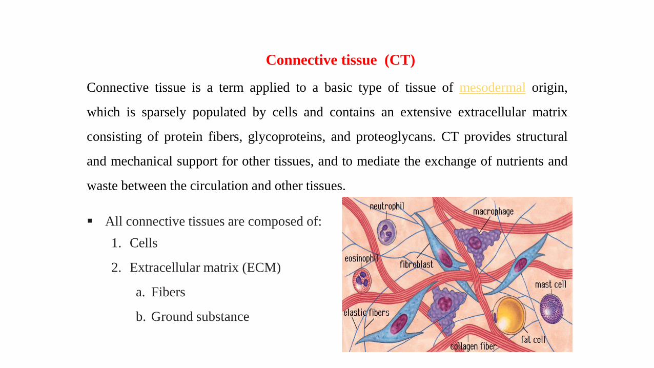
Connective tissue is a term applied to a basic type of tissue of mesodermal origin,
which is sparsely populated by cells and contains an extensive extracellular matrix
consisting of protein fibers, glycoproteins, and proteoglycans. CT provides structural
and mechanical support for other tissues, and to mediate the exchange of nutrients and
waste between the circulation and other tissues.
▪ All connective tissues are composed of:
1. Cells
2. Extracellular matrix (ECM)
a. Fibers
b. Ground substance
Connective tissue (CT)
Functions of CT
Support: Structural support is the major function of connective tissue, which forms
the framework upon which all other body tissues are assembled.
Defense physical: The viscosity of the extracellular matrix, slows the progress of
many bacteria and foreign particles.
Defense immunologic: Foreign bodies that successfully penetrate epithelia are
intercepted by immuno-responsive cells that inhabit the underlying connective
tissue.
Repair: Rapidly closing any breaches in the body's protective barriers.
Storage: Reserves of water, electrolytes, and reserve energy in the form of lipids
are stored in adipocytes.
Transport: CT is thus a crossroads for transporting substances to and from other
tissues.

There are two major groups of cell in connective tissue:
1. Fixed cells (resident cells): Are derived from mesenchyme and
are continuously present in the tissue (e.g., fibroblasts,
adipocytes, mast cell, etc).
2. Free cells (visitant cells): Enter and leave the blood stream to
migrate through and function in connective tissues (e.g.,
neutrophils, eosinophil, basophil, monocyte, lymphocytes, and
plasma cells)
Cells of CT

1. Fibroblasts:
• Fibroblasts are spindle-shaped flat cells
with minute processes spreading out of
the cell body.
• The cells have flattened nucleus
containing 1-2 nucleoli. When fibroblasts
are active, cellular organelles like Golgi
apparatus and rough endoplasmic
reticulum become more prominent.
• Secrete both fibers and ground substance
of the matrix

2. Mast cells.
Mediate immediate hypersensitivity reaction
by releasing immune modulators (Histamine)
from cytoplasmic granules, in response to
antigen binding with cell surface antibodies.
Structure:
• Round to oval-shaped cells.
• Round, usually centrally located nucleus.
• Well-defined cytoplasm filled with
secretory granules containing immune-
modulatory compounds (e.g., histamine).

3. Macrophages.
▪ Derived from blood monocytes; monocytes enter
connective tissue from the bloodstream and rapidly
transform into macrophages that function in
phagocytosis, antigen processing, and cytokine
secretion.
▪ Comprise the mononuclear phagocyte system of
the body; include Kupffer cells in the liver,
alveolar macrophages in the lung, microglia the
central nervous system, Langerhan’s cells in the
skin, and osteoclasts in bone marrow.

Extracellular matrix
• Collagen Fibers: Large fibers made of theprotein collagen and are typically the mostabundant fibers. Promote tissue flexibility.
• Elastic Fibers: Intermediate fibers made ofthe protein elastin. Branching fibers thatallow for stretch and recoil.
• Reticular Fibers: Small delicate, branchedfibers that have same chemical compositionof collagen. Forms structural framework fororgans such as spleen and lymph nodes.
1. Fiber
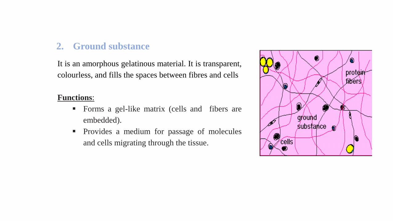
It is an amorphous gelatinous material. It is transparent,
colourless, and fills the spaces between fibres and cells
Functions:
▪ Forms a gel-like matrix (cells and fibers are
embedded).
▪ Provides a medium for passage of molecules
and cells migrating through the tissue.
2. Ground substance
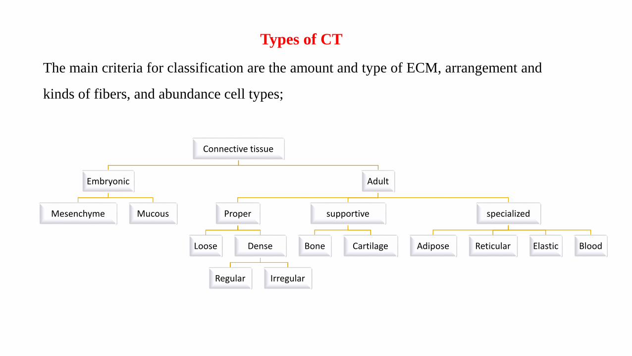
Types of CT
The main criteria for classification are the amount and type of ECM, arrangement and
kinds of fibers, and abundance cell types;
Connective tissue
Embryonic
Mesenchyme Mucous
Adult
Proper
Loose Dense
Regular Irregular
supportive
Bone Cartilage
specialized
Adipose Reticular Elastic Blood

▪ Embryonic CT
• Mesenchyme: Unspecialized CT of early week of embryonic life, which further
differentiates into all CT.
• Mucous: Embryonic connective tissue with abundant ground substance and delicate
collagen fibers; present in the umbilical cord.

a. Loose (areolar) CT b. Dense CT
▪ Highly cellular, numerous cell types
present.
▪ Fewer cells, mostly fibroblasts.
▪ Fewer and smaller caliber collagen fibers
compared with dense.
▪ Highly fibrous with larger caliber collagen
fibers.
▪ Abundant ground substance. ▪ Minimal ground substance.
▪ Highly vascularized. ▪ Poorly vascularized.
▪ diffusion of nutrients, wastes and provides
padding between and around organs and
tissues.
▪ provides strength.
▪ Adult CT
1. True (proper) CT

Dense regular CT Dense irregular CT
▪ The parallel arrangement of bundles of
collagen fibers.
▪ Randomly-arranged collagen fibers
and a few fibroblasts
▪ Forms tendons, ligaments ▪ Forms the capsule of organs and the
dermis of the skin.
▪ Provide strong attachment between
various structures
▪ Provide strength
b. Dense connective tissue
fibroblasts
collagen

2. Supportive connective tissue
a. Cartilage
▪ It is a type of connective tissue which is tough, and flexible.
▪ Like other connective tissues, it consists of cells and ECM.
▪ Unlike other CT, does not contain vessels and nerves.
▪ The strength of cartilage is due to collagen fibers and the resilience is
due to the presence of chondroitin.
▪ Most cartilage is surrounded by the perichondrium. Fibrocartilage is
the exception.

Components of Cartilage
Perichondrium:
▪ Capsule-like sheath of dense connective tissue that
surrounds cartilage.
▪ Harbors the vascular supply for avascular cartilage.
▪ Connects cartilage with the surrounding tissues.
a) Fibrous layer; Outer portion, composed of
dense connective tissue, serves as a source of
reserve cells for the chondrogenic layer.
b) Chondrogenic layer; Inner more cellular
portion contains chondroblasts which create the
major component, the extracellular matrix, of
the cartilage.

❖ Cells:
▪ Chondroblasts
• Lie on the surface of cartilage in the
chondrogenic layer of perichondrium.
• Secrete extracellular matrix around them, thus
becoming.
▪ Chondrocytes
• Are chondroblasts that have surrounded
themselves with matrix.
• Lie within cartilage in potential spaces called
lacunae.
• Secrete and maintain extracellular matrix.
• Are frequently located in isogenous groups, a
cluster of chondrocytes, resulting from the
proliferation of a single chondrocyte.
❖ Extra cellular matrix: Fiber & ground substance
1. Hyaline Cartilage:
Composition :
▪ Presence of isogenous group.
▪ Ground substance appears homogenous & takes blue stain.
▪ Collagen fibers in ground substance have same refractive index as
that of ground substance.
▪ Perichondrium is present in all except in articular cartilage.
Types of Cartilage
Incidence: articular surfaces, wall of large respiratory passages – larynx,
trachea, bronchi, epiphyseal plate, ventral ends of ribs, embryonic temporary
skeleton
Functions: Reduces friction at joints, movement and support


2. Elastic Cartilage:
Composition
▪ Chondrocytes – similar to hyaline
cartilage, housed in lacuna singly or in
pairs.
▪ Extracellular matrix – fibrils of collagen
II and network of fine elastic fibers, less
amount of ground amorphous substance
▪ On the surface - perichondrium is
identifiable.
▪ Itprovides both strength and elasticity to
certain parts of the body.
Incidence: epiglottis, auricle of the ear
,Eustachian tube, cuneiform and corniculate
cartilages in larynx
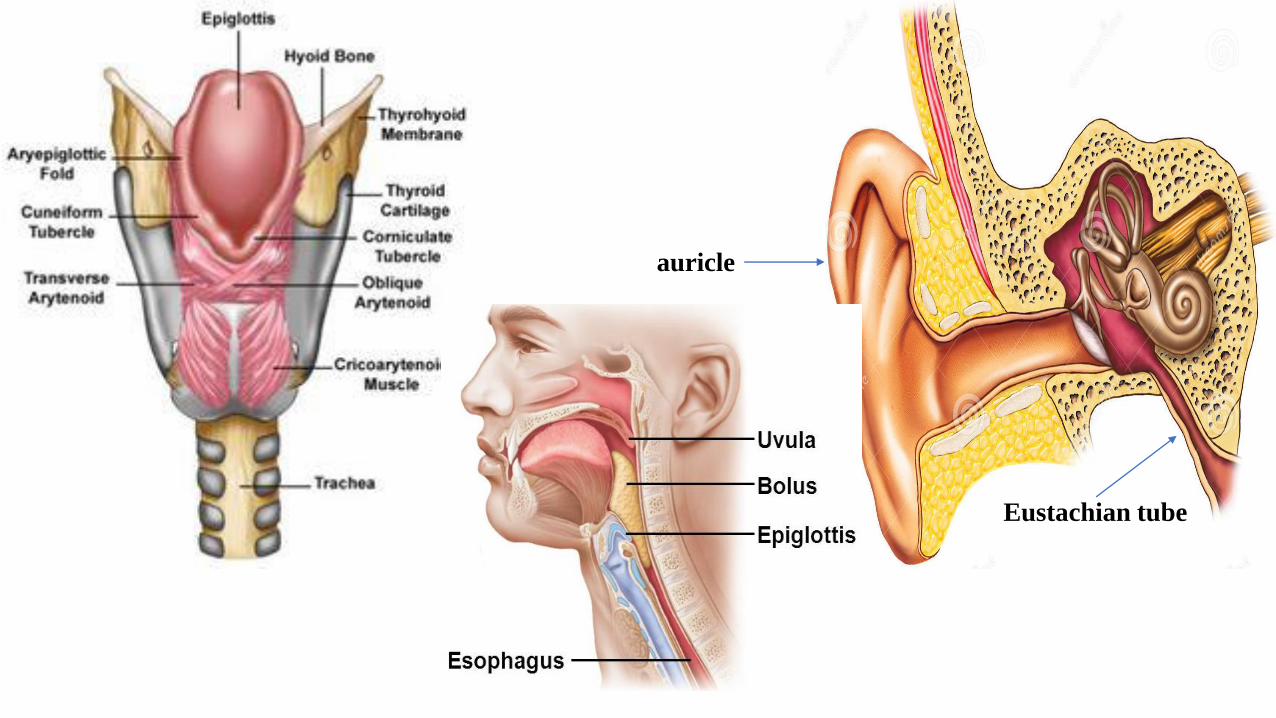
auricle
Eustachian tube

Composition
▪ Chondrocytes – small, spindle-shaped, similar to fibroblasts
▪ arranged singly or in long rows
▪ Extracellular matrix – great number of collagen I fibers – acidophilic, amorphous matrix
less abundant
▪ Perichondrium is not identifiable.
Incidence: Intervertebral disc, articular disc,
pubic symphysis, labrum of ball and socket
joint.
Functions: Shock absorbers,
Provides sturdiness without
impeding movement
3. Fibrocartilage

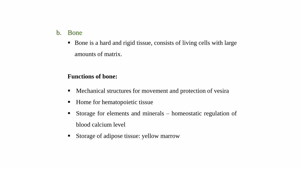
b. Bone
▪ Bone is a hard and rigid tissue, consists of living cells with large
amounts of matrix.
Functions of bone:
▪ Mechanical structures for movement and protection of vesira
▪ Home for hematopoietic tissue
▪ Storage for elements and minerals – homeostatic regulation of
blood calcium level
▪ Storage of adipose tissue: yellow marrow

▪ Cells:
1. Osteo-progenitor cells: Flattened, elongated ovoid nuclei,
derived from mesenchymal cells, located in periosteum,
endosteum and stromal component of BM.
2. Osteoblasts:
▪ They are cuboidal to columnar cells have the epitheloid
appearance with intensely basophilic cytoplasm.
▪ The bone-forming cells derived from osteo-progenitor cells,
which produce and secrete matrix proteins and transport
mineral into the matrix.

3. Osteocytes:
▪ Osteocytes are spider-shaped cells.
▪ Transport materials between blood and bone and to
maintain surrounding matrix; they do not divide or
secrete matrix.
4. Osteoclasts:
▪ Osteoclasts are large, multinucleated cells, have small
projections (microvilli) that is the cell’s active region.
▪ They function in bone resorption by removing local
mineralized matrix.
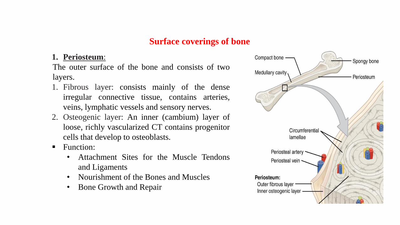
1. Periosteum:
The outer surface of the bone and consists of two
layers.
1. Fibrous layer: consists mainly of the dense
irregular connective tissue, contains arteries,
veins, lymphatic vessels and sensory nerves.
2. Osteogenic layer: An inner (cambium) layer of
loose, richly vascularized CT contains progenitor
cells that develop to osteoblasts.
▪ Function:
• Attachment Sites for the Muscle Tendons
and Ligaments
• Nourishment of the Bones and Muscles
• Bone Growth and Repair
Surface coverings of bone

Endosteum:
▪ It is a thin layer of connective tissue that lines
the inner surfaces of all bones (the cavities
within the bone).
▪ Also contains both osteoblasts, osteoclasts,
and/or osteo-progenitor cells that line all
interior surfaces of bone except for lacunae and
canaliculi.
▪ Serves as a means of bone growth and/or
resorption.
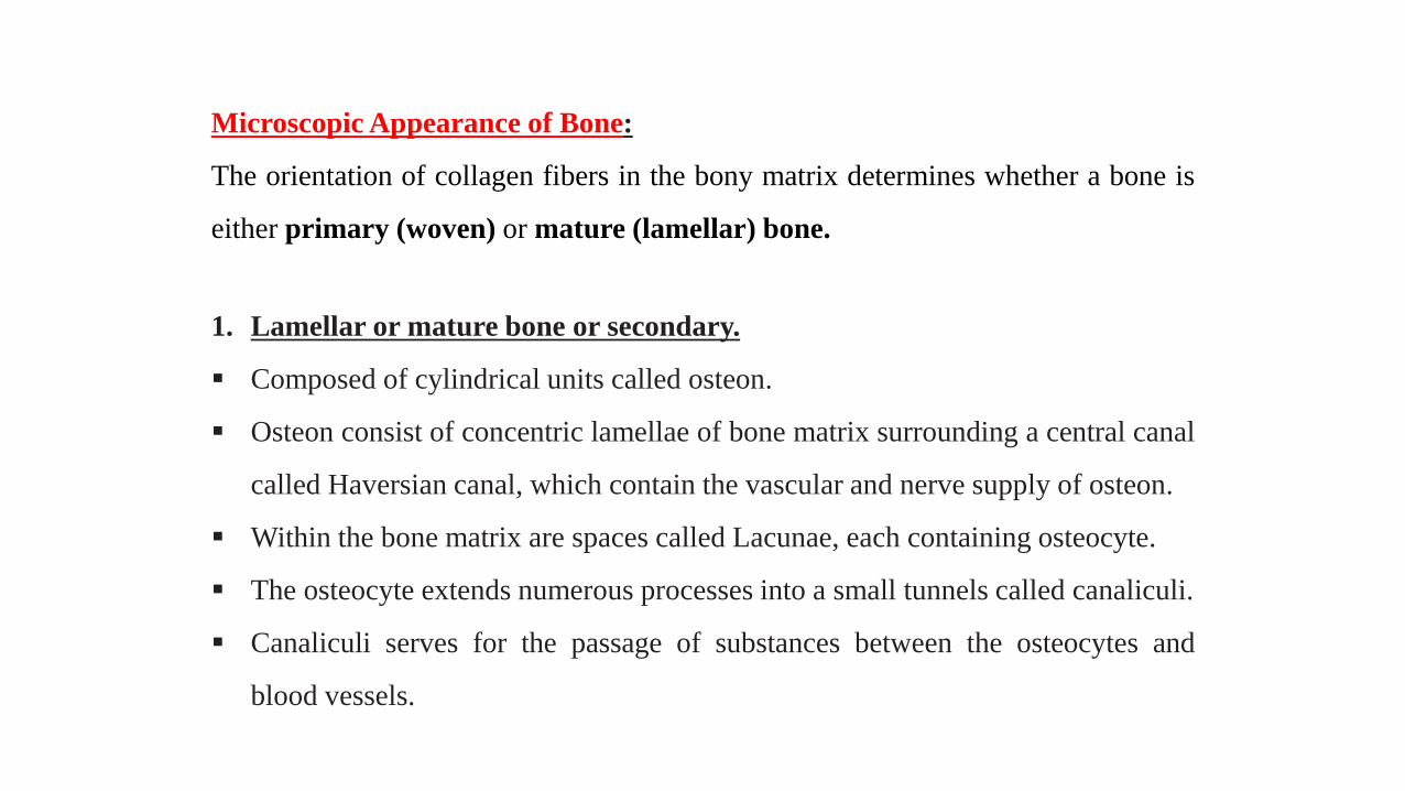
Microscopic Appearance of Bone:
The orientation of collagen fibers in the bony matrix determines whether a bone is
either primary (woven) or mature (lamellar) bone.
1. Lamellar or mature bone or secondary.
▪ Composed of cylindrical units called osteon.
▪ Osteon consist of concentric lamellae of bone matrix surrounding a central canal
called Haversian canal, which contain the vascular and nerve supply of osteon.
▪ Within the bone matrix are spaces called Lacunae, each containing osteocyte.
▪ The osteocyte extends numerous processes into a small tunnels called canaliculi.
▪ Canaliculi serves for the passage of substances between the osteocytes and
blood vessels.


▪ Mature spongy bone is structurally similar to mature compact
bone except that the tissue is arranged as trabeculae.
▪ The matrix of bone is lamellated.
▪ Osteocytes get nutrients directly from circulating blood.

2. Woven or immature bone or primary bone
▪ Possibly will be either spongy or compact. Woven
bone is found on the growing ends of an immature
skeleton or, in adults, at the site of a healing fracture.
▪ Characterized by random deposition of fine collagen
and increased cellularity contains osteocytes that are
more numerous and spherical than those of lamellar
bone.
▪ Relatively low mineral contents.
▪ Seen in embryonic development, fracture, and repair.
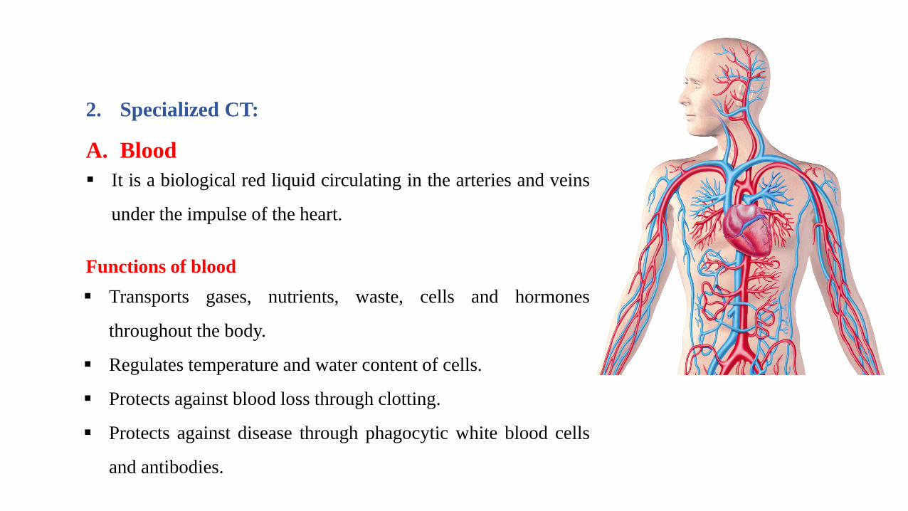
▪ It is a biological red liquid circulating in the arteries and veins
under the impulse of the heart.
A. Blood
Functions of blood
▪ Transports gases, nutrients, waste, cells and hormones
throughout the body.
▪ Regulates temperature and water content of cells.
▪ Protects against blood loss through clotting.
▪ Protects against disease through phagocytic white blood cells
and antibodies.
2. Specialized CT:
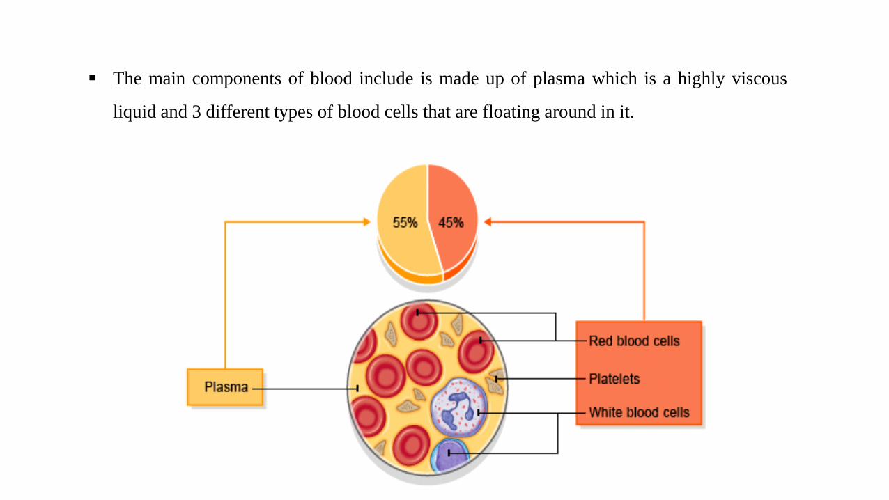
▪ The main components of blood include is made up of plasma which is a highly viscous
liquid and 3 different types of blood cells that are floating around in it.

1. Plasma:
▪ A typical sample of plasma is composed of 90% water, 8% protein, 1%
inorganic salts, 0.5% lipids and 0.1% sugar, the rest being made of
lesser components.
▪ The three main groups of proteins in plasma are the blood coagulation
(fibrinogen) proteins, albumin, and the globulins.
• The globulins can be divided into;
• alpha globulins (proteases, anti-proteases and transport
proteins).
• beta globulins (transferrin, other transport proteins)
• gamma globulins (mainly immunoglobulins).
▪ The plasma proteins are nearly all derived from synthesis in the liver,
with the exception of the immunoglobulins which are synthesised by
plasma cells.

2. Cellular component
a. Erythrocyte (RBC)
▪ RBCs constitute the largest number of cells in the blood
▪ about 5.2 million mm3 (man), 4-5 million (woman)
▪ biconcave disc shape
▪ no nucleus
▪ contain hemoglobin
▪ Life span in blood 120 days
▪ main function is gases transport

b. Platelets (thrombocytes)
• Small, biconvex disks.
• Non-nucleated cell formed in the bone marrow
from the cytoplasm of cells called
megakaryocytes.
• Platelets have a life span of about 10 days.
• Their numbers in circulating blood range from
150 000 to 400 000/mL.
• Function: initiate blood clots

c. Leucocyte WBC
▪ 5000-9000 mm3 (healthy adult)
▪ act mainly outside blood vessels in the tissues
▪ main function is defence.
▪ classification of leukocytes
• classification system is based on appearance of the granule
o Granulocytes
✓ Neutrophil, Basophile and Eosinophil
o Agranulocytes
✓ Lymphocytes and Monocytes

1. Granulocyte
▪ Neutrophil
• the most common type of leucocyte in blood
• Compose 60-70% of the leukocytes
• Life Span: < 1 week
• About 9-12 µm in diameter (thus larger than
RBC).
• Nucleus is polymorphonuclear - has 3-5 lobes
linked together by fine threads of chromatin
• Cytoplasm contains granules, which are
lysosomes that contain enzymes for digestion
of phagocytosed particles, e.g. bacteria.

▪ Eosinophil
• Compose 1-6% of the leukocytes
• Nucleus is bilobed
• Life span < 2 weeks
• Cytoplasm contains ovoid, acidophilic
granules (bright pink), which are larger than
those of neutrophils
• Granules are lysosomes that contain enzymes
that can degrade phagocytosed particles.
• Important in allergic reactions, parasitic
infections, and phagocytosis of Ab-Ag
complexes.

▪ Basophil
• constitute less than 1% of leucocytes.
• Lobulated nucleus often obscured by
granules
• Life Span: 1-2 years
• basophilic cytoplasmic granules (dark
blue), containing heparin (anti-coagulant)
and, histamine (vasodilator).
• Function in allergic reactions and
inflammatory response.

2. Agranulocyte
a. Lymphocytes
• are the smallest cells in the white cell series,
being only slightly larger than erythrocytes.
• constitute 20-30% of leucocytes.
• Life Span: variable (few days to several
years)
• Round dense nucleus.
• Small lymphocytes (inactivate) have very
little cytoplasm.
• Two populations, one that can become T-
lymphocytes and the other B- lymphocytes.
• When activated by encounter with foreign
antigen presented by a macrophage - become
large lymphocytes that are capable of mitosis.
Small lymphocytes
Large lymphocytes
Round nucleus
Ovoid nucleus

Structure:
▪ Oval-shaped cells.
▪ Round, eccentrically located nucleus.
▪ Basophilic cytoplasm due to large amounts of rough
endoplasmic reticulum.
▪ Well-developed Golgi complex appears as a distinct,
unstained region in the cytoplasm near the nucleus
and, for that reason, is often referred to as a
“negative Golgi.”
* Plasma cells.
▪ Secrete antibodies to provide humeral immunity.
▪ Derived from B-lymphocytes.
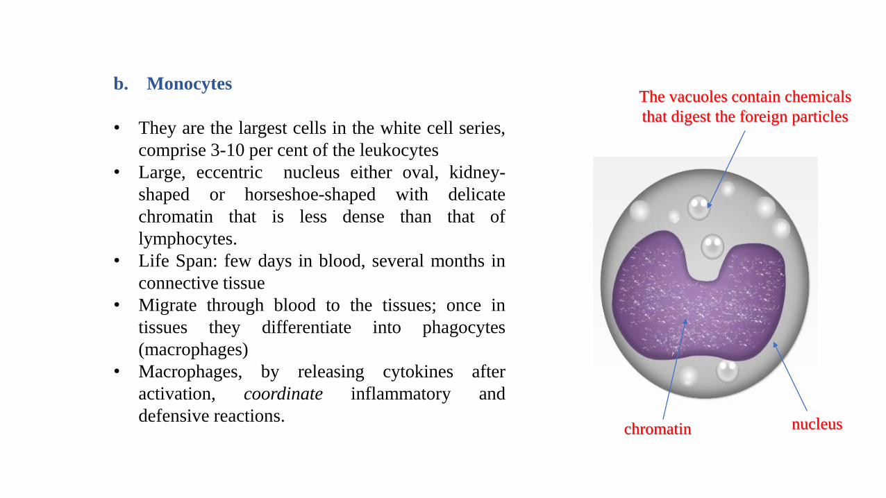
b. Monocytes
• They are the largest cells in the white cell series,
comprise 3-10 per cent of the leukocytes
• Large, eccentric nucleus either oval, kidney-
shaped or horseshoe-shaped with delicate
chromatin that is less dense than that of
lymphocytes.
• Life Span: few days in blood, several months in
connective tissue
• Migrate through blood to the tissues; once in
tissues they differentiate into phagocytes
(macrophages)
• Macrophages, by releasing cytokines after
activation, coordinate inflammatory and
defensive reactions. nucleuschromatin
The vacuoles contain chemicals
that digest the foreign particles
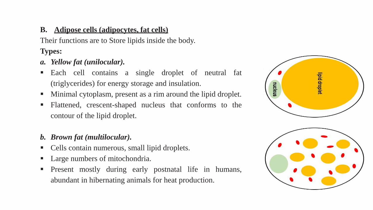
B. Adipose cells (adipocytes, fat cells)
Their functions are to Store lipids inside the body.
Types:
a. Yellow fat (unilocular).
▪ Each cell contains a single droplet of neutral fat
(triglycerides) for energy storage and insulation.
▪ Minimal cytoplasm, present as a rim around the lipid droplet.
▪ Flattened, crescent-shaped nucleus that conforms to the
contour of the lipid droplet.
b. Brown fat (multilocular).
▪ Cells contain numerous, small lipid droplets.
▪ Large numbers of mitochondria.
▪ Present mostly during early postnatal life in humans,
abundant in hibernating animals for heat production.