Embed Size (px)
Citation preview

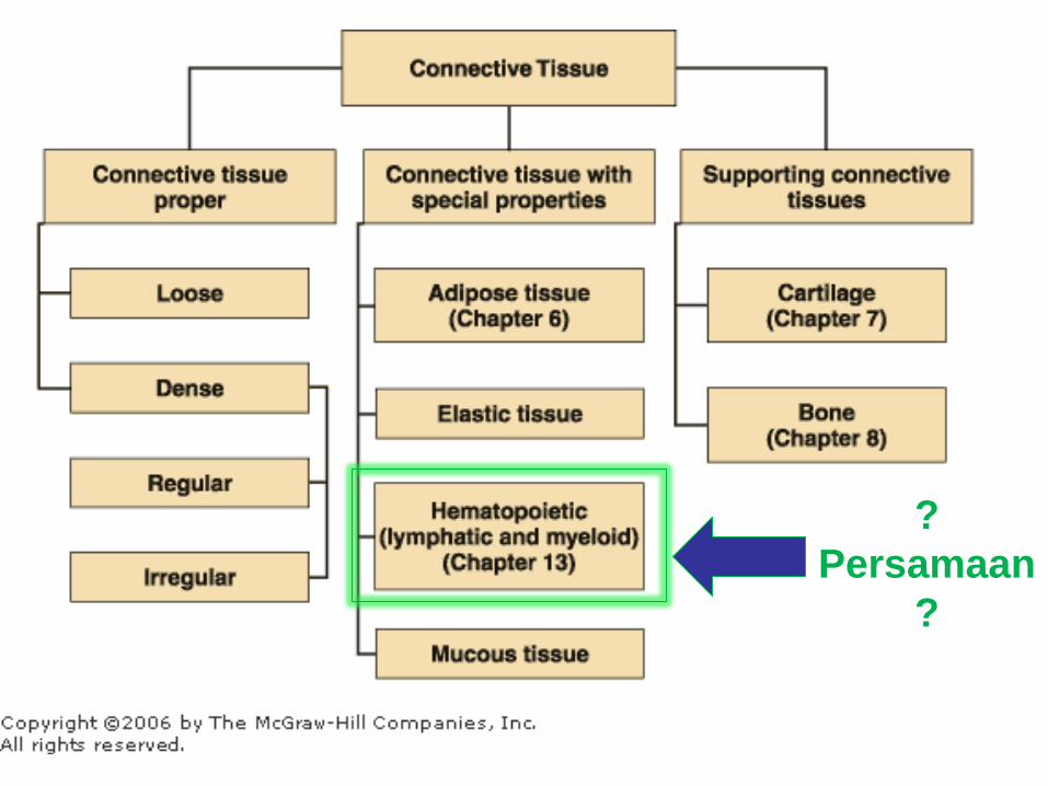
CONNECTIVE TISSUE PART-2
anatomi.lecture.ub.ac.id
?
Persamaan
?
(SYSTEMA LYMPHOPOIETICA)
For for 1st year
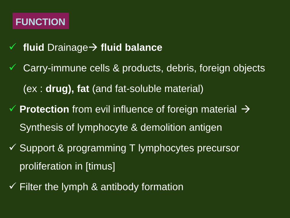
fluid Drainage fluid balance
Carry-immune cells & products, debris, foreign objects
(ex : drug), fat (and fat-soluble material)
Protection from evil influence of foreign material
Synthesis of lymphocyte & demolition antigen
Support & programming T lymphocytes precursor
proliferation in [timus]
Filter the lymph & antibody formation
FUNCTION
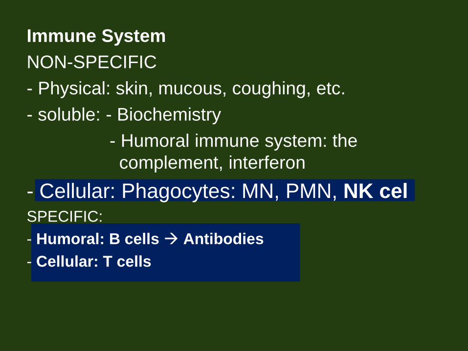
Immune System
NON-SPECIFIC
- Physical: skin, mucous, coughing, etc.
- soluble: - Biochemistry
- Humoral immune system: the
complement, interferon
- Cellular: Phagocytes: MN, PMN, NK cel SPECIFIC:
- Humoral: B cells Antibodies
- Cellular: T cells

The lymphatic system
consists of:
(1) ―vascular‖/vessel
system; capillaries
from various organs and
network of lymph vessels
large vein
(2) lymph fluid
(3) lymph node / lymphoid
organ : composed of
lymphatic cells

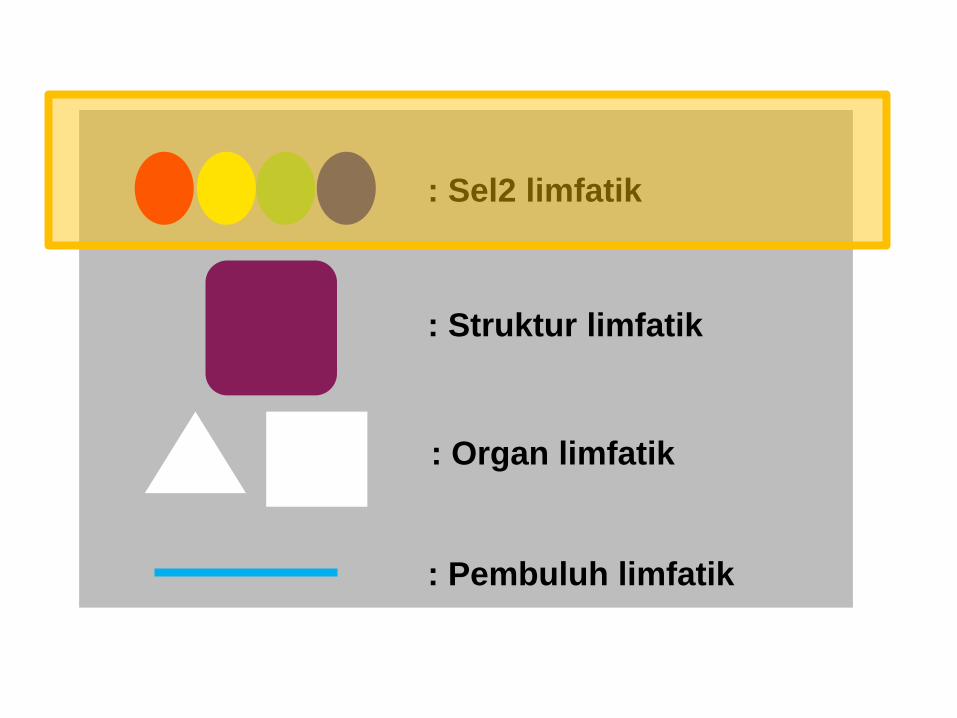
Organization of
lymphatic system
: lymphatic cells
: lymphatic structure
: lymphatic organ
: Lymphatic vessel (With lymph fluid in it)


: Sel2 limfatik
: Struktur limfatik
: Organ limfatik
: Pembuluh limfatik (dgn cairan limfe di dalamnya)

1. SISTEM PEMBULUH
• Contains Lymph FLUID ;
consist of :
Excess tissue fluid
Cellular debris
Lymphocytes
Fat (the gut)
• a closed tube syste
• move one direction
V.subclavia
• Will be further discussed at
CVS
: Sel2 limfatik
: Struktur limfatik
: Organ limfatik
: Pembuluh limfatik

SEL2 SISTEM LIMFATIK
Lymphocyte
NK cell
APC
macrophage
Sel Plasma
Sel Retikuler

Lymphocyte
• can not phagocytosis
• In general there are two types:
- T lymphocytes (T-lymphocytes / T cells)
- B lymphocytes (B-lymphocytes / B cell)
- The B and T cells are the only cells that have the
ability to selectively recognize a specific epitope
(antigenic determinant)
Lymphocyte NK cell APC macrophage Sel Plasma Sel Retikuler

Lymphocytes of Lymphoid
Organ
(T-cell (%) : B-cell (%))
Thymus
100 0
Bone Marrow
10 90
Spleen
45 55
Lymph Nodes
60 40
Blood
80 20


T cell
• responsible for cellular immunity
• require the help of macrophages or other APC
to optimal response
• Cells Effectors:
* T-helper
* T-cytotoxic
* Supressor T-cell
Sel-sel organ limfatik
Lymphocyte NK cell APC macrophage Sel Plasma Sel Retikuler responsible for cellular immunity
• bring the Ag receptors on the surface
receptor
• require the help of macrophages or other
APC to
optimal response
• required for the activation of Ag-MHC
complexes
• Cells Effectors:
* T-helper
* T-cytotoxic
* Supressor T-cel

B Lymphocytes (B cell)
• Responsible for humoral immune
• Cells daughter:
- Plasma cell
- Memory cell
Sel-sel organ limfatik

Plasma Cell
• is the result of B cell effector
differentiation
• secrete immunoglobulin (Ig)
• microscopy: a clock face nucleus
• Present in all lymphatic tissue
Sel-sel organ limfatik
Lymphocyte NK cell APC macrophage Sel Plasma Sel Retikuler

Natural Killer cell
- = Lymphocyte granular
- No receptor
- Can be activated without specific stimulation (no
memory)
Sel-sel organ limfatik
Lymphocyte NK cell APC macrophage Sel Plasma Sel Retikuler

APC (Antigen Presenting Cell)
• cells that display peptides associated with class II MHC
molecules to CD4+ TH cells (komplek antigen small component
presentation express on the cell surface)
• Tipe :
– Professional APC : constitutively express class II MHC
molecules
• = sel yg dpt menimbulkan aktivasi perkembangan limfosit
• Tdd : Dendritic cell, macrophage, & B-lymph.
– Nonprofessional APC : can be induced to express class II MHC
molecules
• =sel yg dpt distimulasi utk presentasi antigen dlm fungsi
efektor
• Ex : endothelial cells, astrocytes, epithelial cells, fibroblast
Sel-sel organ limfatik
Lymphocyte NK cell APC macrophage Sel Plasma Sel Retikuler
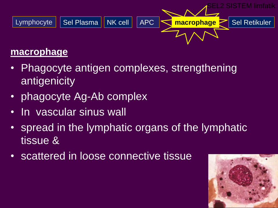
macrophage
• Phagocyte antigen complexes, strengthening
antigenicity
• phagocyte Ag-Ab complex
• In vascular sinus wall
• spread in the lymphatic organs of the lymphatic
tissue &
• scattered in loose connective tissue
SEL2 SISTEM limfatik
Lymphocyte NK cell APC macrophage Sel Plasma Sel Retikuler

Sel Retikuler
• stellata, prosesus beranyaman
• Type :
– Sel Retikuler mesenchymal (dendritic) sbg
APC
– Sel Retikuler epithelial supportive. Di Thymus
Sel-sel organ limfatik
Lymphocyte NK cell APC macrophage Sel Plasma Sel Retikuler

: Sel2 limfatik
: Struktur limfatik
: Organ limfatik
: Pembuluh limfatik

• = kumpulan Lymphocyte2, berbentuk sferis
aggregat2 limfatik sub unit fungsional
• didominasi sel B-helper
• Nodulus primer
– saat prenatal
– Germinal center [-]
– Ag [-]
NODULI LIMFATISI (Lymphatic nodule)
GAMBARAN KHUSUS
(SPECIAL FEATURES)
Nodulus sekunder
setelah lahir
= bentuk aktif dr nodulus
primer oleh paparan Antigen
Terdapat Germinal Centre
banyak limfoblast
Sbg tempat sel memori

.......special features
GERMINAL CENTRE
• Areas that look pale, are in the middle of an noduli
limfatici(Lymphatic nodule)
• Consists of :
- Lymphocyte; aktif proliferasi, p.u berukuran sedang.
Limfoblast [+]
P.u B-cell
- Sel retikuler; relatif besar, dg prosesus panjang
(Dendritic cell), inti pucat dan besar, sitoplasma
basofil
- Sel lain : macrophage, sel plasma

- timbul setelah lahir
- Hilang timbul sesuai stimulasi antigen
- [-] - s/d akan lahir
- bila antigen [-]
- thymectomy saat lahir
GERMINAL CENTRE
GERMINAL
CENTRE

: Sel2 limfatik
: Struktur limfatik
: Organ limfatik
: Pembuluh limfatik

1. Cells
predominantly : Lymphocyte T & B
Other : - Sel plasma - Sel retikuler
- APC - macrophage
2. (structure) Lymphatic Tissue
a. Loose lymphatic tissue Lymphocyte tidak
tersusun rapat.
b. Dense lymphatic tissue Lymphocyte membentuk
aggregat ( lymphatic nodules/follicle)
3. Organ Limfatik
Ex : Limfonodus, Lien, Timus
COMPONENTS OF Lymphatic ORGAN :

• organ limfatik sentral (Primary) :
pembentukan Lymphocyte tidak tergantung antigen
supply T-cell netral atau prekursor Lymphocyte B ke
organ & jaringan perifer
Tdd : Timus dan sumsum tulang
• organ limfatik perifer (Secondary):
pembentukan Lymphocyte tergantung antigen sel2
imunokompeten, bereaksi thd Antigen spesifik
Tdd : Limfonodus, lien, tonsil, aggregat limfatik tidak
berkapsul
Klasifikasi jaringan & organ limfatik :

ORGAN LIMFATIK
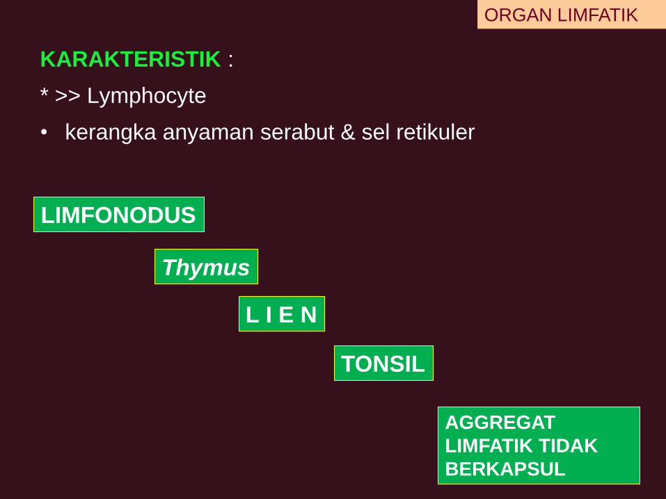
KARAKTERISTIK :
* >> Lymphocyte
• kerangka anyaman serabut & sel retikuler
ORGAN LIMFATIK
Thymus
L I E N
LIMFONODUS
TONSIL
AGGREGAT
LIMFATIK TIDAK
BERKAPSUL
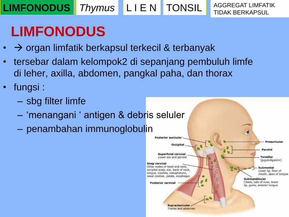
• organ limfatik berkapsul terkecil & terbanyak
• tersebar dalam kelompok2 di sepanjang pembuluh limfe
di leher, axilla, abdomen, pangkal paha, dan thorax
• fungsi :
– sbg filter limfe
– ‗menangani ‗ antigen & debris seluler
– penambahan immunoglobulin
LIMFONODUS
Thymus L I E N LIMFONODUS TONSIL AGGREGAT LIMFATIK
TIDAK BERKAPSUL

LIMFONODUS
Thymus L I E N LIMFONODUS TONSIL AGGREGAT LIMFATIK
TIDAK BERKAPSUL

Struktur
• bentuk seperti kacang
• Tdd cortex dan medulla
• Kapsul trabekula antara nodulus di cortex
• pembuluh darah & pembuluh limfe efferent di hilum
• Pembuluh limfe afferent melalui permukaan convex
LIMFONODUS

CORTEX :
• Lymphocyte tersusun padat 1 lapis noduli limfatici
sekunder
• germinal center [+]
• Lymphocyte tergantung di anyaman jaringan ikat
retikuler
LIMFONODUS

ALIRAN LIMFE
limfe pembuluh afferent
sinus subcapsular
sinus peritrabekular
anyaman anastomose di sinus2 medullary
pembuluh limfe efferent
hilum
LIMFONODUS


• Penyaringan limfe
– limfe afferent membawa debris seluler & antigen
’ditangani’ oleh macrophage & sel2 dendritik folikuler di
sinus2
– Lymphocyte mengalami : kontak dg APC & macrophage di
sinus2
– keluar dr sinus masuk parenchym
LIMFONODUS


Cllinical correlation :
• Lymphadenopathy
• Lymphadenitis

T I M U S (Thymus)
• Hanya membentuk prekursor sel T
• Temporer --mengalami involusi dg pertambahan
umur
• Berat : 35 gr (puber) 25 gr (umur 25 thn) 15
gr (umur 60 thn)
T I M U S L I E N LIMFONODUS TONSIL AGGREGAT LIMFATIK
TIDAK BERKAPSUL

ANATOMI : Thymus

Histofisiologi timus
• pembentukan T-Lymphocyte
( prekursor dibentuk di sum-sum tulang) cortex timus
(= thymocyte)
• Thymocyte :
- proliferasi thymocyte Lymphocyte T
- kebanyakan akan mengalami apotosis difagositosis
oleh macrophage
- p.u tidak bisa bereaksi terhadap antigen
TIMUS

• ―pemrograman‘ :
penyusunan ulang gen reseptor antigen
kemampuan mengenali antigen
menghilangkan sel2 prematur yang
teraktivasi & menjadi Antigen ―self‖‘
TIMUS-Histofisiologi

• sel matur medulla venule postkapiler/pembuluh
limfatik efferent menempati daerah T-dependent di
organ limfatik sekunder differensiasi menjadi T-cell
fungsional
TIMUS-Histofisiologi
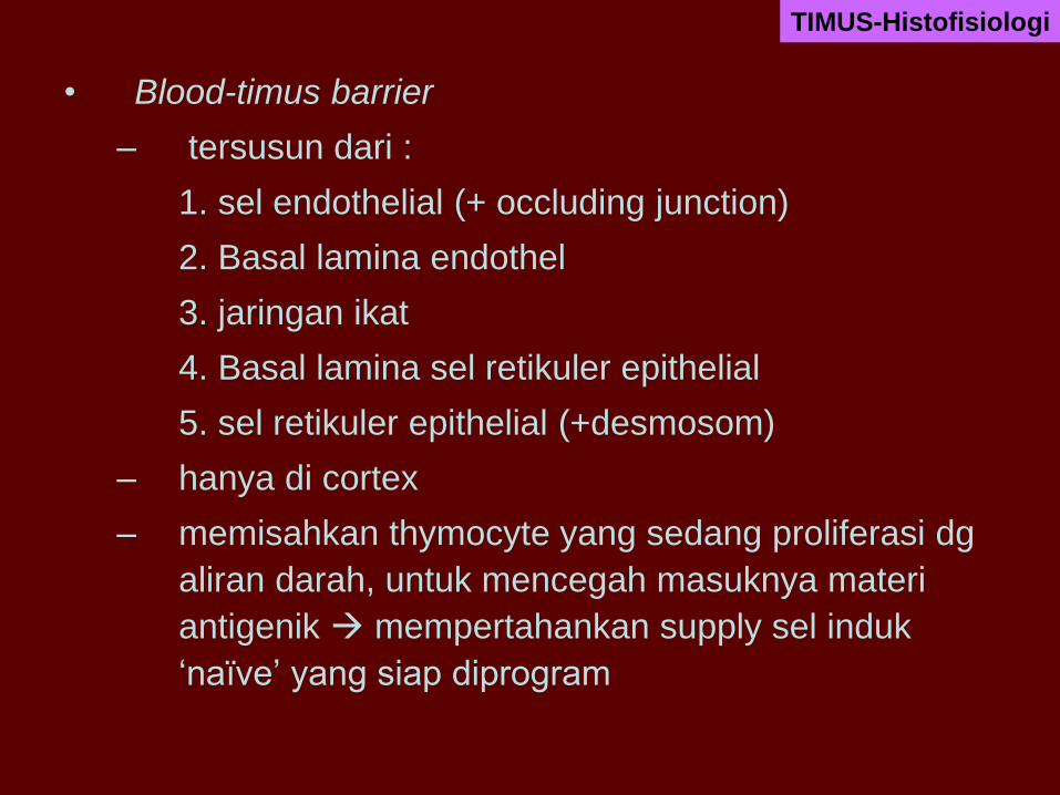
• Blood-timus barrier
– tersusun dari :
1. sel endothelial (+ occluding junction)
2. Basal lamina endothel
3. jaringan ikat
4. Basal lamina sel retikuler epithelial
5. sel retikuler epithelial (+desmosom)
– hanya di cortex
– memisahkan thymocyte yang sedang proliferasi dg
aliran darah, untuk mencegah masuknya materi
antigenik mempertahankan supply sel induk
‗naïve‘ yang siap diprogram
TIMUS-Histofisiologi

L I E N
fungsi :
• tempat pembentukan Lymphocyte teraktivasi
• pertahanan thd benda asing
• tempat destruksi RBC tua
• penyimpanan darah
• berperan dalam metabolisme besi
• Sbg organ hematopoiesis fetal.
Thymus L I E N LIMFONODUS TONSIL AGGREGAT LIMFATIK
TIDAK BERKAPSUL

• Di hipokhondrium; dg sedikit
sampai epigastrium.
• Antara fundus gaster –
diafragma
ANATOMI

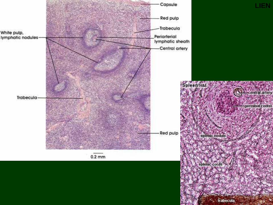
Struktur
• kapsul jaringan ikat padat trabekula splenic pulp
• Pulpa :
white pulp (pulpa putih; pulpa alba): * Noduli Limfatici
(Corpusculum Malpighi), PALS
Red pulp (Pulpa merah; pulpa rubra):
Marginal zone :
• Membentuk penghubung antara pulpa merah dan pulpa putih
• Jaringan limfatik longgar
• Banyak : * macrophage aktif, Antigen darah
– Antigen difagositosis, dijerat oleh sel dendritik (APC)
– Berfungsi mengkonsentrasikan Antigen dipresentasikan
ke Lymphocyte
• pembuluh limfe afferent [-], HEV [-]
LIEN
LIEN

Mekanisme aliran darah
mencapai sinus
• Closed theory
– Dinding kapiler berlanjut
sebagai dinding sinusoid
• Open theory
– Ujung kapiler di Billroth
cord darah keluar,
disaring oleh cord
dinding sinusoid (via
fenestrae)
LIEN : sirkulasi

LIEN : sirkulasi

TONSIL
Macam :
• T.palatina (D-S)
• T.pharyngeal
• T.lingual
Thymus L I E N LIMFONODUS TONSIL AGGREGAT LIMFATIK
TIDAK BERKAPSUL
Ring of Waldeyer

Tonsila palatina/faucial
• 2 buah, di dinding lateral oropharynx, di bawah palatum
molle
• ditutupi epithel squamous complex (non kornifikasi)
• mengandung noduli limfatisi, p.u dengan germinal center
• crypte 10-20 mengandung sel epithel yg desquamasi,
Lymphocyte, dan bakteri
• kapsul jaringan ikat padat : tebal, parsial berfungsi
sebagai barrier untuk mencegah penyebaran infeksi
TONSIL
Tonsila pharyngeal
• tunggal, midline nasopharyx posterior
• kapsul lebih tipis
• crypte [-]
• kapsul jaringan ikat tipis, parsial
• bila hipertrofi adenoid
TONSIL

TONSIL

Tonsila lingualis
• lebih kecil, jumlah lebih banyak
• di pangkal lidah, belakang papilla circumvalata
• ditutupi epithel squamous complex dg sedikit kornifikasi
• germinal center [+]
• Kapsul tidak jelas
TONSIL

TONSIL

Cllinical correlation :
• Tonsilitis

AGGREGAT LIMFATIK TIDAK BERKAPSUL • mrpkn noduli limfatisi
dalam kelompok kecil
atau soliter
• contoh :
– Berkelompok : Peyer‘s
patches di IT
– Soliter : tersebar di
mukosa GIT, UT, UG
• dapat diselubungi selapis
sel retikuler pipih
• kapsul jaringan ikat [-]
Thymus L I E N LIMFONODUS TONSIL AGGREGAT LIMFATIK
TIDAK BERKAPSUL

• MALT ( Mucosa Associated Lymphatic Tissue).
– BALT (Bronchial Associated Lymphatic Tissue).
– GALT (Gut Associated Lymphatic Tissue).
– SALT (Skin Associated Lymphatic Tissue).
AGGREGAT LIMFATIK TIDAK BERKAPSUL

– GALT (Gut Associated Lymphatic Tissue).
AGGREGAT LIMFATIK TIDAK BERKAPSUL
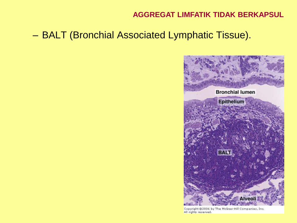
– BALT (Bronchial Associated Lymphatic Tissue).
AGGREGAT LIMFATIK TIDAK BERKAPSUL

dr. Indriati Dwi Rahayu
GENERAL PROPERTIES
THE CELLS
HEMATOPOIESIS

General Properties
- Special connective tissue
• Total volume: + 5 L, + 8 % body weight
• Composition :
√ plasma : the liquid in which the
formed elements, protein, &
hormon are suspended
√ formed element: blood cells
STAINING : Wright, Giemsa, Romanowsky,
Leishman
~ Hematocrite
Copyright © 2009 Pearson Education, Inc., publishing as Pearson Benjamin Cummings Fig 20.1
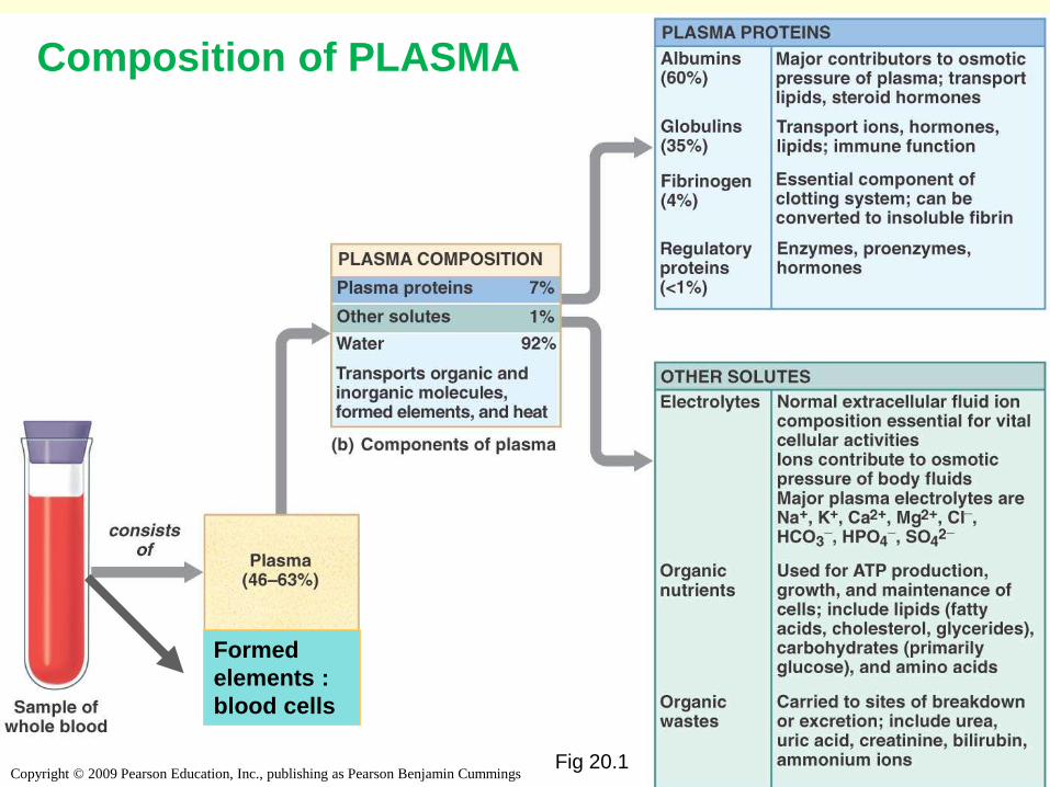
Composition of PLASMA
Formed
elements :
blood cells

• PLASMA
- +55 % blood, homogen
- slightly base
- Composition:
• +90 % water
• +10 % dissolved substance:
1. Anorganic salt : 0.9 %. Ex : Na, K, Ca
2. Organic subs. : 2,1 %. Ex : As.amino, glukosa, peptida,
hormon, lipid
3. Protein plasma : 7 %. Ex; Albumin, Globulin (α,β,γ)
Fibrinogen, prothrombin
SOLID COMPONENT + 45 % : blood cells

General properties
THE BLOOD CELLS
HEMATOPOIESIS

RBC
EOSINOPHYL
BASOPHYL
NETROPHYL
LYMPHOCYTE
MONOCYTE
THROMBOCYTE
L
E
U
C
O
C
Y
T
E
S
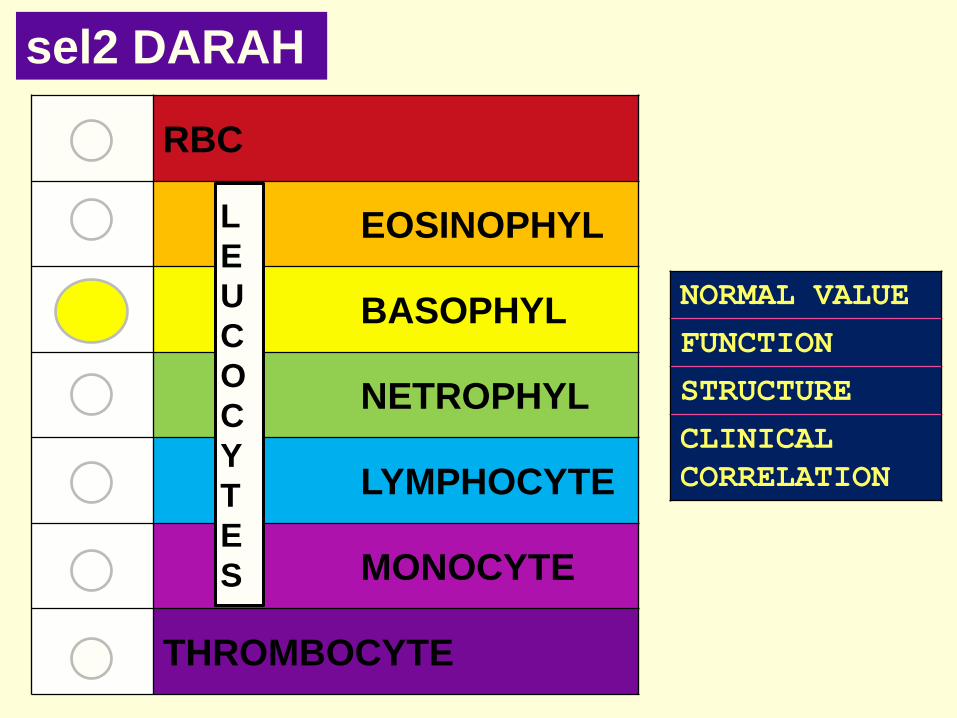
THE BLOOD CELLS
! NORMAL VALUE
FUNCTION
STRUCTURE
CLINICAL
CORRELATION

KOMPONEN PADAT 45 %
1. Red Blood Cell
- Normal value: 4 - 6 X 10⁶ /μL
- Life span : 120 hr lien dan sum2 tulang
- hematocrit is an estimate of the volume of packed
erythrocytes per unit volume of blood. The normal value is
40–50% in men and 35–45% in women.

- FUNCTION :
* O2 transpor (by Hemoglobin)
* acid-base (by Hemoglobin)
* reaction catalisator ( by enzym carbonic anhidrase)
HEMOGLOBIN
* Type :
1. Hb A1 : 97 %
2. Hb A2 : 2 %
3. Hb F : 1 %. (in neonatus 80%)
4. Hb S : abnormal Hb A Sickle cell
anemia

- STRUCTURE :
* Ф : 7 – 8 μm, (fresh preparation : yellow greenish color)
* biconcave ; central: central pallor
* (matur) : nucleus & organella : (-)
* Isotonic sitoplasma; contain Hb
* Plasmalemma : membran protein integral:
Inner Spectrin
Outer contain antigen
* flexible
• Tendention to adhere
Rouleaux formation
(temporary)


- Structure abnormalities
* Anisositosis : RBC in various size
* Macrositer : Ø > 9 µm
* Micrositer : Ø < 6 µm
* Cabot ring = Howell Jolly body : nuclear fragment (> 1 %)
Staining : Brilliant Cresyl Blue to see RER & ribosome
inside the reticulocyte
* Shadow/Ghost blood : pale, round/Spheroid, . E.c hemolysis.
* Crenated : E.c. hypertonic
* Spherocytosis : Spheroid erythrocyte

CLINICAL CORRELATION
Anemia : Hb ↓
may be caused by :
– loss of blood (hemorrhage);
– insufficient production of erythrocytes
– accelerated destruction of blood cells.
Bad oxygenation

RBC
EOSINOPHYL
BASOPHYL
NETROPHYL
LYMPHOCYTE
MONOCYTE
THROMBOCYTE
L
E
U
C
O
C
Y
T
E
S
BLOOD
CELLS
NORMAL VALUE
FUNCTION
STRUCTURE
CLINICAL
CORRELATION

2. LEUCOCYTE
- normal VALUE: 6000 – 10.000 / μL
- classification based on:
~ diameter
~ nuclear shape
~ nuclear- cytoplasm Ratio
~staining

• General characteristic:
- ―real‖ cell nucleus & organella [+]
- amoeboid Motion& diapedesis [+]
- Function in connective tissue. Blood flow only as a
means of transportation
- in the permanent preparations : larger size
- azurophilic granules with lytic enzymes
• classification with special staining diff.count (hitung
jenis)
• Main type : granulocyte & agranulocyte

- Granulocyte
* = PMN (polymorpho nuclear)
* organellS: [mature] lobed nucleus, Golgi, mitokondria,
free ribosome, RER
* specific granules dan azurophilic granules;
* TERDIRI DARI : Eosinophil, Basophil, Netrophil
- Agranulocyte
* mononuclear ; unsegmented
* azurophilic granule ONLY
* TERDIRI DARI : Lymphocyte, Monocyte
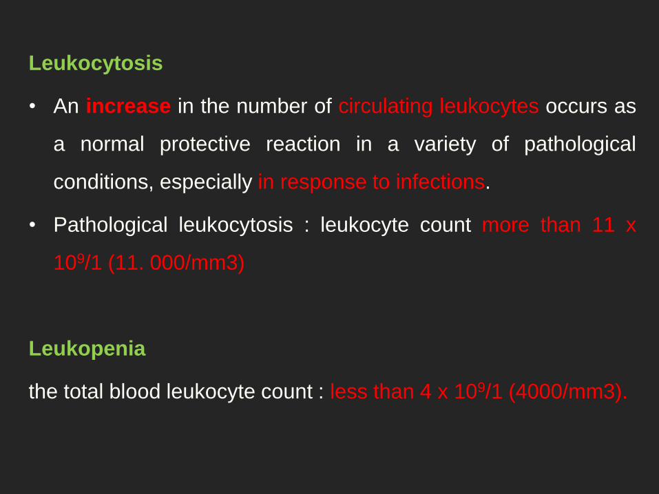
Leukocytosis
• An increase in the number of circulating leukocytes occurs as
a normal protective reaction in a variety of pathological
conditions, especially in response to infections.
• Pathological leukocytosis : leukocyte count more than 11 x
109/1 (11. 000/mm3)
Leukopenia
the total blood leukocyte count : less than 4 x 10⁹/1 (4000/mm3).

Granulocytopenia (neutropenia)
This is a general term used to indicate an abnormal
reduction in the numbers of circulating granulocytes
(polymorphonuclear leukocytes), commonly called
neutropenia because 40 to 75% of granulocytes are
neutrophils. A reduction in the number of circulating
granulocytes occurs when production does not keep
pace with the normal removal of cells or when the life

RBC
EOSINOPHYL
BASOPHYL
NETROPHYL
LYMPHOCYTE
MONOCYTE
THROMBOCYTE
L
E
U
C
O
C
Y
T
E
S
BLOOD CELLS
NORMAL VALUE
FUNCTION
STRUCTURE
CLINICAL
CORRELATION
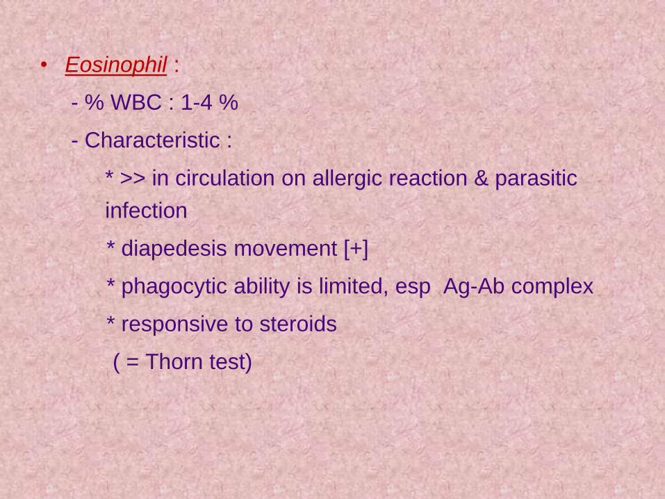
• Eosinophil :
- % WBC : 1-4 %
- Characteristic :
* >> in circulation on allergic reaction & parasitic
infection
* diapedesis movement [+]
* phagocytic ability is limited, esp Ag-Ab complex
* responsive to steroids
( = Thorn test)

STRUCTURE :
• Φ : (circulation) : 9 µm
(tissue) : 14 µm
- Cytoplasm :
* larger granules, refractile, uniform
* granules contain special lisozym + azurophilic
• Nucleus :
- dense chromatin
- lobes: 2, often covered with granules

-Functions:
* Response to parasitic infection
* Modulation in the inflammatory process
* Inactivation of leucotrienes & histamine

CLINICAL CORRELATION:
Eosinophilia : associated with allergic reactions and
helminthic (parasitic) infections.
Corticosteroids can produce a rapid decrease in the
number of blood eosinophils, probably by interfering with
their release from the bone marrow into the bloodstream
Eosinopenia
RBC
EOSINOPHYL
BASOPHYL
NETROPHYL
LYMPHOCYTE
MONOCYTE
THROMBOCYTE
L
E
U
C
O
C
Y
T
E
S
sel2 DARAH
NORMAL VALUE
FUNCTION
STRUCTURE
CLINICAL
CORRELATION

• Basophil :
- % WBC : 0-1 %
- characteristic :
* Similar to mast cells, except its ultrastructure
* The amuboid motion& phagocytosis ability is
limited
- Function :
in the immediate hipersensitivity;
secrete inflammation mediator
• *

- Structure :
* Φ :10-12 µm (smaller than netrophil)
* Cytoplasm :
- less dense
- vary granules size , dark specific granules
- Granules contain heparin, histamine
* Inti :
dense chromatin, pale
3 lobes, S shape, often covered with granules

CLINICAL CORRELATION
• anaphylactic shock
• cutaneus basophil hypersensitivity :
extravascular accumulation due to inflammatory process

RBC
EOSINOPHYL
BASOPHYL
NETROPHYL
LYMPHOCYTE
MONOCYTE
THROMBOCYTE
L
E
U
C
O
C
Y
T
E
S
BLOOD CELLS
NORMAL VALUE
FUNCTION
STRUCTURE
CLINICAL
CORRELATION
Netrofil :
- dominant, 60-70 %
- Can not mitosis
- Role: first line cellular defense: Phagocytosis

- karakteristik :
> Amuboid movement out from blood vessels
~ macrophage active = microphage
> The ability of mitosis [-]
> 2 types of granules (specific & azurophilic)
> Classification (according Schiling): :
~ Segmented neutrophils (57%)
Increased: shift to the right
- Nonsegmented neutrophils (stab) (4%)
Increased : shift to the left

STRUCTURE
• Φ: (circulation): 12 μm
(tissue): 20 μm
• cytoplasm:
- Color: salmon-pink
• Specific granules + Granules Azurofilik
- >> glycogens

nucleus:
• dense chromatin
• Multilobus
• types:
* Hipersegmented (> 5) old
* segmented
* stab
[women] drumstick = Barr body, is
inactive X chromosome (attached to the nucleus)


RBC
EOSINOFIL
BASOFIL
NETROFIL
LIMFOSIT
MONOSIT
TROMBOSIT
HARGA NORMAL
FUNGSI
STRUKTUR
KORELASI
KLINIS
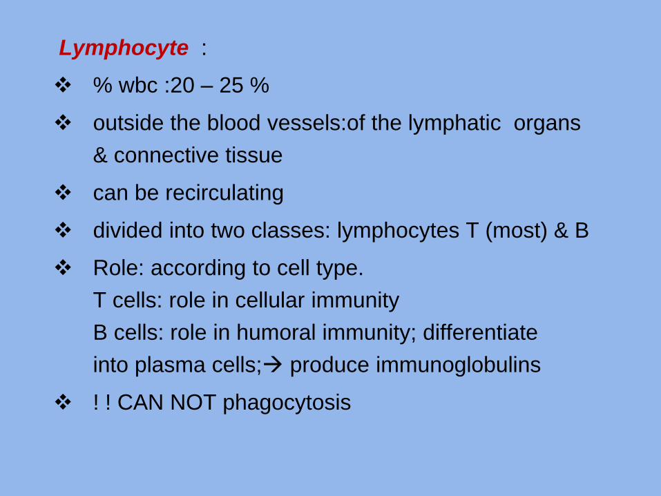
Lymphocyte :
% wbc :20 – 25 %
outside the blood vessels:of the lymphatic organs
& connective tissue
can be recirculating
divided into two classes: lymphocytes T (most) & B
Role: according to cell type.
T cells: role in cellular immunity
B cells: role in humoral immunity; differentiate
into plasma cells; produce immunoglobulins
! ! CAN NOT phagocytosis

Structure :
* Φ: small 6 – 8 µm predominate in the blood
Med-large :9-18 µm
Nucleus :
[small] : - Round / flat, with 1 indentation
- solidHeterochromatis
- Color: blue to purplish black
[med-large] : larger
less heterocromatis
color : reddish purple


CLINICAL CORRELATION
AIDS
• HIV-infected adolescents and adults categorizes
persons on the basis of clinical conditions associated
with HIV infection and CD4+ T lymphocyte counts
SEVERE COMBINED IMMUNODEFICIENCY
• The SCID syndrome is characterized by gross functional
impairment of both humoral and cell-mediated immunity
and by susceptibility to devastating fungal, bacterial,
and viral infections

RBC
EOSINOFIL
BASOFIL
NETROFIL
LIMFOSIT
MONOSIT
TROMBOSIT
HARGA NORMAL
FUNGSI
STRUKTUR
KORELASI
KLINIS
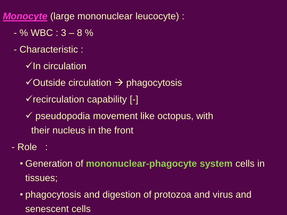
Monocyte (large mononuclear leucocyte) :
- % WBC : 3 – 8 %
- Characteristic :
In circulation
Outside circulation phagocytosis
recirculation capability [-]
pseudopodia movement like octopus, with
their nucleus in the front
- Role :
• Generation of mononuclear-phagocyte system cells in
tissues;
• phagocytosis and digestion of protozoa and virus and
senescent cells
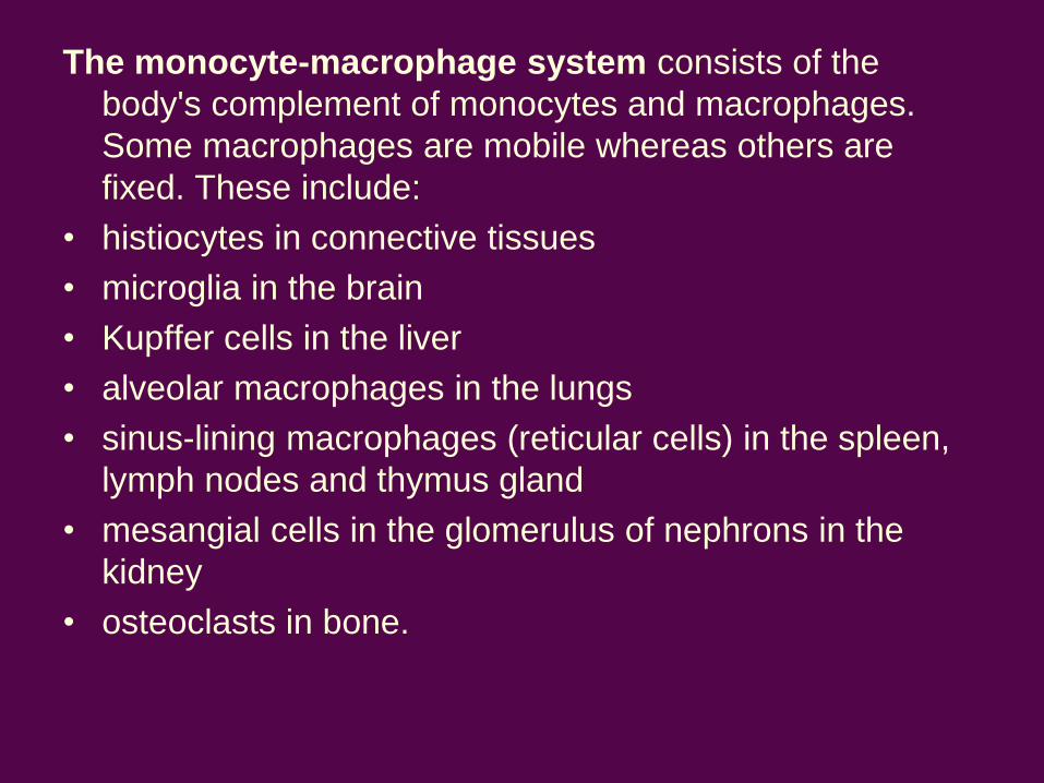
The monocyte-macrophage system consists of the
body's complement of monocytes and macrophages.
Some macrophages are mobile whereas others are
fixed. These include:
• histiocytes in connective tissues
• microglia in the brain
• Kupffer cells in the liver
• alveolar macrophages in the lungs
• sinus-lining macrophages (reticular cells) in the spleen,
lymph nodes and thymus gland
• mesangial cells in the glomerulus of nephrons in the
kidney
• osteoclasts in bone.
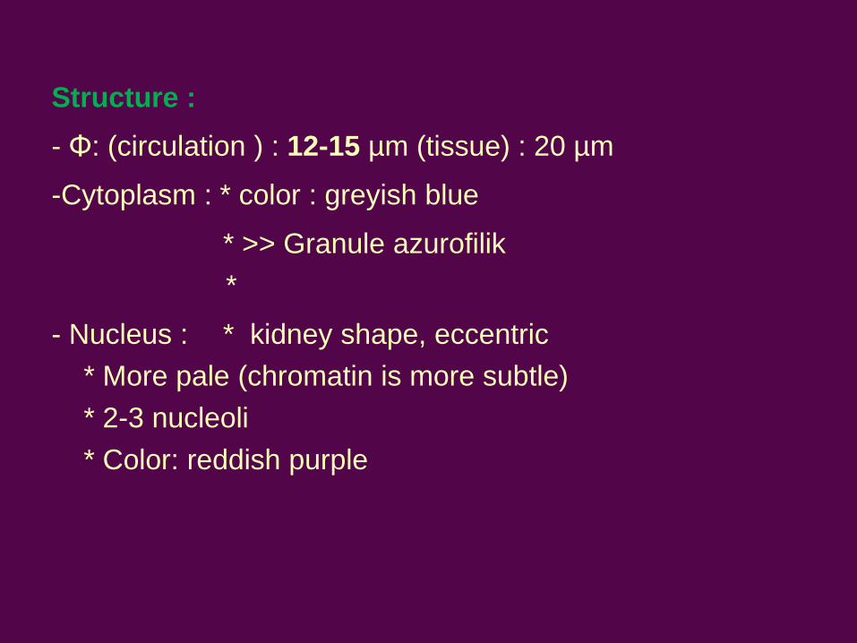
Structure :
- Φ: (circulation ) : 12-15 µm (tissue) : 20 µm
-Cytoplasm : * color : greyish blue
* >> Granule azurofilik
*
- Nucleus : * kidney shape, eccentric
* More pale (chromatin is more subtle)
* 2-3 nucleoli
* Color: reddish purple

CLINICAL CORRELATION
• Monocytopenia
• Monocytosis

RBC
EOSINOFIL
BASOFIL
NETROFIL
LIMFOSIT
MONOSIT
TROMBOSIT
HARGA NORMAL
FUNGSI
STRUKTUR
KORELASI
KLINIS

PLATELET (thrombocyte=thromboplastid)
- FROM megakarocyte ―budding‘ in the bone marrow
- Σ Normal : 200.000-400.000/Μl, lifespan : 8 days
- Function : CLOT FORMATION
• Primary aggregation—Discontinuities in the
endothelium, platelet aggregation platelet plug
• Secondary aggregation—Platelets in the plug
release an adhesive glycoprotein and ADP.
increasing the size of the platelet plug.
• Blood coagulation -- cascade, giving rise to a
polymer, fibrin thrombus.


- Structure :
Ø : 2-5 μm; in group (in the preparation)
disc like Shapes, biconvex
in fresh prep: no color
membrane surface: glycocalyx for adhesion
edge: hyalomere, pale blue color. There is
a marginal bundle
central: dense granulomere, There are
mitokhondria, glycogen granules, and purple
granules.


CLINICAL CORRELATION
THROMBOCYTOPENIA
This is defined as a blood platelet count below 150 x 109/1
(150 000/mm3) but spontaneous capillary bleeding does
not usually occur unless the count falls below 30 x 109/1
(30 000/mm3).
THROMBOCYTOSIS
.


INTRODUCTION
CELLS
HEMATOPOIESIS
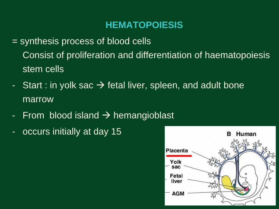
HEMATOPOIESIS
= synthesis process of blood cells
Consist of proliferation and differentiation of haematopoiesis
stem cells
- Start : in yolk sac fetal liver, spleen, and adult bone
marrow
- From blood island hemangioblast
- occurs initially at day 15

• Berproliferasi, membentuk 2 jalur diferensiasi (2 stem
cell):
* Jalur Myeloid RBC, granulosit, monosit, Platelet
~ erythropoiesis
~ granulopiesis
~ monopiesis
~ thrombopiesis
* Jalur lymphoid limfosit dan sel plasma


Terima kasih…….SEMANGAT! (dilarang menghitung jumlah slide)