Embed Size (px)
Citation preview


CONJUNCTIVA: ANATOMY , PHYSIOLOGY AND
SYMPTOMATOLOGY
Dr. Faizur Rahman
Associate Professor
Peshawar Medical College.

ANATOMY
It is the mucous membrane covering the
under surface of the lids and anterior part
of the eyeball upto the cornea.

Parts of conjunctiva
• Palpebral; covering the lids—firmly adherent.
• Forniceal; covering the fornices—loose—thrown into folds.
• Bulbar; covering the eyeball—loosely attached except at limbus.
• Also marginal and limbal parts and plica semilunaris.

Palpebral conjunctiva
• Subtarsal sulcus 2mm from posterior edge of
the lid margin.
• Richly vascular.
• Extremely thin.
• Strongly bound to the tarsal plate.

Conjunctival fornices
• Transitional region between palpebral and bulbar conjunctivae.
• Superior fornix 10 mm from limbus.
• Inferior fornix 8 mm from limbus.
• Lateral fornix 14mm from limbus.
• Medially absent.
• Ducts of lacrimal glands open into lateral part of superior fornix.

Bulbar conjunctiva
• Lies in contact with eyeball.
• Thin, translucent and loosely attached by
connective tissue to sclera and fascia bulbi.
• Conjunctival limbus 1 mm anterior to corneal
limbus.
• Bulbar limbus 1.5 mm behind corneal limbus.

Epithelium
• Stratified columnar epithelium 2 – 5 cells.
• At limbus change into stratified squamous non keratinized epithelium.
• At lid margin non keratinized stratified squamous epithelium changes into keratinized stratified squamous epithelium.
• Goblet cell – mucus.
• Accessory lacrimal glands.

Sub mucosa
• Fine delicate connective tissue.
• Lymphocytes.
• Denser fibrous tissue, blood vessels, nerves,
smooth muscles and accessory lacrimal glands.
• Papillae.

Nerve supply - Sensory
• Bulbar conjunctiva – long ciliary nerves – nasociliary N. – Ophthalmic division of trigeminal N.
• Superior palpebral and forniceal conjunctiva – frontal and lacrimal branches of Ophthalmic division of trigeminal N.
• Inferior palpebral and forniceal conjunctiva – laterally from lacrimal branches of Ophthalmic division of trigeminal N. and medially infraorbital N. – Maxillary division of trigeminal N.
Sympathetic;
• Superior cervical sympathetics to blood vessels.


Blood supply
Arterial supply;
• Posterior conjunctival arteries derived from arterial arcade of lids which is formed by palpebral branches of nasal and lacrimal arteries of the lids.
• Anterior conjunctival arteries derived from the anterior ciliary arteries – muscular br. of ophthalmic artery to rectus muscles.
Venous drainage;
• Palpebral and Ophthalmic veins.


Lymphatic drainage
• Lymph vessels are
arranged as a superficial
and a deep plexus in sub
mucosa.
• Ultimately as in the lids
to the pre auricular and
sub-mandibular lymph
glands.

Lacrimal caruncle
• Small, pinkish ovoid body in lacus lacrimalis at medial canthus of eye.
• Medial to plica semilunaris.
• Modified skin, colorless hair, sebaceous, sweat glands and accessory lacrimal glands.
• Non keratinized stratified squamous epithelium.
• Superior medial palpebral artery.
• Submandibular lymph node.
• Infratrochlear br. of nasociliary N.

Plica semilunaris
• Halfmoon shaped fold of
conjunctiva lateral to and party
under the caruncle.
• Lateral margin is free and
concave.
• Goblet cells, adipose tissue,
smooth muscle fibers and highly
vascular.
• Nictitating membrane.

PHYSIOLOGY
• Smooth surface.
• Secretes mucin and aqueous component of tear film.
• Highly vascular: supplies nutrition to the peripheral cornea.
• Aqueous veins drains from anterior chamber maintenance of IOP.
• Lymphoid tissue helps in combating infections.
• Basic secretion—reflex secretion.

Symptomatology
Non-Specific;
• Lacrimation.
• Irritation.
• Stinging.
• Burning.
• Photophobia.
• Redness.
Specific;
• Pain and FB sensation in corneal involvement.
• Itching in allergic, blephritis and dry eyes.

SIGNS
• Type of discharge.
• Type of conjunctival reaction.
• Presence of membrane/ pseudomembrane.
• Lymphadenopathy.

DISCHARGE
Exudate plus debris plus mucus plus tears.
• Serous; watery exudate in acute viral and acute allergic conjunctivitis.
• Mucoid; mucus discharge in VKC and KCS (dry eyes).
• Purulent; puss in severe acute bacterial conjunctivitis.
• Mucopurulent; puss plus mucus in mild bacterial conjunctivitis and Chlamydial conjunctivitis.

TYPE OF CONJUNCTIVAL REACTION
• Hyperaemia: (Conjunctival injection) Bacterial.
• Sub-conjunctival Haemorrhage: Viral.
• Bleeding:
• Chemosis: (Oedema)
• Scarring: Trachoma, cicatricial pemphigoid, atopic conjunctivitis and prolong use of topical drops.
• Follicular reaction.
• Papillary reaction.
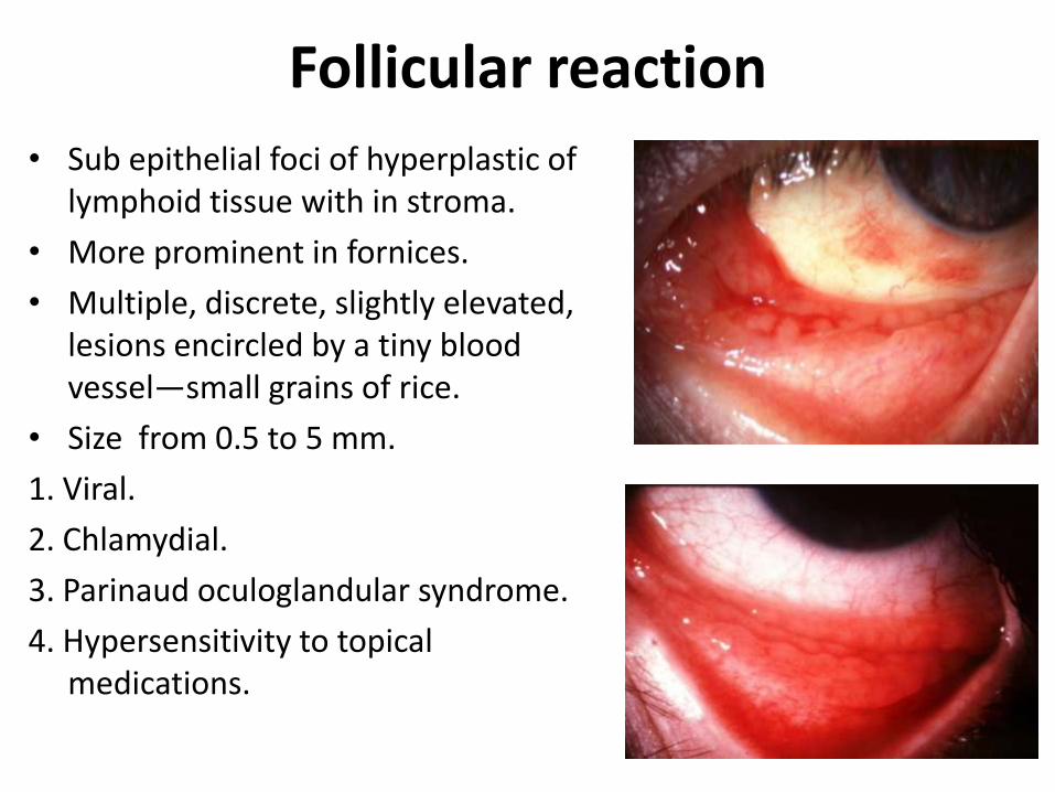
Follicular reaction
• Sub epithelial foci of hyperplastic of lymphoid tissue with in stroma.
• More prominent in fornices.
• Multiple, discrete, slightly elevated, lesions encircled by a tiny blood vessel—small grains of rice.
• Size from 0.5 to 5 mm.
1. Viral.
2. Chlamydial.
3. Parinaud oculoglandular syndrome.
4. Hypersensitivity to topical medications.

Follicular reaction

Papillary reaction• Hyperplastic conjunctival epithelium.
• Can develop in palpebral conjunctiva (firmly attached) and limbus.
• Papilla may mask follicles.
• Giant papilla (confluence)
• Non-specific; (less diagnostic)
1. Chronic blephritis.
2. Allergic conjunctivitis.
3. Bacterial conjunctivitis.
4. Contact lens wears.
5. Superior limbic keratoconjunctivitis.
6. Floppy eyelid syndrome.

Pseudomembrane
• Outside epithelium.
• Coagulated exudate adherent to the inflammed epithelium.
• Can be easily pealed off.
• Causes;
1. Severe adenoviral infection.
2. Ligneous conjunctivitis.
3. Gonococcal conjunctivitis.
4. Stevens-Johnson syndrome.

Membrane
• Includes epithelium.
• Infiltrate the superficial layers of conjunctival epithelium.
• Epithelium is injured if removal attempted.
• Causes;
1. Diphtheria.
2. Beta-hemolytic steptococci.

Lymphadenopathy
• Pre auricular and sub mandibular.
1. Viral infection.
2. Chlamydial infection.
3. Severe bacterial infections. (Gonococcal)
4. Parinaud oculoglandular syndrome.

Laboratory Investigations
Indications:
• Sever purulent conjunctivitis.
• Follicular conjunctivitis: viral vs chlamydial.
• Conjunctival inflammation.
• Neonatal conjunctivitis.

Laboratory Investigations—cont…
• Cultures.
• Cytological investigations.
• Inoculation.
• Detection of viral and chlamydial antigens.
• Impression cytology for ocular surface neoplasia, dry eyes, ocular cicatricial pemphigoid, limbal stem cells failure, infection.
• Polymerase chain reaction: small quantity of DNA for adenovirus, herpes simplex, chlamydia trachomatis.

Differential diagnosis of red eye
• Conjunctival– Blepharoconjunctivitis– Bacterial conjunctivitis– Viral conjunctivitis– Chlamydial conjunctivitis– Allergic conjunctivitis– Toxic/chemical reaction– Dry eye– Pinguecula/pteyrgium
• Lid diseases– Clalazion– Sty– Abnormal lid function
• Corneal disease– Abrasion– Ulcer
• Foreign body
• Dacryoadenitis• Dacryocystitis• Masquerade syndrome• Carotid and dural fistula• Acute angle glaucoma• Anterior uveitis• Episcleritis/scleritis• Subconjunctival hemorrhage• Factitious


THANK YOU


![Vasa aberrantia connecting the brachial and radial arteries aberrantia connecting the brachial and radial arteries 77 References [1] Hollinshead WH. Anatomy for surgeons: The back](https://img.dokumen.tips/doc/110x75/5a9f52337f8b9a62178c8d5c/pdfvasa-aberrantia-connecting-the-brachial-and-radial-arteries-aberrantia-connecting.jpg)


![1. anatomy of Respiratory Systembadripaudel.com/badri/images/LecturesElective/1/2/1... · 6/8/12 2 ! Anterior!intercostal!arteries!! 1]6!:!from!internal!thoracic!arteries!! 79:from](https://img.dokumen.tips/doc/110x75/5fd423a19c712976db423b83/1-anatomy-of-respiratory-6812-2-anteriorintercostalarteries-16frominternalthoracicarteries.jpg)