Embed Size (px)
Citation preview

���������������
���� ���������� �
����
���������� �������������� ������������������� ������� ����������������������������
�������� �������
���������������������� ����������� ������������ �� ���������
������ � ���������
���� ���� �!"���#� $%& $� ��� %��' ��(�)���)*�)��)+,� �"�'��

����������������������� ������� ��������������������������������������������������������������������������� ������!��"##$����%!&##�'�������������������������� ������ ������������������������ ��������������������������������()����*
��������+���������,��-*�"##$*�.)����������'�*�/����������������)��������������'����������������������������*�/���� ������������ ���������*����������� ������������� ������������������� �������� �������������������������010*�02���*� ������* !"#�$324$%45504351!45*
6��������������'���������������)����������'��7��������������������������7������������������������������������������'��������������������)���������������������������������)4�����8�������'���'�����'��'����*
���� ���7��������������������7����������)�'�"2#���������7�����)����������'�����)�%$$54$1*�6��������)�� ��������7���%*09*�6�����7��� ��)���������)������������������������*
���� ��������������������)�������50��7�����������������'��7��������%"�����'��)�*���)�'�������������'������������������������������������:--4;4�������<����'�'������������������7�����������)��)��7����������7��������������������������*
���� ��������������!5���������:03�'���<��'����������������������������������)��'�0*5���������� ����--4;4��������7���������� ������������ ����'���� :"%��������<� ���� �� ���������������� '���*� ;� ��������� ���7��� ��������� ������ ��� '��'�� �������� :==/<� 7�����������*���������==/������������������������������'������������*
���� ���������7����������������������7����>��?����(�������=���������������������:%#'���<��14%5�������'��)���7���������������'�������*�/�����������������������������������'����7����������������������������������''����7�������������������0@%#�'���*
���� ����������������'4������������������2!���������:1!��������"#�'������<�������)��'10������������)���=4!1����(A45����������)��// ��'��������������*�/)�����)����������������)�����7��������*�=��������������������7������������������������*�������������'����������������)�������'�������'����������'����*
�������&� 6��� �������� '� �)������ ����'�� �� �7���� ��� ��)���� ���� �� ����������������� �������*� �����)������ ��� ��������� '�� ��������)� ���������� '� ���� ���4��������� ���� ������'�����������������B�������B�:'����<��������*����������� ��������7��������>��?����(�������=�����������������������������������������''����������*�C)4������'���������������'��������������������������'����������'�����'������)�������������'���������������������)�������'������������������������'�8�������'���'�*
��� ��!�.)����������'������������������)�������'��'�������������������������������������������>��?����(�������=����������������������������''�����������������������������)�����)�������''�����
"�� �#����$������� %����������� �������������������& ���������%�������������� ����%�������������������� ����
D�,�����-�+��������"##$
>��;�%15%41"#1>��;�$324$%45504351!45��&�&��&��&����4%#1!11�:����&@@��*��*��@������E��F��&�&��&��&����4%#1!11<

Experience is wisdom, everything else is information Unknown To Sara
Björn Karl


List of Papers
I Wallander, H., Hovelius, L., Michaelsson, K. (2006) Incidence of con-genital clubfoot in Sweden. Acta Orthop 77(6):847-52.
II Aurell, Y., Johansson, A., Hansson, G., Wallander, H., Jonsson, K.
(2002) Ultrasound anatomy in the normal neonatal and infant foot: an anatomic introduction to ultrasound assessment of foot deformities. Eur Radiol 12(9): 2306-12.
III Wallander, H., Aurell, Y., Hansson, G. (2007) No association between
residual forefoot adduction and the position of the navicular in clubfeet treated by posterior release. J Pediatr Orthop 27(1): 60-6.
IV Wallander, H., Hansson, G., Tjernström, B. (1996) Correction of persis-
tent clubfoot deformities with the Ilizarov external fixator. Experience in 10 previously operated feet followed for 2-5 years. Acta Orthop Scand 67(3): 283-7.
V Wallander, H., Larsson, S., Bjönness, T., Hansson, G. (2009) Patient
Reported Outcome at 62-67 years of age in 83 patients treated for Con-genital Clubfoot. Journal of Bone and Joint Surgery (Br.), accepted.
Reprints were reproduced with permission from the respective publishers.

Contents
Introduction ................................................................................................... 11 Epidemiology and pathogenesis ............................................................... 11 Anatomy of normal infant foot ................................................................. 13 Pathoanatomy of congenital clubfoot ....................................................... 13 Classification ............................................................................................ 16 Initial treatment ........................................................................................ 16
Ponseti method .................................................................................... 17 French method ..................................................................................... 18
Surgical treatment .................................................................................... 18 Recurrence and persistence of clubfoot deformities ................................ 19 Evaluation of treatment ............................................................................ 20
Aims of the thesis.......................................................................................... 22
Methods ........................................................................................................ 23 Overview of populations in the papers ..................................................... 23 Population Study (Paper I) ....................................................................... 24 Ultrasonography (Papers II, III) ............................................................... 24 Clinical evaluation (Papers III, IV) .......................................................... 25 Patient reported outcome measures (Paper V) ......................................... 25 Norm group (Paper V) .............................................................................. 26 Global subjective evaluation (Papers III, IV, V) ...................................... 26 Statistical methods .................................................................................... 27
Summary of papers ....................................................................................... 28 Paper I: Incidence of congenital clubfoot in Sweden ............................... 28 Paper II: Ultrasound anatomy in the normal neonatal and infant foot: an introduction to ultrasound assessment of foot deformities .................. 29 Paper III: No association between residual forefoot adduction and the position of the navicular in clubfeet treated by posterior release. ............ 30 Paper IV: Correction of persistent clubfoot deformities with the Ilizarov external fixator ............................................................................ 30 Paper V: Patient Reported Outcome at 62-67 years of age in 83 patients treated for Congenital Clubfoot .................................................. 32
Discussion ..................................................................................................... 35
Conclusions ................................................................................................... 40
Future perspectives ....................................................................................... 41
Acknowledgements ....................................................................................... 42
References ..................................................................................................... 43

Abbreviations
AAOS Foot and Ankle Questionnaire.
Region specific questionnaire developed by the American Association of Orthopaedic Surgeons
C-C distance Calcaneus-cuboid distance
CTEV Congenital talipes equinovarus;
Congenital clubfoot
EQ-5D EuroQol
FFA Forefoot adduction
MM-N distance Medial malleolus to navicular distance
MRI Magnetic Resonance Imaging
QoL Quality of Life
SF-36 Medical Outcome Study Short Form 36 Health Survey
TC angle Talo-calcaneal angle
TC Index Talo-calcaneal index, sum of TC angle AP (ante-
rioposterior) and lateral
T-MT-1 angle Talo-first metatarsal angle


11
Introduction
Epidemiology and pathogenesis In a regular healthcare system, where the vast majority of children are born in hospitals, the congenital clubfoot (CTEV, Congenital talipes equinovarus) diagnosis is easily set at birth by health professionals. The probability of overlooking, or mistaking, this disorder for another, is very small; therefore, practically all patients with congenital clubfoot are expected to be identified [1, 2]. From this viewpoint, where newborn children are diagnosed with congenital clubfoot, further steps are needed, including proper coding, regis-tration, and reporting to a database before estimation of incidence is reliable. Under- and overestimation of the number of children with a preliminary diagnosis of congenital clubfoot may still occur. Overestimation is based on two forms of clubfoot: mild forms of fully reducible feet with no need for regular treatment; and complex forms with additional malformations of neu-rological origin. There is always an underestimation of the true incidence, due to under-reporting and missing cases, as reported by Severin (1956) and Wal-lander et al (2006) [3, 4].
The etiology of clubfoot remains unknown and is controversial, and nu-merous theories on causative factors are proposed. These include: nerve lesion[5]; muscular abnormality[6];vascular defect[7],neuromuscular de-fect[8];regional growth disturbance [9, 10]; intrauterine extrinsic pressure induction, either primary [11] or secondary to early amniocentesis [12-14]; a genetic component, either alone [15] or with a gene-environment interaction [16]; connected to maternal smoking [16, 17]; seasonal variation [2, 18, 19] (although this hypothesis has been rejected by others [20, 21]). Anatomic observations [6] conclude the findings are of primary cause, although it may not be possible to distinguish primary from secondary abnormalities[9].
Although long held proposals of intra-uterine compression as a causative factor is unsubstantiated , Wynne-Davies still claims[11] “a feeling remains that mechanical pressure may be of some significance”(p 446). Evidence for this theory is criticised [9] for being anecdotal. Kavashima and Uhthoff [10] claim interruption in the development of a normal foot during the ninth week of gestation might be responsible for the deformity. This hypothesis of a regional growth disturbance [9, 10] is supported by two observations. First, a disproportionate amount of type I fibres in the posterior and medial muscle groups [8], which suggests the presence of a neural abnormality as muscle

12
fibre types are neurally determined. Second, tendon sheaths of toe flexors and posterior tibial tendon have signs of cellular hypoplasia with smaller cell and cytoplasmic volume [9].
Ethnic differences in CTEV occurrence is reported [22-24], with the low-est (0.6‰) incidence among the Chinese population, the highest (6.8‰) in the Polynesian region and a cumulative incidence of approximately one per 1000 live births among Caucasians. Studies on ethnic groups, populations, and families suggest a genetic component as one causative factor of congeni-tal clubfoot. However, the mode of heritance does not follow a classic pat-tern [24] and both genetic and environmental factors are probably involved: the presence of a genetically determined connective tissue defect and a tran-sient deforming force is supposed [24] to allow the mobile foot to be pushed into equinovarus position at a vulnerable time of its development.
Twin studies are useful for determining whether the cause of a disorder is genetic or not. As monozygotic twins have identical genes, dizygotic twins share 50% of the genes, and all twins share the antenatal environment, it is possible to assess the effect of genetic factors on congenital disorders. In-creased rates of doubly affected monozygotic twins, compared with dizy-gotic twins (concordance), would indicate a genetic etiology for a disorder. This finding, confirmed by Engel et al. [25], suggests if a monozygotic twin is born with a clubfoot, the risk for the second to have the disorder is esti-mated to be one in three, strongly indicating a partly genetic etiology. The risk of having clubfoot in first-degree relative to a person with idiopathic clubfoot is higher than in the normal population. A sibling of a child with idiopathic clubfoot is reported [26] to have 2% to 4% risk of having a club-foot. Through molecular genetic investigations, the search for predisposing genes has intensified. One single gene together with some unmeasured factor is proposed [27] but recently a possibly polygenic cause was suggested [28]. This polygenic threshold theory is supported by the finding of the Carter effect, i.e. females require a greater genetic load to inherit the disorder than males. Work by Wang et al. [15] demonstrates a link to gene Hoxd13, which is a gene directly regulating skeletal muscle protein (Fhl1).
The strongest evidence for an environmental, though intrauterine, cause is from research on the outcome of amniocentesis [12-14]. Early amniocentesis (EA=11-12+6 gestational weeks) was associated with ten times higher rate of clubfoot than mid-trimester amniocentesis (MA=15-16+6), or if this pro-cedure was not performed [14]. This prospective randomised trial with the largest number of women enrolled had the same sex and laterality distribu-tion as in other reports on clubfoot. This indicated the possibility of a similar mechanism without previous amniocentesis. Further, transient leakage of amniotic fluid could have contributed to the development of clubfoot by deformation, although it does not provide a full explanation.
Seasonal variation is observed [2, 18, 19], with peak rates of clubfoot co-inciding with observed peaks of enterovirus infection, suggesting a connec-
13
tion with a virus-causing agent. However, others [20, 21] have failed to prove this connection.
Smoking during pregnancy is associated with an increased risk for certain birth defects, and among them especially strong association to oral cleft preva-lence has been demonstrated [29]. Isolated association between maternal smoking and clubfoot is reported [17]. Furthermore, evidence for a magnitude effect through joint exposure through maternal exposure and family history is larger than would be expected for an additive effect [16] and a potential im-portant interaction between these two, or other, variables is suggested [16].
Anatomy of normal infant foot At the time of birth, the child’s foot has reached a stage of development that is clearly recognised as a version of the adult form. This occurs in the absence of most biomechanical and functional restrictions that later play a role during plantigrade standing and bi-pedal gait. The configuration of normality is reached primarily by genetic influences and in part by the intrauterine envi-ronment. After birth, the effects of movement and gravity are uninfluenced by the intrauterine conditions. However, normality is a relative term. Even at birth, a normal foot can be held for a short time in a moulding abnormal posi-tion, resembling the clubfoot deformity, due to previous intrauterine pressure.
The ossification centres of the hindfoot (talus and calcaneus) is present at birth, while the navicular, the three cuneiforms and the cuboid are absent in the newborn. The navicular is the last bone to be ossified, normally at the age of 3-4 years.
Pathoanatomy of congenital clubfoot Clinical inspection of a newborn child with a clubfoot reveals the fundamen-tal parts of the deformity (Figure 1). The hindfoot is held in a firm position of equinus, with a tight heel cord. There is retraction and atrophy of the gas-trosoleus muscles; the calcaneus is inverted in varus position; and the fore-foot is held in adduction and supination, but still less supinated than the hindfoot, producing a cavus deformity with a medial and a posterior skin crease, more pronounced in severe cases [30].
On palpation, the change in kinematics is apparent [31] . Decreased subta-lar motion reflects the severe shortening of the medial and posterior tarsal ligaments and the tightness of the tibialis posterior and gastrosoleus muscles. On the dorsal part of the foot, the head of the talus is palpated uncovered by the navicular, which is medially positioned, close to the medial malleolus [31].. The active and passive motion of the forefoot, from the Lisfranc joint and distally, is relatively preserved with only slight restriction.

14
A B
Figure 1. A: Newborn with bilateral congenital clubfoot. B: After manipulation and first casting of left foot.
Investigations on muscle and ligament specimens diverge. A change in composition of muscles samples, with a type I fibre predominance in soleus, tibialis posterior, long toe flexors, and peroneal muscles, compared to the non-affected leg, is reported [8, 32]. Others report increased fibrosis in af-fected muscles, indicating a neurogenic defect [33]. However, in a recent report on this issue [34], there was normal composition of type I and type II fibres in over 99% of 431 muscle samples obtained during posteriomedial release. Further, examinations of ligaments revealed more collagen fibres than normal [6].
In anatomical dissections, the shape and position of the tarsal bones are well described [6, 35-37]: the talus is deformed with medial angulation of the neck and the head is dome-shaped. The calcaneus is medially rotated underneath the talus and both the calcaneus and talus are in plantar flexion, producing varus deformity of the hindfoot. On plain radiographs, this derota-tion between talus and calcaneus produces a decreased talo-calcaneal (TC) angle on both AP view and lateral view, sometimes close to zero. The posi-tion of the distal part of the calcaneus beneath the head of the talus prevents the talus from pronation and dorsiflexion, thereby, blocking the talus in an equinus position [38]. Contradictory opinions on the position of the talus in the ankle mortise are presented: most investigators [35, 39] propose that the talar body is externally rotated in the ankle joint, whereas, others claim that the talus is neutral [6] or even internally rotated [40]. The change in the rela-tionship between the talus and calcaneus is illustrated by 3D-CT [41]: rela-tive to the bi-malleolar axis, the clubfoot talus has a 14º external rotation of body and 45º internal rotation of the neck, whereas, in the normal foot, 5° internal rotation of body and 25° internal rotation of neck are seen.
On the medial and plantar side of the foot, there are anatomical findings [6] with thick and tight tibio-navicular ligament, calcaneo-navicular liga-ment, and tibialis posterior tendon sheath. This “medial tie” is described as crucial for the development of the midfoot part of the deformity [38]. By putting the transducer on the medial part of the clubfoot during ultrasono-graphy, this fibrous mass corresponds to findings of increased soft-tissue

15
thickness [42, 43]. On the lateral side, the fibulo-calcaneal ligament is short and thick, forming a fibrotic mass merged with the peroneal tendon sheaths.
Advances in imaging techniques (Magnetic Resonance Imaging (MRI) and Ultrasonography (US)) have enabled scientists to investigate larger se-ries of patients with clubfoot than is possible with invasive methods, and to compare these with normal feet. With 3D MRI, the volume of talus and cal-caneus is calculated as 15-40% less in unilateral clubfoot than in the normal foot [44, 45].
The dynamics of the medial part of the clubfoot is visualised by both MRI and US, with an increase in the distance between the medial malleolus and the navicular during manipulation. This has been shown both in the French functional method [46] and in the Ponseti method [47-49]. Especially in more severely deformed feet, a “spurious” (false) correction is reported, defined as a not fully reduced navicular over the head of the talus, together with a transversal break in the naviculo-cuneiform joint [49, 50], and still with proper alignment of the forefoot.
With MRI, increased medial deviation of the talar neck angle [51, 52] and a medial position of the navicular have been identified [51, 52]. These find-ings are confirmed by ultrasonography [42, 47, 53, 54]. A more specific talo-navicular relationship is described, emanating from definition of the align-ment of the talus: Kamegaya et al. [51] define the long axis of the body of the talus as perpendicular to the transmalleolar line, according to the descrip-tion from investigations on foetal specimens [55]. With a talar neck angle pointing more medially in the clubfeet than in normal feet, the navicular is positioned either medially or laterally in relation to the talar head. The greater the severity, the greater was the medial shift and the angulation of the neck of the talus [51]. Also with MRI images, perpendicular to the joint, the calcaneo-cuboid relationship has been described [56] with wedge-shaped distal calcaneus articular surface contributing to increased forefoot adduc-tion.Flattening of the talar head (flat-top talus) is described as a deformity secondary to forced dorsal flexion, and not a primary lesion [38]. This can occur if dorsal flexion is tried before the calcaneus is rotated out of supina-tion and posterior elements are still too tight to allow the talus to rotate in the ankle joint (a nutcracker effect). On radiographs, talar flattening is common and can appear to be more pronounced than in reality, due to improper posi-tioning when taking a lateral view.
There are contradictory reports on whether there is increased tibial torsion or not in CTEV [38, 57-59]. A mean external torsion of the tibia of 18º in children with clubfoot is reported [59], which is significantly less than the mean 40º in the contralateral normal side; however, other authors [38, 57, 58] claim there is no tibial torsion in patients with clubfeet.

16
Classification For grading severity of the initial deformity and for prognostic purposes, a consistent system for classification is essential. For these purposes, the pro-tocols of Pirani [60] and Dimeglio [30] are mainly used, both of which are proved to be valid and reliable [61, 62]. There is an observer’s learning phase before application in clinical practice and both classification systems rely equally on clinical examination. The Pirani score assesses six clinical signs, which are scored as normal (0), moderate (0.5), or severe (1), with maximum total score (severe deformity) of six points. The Dimeglio score is a 20-point classification and consists of “four essential” parameters (equinus, varus, derotation of the calcaneo-forefront block (supination), and forefoot adduction), which are graded according to stiffness (total 16 points: severe stiffness), additional clinical signs yield 0-4 points. Based on a total score, each foot is graded I-IV.
Initial treatment The first written description of clubfoot treatment was by Hippocrates (circa 400 B.C.) [63], and is similar to current non-operative methods. Hippocrates declared two important principles in the treatment of clubfoot, which subse-quent generations have claimed as new. Hippocrates explained that the vast majority of cases can be successfully treated with serial manipulation, and that treatment should begin as early as possible before the deformity of the bones is well established. He also understood the inadequacy of restoring the foot to its normal position, but that it must be over corrected and then held in this position afterwards to prevent recurrence.
The adaptation of connective tissue to stress is considered related to dy-namic, intermittent loading, and as a result, static stress is regarded harmful to tissue adaptation [64]. However, a change in pathology during conserva-tive treatment for the Ponseti method is illustrated [56]: marked improve-ment or complete correction is found by MRI on the abnormal relationships of the tarsal bones and individual shapes of the cartilaginous anlagen. The hypothesis is that changes in mechanical loading of fast growing tissues result in changes in growth pattern [56]. These findings have been corrobo-rated: alteration in magnitude and direction of static (rather than dynamic) loading play a crucial role in accelerating the growth and development of cartilaginous anlagen in the infant clubfoot [65].
There are currently two main non-surgical approaches for primary correc-tion of idiopathic clubfoot: the Ponseti and the French methods. Effective use of either of these techniques can alleviate the need for extensive soft tissue release in most children, thus avoiding the risk for potential over cor-rection, stiffness, and pain.
17
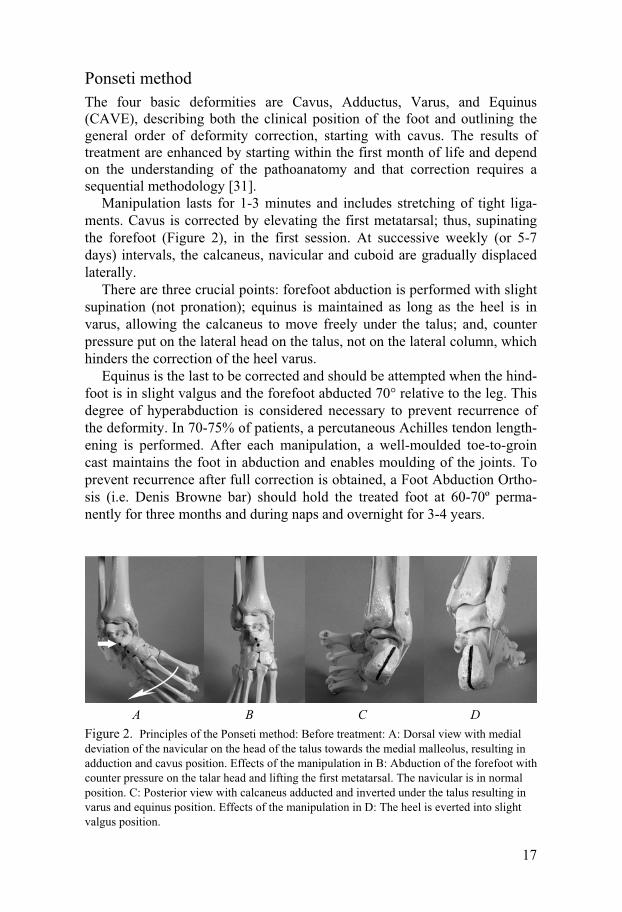
Ponseti method The four basic deformities are Cavus, Adductus, Varus, and Equinus (CAVE), describing both the clinical position of the foot and outlining the general order of deformity correction, starting with cavus. The results of treatment are enhanced by starting within the first month of life and depend on the understanding of the pathoanatomy and that correction requires a sequential methodology [31].
Manipulation lasts for 1-3 minutes and includes stretching of tight liga-ments. Cavus is corrected by elevating the first metatarsal; thus, supinating the forefoot (Figure 2), in the first session. At successive weekly (or 5-7 days) intervals, the calcaneus, navicular and cuboid are gradually displaced laterally.
There are three crucial points: forefoot abduction is performed with slight supination (not pronation); equinus is maintained as long as the heel is in varus, allowing the calcaneus to move freely under the talus; and, counter pressure put on the lateral head on the talus, not on the lateral column, which hinders the correction of the heel varus.
Equinus is the last to be corrected and should be attempted when the hind-foot is in slight valgus and the forefoot abducted 70° relative to the leg. This degree of hyperabduction is considered necessary to prevent recurrence of the deformity. In 70-75% of patients, a percutaneous Achilles tendon length-ening is performed. After each manipulation, a well-moulded toe-to-groin cast maintains the foot in abduction and enables moulding of the joints. To prevent recurrence after full correction is obtained, a Foot Abduction Ortho-sis (i.e. Denis Browne bar) should hold the treated foot at 60-70º perma-nently for three months and during naps and overnight for 3-4 years.
A B C D
Figure 2. Principles of the Ponseti method: Before treatment: A: Dorsal view with medial deviation of the navicular on the head of the talus towards the medial malleolus, resulting in adduction and cavus position. Effects of the manipulation in B: Abduction of the forefoot with counter pressure on the talar head and lifting the first metatarsal. The navicular is in normal position. C: Posterior view with calcaneus adducted and inverted under the talus resulting in varus and equinus position. Effects of the manipulation in D: The heel is everted into slight valgus position.

18
French method According to Bensahel et al [66], the primary factors responsible for club-foot are retraction of tibialis posterior muscle and weak peroneal muscles, together with a fibrous zone in the medial part of the midfoot. This func-tional method consists of daily manipulation by a skilled physiotherapist, followed by temporary immobilisation of the foot by non-elastic adhesive strapping. The schedule continues for approximately 2 months, followed by manipulation three times a week for an additional 6 months. Nighttime splinting is recommended for an additional 2-3 years.
The focus is to relax the stiff tissues by progressive passive manipulation, active muscle stimulation, taping and splinting. This is done for 30 minutes per foot, and the steps are detailed and precise, but the manipulation is gentle and smooth to avoid defence reactions with further contractions occurring.
The first step in reducing the deformities is progressive release of the navicular from the medial malleolus: the medial tissues are stretched by this manoeuvre. Second, forefoot adduction is reduced by stretching the joints along the medial ray of the foot. To strengthen the pronation forces, the cu-taneous reflexes are stimulated. The final step corrects the equinus by loos-ening posterior structures. Continuous passive motion (CPM) with the aim to maintain the passive range of motion and to soften the tissues during sleep, was elaborated further for use in clubfoot treatment in the early 1990s [67].
Surgical treatment Irrespective of the treatment protocol used, it should not delay the child from starting to ambulate. Beginning from birth with manipulation and casting, or with functional treatment, and continuing during the first months of life al-lows the foot to grow, and the clinician can follow the correction progress. By the age of 3-4 months, a plateau in progress is often reached. From this age onwards, there have been disappointing results from this program with non-surgical treatment, and historically up to 50% of clubfeet have not re-sponded adequately. By then a decision about surgical release in order to achieve a plantigrade foot is advocated [35]. This surgical approach always includes various techniques of soft tissue release, starting with the hindfoot. Ankle and subtalar joint capsule are opened together with dividing posterior ligaments and lengthening flexor tendons and the tibialis posterior tendon. The second stage, a more extended subtalar release with division of lateral and/or medial tether, also corrects residual forefoot adduction.
The timing and extent of surgery is a matter of debate. Limited soft tissue release “à la carte” at the age of three months, proposed by Bensahel [66], contrasts to the complete subtalar release at the age of six months or more suggested by Simons [68]. Extensive soft tissue releases are proposed to

19
achieve full correction and prevent relapse in one single procedure. How-ever, about 25% of the operated feet still relapse and require additional sur-gical intervention [69, 70]. Ponseti claims [71] that after extensive soft-tissue release there is substantial risk for stiff and painful feet.
Several reasons for these disappointing results are suggested, such as ex-cessive scarring and inadequate post-operative management. Repeat ex-tended soft tissue releases seldom solve these difficult problems. When de-formities are fixed and from 3-4 years of age, bony procedures are needed. The strategy for this includes balancing the medial and lateral column of the foot with shortening of the lateral side relative to the medial by osteotomies. With knowledge of pathoanatomy, adherence to the principles of hyperab-duction, and with endurance, the frequency of necessary extensive surgical treatment decreases from more than 50% to approximately 10-15%.
Recurrence and persistence of clubfoot deformities Recurrence (or relapse) is by definition the development of one or more of the original deformities (equinus, varus, adductus and cavus) after full cor-rection of the clubfoot is obtained. The cause of recurrence is unknown, but is logically the same mechanism that initially caused the deformities to de-velop and is related to rapid growth of the foot. Recurrence is rare after the age of 4-5 years and almost never occurs after 7 years of age. After correc-tion with the Ponseti technique, slight recurrence of cavus and adduction is seldom a problem [72]. The more important relapses involve the hindfoot with equinus and varus, and appear related to the retraction of posterior and medial tarsal ligaments and musculo-tendinous units.
Relapse is often effectively and easily treatable when discovered at an early stage. Commonly, this is seen when parents are non-compliant with the Foot Abduction Orthosis (FAO) regimen, and the heel slips out of the shoe. The principles for corrective manoeuvres are the same as for the original treatment [72]. Two or three repeated castings, with up to two-week inter-vals, are usually needed to correct the varus position of the heel and forefoot adduction. A percutaneous Achilles tendon lengthening can be performed up to one year of age, although the upper age limit for this procedure is un-known. The ambulating child with dynamic supination, and a fully corrected foot at static examination, can be treated with anterior tibial tendon transfer [73]. A foot with some residual deformity and retained mobility may be preferable to a cosmetically corrected foot that has increased long-term stiff-ness. Severe degrees of persistent deformities in older children walking on the lateral part of the foot, or with equinus, have been treated with repeated, or more radical, soft-tissue releases. These procedures cause increased stiff-ness and the risk of complications [74, 75]. With bony procedures, such as Dillwyn Evans [76, 77] and Dwyer [78, 79] procedure, there is a risk of
20
making an already small foot even smaller, even if good results are pre-sented. The Ilizarov technique is proposed [80-83] as an alternative for re-ducing complications after repeat surgery. The application of this form of external fixator on the foot can achieve correction by gradual distraction of the joints that allow realignment of the foot without the need for an open operation. The best results were reported [83, 84] in children who had re-tained some mobility of the foot. In order to maintain the correction in these feet, a tibialis anterior tendon transfer is recommended. Combination with osteotomies in conjunction with the Ilizarov technique is recommended for children over eight years of age or for rigid feet [83].
The frequency of recurrence decreases with increasing parental awareness about the need for continuing night FAO bracing up to 3-4 years of age. In a review of the Ponseti treatment [73], the authors concluded that in 157 pa-tients (256 feet), treated between 1991-2001, there was a relapse in seven-teen of the patients (11%). With night bracing compliance, there was relapse in 2/140 patients (1%), and with non-compliance, relapse was found in 15/17 patients (89%). In their report on 51 consecutive infants with 86 idiopathic clubfoot, Dobbs and associates [85] conclude that brace wear non-compliance is the factor most strongly related to relapse, with an odds ratio of 183 (p<0.001). Therefore, ensuring the patients and parents comply with bracing is the most effective method of preventing relapses.
Evaluation of treatment The methods for evaluation differ between children and adults. Small chil-dren are assessed clinically, by radiographic measurements and by functional measures.
In small children, clinical observation includes observation of standing, gait, and sometimes running [86]. The foot is examined with a goniometer to assess position and passive and active range of motion for ankle movements. Subtalar motion is difficult to measure and can be arbitrarily rated in inter-vals. Forefoot adduction and preferably supination are recorded by an image of the weight bearing area on a glass plate.
Radiographic measurements from 3-4 years of age require standardising with the foot weight bearing and the tibia and the X-beam inclined at 30º each. With AP and lateral views, different angles are traditionally measured, including The Beatson Combined Talo-Calcaneal angle (TC Index) [87, 88] and Talo-first metatarsal angle (T-MT-1 angle). However, in young children, valid conclusions about anatomic relationships from radiographs are difficult [35], as a large proportion of the “bony architecture” is still cartilaginous.
Lehman and other investigators have developed different grading sys-tems, some employing a 100-point scale, measuring foot size, shape, range

21
of motion and radiographic parameters and in addition assessment of func-tion and pain [69]. Laaveg and Ponseti [89] were the first to introduce func-tional assessment with a focus on satisfaction, function, pain, and position of foot when standing, and in passive motion and gait.
Irrespective of these various methods, there is still no firm consensus on the best method for assessing the results of clubfoot treatment in children. Much of this controversy derives from the general challenges in evaluating congenital paediatric conditions, which include:
• Low incidence in the population • Difficulty in rating pre-treatment severity (classification) • Variable time and procedures when surgery is performed • Physical function is not a static condition in childhood, but is vari-
able and has marked age dependency.
In addition to the measurement tools used on children, another common issue for adults is whether the measure should be disease-specific, regional (i.e. the foot and ankle), or including a broader (generic) instrument. For the last 10-15 years, focus has been on quality of life (QoL) in terms of patients‘ own self-reported assessment of the situation. QoL might appear a complex summary of interacting objective and subjective dimensions; however, the concept of QoL can be considered as a uni-dimensional concept, despite the multiplicity, diversity and complexity of its causes [90]. As a result, a one-dimensional rating, such as ’How do you feel about your life as a whole’ can be a consequence of global assessment of a numerous number of diverse factors and causes.
Another issue under debate is whose perspective the outcomes should ex-plore: this is a concern for all age groups of patients. The importance of pa-tient perspective is recognized as this is considered the most important crite-rion for judgment. Valid and reliable patient-reported outcome measures for use on children with foot and ankle disorders are scarce. The Juvenile Arthri-tis Foot Disability Index is condition-specific for juvenile arthritis [91]. Roye et al [92] defined a 10-item Disease Specific Instrument (DSI) that can cap-ture patient-based attitudes, but further validation is recommended. In a re-cent report [93], the Oxford ankle foot questionnaire, comprising 15 items divided into three subscales (Physical, School and Play and Emotional) was shown to be valid and reliable, and is available for clinical practice.

22
Aims of the thesis
The overall aim of this thesis on congenital clubfoot was to estimate the incidence with a national perspective, analyse residual deformities and their management, and to evaluate patient reported long-term quality of life and foot function. The specific aims of the papers were: 1. To estimate the current incidence of congenital clubfoot in Sweden and analyse epidemiological factors. 2. To standardise ultrasonography for assessment of infant foot anatomy and to apply this protocol on a cohort of children with clubfoot. 3. To investigate the association between the medial position of the navicular and residual forefoot adduction in clubfoot and its clinical implications. 4. To evaluate the Ilizarov method for the use on relapsed clubfoot deformi-ties after previous surgery, in terms of patient satisfaction, clinical outcome, and complications. 5. To assess patient reported quality of life and foot and ankle function after 60 years of age in patients treated for congenital clubfoot.
23
Methods
Overview of populations in the papers Paper I – 292 patients registered at 44 Orthopaedic Departments in Sweden.
Paper II – 54 healthy children examined at the Department of Radiology at Halmstad Central Hospital.
Paper III – 35 patients undergoing posterior soft tissue release at the Ortho-paedic Departments of Kristianstad Central Hospital, Växjö Central Hospi-tal, Norrköping Central Hospital, and Karlstad Central Hospital.
Paper IV – 7 patients undergoing corrective treatment with the Ilizarov ex-ternal fixator at the Orthopaedic Department of Uppsala University Hospital.
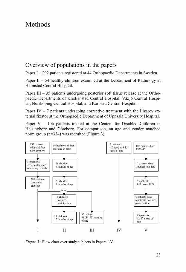
Paper V – 106 patients treated at the Centers for Disabled Children in Helsingborg and Göteborg. For comparison, an age and gender matched norm group (n=334) was recruited (Figure 3).
Figure 3. Flow chart over study subjects in Papers I-V.
I II III IV V
54 healthy children assessed at birth
28 children 4 months of age
23 children 7 months of age
3 children declined participation
51 children 12 months of age
35 patients 54 (38-72) months of age
7 patients (10 feet) at 6-15 years of age
106 patients born 1939-45
10 patients dead 1 patient lost data
95 patients follow-up 1974
6 patients dead 6 patients declined participation
83 patients 62-67 years of age
3 positional 3 ”teratological” 6 missing records
292 patients with clubfoot born 1995-96
280 patients congenital clubfoot

24
Population Study (Paper I) The annual number of newborn children with CTEV was compared with the number of all live births during 1995 and 1996. For this purpose, official reports concerning native data from the Swedish Board of Statistics (SCB) were used. The cumulative incidence (also known as incidence proportion) was calculated as number of newborn children with CTEV (teratological clubfeet excluded) divided by the number of native births (the size of the population initially at risk). Abortive cases and stillbirths were not included in the population at risk. To assess geographical distribution of clubfoot de-formities, the country was divided into six regions, commonly used by Health Service Authorities for administrative purposes.
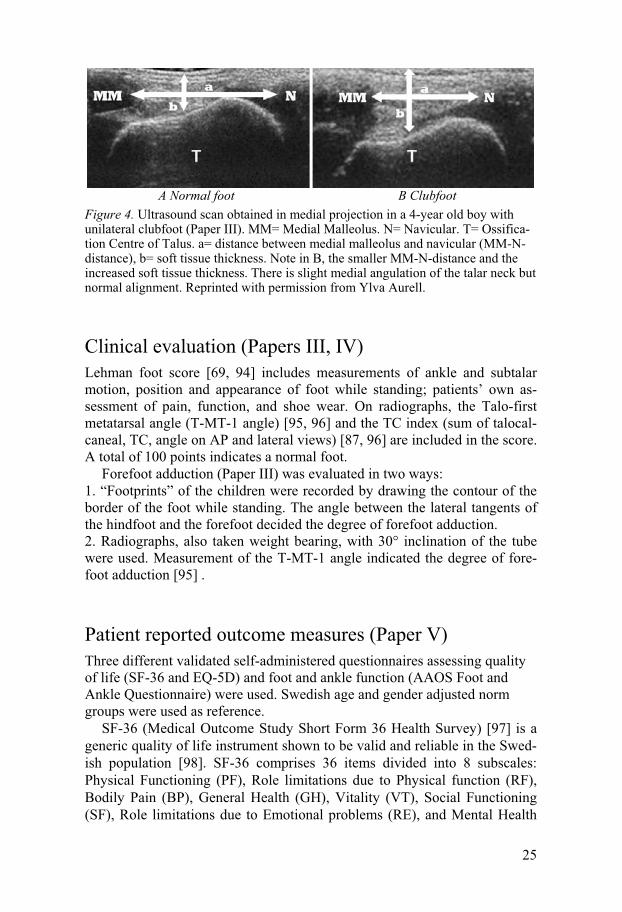
Ultrasonography (Papers II, III) In both Papers II and III, one experienced examiner performed all sono-graphical examinations. In Paper II, three standardised projection planes [42] were used: medial, lateral and dorsal.
Medial projection: The transducer was placed in a slightly oblique posi-tion with visualisation of the anatomical landmarks: medial and lateral mal-leolus, talus and navicular. For evaluation of navicular position, the shortest distance between the medial malleolus and the medial part of the navicular was measured (medial malleolus to navicular distance, MM-N-distance). The soft tissue thickness was measured as the perpendicular distance from the skin surface to the medial border of the cartilaginous anlagen of the talus at its midpoint.
Lateral projection: The transducer was placed along the lateral border of the foot parallel to the plantar surface. The alignment between the calcaneus and cuboid was evaluated by measuring the perpendicular distance from the tangent of the calcaneus to the lateral border of cartilaginous anlagen of the cuboid (calcaneus-cuboid distance, C-C-distance).
Dorsal projection: The transducer was placed on the dorsum of the foot, imaging the relationship between the talus and the navicular in a sagittal plane. The length of the talus was measured.
In Paper III, the examiner held the foot in a close to neutral position with-out pushing the foot in either adduction or abduction. The medial view was used [42] to evaluate the talonavicular alignment by measuring the shortest distance between the medial malleolus and the medial proximal part of the navicular (MM-N-distance) (Figure 4).
25
A Normal foot B Clubfoot
Figure 4. Ultrasound scan obtained in medial projection in a 4-year old boy with unilateral clubfoot (Paper III). MM= Medial Malleolus. N= Navicular. T= Ossifica-tion Centre of Talus. a= distance between medial malleolus and navicular (MM-N-distance), b= soft tissue thickness. Note in B, the smaller MM-N-distance and the increased soft tissue thickness. There is slight medial angulation of the talar neck but normal alignment. Reprinted with permission from Ylva Aurell.
Clinical evaluation (Papers III, IV) Lehman foot score [69, 94] includes measurements of ankle and subtalar motion, position and appearance of foot while standing; patients’ own as-sessment of pain, function, and shoe wear. On radiographs, the Talo-first metatarsal angle (T-MT-1 angle) [95, 96] and the TC index (sum of talocal-caneal, TC, angle on AP and lateral views) [87, 96] are included in the score. A total of 100 points indicates a normal foot.
Forefoot adduction (Paper III) was evaluated in two ways: 1. “Footprints” of the children were recorded by drawing the contour of the border of the foot while standing. The angle between the lateral tangents of the hindfoot and the forefoot decided the degree of forefoot adduction. 2. Radiographs, also taken weight bearing, with 30° inclination of the tube were used. Measurement of the T-MT-1 angle indicated the degree of fore-foot adduction [95] .
Patient reported outcome measures (Paper V) Three different validated self-administered questionnaires assessing quality of life (SF-36 and EQ-5D) and foot and ankle function (AAOS Foot and Ankle Questionnaire) were used. Swedish age and gender adjusted norm groups were used as reference.
SF-36 (Medical Outcome Study Short Form 36 Health Survey) [97] is a generic quality of life instrument shown to be valid and reliable in the Swed-ish population [98]. SF-36 comprises 36 items divided into 8 subscales: Physical Functioning (PF), Role limitations due to Physical function (RF), Bodily Pain (BP), General Health (GH), Vitality (VT), Social Functioning (SF), Role limitations due to Emotional problems (RE), and Mental Health
26
(MH). Each sub scale has a range 0-100 (optimal). Two summary scales can also be obtained: The Physical Component Summary Score (SF-36 pcs) and the Mental Component Summary Score (SF-36 mcs). These two summary scales capture about 85% of the reliable variance of the 8-scale SF-36.
EQ-5D (EuroQol) [99] consists of two parts: The first part is the EQ-5D Index, where respondents assess their health status in five dimensions: mo-bility, self-care, usual activities, pain/discomfort, and anxiety/depression. Each dimension has three levels: no problem, moderate problems, or severe problems. With this classification, 243 health states can be generated and with an algorithm, each person is assigned an index from 0.00-1.00. Index values, based on the UK EQ-5D tariff values [100] are established for a Swedish population [101, 102] .
The second part consists of the EQ VAS (Visual Analogue Scale), which is a 0-100 labelled scale, where the respondents mark the alternative repre-senting their current health state, with 100 representing best possible health status.
The AAOS Foot and Ankle Questionnaire [103] was developed by the American Association of Orthopaedic Surgeons, where the Foot and Ankle Module combines items from the Lower Limb Scale with additional items specifically assessing symptoms and functional status related to foot and ankle problems. The questionnaire consists of two scales: The Foot and An-kle Core Scale (twenty items and four subscales measuring pain, function, stiffness, swelling, and giving way symptoms) and the Shoe Comfort Scale (five items assessing the ability to comfortably wear a variety of shoe types). The questionnaire is designed to assess various foot and ankle conditions and evaluate different treatment modalities.
Norm group (Paper V) For the AAOS Foot and Ankle Questionnaire, an age- and gender-adjusted reference group was recruited. An invitation and the questionnaires were sent to a representative group (n= 500) with a mean age of 64 years, and living in the same area as the study group. In all, 206 responded to the first invitation. The second invitation generated a further 102 responses. A third letter together with the questionnaires was sent, providing another 26 re-sponses. The control group consisted of 334 persons with an average age of 64 years (range 62-67).
Global subjective evaluation (Papers III, IV, V) In Paper III, at follow-up examination, parents were asked to provide an overall estimation of the results of the treatment by answering the question:

27
“Are you satisfied with your child’s foot function and appearance?” The parents were given three alternative answers: (1) Yes, I am satisfied; (2) I am not sure or my opinion is between; and (3) No, I am not satisfied.
In Paper IV, the patients and their parents completed a short questionnaire including questions on gait, pain, appearance, and their overall opinion on the result of the treatment.
In Paper V, the patients were asked to evaluate their own experience of the cosmetic appearance of their treated foot/feet by answering the following question: “Do you find your treated foot/feet looking normal?” which was one of the questions asked by Bjönness [104]in 1974. The alternatives for answering were: “Yes” or “No”.
Statistical methods Differences in distribution were tested by Student’s t-test for normally dis-tributed continuous variables (Papers II, III), otherwise continuous variables were tested with the non-parametric Mann-Whitney U-test (Papers III, V). For paired observations (Paper I, unilateral clubfoot vs contralateral normal foot) a paired t-test was performed.
Grouped data were compared with &² test (Papers I, III and V). In Paper III, categorical data were analysed with Fisher exact test.
For correlation analysis, the Pearson correlation coefficient was used when variables were approximately normally distributed and independent between groups (Paper III), and Spearman rank correlation was used for non-normal distribution (Paper III).
For analysis of seasonal distribution (Paper I), comparing children with clubfoot with all other newborn children during the same time interval, an average vector for each month represented the mean birth month. The Rayleigh z-test was used for analysis of uniformity, and Watson’s non-parametric test was used for testing whether mean vector was equal between clubfoot group and other newborn children. Linear regression was used to describe the relationship between an independent variable (i.e foot and an-kle function) and the predicted value of quality of life (SF-36 and EQ-5D) (Paper V).
Mean ± Standard deviation and 95% confidence interval (CI) were calcu-lated. Probability values of <0.05 (two-sided test) denoted statistical signifi-cance.

28
Summary of papers
Paper I: Incidence of congenital clubfoot in Sweden
Is the incidence of clubfoot consistent with previous reports from Sweden?
In a prospective study during the years 1994-1995, the reported cases of congenital clubfoot from all 44 public Orthopaedic Departments in Sweden, were collected (teratological forms were not included) and the childrens’ medical records were scrutinised. Official reports of native data from the Swedish Board of Statistics were used for comparison. The cumulative inci-dence was calculated as the ratio between the annual number of children with clubfoot and the number of native births during the same period. Out of 292 reported preliminary cases of clubfoot, 280 children could be identified with the correct diagnosis of congenital clubfoot. The cumulative incidence of the two years was similar: 1.5‰ and 1.3‰ (p for difference=0.2), giving the average incidence of 1.4‰ (95% CI 1.2-1.6, Table 1).
Table 1. Number of children with congenital clubfoot in relation to number of native births
No of children Cumulative incidence Year with clubfoot born alive per thousand (95% CI)
1995 157 103 422 1.5 (1.3-1.8) 1996 123 95 297 1.3 (1.1-1.5) Total 280 198 719 1.4 (1.2-1.6) Boys 198 101 874 1.9 (1.7-2.2) Girls 82 96 845 0.8 (0.7-1.1)
There were 198 boys (72%) and 82 girls (28%). Bilateral clubfoot was iden-tified in 46% of the cases. No gender differences were found. Analysis of seasonal variation revealed no significant statistical difference between the study population with congenital clubfoot and the group of all live births in Sweden. To assess geographical distribution, the country was divided into six regions, commonly used for administrative purposes. There was a statis-tically heterogeneity with a lowest incidence of 1‰ (95% CI 0.7-1.4) in one region, compared to 2.0‰ (95% CI 1.5-2.6) in the region with the highest incidence (p=0.007,Table 2).

29
Table 2. Cumulative incidence of congenital clubfoot during 1995 and 1996 in six regions in Sweden
No of children with No of native births Cumulative incidence Region Clubfoot over 2 years per thousand (95% CI)
1 62 30 775 2.0 (1.5-2.6) 2 73 45 248 1.6 (1.3-2.6) 3 30 21 660 1.4 (0.9-2.0) 4 52 45 704 1.1 (0.8-2.0) 5 35 35 022 1.0 (0.7-1.4) 6 28 18 848 1.5 (1.0-2.1)
Paper II: Ultrasound anatomy in the normal neonatal and infant foot: an introduction to ultrasound assessment of foot deformities
Can the infant foot anatomy be visualised in a reliable manner by ultrasono-graphy?
In order to reproduce anatomical findings with good reliability, the infant foot was investigated with ultrasonography (US). The feet of 54 newborn healthy children (24 boys, 30 girls) were examined with US in three defined projections (medial, lateral, dorsal) and 51 of these children were followed prospectively at the age of 4,7 and 12 months (three were lost on follow-up). In the medial projection, the MM-N distance, as a view of the position of the navicular in relation to the medial malleolus, measured 9±1 mm in the newborn and 12±1 mm at the age of 12 months (p<0.001). The soft tissue thickness increased during the same period from 5±1 mm to 8±1 mm (p<0.001). The length of talus, measured by the dorsal projection at birth and at 7 months of age, was larger at 7 months of age (23 mm) than in newborn children (18 mm). The reliability test for intra-observer agreement was better than for inter-observer agreement (Table 3).
Table 3. Intervals for intra-and inter-observer correlation (r; p<0.01). MM-N-distance Soft tissue thickness
Intra-observer correlation (r) 0.85-0.87 0.68-0.94Inter-observer correlation (r) 0.65-0.81 0.55-0.93

30
Paper III: No association between residual forefoot adduction and the position of the navicular in clubfeet treated by posterior release.
Is a pronounced medial position of the navicular associated with residual forefoot adduction?
Thirty-five children (27 boys, 8 girls) were reviewed at a mean age of 4.5 years after surgical treatment with posterior release. The forefoot adduction (FFA) was assessed by two methods: first by measurement of the angle be-tween the hindfoot and the forefoot on the lateral side, with the children standing (“footprints”), and second, by measurement of the T-MT-1 angle on radiographs. Foot and ankle function was evaluated by the Lehman foot score, and the parents were asked about their degree of satisfaction with the overall outcome. In the 21 patients with unilateral clubfeet, the MM-N dis-tance (Figure 3) was significantly shorter on the affected side than on contra-lateral normal feet (p<0.001). There was no correlation between the MM-N distance and FFA, either on “footprints” or on radiographs. Patients with smaller FFA had better subjective outcome and better results on Lehman foot score (Table 4).
Table 4. Correlation between forefoot adduction (FFA) and various parameters in 49 clubfeet.
Variable Clubfeet Forefoot Adduction (FFA) N Footprints, p-value Radiographs, p-value
Lehman foot score'70 points 44 <0.001 <0.001 <70 points 5
Parents‘ evaluationSatisfactory 35 0.009 0.085
Unsatisfactory-intermediate 14
MM-N-distance 49 0.690 0.390
Paper IV: Correction of persistent clubfoot deformities with the Ilizarov external fixator
Is there a place for external fixators for correction of residual foot deformi-ties after previous surgery in children with congenital clubfoot? In order to review the experience, technical aspects, and complications of distraction treatment with the Ilizarov External Fixator, seven patients, all

31
boys (aged 6-15 years and with a total of 10 clubfeet) were retrospectively reviewed at more than two years follow-up (Figure 5). The number of previ-ous surgical procedures varied from 1-7. Severe equinus (>30°) was identi-fied in 9/10 feet. Gradual axial distraction was followed by realignment with the frame “non-constrained”, i.e. without the use of hinges, except for the first case. The period in the fixator was 6-12 weeks, followed by immobilisa-tion in a below-knee cast for 8-12 weeks. There were two serious complica-tions: one had a partial separation of the distal tibia physis, and one had a ventral subluxation of the ankle joint.
On the patients’ subjective evaluation, six out of seven patients reported satisfaction with the overall result of the treatment and the same patients had better walking capacity and better cosmetic appearance of their feet. In eight out of 10 feet, the patients experienced the treatment had facilitated finding fitting shoes, but only 4/10 experienced less stiffness. On clinical examina-tion of nine feet with severe equinus deformity pre-operatively, five had plantigrade position at follow-up (Table 5). The overall result according to Lehman Foot Score was fair in 3 feet and poor in the remaining 7 feet.
Figure 5. Application of Ilizarov External Fixator for correction of residual deformity.
A B C D
Figure 6. Six-year old boy (same as seen in Figure 5) with residual deformities (equinus, cavus, adductus). A-B: before treatment with Ilizarov External Fixator. C-D: at follow-up. Lehman Foot score: Fair.

32
Table 5. Assessment of pre-and postoperative equinus after treatment of residual deformity with Ilizarov external fixator. Equinus deformity Plantigrade mild (5-20º) equinus severe (>20º) equinus Pre- operatively 1 0 9 At follow-up 5 5 0
Paper V: Patient Reported Outcome at 62-67 years of age in 83 patients treated for Congenital Clubfoot How do patients with clubfoot estimate their foot and ankle function and general quality of life, in a long-term and gender perspective? The long-term Patient Reported Outcome (PRO) in congenital clubfoot was evaluated in 83 patients, (63 males and 20 females: mean age 64 years) through three self-administered questionnaires assessing general quality of life (SF-36 and EQ-5D) and specific foot and ankle function (AAOS Foot and Ankle Questionnaire). Swedish age and gender adjusted norm groups were used as reference.
Male patients scored better than female patients did, according to both SF-36 pcs (p=0.005), SF-36 mcs (p=0.011), EQ-5D VAS (<0.001) and AAOS Foot and Ankle Score Core Scale (<0.001).
In SF-36 (Table 6), the male patients scored better than the male norm group in seven out of eight domains (p<0.05), and female patients (Table 7) scored worse than the female norm group in two domains (p<0.05).
Table 6. SF-36 scores for male patients with clubfoot compared with a gender and age-matched norm group.
Domain Score for patients (n=63)
Score for norm group (n=144) p-value
Physical function 82.5±18.9 78.4±24.1 ns
Role Physical 81.0±35.0 69.2±39.6 0.024
Bodily Pain 78.5±23.0 66.2±25.0 <0.001
General Health 78.3±15.8 66.6±23.3 <0.001
Vitality 79.3±18.8 68.6±22.6 <0.001
Social Function 94.4±16.2 89.2±19.4 0.032
Role Emotional 91.3±25.7 79.6±33.2 0.003
Mental Health 89.3±13.2 82.6±17.3 0.002

33
Table 7. SF-36 scores for female patients with clubfoot compared with a gender age-matched norm group.
Domain Score for patients (n=20)
Score for norm group (n=42) p-value
Physical function 62.3±28.8 84.4±17.2 <0.001
Role Physical 67.6±45.7 77.6±36.6 ns
Bodily Pain 57.9±27.8 75.1±25.3 0.028
General Health 63.5±25.2 71.3±22.6 ns
Vitality 58.4±26.6 68.3±22.5 ns
Social Function 88.2±19.9 92.7±15.0 ns
Role Emotional 72.2±43.2 84.6±31.4 ns
Mental Health 76.2±17.1 77.5±21.5 ns
In both EQ-5D Index and VAS male patients scored better than the male
norm group (p=0.027 and p=0.013 respectively, Table 8). Female patients scored worse than the female norm group in both EQ-5D Index and VAS scores, with a significant difference on VAS score (p<0.001, Table 8).
Table 8. EQ-5D Index and VAS (Visual Analogue Scale) scores for patients with clubfoot compared with a gender and age-matched norm group.
Patients and
Norm groups Index p-value VAS p-value
1. Male patients 0.88±0.15 84±1.7¹
(n=63) 0.027 0.013
2. Male norm group 0.82±0.19 78±1.8
(n=376) 1. Female patients 0.74±0.48 70±4.5²
(n=20) 0.410 <0.001
2. Female norm group 0.79±0.22 78±1.6
(n=393)
¹n=62 on this issue, ²n=19 on this issue On the AAOS Core Scale, both male and female patients had a worse
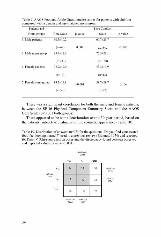
outcome than their corresponding male norm (p=0.002) and female norm groups (p<0.001, Table 9).
34
Table 9. AAOS Foot and Ankle Questionnaire scores for patients with clubfoot compared with a gender and age-matched norm group.
Patients and Shoe Comfort
Norm groups Core Scale p-value Scale p-value
1. Male patients
90.3±10.2 60.7±29.7
(n=62) 0.002 (n=53) <0.001
2. Male norm group
95.7±11.6 76.5±29.1
(n=231) (n=158)
1. Female patients
78.2±19.0 43.3±13.9
(n=19) (n=12)
2. Female norm group
94.2±11.8 <0.001 58.7±29.7 0.104
(n=59) (n=43)
There was a significant correlation for both the male and female patients,
between the SF-36 Physical Component Summary Score and the AAOS Core Scale (p<0.001 both groups).
There appeared to be some deterioration over a 30-year period, based on the patients’ subjective evaluation of the cosmetic appearance (Table 10).
Table 10. Distribution of answers (n=73) for the question: “Do you find your treated foot/ feet looking normal?” used in a previous review (Bjönness 1974) and repeated for Paper V (Chi-square test on observing the discrepancy found between observed and expected values: p-value <0.001)
73
57
16
34
32
2
39
25
14
Wallander 2009
Yes No Total
Yes Bjönness 1974 No Total
Total No 1974
Total Yes 1974
Total Yes 2006
Total No 2006
35
Discussion
This thesis focuses on a number of diverse parts of the concept of congenital clubfoot. The introductory part addresses the diagnosis, registration and re-porting of incidences of congenital clubfoot and its possible causes. The importance of epidemiology, including research on incidence, is emphasised by Engesaeter [105], as from this kind of research, additional etiological factors may be determined, leading to a basis for better management and perhaps even future prevention. At the time of the present study (Paper I), there was no better including method for newborn children with congenital clubfoot than contacting all Orthopaedic departments in Sweden and scruti-nising medical records to identify children with the correct diagnosis. Since 1999, there has been continuous reporting of congenital clubfoot to the Swedish Registry of Congenital Malformations [106]. Between 1999 and 2004, the reported incidence in this registry was approximately 0.7‰ and this reported number is compared with the estimated incidence of 1.4‰ [4], indicating that there is still under-reporting of the actual number of cases in Sweden. Compared with previous studies from Scandinavia, and even a re-cent report from southern Sweden [107], there was a higher cumulative inci-dence presented in Paper I. Even so, congenital clubfoot represents an un-common congenital malformation with an estimated annual number of 100-150 cases in Sweden. One important aspect, from a global perspective, is that approximately 100 000 children are born world-wide each year with clubfoot [108]. Of these children, 80% live in developing countries and many of them will never receive any treatment. Globally, these neglected clubfeet are the most serious reason for physical, social, and psychological disability among patients with congenital musculo-skeletal defects.
The etiology of clubfoot remains controversial. Several interacting factors may produce similar expressions in terms of the type of deformities in-volved, although severity varies. A common link between the diverse results from genetic and clinical research and findings on anatomical dissections and histological sections is difficult to determine, and some of the findings are probably secondary. The hypothesis of a “developmental arrest” with regional growth disturbance, as suggested by Dietz [9], could be this connec-tion, as reports [12, 13]on an increased risk, up to 10 times [14], with early amniocentesis are prominent. This procedure takes place during a short pe-riod in the first trimester, which is known to be critical for the position and growth of the foot. This “window of vulnerability” is open for both exogenic

36
(environmental) and genetic influences. The proposed mechanism of vascu-lar disruption [14] fits into this pattern, with disturbed growth of tarsal bones and increased fibrosis on the medial part of the foot as a consequence.
Increased incidence in southern Sweden is not easily explained by a sin-gle causative factor, but could be an effect of both exogenic factors and im-migration. We had the intention to examine whether there was any aggrega-tion of children with clubfoot in urban, compared to rural, areas (the same idea as for Kroogsgaard et al.[1]). However, there was no objective means of dividing the country into the areas required by this principle.
The second part dealt with imaging (Paper II), primarily with the aim of obtaining a standardised method of US for describing both the talo-navicular and the calcaneo-cuboidal relationships. This method has the possibility to provide better understanding of the pathology behind this condition and al-lows to follow treatment progress . The focus in Paper III was primarily on the medial part of the midfoot and the relation between the talus and the navicular, the position of the navicular, and its possible implication on resid-ual forefoot adduction. It was hypothesised that a small distance between the navicular and the medial malleolus would be associated with a more pro-nounced forefoot adduction, but this hypothesis was rejected. Ponseti [31] noted this negative association, called “spurious” correction, in adults, with the observation that inadequate “push” of the navicular in a lateral direction was sometimes still associated with a straight forefoot. Comparable studies on how treatment modifies skeletal pathology are scarce. Ippolito et al. [109] investigated 3-dimensional remodelling of skeletal pathology in two groups of patients, both at 25 years of age: the first group treated with extensive posteriomedial release (32 patients, 47 clubfeet), and the second group treated by the Ponseti manipulation technique, followed by limited posterior release (32 patients, 49 clubfeet). The navicular in both groups was in a me-dial position, and more pronounced in the “Ponseti-group”. Conversely, the group treated with less extensive posterior release had a better correction of both forefoot adduction and cavus. This finding indicates that correction of forefoot adduction can be obtained, despite residual medial position of the navicular. This was corroborated by the results in Paper III.
The measurement of MM-N-distance can be erroneously labelled medial “subluxation” of the navicular. However, “subluxation”, or displacement, would mean that the talus always has a normal alignment and a normal struc-ture, which is incorrect [38]. The talus is often altered in shape and direction with medial angulation of the neck and lateral pointing talar body, and is con-firmed by US [42] and MRI [51, 52]. Therefore, MM-N-distance is an indirect measure of midfoot pathology, and is apparent in comparisons with the contra-lateral normal foot in unilateral cases. Further, US could not be satisfactorily applied on the calcaneo-cuboidal joint where the joint surface appeared only medially angulated and not with medial displacement of the cuboid. Therefore, the lateral part of the clubfoot might be better described by MRI.

37
Another issue still under debate is whether US has further practical appli-cations and will change treatment protocols. With continuous technical de-velopment and more longitudinal series of patients, the understanding of treatment dynamics will develop, preferably assessed with US as a dynamic method. Also with MRI these aspects are demonstrated and Pirani[56] and Brand et al [65] determined marked and rapid remodelling potentials when deformities were successively corrected.
In Paper III, finally, a correlation between increased forefoot adduction and negative outcome in foot score and subjective outcome was determined, which indicated the need for hyperabduction in primary treatment, early management of relapses, and efforts to balance the medial and lateral part of the foot.
The history of medicine is replete with cyclical trends of various treat-ment protocols. In orthopaedics, this is expressed by exchanging surgical and non-surgical approaches to problems. Clubfoot management is no ex-ception, with the pendulum currently swinging in the direction of prolonged non-surgical treatment, which for surgeons might appear laborious. Even if the majority of children with clubfoot can be managed without extensive surgery, there will still be a number of reluctant clubfeet, atypical clubfeet, and non-compliants to treatment protocols. All these examples represent challenges for both the health system and clinicians. Surgery will remain an important tool for solving these problems.
For correcting difficult persistent or recurrent deformities there is cur-rently no obvious method of choice. For this purpose the Ilizarov distraction treatment was reviewed (Paper V).The question remains as to whether the Ilizarov method can still be of use as a salvage procedure. It is a demanding procedure for all involved, which emphasises the importance of thorough advance information, including the whole family, and appropriate selection of children. The results according to the Lehman foot score were disappoint-ing and did not concur with the patients’ subjective evaluation. A similar disappointing outcome is reported by Freedman et al. [110] in a study on 21 resistant clubfeet in 17 patients and more than 6 years follow-up: according to a four-graded scale, there were 86% fair or poor results. The authors con-cluded that after treatment with the Ilizarov method, there would still be a substantial need for revision surgery; indicating the method is not as promis-ing as has been stated. Revision rate as an end-point, together with relatively long follow-up are clear factors speaking in favour of the conclusions of Freedman et al. [110]. However, in the selection of patients suitable for distraction treatment, there is sometimes an obvious need for additional pro-cedures in order to prevent recurrence, such as corrective osteotomies in stiff feet and transfer of anterior tibial tendon in flexible feet. Nevertheless, the ultimate goal for all treatment is to reduce the need for extensive and repeat surgery, as this is better for both the patient and society.

38
Improved quality of life (QoL) is often mentioned as an outcome goal of medical intervention, especially in patients with chronic health conditions. There is still no gold standard for measuring QoL and some researchers have narrowed the concept of QoL into health-related factors (causes) and which are applied to medical research and medical outcome, so called HRQoL (Health-related Quality of Life). Beckie and Hayduk [90] strongly argue for retaining QoL as a “global personal assessment “: their concept is that QoL is a dependant variable, where the predictor variables include several factors, including health and psychological/social causes among other aspects. In that respect, congenital disorders with apparent physical impairment have a different impact on patients’ subjective satisfaction with life than acquired conditions of similar magnitude. Therefore, the use of generic outcome measures for evaluating general QoL combined with regional measures are recommended for the study of long-term outcome of congenital clubfoot and other paediatric disorders. Another critical issue is whose perspective the outcome rating scale should explore [111]. The patient’s perspective is now recognised [112], as this is the most important criterion for judging the effec-tiveness of a treatment in many chronic health problems. Therefore, patient-based instruments, such as self-reported questionnaires, are appropriate tools.
Besides the importance of valid instruments, another pre-requisite for re-liable conclusions is the use of gender and age-matched norm groups. Gen-der aspects in the treatment of CTEV are only occasionally addressed. In a study on 170 patients (252 clubfeet) with an average follow-up of 16 years [113], female patients are less satisfied and more concerned about the shape of their feet and legs than male patients are, although it is not mentioned whether there was any difference in physical function. When 83 patients, treated for CTEV, were evaluated at a mean age of 64 years (Paper V), an apparent gender related difference was found. Female patients with clubfoot had a poorer outcome, according to SF-36, EQ-5D and measure of ankle and foot function, than male patients did. Assessment of the same patients by Bjönness (pers. comm.) revealed the same difference at his follow-up in 1974. One possible explanation could be that women are more concerned than men by the lack of symmetry with a unilateral deformity, and generally by non-normal lower leg cosmetics. However, there was no difference in foot function between either female patients or male patients with unilateral deformity and bilateral deformity. No difference between female and male patients was determined on the SF-36 mental component summary score, indicating that mental function was preserved and no valid explanation for gender difference was apparent, in terms of either general physical QoL or ankle and foot function.
The principles developed by Ponseti [31] aim to correct all components of the deformity by gentle manipulation and well-moulded serial plaster casts, often facilitated by percutaneous Achilles tendon lengthening. When initi-

39
ated soon after birth, and by maintaining the treated foot in an outward rota-tion for up to 4 years of age in order to prevent relapses, more than 80% of the feet yield good functional long-term results [88].Only a small percentage of patients with very short and stiff feet require extensive surgical treatment. The treatment principles applied in the group of patients in this study (Paper V) differed from the Ponseti technique in two main ways. First, treatment started at a mean age of four months, and this substantial delay possibly ren-dered manipulations more difficult. Second, primary surgical treatment, in-stead of prolonged non-surgical or less extensive surgery, possibly resulted in more stiffness, undercorrection, and pain. In that respect, improvement of the QoL and functional outcome can be expected in the future.
One important aspect, and challenge, in the use of quality of life measures is translating and interpreting statistically significant differences, or changes, into clinically important ones [114]. The use of self-reported measures raises questions on interpretation: “What do the results mean?” Statistical signifi-cance in health-related measures is not synonymous with clinically important differences, for the patient or for the physician: questions that are more ap-propriate would be “How much change is enough to be important?” or “How much of a difference makes a difference?”

40
Conclusions
Clubfoot incidence calculation by including patients from all orthopaedic departments in Sweden reveals higher figures than in previous reports. Regional heterogeneity proposes environmental factors together with ethni-cal admixture as possible causes of higher incidence figures. With ultrasonography it is possible to reliably visualise non-ossified bones and their relationships, to illustrate progress of initial treatment, and to dis-cover deviations from the normal treatment protocol, i.e. in “atypical club-feet”. Through manipulation and limited surgery it is possible to reduce deformi-ties and achieve a straight forefoot, with the navicular still in a medial posi-tion (“spurious” correction). The Ilizarov technique has an acceptable outcome on residual equinus de-formity, but does not increase ankle joint range of motion. The Ilizarov technique offers a salvage treatment in older children after pre-vious surgery with complex residual or recurrent deformities with subjective improvement of function and cosmetics. Both female and male patients at the age of 60 years or more experience decreased foot and ankle function, the outcome being worse for female pa-tients. For both female and male patients, the foot and ankle problems have a nega-tive influence on physical aspects of quality of life. There are important gender differences in the pattern of self-estimated gen-eral quality of life, in that female patients experience a negative influence on physical aspects, whereas male patients assess their quality of life compara-ble or better than their adjusted norm groups.

41
Future perspectives
Clubfoot is one of the most common orthopaedic birth defects and the over-riding goal of management is to achieve a pain-free, plantigrade and flexible foot. Joint-sparing treatment principles, as proposed by for example Ponseti, with manipulation, serial casting and percutaneous tenotomy of the Achilles tendon, offer a promising future perspective. This method is technically non-demanding and therefore of special importance for those parts of the world where health facilities are scarce. For all healthcare systems there are merely logistical challenges to initiate and fulfil treatment which last for several years during childhood.
In Sweden the treatment and evaluation of children with congenital club-foot are currently spread among a large number of orthopaedic units in Swe-den. There is still much to learn about clubfoot, but the knowledge risks dilution through the low number of cases with each orthopaedic surgeon. Therefore, nationwide data collection and the establishment of a quality reg-ister would be the logical way to improve management principles and to structure evaluation, including quality of life issues.
There is a need for further development of imaging and ultrasonography appears to offer dynamic aspects of pathoanatomy at a low cost. The learn-ing curve for this method indicates the need for larger series of children for each examiner, starting with “easier” cases. Imaging has a limited role in initial treatment, but might in the future enable clinicians to understand the morphology in complicated or atypical cases, or when progress in treatment has come to a halt.
Current surgery rates are decreasing, with less need for early extensive soft tissue releases. The Ilizarov technique is an important contribution to salvage treatment in older children, but the method is time-consuming, com-plex, and fraught with complications. For this reason, the Ilizarov method needs more precise indications and needs comparison with other surgical methods.
There are many reports on short-term clubfoot outcome with the use of non-validated rating systems that may not capture the important questions or the relevant physical findings, especially from the patient’s perspective. Gender aspects are still rare in orthopaedic literature, and the clubfoot diag-nosis is no exception. There is an obvious need to focus on children and adolescents treated for clubfoot, and for considering issues that are important for those specific ages.

42
Acknowledgements
I would like to express my gratitude to everyone who have supported and encouraged me in my research and clinical work, and especially I would like to thank:
Göran Hansson, Associate professor, Department of Pediatric Surgery, The Queen Silvia Children’s Hospital, Göteborg, principal tutor.
Sune Larsson, Professor, Department of Orthopaedics, Uppsala University Hos-pital, co-tutor.
Ylva Aurell, MD, PhD, Department of Radiology, Sahlgrenska University Hospital, Mölndal, co-author.
Terje Bjönness, MD, PhD, Kungälv, co-author.
Lennart Hovelius, Professor, Depart-ment of Orthopaedics, Gävle County Hospital, co-author.
Arne Johansson, MD, Department of Orthopaedics, Kärnsjukhuset, Skövde, co-author.
Kjell Jonsson, Professor, Department of Radiology, Lund University Hospital, co-author.
Karl Michaelsson, Associate Professor, Department of Orthopaedics, Uppsala University Hospital,co-author.
Björn Tjernström, MD, PhD, Depart-ment of Orthopaedics, Uppsala Univer-sity Hospital, co-author.
All colleagues and friends at Swedish Paediatric Orthopaedic Society.
All colleagues and personnel at the De-partment of Orthopaedics, Gävle County Hospital for patience and support.
Bo Lundberg, former Associate Profes-sor, Department of Orthopaedics, Gävle County Hospital, for evoking my inter-est in clubfoot and research, and teach-ing me that there is always more to learn and discuss.
Past and present heads of the Depart-ment of Orthopaedics, Gävle County Hospital.
Sue Pajuluoma, for superb linguistic check.
Sven Östberg, for layout and illustra-tions.
Centre for Research & Development, Uppsala University/County Council of Gävleborg (CFUG): Marieann Högman, Hans Högberg, Lennart Fredriksson, Inga-Lill Stenlund,Vivi-Anne Rahm, Lawrence Teeland and research fellows: you were there when I needed it most.
And last but not least: To my beloved family (Sara, Björn, Karl and my wife Åsa), all friends and relatives.
This study was supported by grants from: Stiftelsen för Skobranschens Utveck-lingsfond,
HKH Kronprinsessan Lovisas förening för barnsjukvård/Axel Tielmans min-nesfond, and
Centre for Research & Development, Uppsala University/ County Council of Gävleborg.

43
References
1. Krogsgaard, M.R., et al., Increasing incidence of club foot with higher popula-tion density: incidence and geographical variation in Denmark over a 16-year period--an epidemiological study of 936,525 births. Acta Orthop, 2006. 77(6): p. 839-46.
2. Barker, S.L. and M.F. Macnicol, Seasonal distribution of idiopathic congenital talipes equinovarus in Scotland. J Pediatr Orthop B, 2002. 11(2): p. 129-33.
3. Severin, E., [The frequency of congenital hip dislocation and congenital equi-novarus in Sweden.]. Nord Med, 1956. 55(7): p. 221-3.
4. Wallander, H., L. Hovelius, and K. Michaelsson, Incidence of congenital club-foot in Sweden. Acta Orthop, 2006. 77(6): p. 847-52.
5. Feldbrin, Z., et al., Muscle imbalance in the aetiology of idiopathic club foot. An electromyographic study. J Bone Joint Surg Br, 1995. 77(4): p. 596-601.
6. Ippolito, E., Update on pathologic anatomy of clubfoot. J Pediatr Orthop B, 1995. 4(1): p. 17-24.
7. Hootnick, D.R., et al., Confirmation of arterial deficiencies in a limb with ne-crosis following clubfoot surgery. J Pediatr Orthop B, 1999. 8(3): p. 187-93.
8. Handelsman, J.E. and M.A. Badalamente, Neuromuscular studies in clubfoot. J Pediatr Orthop, 1981. 1(1): p. 23-32.
9. Dietz, F.R., I.V. Ponseti, and J.A. Buckwalter, Morphometric study of clubfoot tendon sheaths. J Pediatr Orthop, 1983. 3(3): p. 311-8.
10. Kawashima, T. and H.K. Uhthoff, Development of the foot in prenatal life in relation to idiopathic club foot. J Pediatr Orthop, 1990. 10(2): p. 232-7.
11. Wynne-Davies, R., Family Studies and the Cause of Congenital Club Foot. Talipes Equinovarus, Talipes Calcaneo-Valgus and Metatarsus Varus. J Bone Joint Surg Br, 1964. 46: p. 445-63.
12. Cederholm, M., B. Haglund, and O. Axelsson, Infant morbidity following amniocentesis and chorionic villus sampling for prenatal karyotyping. BJOG, 2005. 112(4): p. 394-402.
13. Nikkila, A., et al., Early amniocentesis and congenital foot deformities. Fetal Diagn Ther, 2002. 17(3): p. 129-32.
14. Farrell, S.A., et al., Club foot, an adverse outcome of early amniocentesis: disruption or deformation? CEMAT. Canadian Early and Mid-Trimester Am-niocentesis Trial. J Med Genet, 1999. 36(11): p. 843-6.
15. Wang, L.L., et al., HOXD13 may play a role in idiopathic congenital clubfoot by regulating the expression of FHL1. Cytogenet Genome Res, 2008. 121(3-4): p. 189-95.
16. Honein, M.A., L.J. Paulozzi, and C.A. Moore, Family history, maternal smok-ing, and clubfoot: an indication of a gene-environment interaction. Am J Epi-demiol, 2000. 152(7): p. 658-65.
17. Skelly, A.C., et al., Talipes equinovarus and maternal smoking: a population-based case-control study in Washington state. Teratology, 2002. 66(2): p. 91-100.
18. Pryor, G.A., et al., Seasonal variation in the incidence of congenital talipes equinovarus. J Bone Joint Surg Br, 1991. 73(4): p. 632-4.

44
19. Robertson, W.W., Jr. and D. Corbett, Congenital clubfoot. Month of concep-tion. Clin Orthop Relat Res, 1997(338): p. 14-8.
20. Loder, R.T., et al., Lack of seasonal variation in idiopathic talipes equinovarus. J Bone Joint Surg Am, 2006. 88(3): p. 496-502.
21. Carney, B.T. and T.R. Coburn, Demographics of idiopathic clubfoot: is there a seasonal variation? J Pediatr Orthop, 2005. 25(3): p. 351-2.
22. Cartlidge, I., Observations on the epidemiology of club foot in Polynesian and Caucasian populations. J Med Genet, 1984. 21(4): p. 290-2.
23. Ching, G.H., C.S. Chung, and R.W. Nemechek, Genetic and epidemiological studies of clubfoot in Hawaii: ascertainment and incidence. Am J Hum Genet, 1969. 21(6): p. 566-80.
24. Wynne-Davies, R., Genetic and environmental factors in the etiology of talipes equinovarus. Clin Orthop Relat Res, 1972. 84: p. 9-13.
25. Engell, V., et al., Club foot: a twin study. J Bone Joint Surg Br, 2006. 88(3): p. 374-6.
26. Wynne-Davies, R., Talipes Equinovarus. A Review of Eighty-Four Cases after Completion of Treatment. J Bone Joint Surg Br, 1964. 46: p. 464-76.
27. Rebbeck, T.R., et al., A single-gene explanation for the probability of having idiopathic talipes equinovarus. Am J Hum Genet, 1993. 53(5): p. 1051-63.
28. Kruse, L.M., M.B. Dobbs, and C.A. Gurnett, Polygenic threshold model with sex dimorphism in clubfoot inheritance: the Carter effect. J Bone Joint Surg Am, 2008. 90(12): p. 2688-94.
29. Khoury, M.J., M. Gomez-Farias, and J. Mulinare, Does maternal cigarette smoking during pregnancy cause cleft lip and palate in offspring? Am J Dis Child, 1989. 143(3): p. 333-7.
30. Dimeglio, A., et al., Classification of clubfoot. J Pediatr Orthop B, 1995. 4(2): p. 129-36.
31. Ponseti, I.V., Treatment of congenital club foot. J Bone Joint Surg Am, 1992. 74(3): p. 448-54.
32. Gray, D.H. and J.M. Katz, A histochemical study of muscle in club foot. J Bone Joint Surg Br, 1981. 63-B(3): p. 417-23.
33. Isaacs, H., et al., The muscles in club foot--a histological histochemical and electron microscopic study. J Bone Joint Surg Br, 1977. 59-B(4): p. 465-72.
34. Herceg, M.B., et al., Histologic and histochemical analysis of muscle speci-mens in idiopathic talipes equinovarus. J Pediatr Orthop, 2006. 26(1): p. 91-3.
35. Carroll, N.C., Pathoanatomy and surgical treatment of the resistant clubfoot. Instr Course Lect, 1988. 37: p. 93-106.
36. Hjelmstedt, A. and B. Sahlstedt, Talar deformity in congenital clubfeet. An anatomical and functional study with special reference to the ankle joint mobil-ity. Acta Orthop Scand, 1974. 45(4): p. 628-40.
37. Irani, R.N. and M.S. Sherman, The pathological anatomy of idiopathic club-foot. Clin Orthop Relat Res, 1972. 84: p. 14-20.
38. Howard, C.B. and M.K. Benson, Clubfoot: its pathological anatomy. J Pediatr Orthop, 1993. 13(5): p. 654-9.
39. McKay, D.W., New concept of and approach to clubfoot treatment: section I-principles and morbid anatomy. J Pediatr Orthop, 1982. 2(4): p. 347-56.
40. Goldner, J.L., Congenital talipes equinovarus--fifteen years of surgical treat-ment. Curr Pract Orthop Surg, 1969. 4: p. 61-123.
41. Herzenberg, J.E., et al., Clubfoot analysis with three-dimensional computer modeling. J Pediatr Orthop, 1988. 8(3): p. 257-62.
42. Aurell, Y., et al., Ultrasound anatomy in the neonatal clubfoot. Eur Radiol, 2002. 12(10): p. 2509-17.

45
43. Shiels, W.E., 2nd, et al., Focused dynamic sonographic examination of the congenital clubfoot. Pediatr Radiol, 2007. 37(11): p. 1118-24.
44. Cahuzac, J.P., et al., Assessment of hindfoot deformity by three-dimensional MRI in infant club foot. J Bone Joint Surg Br, 1999. 81(1): p. 97-101.
45. Itohara, T., et al., Assessment of the three-dimensional relationship of the ossi-fic nuclei and cartilaginous anlagen in congenital clubfoot by 3-D MRI. J Or-thop Res, 2005. 23(5): p. 1160-4.
46. Richards, B.S. and M. Dempsey, Magnetic resonance imaging of the congenital clubfoot treated with the French functional (physical therapy) method. J Pediatr Orthop, 2007. 27(2): p. 214-9.
47. Kuhns, L.R., et al., Ultrasound of the navicular during the simulated Ponseti maneuver. J Pediatr Orthop, 2003. 23(2): p. 243-5.
48. Aurell, Y., et al., Ultrasound assessment of early clubfoot treatment: a com-parison of the Ponseti method and a modified Copenhagen method. J Pediatr Orthop B, 2005. 14(5): p. 347-57.
49. Desai, S., A. Aroojis, and R. Mehta, Ultrasound evaluation of clubfoot correc-tion during Ponseti treatment: a preliminary report. J Pediatr Orthop, 2008. 28(1): p. 53-9.
50. Lowe, L.W. and M.A. Hannon, Residual adduction of the forefoot in treated congenital club foot. J Bone Joint Surg Br, 1973. 55(4): p. 809-13.
51. Kamegaya, M., et al., MRI study of talonavicular alignment in club foot. J Bone Joint Surg Br, 2001. 83(5): p. 726-30.
52. Itohara, T., et al., Assessment of talus deformity by three-dimensional MRI in congenital clubfoot. Eur J Radiol, 2005. 53(1): p. 78-83.
53. Gigante, C., E. Talenti, and S. Turra, Sonographic assessment of clubfoot. J Clin Ultrasound, 2004. 32(5): p. 235-42.
54. Wallander, H., Y. Aurell, and G. Hansson, No association between residual forefoot adduction and the position of the navicular in clubfeet treated by pos-terior release. J Pediatr Orthop, 2007. 27(1): p. 60-6.
55. Howard, C.B. and M.K. Benson, The ossific nuclei and the cartilage anlage of the talus and calcaneum. J Bone Joint Surg Br, 1992. 74(4): p. 620-3.
56. Pirani, S., L. Zeznik, and D. Hodges, Magnetic resonance imaging study of the congenital clubfoot treated with the Ponseti method. J Pediatr Orthop, 2001. 21(6): p. 719-26.
57. Cuevas de Alba, C., et al., Computed tomography for femoral and tibial torsion in children with clubfoot. Clin Orthop Relat Res, 1998(353): p. 203-9.
58. Herold, H.Z. and C. Marcovich, Tibial torsion in untreated congenital clubfoot. Acta Orthop Scand, 1976. 47(1): p. 112-7.
59. Krishna, M., et al., Tibial torsion measured by ultrasound in children with talipes equinovarus. J Bone Joint Surg Br, 1991. 73(2): p. 207-10.
60. Pirani, S., et al., A method of evaluating the virgin clubfoot with substantial inter-observer reliability, in POSNA. 1995: Miami, Florida.
61. Flynn, J.M., M. Donohoe, and W.G. Mackenzie, An independent assessment of two clubfoot-classification systems. J Pediatr Orthop, 1998. 18(3): p. 323-7.
62. Wainwright, A.M., et al., The classification of congenital talipes equinovarus. J Bone Joint Surg Br, 2002. 84(7): p. 1020-4.
63. Hippokrates, On the Articulations, part 62 Written 400 B.C.E http://classics.mit.edu//Hippocrates/artic.html.
64. Bonnel, F., et al., Effects of compression on growth plates in the rabbit. Acta Orthop Scand, 1983. 54(5): p. 730-3.
65. Brand, R.A., et al., Cartilage anlagen adapt in response to static deformation. Med Hypotheses, 2006. 66(3): p. 653-9.
46
66. Bensahel, H., et al., Results of physical therapy for idiopathic clubfoot: a long-term follow-up study. J Pediatr Orthop, 1990. 10(2): p. 189-92.
67. Dimeglio, A., et al., Orthopaedic treatment and passive motion machine: con-sequences for the surgical treatment of clubfoot. J Pediatr Orthop B, 1996. 5(3): p. 173-80.
68. Simons, G.W., Complete subtalar release in club feet. Part II--Comparison with less extensive procedures. J Bone Joint Surg Am, 1985. 67(7): p. 1056-65.
69. Lehman, W.B., et al., Re-do clubfoot: surgical approach and long-term results. Bull N Y Acad Med, 1990. 66(6): p. 601-17.
70. Tarraf, Y.N. and N.C. Carroll, Analysis of the components of residual deformity in clubfeet presenting for reoperation. J Pediatr Orthop, 1992. 12(2): p. 207-16.
71. Ponseti, I.V., Clubfoot management. J Pediatr Orthop, 2000. 20(6): p. 699-700. 72. Dietz, F.R., Treatment of a recurrent clubfoot deformity after initial correction
with the Ponseti technique. Instr Course Lect, 2006. 55: p. 625-9. 73. Morcuende, J.A., et al., Radical reduction in the rate of extensive corrective sur-
gery for clubfoot using the Ponseti method. Pediatrics, 2004. 113(2): p. 376-80. 74. Davidson, R.S., Clubfoot salvage: a review of the past decade's contributions. J
Pediatr Orthop, 2003. 23(3): p. 410-8. 75. Crawford, A.H. and A.K. Gupta, Clubfoot controversies: complications and
causes for failure. Instr Course Lect, 1996. 45: p. 339-46. 76. Evans, D., Treatment of the unreduced or 'relapsed' club foot in older children.
Proc R Soc Med, 1968. 61(8): p. 782-3. 77. Graham, G.P. and C.M. Dent, Dillwyn Evans operation for relapsed club foot.
Long-term results. J Bone Joint Surg Br, 1992. 74(3): p. 445-8. 78. Kumar, P.N., P.W. Laing, and L. Klenerman, Medial calcaneal osteotomy for
relapsed equinovarus deformity. Long-term study of the results of Frederick Dwyer. J Bone Joint Surg Br, 1993. 75(6): p. 967-71.
79. Dwyer, F.C., Treatment of the relapsed club foot. Proc R Soc Med, 1968. 61(8): p. 783.
80. Grill, F. and J. Franke, The Ilizarov distractor for the correction of relapsed or neglected clubfoot. J Bone Joint Surg Br, 1987. 69(4): p. 593-7.
81. Grant, A.D., D. Atar, and W.B. Lehman, The Ilizarov technique in correction of complex foot deformities. Clin Orthop Relat Res, 1992(280): p. 94-103.
82. El Barbary, H., H. Abdel Ghani, and M. Hegazy, Correction of relapsed or neglected clubfoot using a simple Ilizarov frame. Int Orthop, 2004. 28(3): p. 183-6.
83. Bradish, C.F. and S. Noor, The Ilizarov method in the management of relapsed club feet. J Bone Joint Surg Br, 2000. 82(3): p. 387-91.
84. Wallander, H., G. Hansson, and B. Tjernstrom, Correction of persistent club-foot deformities with the Ilizarov external fixator. Experience in 10 previously operated feet followed for 2-5 years. Acta Orthop Scand, 1996. 67(3): p. 283-7.
85. Dobbs, M.B., et al., Factors predictive of outcome after use of the Ponseti method for the treatment of idiopathic clubfeet. J Bone Joint Surg Am, 2004. 86-A(1): p. 22-7.
86. Andriesse, H., G. Hagglund, and G.B. Jarnlo, The clubfoot assessment protocol (CAP); description and reliability of a structured multi-level instrument for fol-low-up. BMC Musculoskelet Disord, 2005. 6: p. 40.
87. Beatson, T.R. and J.R. Pearson, A method of assessing correction in club feet. J Bone Joint Surg Br, 1966. 48(1): p. 40-50.
88. Laaveg, S.J. and I.V. Ponseti, Long-term results of treatment of congenital club foot. J Bone Joint Surg Am, 1980. 62(1): p. 23-31.

47
89. Munshi, S., R.A. Varghese, and B. Joseph, Evaluation of outcome of treatment of congenital clubfoot. J Pediatr Orthop, 2006. 26(5): p. 664-72.
90. Beckie, T.M. and L.A. Hayduk, Measuring quality of life. Social Indicators Research, 1997. 42(1): p. 21-39.
91. Andre, M., S. Hagelberg, and C.H. Stenstrom, The juvenile arthritis foot dis-ability index: development and evaluation of measurement properties. J Rheu-matol, 2004. 31(12): p. 2488-93.
92. Roye, B.D., et al., Patient-based outcomes after clubfoot surgery. J Pediatr Orthop, 2001. 21(1): p. 42-9.
93. Morris, C., et al., The Oxford ankle foot questionnaire for children: scaling, reliability and validity. J Bone Joint Surg Br, 2008. 90(11): p. 1451-6.
94. Atar, D., et al., Revision surgery in clubfeet. Clin Orthop Relat Res, 1992(283): p. 223-30.
95. Fridman, M.W. and H.S. de Almeida Fialho, The role of radiographic meas-urements in the evaluation of congenital clubfoot surgical results. Skeletal Ra-diol, 2007. 36(2): p. 129-38.
96. Vanderwilde, R., et al., Measurements on radiographs of the foot in normal infants and children. J Bone Joint Surg Am, 1988. 70(3): p. 407-15.
97. Ware, J.E., Jr. and C.D. Sherbourne, The MOS 36-item short-form health sur-vey (SF-36). I. Conceptual framework and item selection. Med Care, 1992. 30(6): p. 473-83.
98. Sullivan, M. and J. Karlsson, The Swedish SF-36 Health Survey III. Evaluation of criterion-based validity: results from normative population. J Clin Epide-miol, 1998. 51(11): p. 1105-13.
99. Brooks, R., EuroQol: the current state of play. Health Policy, 1996. 37(1): p. 53-72.
100. Dolan, P., et al., The time trade-off method: results from a general population study. Health Economics, 1996. 5(2): p. 141-54.
101. Burstrom, K., M. Johannesson, and F. Diderichsen, Swedish population health-related quality of life results using the EQ-5D. Quality of Life Research, 2001. 10(7): p. 621-35.
102. Bjork, S. and A. Norinder, The weighting exercise for the Swedish version of the EuroQol. Health Econ, 1999. 8(2): p. 117-26.
103. Johanson, N.A., et al., American Academy of Orthopaedic Surgeons lower limb outcomes assessment instruments. Reliability, validity, and sensitivity to change. J Bone Joint Surg Am, 2004. 86-A(5): p. 902-9.
104. Bjonness, T., Medfödd klumpfot, in Department of Orthopaedics. 1974, Goth-enburg: Gothenburg. p. 61.
105. Engesaeter, L.B., Increasing incidence of clubfoot: changes in the genes or the environment? Acta Orthop, 2006. 77(6): p. 837-8.
106. http://www.socialstyrelsen.se/NR/rdonlyres/A842A8FB-7BC9-43D2-A087-07901BBD338E/6708/20064210.pdf.
107. Danielsson, L.G., Incidence of congenital clubfoot in Sweden. 128 cases in 138,000 infants 1946-1990 in Malmo. Acta Orthop Scand, 1992. 63(4): p. 424-6.
108. http://www.usbjd.org/projects/project_op.cfm?dirID=115. 109. Ippolito, E., et al., The influence of treatment on the pathology of club foot. CT
study at maturity. J Bone Joint Surg Br, 2004. 86(4): p. 574-80. 110. Freedman, J.A., H. Watts, and N.Y. Otsuka, The Ilizarov method for the treat-
ment of resistant clubfoot: is it an effective solution? J Pediatr Orthop, 2006. 26(4): p. 432-7.
48
111. Eechaute, C., et al., The clinimetric qualities of patient-assessed instruments for measuring chronic ankle instability: a systematic review. BMC Musculoskelet Disord, 2007. 8: p. 6.
112. Parker, J., et al., The problem with measuring patient perceptions of outcome with existing outcome measures in foot and ankle surgery. Foot Ankle Int, 2003. 24(1): p. 56-60.
113. Hutchins, P.M., et al., Long-term results of early surgical release in club feet. J Bone Joint Surg Br, 1985. 67(5): p. 791-9.
114. Beaton, D.E., M. Boers, and G.A. Wells, Many faces of the minimal clinically important difference (MCID): a literature review and directions for future re-search. Curr Opin Rheumatol, 2002. 14(2): p. 109-14.


��� -�,�(*�* -.*����**���������� �������������� ������������������� ������� ����������������������������
�+�() /0� ���� �� 0� 1����2 �� ��+���
� +���(�� +**�(��� �(�3 0� 1����2 �� ��+���4 -..*���-�,�(*24 * �*����2 � *�33�(2 �� � ��3��( �� .�.�(*5 � ��6��.�* �� 0� ��3.��� +**�(��� �(� 7�. � 3�8�( �6�+*0(�*��(�0 ��(�(�*4 60�� 0� *�33�(2 ����� * +*(���+��(�������2 0(���0 0� *�(�* ���� ��3.(�0��*,���33�(�* �� -..*��� �**�(���* �(�3 0� 1����2 ����+���5 9:(�( � ;����(24 !""�4 0� *�(�* 6�* .���*0�+��+�( 0� �� <��3.(�0��*,� ��33�(�* �� -..*����**�(���* �(�3 0� 1����2 �� ��+���=5>
�*(����) .�������*5��5*��(�)���)*�)��)+,� �"�'��
���������������
���� ���������� �
����


![A polyaxial fixation brace for the treatment of idiopathic ...CTEV, also known as clubfoot, is the fifth most common congenital malformation in children [1]. CTEV consists of four](https://img.dokumen.tips/doc/110x75/60bc26e493380344804f2a3f/a-polyaxial-fixation-brace-for-the-treatment-of-idiopathic-ctev-also-known.jpg)


![Treatment Of Congenital Clubfoot Using The Ponseti · PDF fileIris Lohan Treatment Of Congenital Clubfoot Using The Ponseti Method Workshop Manual [2nd Edition]](https://img.dokumen.tips/doc/110x75/5a9d8bd67f8b9a21688c2ac8/treatment-of-congenital-clubfoot-using-the-ponseti-lohan-treatment-of-congenital.jpg)