Embed Size (px)
Citation preview

05/09/2011
1
João Flávio Nogueira, MDSinus & Oto Centro - Fortaleza, Brazil
CONCEPTS OF FUNCTIONAL ENDOSCOPIC EAR SURGERY
DISCLOSURE
• Research grant ACCLARENT/J&J
• Support by Karl Storz (Silver Books)

05/09/2011
2
Antonio Maria Valsalva(1666 – 1723)
Nicolas Deleau1797 - 1862
Adam Politzer1835 - 1920

05/09/2011
3
Joseph Toynbee1815 - 1866
The healthy middle ear is a sterile site Tube-Tympanum-Mastoid immunity
T.T.M.A.L.T.
Available at: http://flylib.com/books/en/2.953.1.20/1/
Ventilation
Muco-cilliary
Immunological
Nose-Pharynx-Tube-Tympanum-Mastoid Unit
3 units
Nose-Pharynx-Tube-Tympanum-Mastoid Unit
• Innate immunity• Muco-cilliary clearance
– Mucins– Surfactant– Acquaporins
• Peptides and Antimicrobial Proteins– Lysozime (Lz)– Lactoferrin (Lf)– Defensins– Collectins and surfactant
(SP-A, SP-D)

05/09/2011
4
Differences of the Nose-Pharynx-Tube-Tympanum-Mastoid Unit
Ventilation
Ventilation Ventilation
Emb
riolo
gical path
way
Ventilation
Embriological pathway
Hammar JAStudien über die Entwicklung des Vorderdarms und einiger angrenzenden Organe. 1. Abt:
allegemeine Morphologie der Schlundspalten beim Menschen. Entwicklung des Mittelohrraumes und des äusseren Gehötganges.
Arch Mikrosk Anat 1902; 59:471-628
The EmbriologyBetween the third and seventh foetal month the gelatinous tissue of the middleear cleft is gradually absorbed. At the same time the primitive tympanic cavity
develops by a growth, into the cleft, of an endothelial lined fluid pouch extendingfrom the eustachian tube. Four primary sacs or pouches then bud out. They aresaccus anticus, saccus medius, saccus superior and saccus posticus (Hammar,
1902). Where these pouches contact each other mucosal folds are formed.Between the mucosal layers of the folds are remnants of the mesoderm, includingblood vessels supplying the “viscera” of the tympanic cavity

05/09/2011
5
Proctor BThe development of the middle ear spaces and their surgical significance. The Journal of
Laryngology and Otology 1964
In this sense we can define the intra-tympanic ventilation routes as the result of the path travelled by four
pockets of the middle ear embryological cavitation process
Saccus Posticus – Sinus Tympani
Saccus Superior Saccus Anticus
Saccus Medius

05/09/2011
6
Marchione D, et al. The Contribution of Selective Dysventilation to Attical Middle Ear Pathology. Medical Hypoteses; 2011: in press
Ventilation Routes Middle Ear

05/09/2011
7
MACSMucosal respiration – Gas diffusion
The tympanum is a single, large air cell that contains the
ME ossicles.
The MACs are multiple
partitioned cellularairspaces thatincreases ME
volume
LUNGS x MACS
05/09/2011
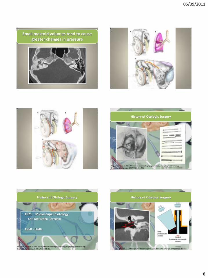
8
Small mastoid volumes tend to cause greater changes in pressure
History of Otologic Surgery
Nogueira JF Jr, et al. A brief history of otorhinolaryngolgy: otology, laryngology and rhinology. Braz J Otorhinolaryngol. 2007;73(5):693-703.
• 1921 – Microscope in otology
– Carl Olof Nylen (Sweden)
• 1950 - Drills
Nogueira JF Jr, et al. A brief history of otorhinolaryngolgy: otology, laryngology and rhinology. Braz J Otorhinolaryngol. 2007;73(5):693-703.
History of Otologic Surgery
Tarabichi M. Endoscopic middle ear surgery. Ann Otol Rhinol Laryngol. 1999;108:39–46.
History of Otologic Surgery

05/09/2011
9
Tarabichi M. Endoscopic middle ear surgery. Ann Otol Rhinol Laryngol. 1999;108:39–46.
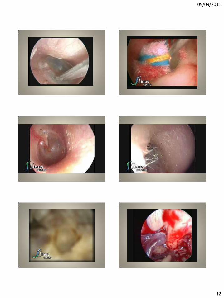
Endoscopic Otologic Surgery

05/09/2011
10

05/09/2011
11
05/09/2011
12

05/09/2011
13

05/09/2011
14
"In clear contrast to the impact of the introduction of endoscope in most surgical disciplines, the practice of ear surgery has changed little and it continues to be the domain for the microscope. Depending on the task at hand, there are many distinctions that would make the endoscope a better instrument than the microscope and vice-versa. We all need to master working with both instruments to better understand and treat pathologies of the ear. The objective of the IWGEES is to neutralize these longstanding biases toward the microscope and to get all of us to use the best instrument in the best way possible to help our patients."
Muaaz Tarabichi – American Hospital of Dubai