Embed Size (px)
Citation preview

Received: 30 August 2009, Revised: 7 December 2009, Accepted: 9 December 2009, Published online in Wiley InterScience: 19 February 2010
Computational modeling of humanparaoxonase 1: preparation of protein models,binding studies, and mechanistic insightsToby T. Sanana, Sivaramakrishnan Muthukrishnana, Jeremy M. Becka,Peng Taoa, Carrigan J. Hayesa, Tamara C. Ottob, Douglas M. Cerasolib,David E. Lenzb and Christopher M. Hadada*
The enzyme human paraoxonase 1 (huPON1) has demonstrated significant potential for use as a bioscavenger fortreatment of exposure to organophosphorus (OP) nerve agents. Herein we report the development of protein modelsfor the human isoform derived from a crystal structure of a chimeric version of the protein (pdb ID: 1V04) and ahomology model derived from the related enzyme diisopropylfluorophosphatase (pdb ID: 1XHR). From thesestructural models, binding modes for OP substrates are predicted, and these poses are found to orient substratesin proximity to residues known to modulate specificity of the enzyme. Predictions are made with regard to the rolethat residues play in altering substrate binding and turnover, in particular with regard to the stereoselectivity of theenzyme, and the known differences in activity related to a natural polymorphism in the enzyme. Potentialmechanisms of action of the protein for catalytic hydrolysis of OP substrates are also evaluated in light of theproposed binding modes. Copyright ß 2010 John Wiley & Sons, Ltd.
Supporting information may be found in the online version of this paper
Keywords: bioscavenger; catalytic hydrolysis; organophosphorus compounds; paraoxonase
INTRODUCTION
The human enzyme paraoxonase 1 (huPON1) possesses the
ability to hydrolyze a variety of substrates, including organopho-
sphorus (OP) nerve agents, in a catalytic manner – a capability for
which it has received considerable attention as a potential
bioscavenger of chemical warfare agents.[1–4] Predictions on the
basis of the rate of inhibition of acetylcholinesterase in vivo (in
animal models) suggest that the wild-type natural form of
huPON1 (WT-huPON1) does not have sufficient catalytic activity
to act as a bioscavenger. However, variants of huPON1 with
enhanced kinetic activity for deactivation of OP agents could
provide protection against nerve agents in vivo. It has been
estimated that an in vivo concentration of a nerve agent of 0.1mM
is capable of eliciting a toxic response.[5] This estimate suggests
that a decrease in KM of two orders of magnitude (e.g., a KM of
1.0–10mM), coupled with a 100 fold increase in kcat of
WT-huPON1 may be required to achieve the levels of catalytic
activity needed for an efficient bioscavenger. Given this dual
challenge of a need to alter both KM and kcat simultaneously, in
silico design can provide an efficient approach to identifying
potential variants with the necessary properties. Attempts to
achieve such an enhancement using state-of-the-art protein
engineering techniques are in progress; however, this work is
complicated by the dearth of information regarding the nature of
the active site of the human enzyme, as only a single crystal
structure has been reported, featuring an apo-version (with no
inhibitor or substrate bound) of a recombinant, gene-shuffled
variant of the enzyme. The putative active site has been identified
primarily throughmutagenesis studies which have demonstrated
altered reaction selectivity.[6] Using such methods to identify the
catalytic mechanism, however, is complicated by the presence of
two bound calcium ions for the enzyme; many mutations in the
calcium binding regions result in loss-of-function, which can be
either due to a change in the protein folding or due to disruptions
of the reaction mechanism.
The human form of the protein is extremely difficult to purify
to homogeneity while maintaining functionality, and attempts
to obtain structural information of the wild-type (WT) protein
by crystallographic methods have been unsuccessful to date.
The sole crystal structure published at present is that of a
recombinant, gene-shuffled variant derived from the human,
rabbit, rat, and mouse paraoxonases; this variant, known as G2E6,
shares 86% sequence homology with the WT human protein
(PDB entry 1V04).[7] The crystal structure of G2E6 has been
resolved at 2.2 A resolution, with two fragments remaining
(www.interscience.wiley.com) DOI 10.1002/poc.1678
Special Issue Article
* Correspondence to: C. M. Hadad, Department of Chemistry, 100 West 18th
Avenue, Ohio State University, Columbus, OH 43210, USA.
E-mail: [email protected]
a T. T. Sanan, S. Muthukrishnan, J. M. Beck, P. Tao, C. J. Hayes, C. M. Hadad
Department of Chemistry, 100 West 18th Avenue, Ohio State University,
Columbus, Ohio 43210, USA
b T. C. Otto, D. M. Cerasoli, D. E. Lenz
Physiology and Immunology Branch, Research Division, US Army Medical
Research Institute of Chemical Defense, 3100 Ricketts Point Rd, Aberdeen
Proving Ground, Maryland 21010, USA
J. Phys. Org. Chem. 2010, 23 357–369 Copyright ß 2010 John Wiley & Sons, Ltd.
357

unresolved: the N-terminal 15 residues of the protein as well as
a flexible loop from residues 72–79, both of which are in the
putative high-density lipoprotein (HDL) binding domain. The
center of the b-propeller contains two bound calcium ions,
which have been designated ‘catalytic’ and ‘structural’ on the
basis of mutagenesis studies. In the crystal structure, a single
phosphate ion is coordinated to the ‘catalytic’ calcium ion (refer
Fig. 1).
Due to this lack of a crystal structure of a complete form of the
human enzyme, the mechanism of action of huPON1 for
hydrolysis of organophosphorus compounds remains in doubt.
Postulated mechanisms include general base catalysis of
substrate by activated water molecules (the source of which is
also debated) or direct hydrolysis by a nucleophilic residue in the
active site pocket. Possible sources for the generation of
hydroxide include either the H115/H134 and D269/H285 dyads,
or direct activation of water by coordination to the ‘catalytic’
calcium ion (Fig. 2).[1–4,8]
With regard to hydrolysis of organophosphorus compounds,
there have been results from mutagenesis studies which cast
doubt onto several of the postulated sources of the nucleophile.
For example, mutations at H115 have been identified which
preserve OPase activity, including H115W, suggesting that a
catalytic dyad of these residues is not operative in the
mechanism.[6] A recent publication[9] of a neutron-scattering
structure of DFPase, an enzyme highly similar in structure to
PON1, shows the coordination of an intact water molecule to the
calcium ion. This result suggests that coordination to the calcium
ion alone may be insufficient to generate hydroxide in the active
site. The authors postulated that the reaction mechanism for
PON1 involved the direct hydrolysis of substrate by D269, which
is likely to be in proximity to a substrate coordinated via the
phosphoryl oxygen to calcium. We believe, however, that the
coordination of D269 to calcium reduces its nucleophilicity, and
further, that this carboxylate residue is too distant from the
phosphorus center to participate in hydrolysis. The lack of
demonstrated ‘aging’[10] in PON1, or the isolation of any covalent
intermediates of the enzyme with either substrates or inhibitors,
further discounts a direct hydrolysis mechanism by an active-site
residue; in the cholinesterases, following the formation of the
OP-enzyme adduct, the secondary expulsion of alkyl groups
occurs as an internally catalyzed process known as aging.[11] In
contrast, in carboxylesterases, a covalent conjugate with OPs is
formed without a subsequent aging step, but the outcome is a
stable complex of nerve agent-adducted enzyme.[12] Accordingly,
we postulate that the most likely mechanism of the protein for
OPase activity is general base catalysis, with hydroxide generated
in proximity to E53, D269, and/or H285.
The use of computational modeling to study enzymes is a
rapidly growing field, although the majority of work being done
in the field is in the area of small molecule design and
optimization. However, small molecules are not the only possible
drug leads, and the emerging field of protein engineering also
benefits substantially from computational approaches, in
particular for the optimization of substrate binding and catalysis
of protein-based biological drugs. Computational techniques can
be used to model and predict, or rationalize after the fact, the
effects of mutations in proteins, and can evaluate postulated
enzymatic reaction mechanisms.[13–15] However, for these
methods to work, there must first be an accurate computational
model of the enzyme of interest, which can be difficult,
particularly for membrane-bound proteins or ones which are
difficult to crystallize. In addition, the validation of computational
models via measurable experimental properties is a constant
requirement. In this article, we describe in detail the methods
involved in the preparation of a structural model of huPON1 and
our attempt to understand the dynamics of the structure and
ligand binding using molecular dynamics simulations, molecular
docking, and estimation of free energies of binding using several
methodologies.
MATERIALS AND METHODS
Model preparation
Human paraoxonase 1 (huPON1) is a 355 residue, monomeric
protein, with a six-bladed b-propeller structure (Fig. 1), which in
serum is associated with high density lipoprotein (HDL).[16] The
HDL association is believed to be limited to the loops on the top
portion of the protein.[6,16]
Preparation of a computational model of the G2E6 variant of
huPON1 was performed using the AMBER 9 suite of programs.
Protonation states of titratable residues were assigned using the
program pdb2pqr,[17] with a solvent pH of 7.0. The FF03[18] force
field was employed for modeling of the protein; simulations of
ligands were performed using parameters assigned using the
GAFF[19] force field. Parameters for the calcium ions were derived
from the published literature, as implemented in the AMBER FF03
force field; these parameters have been calibrated to reproduce
the experimental free energy of solvation of calcium ions.[20] To
generate structures for huPON1, the final structure of G2E6
following 20 ns of MD simulations was used as a starting point for
additional MD simulations on a model with restored human WT
residues using in silico mutagenesis.
MD simulations
The total number of residues in the G2E6 protein model is 340,
along with two calcium ions; the N-terminal 15 residues were
unresolved in the crystal structure as described above, and were
Figure 1. X-ray crystallographic structure of the G2E6 variant of
human paraoxonase 1 (huPON1), resolved at 2.2 A resolution (pdb ID
1V04).[10] This figure is available in color online at www.interscience.
wiley.com/journal/poc
www.interscience.wiley.com/journal/poc Copyright ß 2010 John Wiley & Sons, Ltd. J. Phys. Org. Chem. 2010, 23 357–369
T. T. SANAN ET AL.
358

not reconstructed prior to MD. Themissing residues from 72 to 79
were reconstructed by comparison with a published homology
model of PON1 designed by comparison with the related enzyme
DFPase (pdb ID: 1XHR), and the reconstructed surface loop was
incorporated into the available crystal structure of PON1 prior to
extensive MD simulations.[6,21] A total of 24074 TIP3P[22,23] waters
were added in a rectangular periodic box of 91� 105� 96 A3. The
protein contains a single disulfide bond, between cysteines 42
and 353, which was included in modeling studies. Energy
minimizations and molecular dynamics simulations were per-
formed using the sandermodule within AMBER 9.[24,25] A five-step
equilibration procedure was employed in preparing the model
for production MD simulations. First, a 1000-step energy
minimization was performed. This was followed by a four-step
warming regime with constant volume, in which the protein was
heated to 300 K over 4 ps of MD simulations, with a 2 fs time step
in each case. No restraints were employed on the protein
structure during this heating regime, and no instabilities were
observed from analyses of the subsequent MD trajectories. These
equilibration steps were followed by production MD (NPT)
simulations. The temperature and pressure of the system were
regulated using the Berendsen[26] scheme of heat bath coupling,
and a coupling time of 1.0 ps. While the Berendsen temperature
bath has been demonstrated to result in instabilities in certain
types of simulations,[27] the use of explicit solvation and coupling
to the temperature bath largely remove these concerns, and the
method has been successfully used in the study of various
proteins.[28] The SHAKE[29] algorithm was employed to constrain
bonds involving hydrogen atoms, and the particle-mesh Ewald
(PME) method[30] was used with a 10.0 A cutoff for non-bonded
interactions. Periodic boundary conditions were employed in all
simulations.
Figure 2. Potential reaction mechanisms for huPON1[1–4,8]
J. Phys. Org. Chem. 2010, 23 357–369 Copyright ß 2010 John Wiley & Sons, Ltd. www.interscience.wiley.com/journal/poc
COMPUTATIONAL MODELING OF HUPON1
359

Docking simulations
Molecular docking simulations were performed using Autodock
4.0.[31] A total of eight ‘snapshots’ were taken from the MD
trajectories at 0.5 ns intervals over the last 4 ns of the 20 ns of
production MD, and these eight snapshots were used for the
docking simulations. Automated receptor preparation was
performed, including merging of non-polar hydrogens, and
solvent molecules were removed. Autodock 4.0 allows for limited
receptor side-chain flexibility, and we chose residues identified as
the largest hits in initial rigid docking to be flexible: K70, H115,
F222, I291, F292, and V346. The applied scoring grid (refer
Supporting Information) had a 0.375 A grid spacing, with
dimensions of 18.75� 15.00� 15.00 A, centered above the
catalytic calcium ion, to ensure the treatment of all possible
binding poses. To generate charges for the OP ligands, each
structure was optimized at the B3LYP/6-31þG(d,p) level of theory,
and then ChelpG[32] nuclear-centered atomic charges were
obtained at the B3LYP/6-311þG(d,p)//B3LYP/6-31þG(d,p) level of
theory using Gaussian03.[33–41] Each of these calculations used
the standard basis sets available in Gaussian. The combination
of the B3LYP functional and augmented basis sets is generally
considered adequate for the accurate prediction of atomic
charges using the ChelpGmethod.[42,43] For the docking protocol,
non-polar hydrogens were merged into adjacent heavy atoms.
Additional details on the docking protocol are included in the
Supporting Information.
After analysis of the various docking poses for different OP
ligands bound into the active-site snapshots, subsequent MD
simulations were performed on select calcium-bound receptor–
ligand complexes obtained from the docking simulations. A
similar minimization protocol was utilized as described above for
the initial G2E6 model; the only difference was the inclusion of a
moderate, flattened parabolic restraint on the calcium–
phosphoryl oxygen bond coordinate, from 2.5 to 4.0 A, to allow
for enhanced relaxation of the ligand–receptor contacts prior to
the possible dissociation of substrate. This was found to improve
on the sub-optimal treatment of receptor flexibility in the
docking protocol, and resulted in a reduction in the number of
dissociative poses. A total of 4 ns of unrestrained MD simulations
were performed on each ligand–receptor complex. A series of
coordinate snapshots was extracted from the production MD
trajectory from the terminal 1.5 ns of simulations, at 10 ps
intervals, from which individual trajectories were generated for
the unbound ligand, free receptor, and complex. Poisson–
Boltzmann (MM-PBSA)[44] and Generalized Born (MM-GBSA)[45]
simulations were performed on these snapshots, using the sander
module to calculate individual components of the free energy for
each component as in Eqn (1). From the individual results, the
overall free energy of binding was calculated using Eqn (2).
G ¼ Ghyd þ EMM ÿ TSsolute (1)
DGbind ¼ Gcomplex ÿ ðGreceptor þ GligandÞ (2)
The non-polar (SA) terms were estimated using the MSMS
algorithm[46] using the equation GSA¼ g SASAþb, with g and b
set to 0.00542 kcal/(mol Aÿ2) and 0.92 kcal/mol, respectively, and
using a probe radius of 1.4 A for estimating the solvent accessible
surface area for the polar (Gpolar) energy terms, the Generalized
Born and Poisson–Boltzmann methods were both utilized as
implemented in the AMBER software package. In the GB
calculations, dielectric constants of 1 and 78.5 were utilized
with AMBER mbondi2 radii. The TSsolute term represents
temperature and solute entropy, and in these calculations this
term was omitted. As the binding energies were only compared
within ligand families, the effect of entropy changes was
estimated to be minimal. Such methods have been employed
in estimating binding energies for organic molecules with good
agreement with experimental data.[47,48]
Umbrella sampling was employed to study the potential
energy surface for removal of the substrates from coordination to
the calcium ions. A 30 kcal/mol A2 force constant was placed on
the calcium–phosphoryl oxygen bond distance, which was
restrained at 0.2 A intervals from 2.0 to 7.0 A; thus, a total of 26
parallel trajectories were simulated over 100 ps for each
ligand-bound orientation. The Weighted Histogram Analysis
Method (WHAM)[49] was used to analyze the output of the
parallel, restrained trajectories, from which the free energy as a
function of distance was predicted.
RESULTS
Preparation of the G2E6 model
Generation of the G2E6-based model of huPON1 required
reconstruction of the loop from residues 72–79. This was
accomplished by adaptation of the loop structure contained
within a homology model of PON1 previously developed from
the related protein diisopropylfluorophosphatase (DFPase),[6,21]
which has a similar six-bladed, b-propeller structure. A total of
20 ns of MD simulations were performed, following the
procedure described above, on the G2E6 (chimeric) model
system. To measure the stability of the model, the structural
and energetic properties were monitored over the course of
the MD simulations (refer Supporting Information). The root-
mean-squared deviation (RMSD) from the starting coordinates
was monitored for all atoms as a function of time (Fig. 3a), the
results of which suggested that the initial portion of the
simulation resulted in fairly substantial structural changes, but
after�10 ns, the structure of the protein was largely stable for the
remainder of the MD simulation. An additional analysis of the
a-carbon RMSD of each individual residue relative to the starting
structure was performed to provide insight into the location of
the most mobile residues in the protein (Fig. 3b).
As can be seen in Fig. 3, the regions with the largest changes
relative to the crystal structure of the chimeric protein are the
loops on the ‘top’ of the protein, corresponding to the putative
HDL binding domain, as well as the more flexible turns units on
the periphery of the b-propeller. The missing loop in the crystal
structure, from residues 72–79 (Loop H1), was found to display
the highest amounts of flexibility, along with the N-terminal
domain (neglecting the unresolved 15 N-terminal residues) and
an additional flexible surface loop (Loop H2). The lack of
resolution of these loops in the available crystallographic
structure of G2E6 also supports their inherent flexibility.
Calcium Ion coordination and the active site
The ‘catalytic’ calcium ion for G2E6 in the published structure for
the chimeric isoform[7] is coordinated by five basic residues (E53,
N168, N224, D269, N270), as well as one crystallographic water
molecule, and with a phosphate from the buffer coordinated to
the metal ion. In preparing the computational model system, the
phosphate anion was removed prior to the MD simulations.
www.interscience.wiley.com/journal/poc Copyright ß 2010 John Wiley & Sons, Ltd. J. Phys. Org. Chem. 2010, 23 357–369
T. T. SANAN ET AL.
360

Following 20 ns of MD, some changes were observed in the
coordination environment of the calcium ion. The calcium ion
itself was observed to move <1 A toward the D269 portion of
the pocket, and, while the coordination sphere was preserved,
some movement of the basic residues around the calcium was
observed (Fig. 4). In particular, the backbone carbonyl of N224
was found to swing in and coordinate with the calcium ion, which
was coupled with movement of the side-chain amide upward
toward the now-vacant coordination site that the phosphate
anion had occupied. A similar movement was observed for the
carboxylates of both E53 and D269 as well. Overall movement of
the a-carbons of the calcium coordinating residues was
approximately 1 A relative to the G2E6 crystal structure.
In proximity to the catalytic calcium ion of PON1 are two residue
pairs within the hydrogen bond distance: the D269/H285 and
H115/H134 dyads, both ofwhich have been suggested as possible
bases for activation of water for the hydrolysis of substrate (Figs 1
and 4). Over the course of the MD simulations, the D269/H285
dyad demonstrates a dynamic hydrogen bond between the
protonated e nitrogen of H285 and the carboxylate of D269, with
the fluxional motion primarily in the histidine side-chain; hÿ 1
coordination of the catalytic calcium ion by the other carboxylate
oxygenof D269 is preserved throughout the simulation (Fig. 5c). In
theotherdyad, hydrogenbondingbetween the dnitrogenofH115
and the e hydrogen of H134 is conserved over the simulation, with
a secondhydrogenbondbetweenE53and the ehydrogenofH115
also formed early in the trajectory (Fig. 5a,b). The latter interaction
is not observed in the crystal structure, with a distance between
the two residues of 3.7 A.
In the G2E6 variant of human paraoxonase, the vast majority of
the substitutions relative to humanWT protein are on the exterior
of the protein, distal from the active site itself. These substitutions
tend to be non-polar-to-polar mutations which presumably play a
role in improving folding and solubility.[7] Within the active site
periphery, only two residues are modified in the variant, at
positions 166 and 192. In the human WT protein, residue 192 is
polymorphic, with isoforms having either arginine or glutamine,
with detectible differences in sensitivity to organophosphorus
compounds resulting from the polymorphism.[50] In G2E6,
residue 192 has been modified to lysine, with a second
modification at 166 from N to S. Interestingly, in the X-ray
crystal structure of G2E6,[7] the residues are only 2.9 A apart,
suggesting that the N166S mutation might be compensatory. In
the MD trajectory for the G2E6 model, K192 is observed to
hydrogen-bond with both S166 (as observed in the MD
simulation) as well as D183; the latter is a residue in closer
proximity to the active site cavity. The loop on which K192 resides
is flexible enough that these interactions are fluxional over the
course of MD, possibly corresponding to active site breathing
modes (Fig. 6, and Supporting Information).
Figure 3. (a) All atom RMSD of the chimeric G2E6 model over 20 ns of MD. (b) RMSD of a carbons in the G2E6 model after 20 ns of MD simulations,
relative to the X-ray crystal structure of the protein, in angstroms (A). (c) G2E6 protein model following 20 ns of MD with a colored scale illustrating the
RMSD relative to the X-ray crystal structure, in A (pdb ID 1V04). This figure is available in color online at www.interscience.wiley.com/journal/poc
Figure 4. Putative active site of the G2E6 variant of huPON1 following
20 ns of MD simulations (gray) and the orientation of these residues in the
X-ray crystal structure (white)
J. Phys. Org. Chem. 2010, 23 357–369 Copyright ß 2010 John Wiley & Sons, Ltd. www.interscience.wiley.com/journal/poc
COMPUTATIONAL MODELING OF HUPON1
361

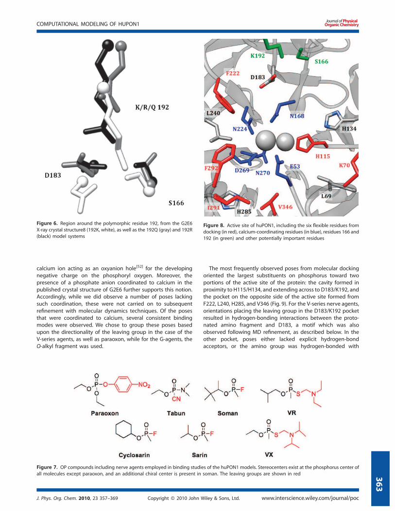
With no other modifications to the putative active site, residue
192 is highly suggestive for playing a role in the considerable
differences in OPase activity between the WT and G2E6 variants
of huPON1.[51] Accordingly, we performed MD simulations on
G2E6 K192R and K192Q mutants, to examine the effects on the
hydrogen bonding and electrostatic interactions in this pocket of
the active site.
Examination of the G2E6 K192R mutant model over the MD
trajectory suggests a similar hydrogen-bonding interaction to
that observed in the G2E6 ‘WT’ (K192) protein model; R192 is
observed to hydrogen bond extensively with both D183 and
N166, with a high degree of occupancy, and the arginine mutant
is also similar to the lysine in G2E6 WT in terms of electrostatic
interactions. In contrast, when Q192 was simulated, the distance
to D183 was too long for hydrogen bonding to occur, leaving
both D183 and S166 available to hydrogen bond with other
neighboring residues (Fig. 6). Accordingly, we postulate that
the differences in activity of huPON1 WT and the G2E6 variant
may result from alterations in the electrostatic and hydrogen-
bonding interactions available in the region around residues 183
and 192.
Molecular binding studies of nerve agents
In the G2E6 model of PON1, molecular docking simulations were
performed to study the binding of a variety of nerve agent
substrates (Fig. 7) into the active site of the protein. This included
a number of known nerve agents, as well as paraoxon. The
assignment of stereochemistry at phosphorus in nerve agent
compounds remains ambiguous, apparently owing to disagree-
ment regarding the priority rules as applied to phosphorus
centers. In this work the phosphoryl (P¼O) bond is given higher
priority than any O-alkyl substituents.
Receptor flexibility was restricted to six residues bracketing the
putative active site region: K70, H115, F222, I291, F292, and V346
(Fig. 8). These residues were chosen based on their proximity to
the active site and their ability to modulate the active site cavity.
To allow for additional sampling of receptor movement, docking
simulations were prepared for eight snapshots of the receptor
model spanning 4 ns at 0.5 ns intervals (from the final 4 ns of the
simulation); each of these models was used for complete docking
simulations with all of the ligands, including stereoisomers and
possible protonation states. For the V-series nerve agents, the
amino fragment was protonated in accordance with the likely pKain serum; for tabun, a similar assignment was notmade due to the
proximity of the amino group to the phosphorus center.
Evaluation of the bound orientations obtained showed a range
of poses spanning the active site region, both coordinated to the
catalytic calcium ion and more localized in the HDL binding
domain on the upper portion of the protein. A consistent theme
in the postulated reaction mechanisms of PON1 is that
coordination of the substrate to the ‘catalytic’ calcium ion is a
prerequisite for hydrolysis, with polarization of the phosphor-
us–oxygen bond lowering the reaction barrier, and with the
Figure 5. (a) E53–H115, (b) H115–H134, and (c) D269–H285 hydrogen-bonding distances over 20 ns of molecular dynamics simulations. The H115
hydrogen bonds have over 90% occupancy over the terminal 10 ns, while the D269–H285 bond is more fluxional
www.interscience.wiley.com/journal/poc Copyright ß 2010 John Wiley & Sons, Ltd. J. Phys. Org. Chem. 2010, 23 357–369
T. T. SANAN ET AL.
362
calcium ion acting as an oxyanion hole[52] for the developing
negative charge on the phosphoryl oxygen. Moreover, the
presence of a phosphate anion coordinated to calcium in the
published crystal structure of G2E6 further supports this notion.
Accordingly, while we did observe a number of poses lacking
such coordination, these were not carried on to subsequent
refinement with molecular dynamics techniques. Of the poses
that were coordinated to calcium, several consistent binding
modes were observed. We chose to group these poses based
upon the directionality of the leaving group in the case of the
V-series agents, as well as paraoxon, while for the G-agents, the
O-alkyl fragment was used.
The most frequently observed poses from molecular docking
oriented the largest substituents on phosphorus toward two
portions of the active site of the protein: the cavity formed in
proximity to H115/H134, and extending across to D183/K192, and
the pocket on the opposite side of the active site formed from
F222, L240, H285, and V346 (Fig. 9). For the V-series nerve agents,
orientations placing the leaving group in the D183/K192 pocket
resulted in hydrogen-bonding interactions between the proto-
nated amino fragment and D183, a motif which was also
observed following MD refinement, as described below. In the
other pocket, poses either lacked explicit hydrogen-bond
acceptors, or the amino group was hydrogen-bonded with
Figure 8. Active site of huPON1, including the six flexible residues from
docking (in red), calcium-coordinating residues (in blue), residues 166 and
192 (in green) and other potentially important residues
Figure 6. Region around the polymorphic residue 192, from the G2E6
X-ray crystal structure8 (192K, white), as well as the 192Q (gray) and 192R
(black) model systems
Figure 7. OP compounds including nerve agents employed in binding studies of the huPON1 models. Stereocenters exist at the phosphorus center of
all molecules except paraoxon, and an additional chiral center is present in soman. The leaving groups are shown in red
J. Phys. Org. Chem. 2010, 23 357–369 Copyright ß 2010 John Wiley & Sons, Ltd. www.interscience.wiley.com/journal/poc
COMPUTATIONAL MODELING OF HUPON1
363

D269 (which also coordinates the active-site calcium). Similar
poses were predicted for binding of paraoxon in the active site,
with the exception that hydrogen bonding with D183 was not
observed; rather, orientation of the p-nitrophenoxide moiety
toward that portion of the active site resulted in either hydrogen
bonding with K192, or p–p stacking with H134. Of the two
principal binding modes, orientation of the thioalkylamino or
p-nitrophenoxide leaving group toward D183/K192 also places it
anti to the D269/H285 pocket, in a pose suitable for hydrolysis by
a nucleophile generated in that vicinity. However, the alternative
pose positioning the leaving group in proximity to L240/F222 is
not expected to be productive for hydrolysis.
For the G-series nerve agents, which lack the large, polar
leaving group that paraoxon and the V-series agents possessed,
the binding modes predicted with docking were somewhat
different. For sarin, the lowest energy docking pose for the PSenantiomer oriented the O-isopropyl fragment in proximity to
F222 and L240, with the leaving group fluoride anti to D269. In
contrast, for the PR enantiomer, the same orientation of the
isopropyl group results in the methyl substituent orienting anti to
D269 (Fig. 9). To orient the leaving group fluoride anti to D269 for
the PS isomer would require placing the O-isopropyl group into
the somewhat more congested active site region around V346.
Similarly, for cyclosarin, the PR isomer also fails to orient the
leaving group anti to calcium in any of the docking poses (refer
Supporting Information). These findings are consistent with the
preference of WT huPON1 for the PS configuration of the G-series
nerve agents, and with prior computational studies on
fluorogenic OP-analogs docked into the PON1 crystal structure.[8]
It should be noted that while poses were obtained from
docking which were consistent with certain mechanistic
hypotheses, the energetic ordering of binding poses was not
sufficiently large to distinguish them beyond the margin of error
of the docking protocol (refer Supporting Information). Energies
for docking poses were generally within 2–3 kcal/mol for the 10
lowest energy binding modes predicted for a given substrate
molecule, including orientations placing the substrate within the
HDL binding domain; furthermore, the energetic ordering for the
two modes was not consistent across the ligand library, even for
the V-series agents. This is, we suspect, a limitation of the docking
protocol when applied to a very open active-site cavity without
the possession of a known binding mode from which to prune
the output of the calculation.
While the energetic output from the docking simulations was
not useful in a quantitative sense as a prediction of binding and
turnover of substrate, the conformational sampling of the active
site was useful in terms of both identifying general binding
modes for substrates, and in suggesting residues for which
mutations might alter the substrate specificity of the protein. A
tally of the ligand–receptor contacts by distance suggested a
number of residues in the active site that are in proximity to the
calcium ion, as well as in the peripheral regions, which could be
involved in binding specificity for organophosphorus agents.
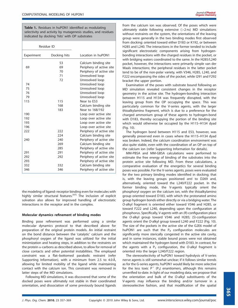
Interestingly, a number of residues from this list have already
been identified as modulating substrate specificity in mutagen-
esis studies, which further supports the notion that the docked
orientations are sampling valid substrate binding modes, despite
the coarse nature of the methodology (Table 1). To improve the
treatment of receptor flexibility, and to obtain more reliable
binding energies for comparison of poses, we employed
molecular dynamics simulations to study the ligand–receptor
complexes. This methodology has been successfully employed in
Figure 9. Representative binding modes for V-agents ((PR)-VR, top) and G-agents (PS and PR sarin, bottom) in the G2E6 model active site observed from
molecular docking
www.interscience.wiley.com/journal/poc Copyright ß 2010 John Wiley & Sons, Ltd. J. Phys. Org. Chem. 2010, 23 357–369
T. T. SANAN ET AL.
364
the modeling of ligand–receptor binding even for molecules with
highly similar structural features.[47] The inclusion of explicit
solvation also allows for improved handling of electrostatic
interactions in the receptor and in the complex.
Molecular dynamics refinement of binding modes
Binding pose refinement was performed using a similar
molecular dynamics protocol to that described above for
preparation of the original protein models. An initial restraint
on the bond distance between the ‘catalytic’ calcium and the
phosphoryl oxygen of the ligand was utilized for the initial
minimization and heating steps, in addition to the restraints on
the protein a-carbons as described above, to allow for removal of
close contacts and other potential instabilities. The employed
constraint was a flat–bottomed parabolic restraint (refer
Supporting Information), with a minimum from 2.5 to 4.0 A,
allowing for limited mobility of the ligand while maintaining
contact with the calcium ion. This constraint was removed in
latter steps of the MD simulations.
Following MD simulations, it was discovered that some of the
docked poses were ultimately not stable in their coordinated
orientation, and dissociation of some previously bound ligands
from the calcium ion was observed. Of the poses which were
ultimately stable following extensive (>2 ns) MD simulations
without restraints on the system, the orientations of the leaving
group were generally in the two binding modes first observed
from docking: oriented toward either D183 or K192, or between
H285 and L240. The interactions in the former tended to include
significant electrostatic components arising from hydrogen-
bonding interactions with the charged residues in the pocket, or
with bridging waters coordinated to the same. In the H285/L240
pocket, however, the interactions were primarily simple van der
Waals interactions; the peripheral residues in the latter pocket
tend to be of the non-polar variety, with V346, H285, L240, and
F222 encompassing the sides of the pocket, while I291 and F292
bracket the upper portion.
Examination of the poses with substrate bound following an
MD simulation revealed consistent changes in the receptor
geometry in the active site. The hydrogen-bonding interaction
between H115 and H134 was frequently disrupted, with the
leaving group from the OP occupying the space. This was
particularly common for the V-series agents, with the larger
thioalkylamino fragment, which is due to a preference for the
charged ammonium group of these agents to hydrogen-bond
with D183, thereby occupying the portion of the binding site
which would otherwise be occupied by the H115–H134 dyad
(Fig. 10).
The hydrogen bond between H115 and E53, however, was
generally preserved even in cases where the H115–H134 dyad
was broken. Indeed, the calcium coordination environment was
also quite stable, even with the coordination of an OP on top of
the calcium ion (refer Supporting Information for details).
MM-PBSA and MM-GBSA calculations were performed to
estimate the free energy of binding of the substrates into the
protein active site following MD. From these calculations, a
comparative evaluation of the energetics for several binding
poses was possible. For the V-series agents, poses were evaluated
for the two primary binding modes identified in docking; that
is, with the leaving groups positioned anti to D269 and,
alternatively, oriented toward the L240/F222 pocket. In the
former binding mode, the V-agents typically orient the
phosphoryl oxygen on the calcium ion, with the thioalkylamino
group oriented toward D183, with which the protonated amino
group hydrogen bonds either directly or via a bridging water. The
O-alkyl fragment is oriented either toward V346 and H285, or
toward F222 and L240, depending upon the configuration at
phosphorus. Specifically, V-agents with an (R)-configuration place
the O-alkyl group toward V346 and H285; (S)-configuration
agents orient the O-alkyl group toward L240 and F222 (Fig. 10).
The size of the pockets in the active site of the G2E6 model of
huPON1 are such that the PR configuration molecules are
significantly more sterically congested in the active site cavity,
and in some instances, stable bound poses were not obtained
which maintained the hydrogen bond with D183. In contrast, for
the agents with a PS configuration, the O-alkyl fragment is
oriented into the larger L240/F222 region.
The stereoselectivity of huPON1 toward hydrolysis of V-series
nerve agents is still somewhat unclear; if it follows similar trends
as for the G-series agents, huPON1 would likely be more selective
for the less toxic Pþ (PS) enantiomers, although this remains
unverified to-date. In light of our modeling data, we propose that
the orientation and bulk of the O-alkyl substituents of the
V-agents may influence the binding and/or turnover in a
stereoselective fashion, and that modification of the spatial
Table 1. Residues in huPON1 identified as modulating
selectivity and activity by mutagenesis studies, and residues
indicated by docking ‘hits’ with OP substrates
Residue ID
Location in huPON1Experiment Docking hits
53 Calcium binding site
69 69 Periphery of active site
70 Periphery of active site
71 Unresolved loop
72 Unresolved loop
74 Unresolved loop
75 Unresolved loop
76 Unresolved loop
79 Unresolved loop
115 115 Near to E53
168 Calcium binding site
183 Near to 168/192
190 Loop over active site
192 Loop over active site
193 Loop over active site
196 Loop over active site
222 222 Periphery of active site
224 Calcium binding site
240 240 Periphery of active site
269 Calcium binding site
285 Periphery of active site
291 291 Periphery of active site
292 292 Periphery of active site
293 Periphery of active site
332 332 Calcium binding site
346 346 Periphery of active site
J. Phys. Org. Chem. 2010, 23 357–369 Copyright ß 2010 John Wiley & Sons, Ltd. www.interscience.wiley.com/journal/poc
COMPUTATIONAL MODELING OF HUPON1
365

constraints in proximity to L69 and V346 might modulate the
turnover of the PR enantiomers. The lowest energy binding
modes were predicted to orient the O-alkyl fragments in
proximity to D269/E53, which suggests that interactions with
and/or occlusion of the approaching nucleophile, be it one of the
carboxylates or a coordinated water molecule, by the O-alkyl
fragments of the V-agents could be impeding turnover and
reducing kcat. The reader should note, however, that these
predictions are based only on binding information. This is
consistent with both the direct hydrolysis of substrate by D269, as
well as the general-base hydrolysis mechanisms for huPON1.
Interestingly, work by Furlong et al.[50] on huPON1 ‘status’
suggests that the polymorphic residue 192, which in G2E6 is
modified to lysine from arginine or glutamine in the WT human
protein, significantly alters the activity of PON1 for hydrolysis of
OP pesticides. In the context of the study, it was found that 192R
was more protective than 192Q for exposure to paraoxon. In light
of the predicted binding modes for the V-series agents orienting
the protonated amino fragment in proximity to D183 and K192,
we propose a molecular-level rationalization for this modulation:
competition for the hydrogen bond to D183, which is reduced in
the 192Q polymorph. In contrast, the lowest energy binding
modes for paraoxon placed the p-nitrophenoxide group in
proximity to K192 itself, hydrogen bonding directly with the
protonated amino group, and with an alternative pose with
similar energy orienting the leaving group parallel to H134 in a
p-stacking mode (Fig. 11). An alternative theory regarding the
K192mutation, suggesting that it mitigates charge repulsion with
D183 via hydrogen bonding, is also consistent with our modeling
results.[53]
These results suggest the role of residue 192 in the active site
for substrates with large, polar/charged groups may be related to
orientation of the substrate prior to hydrolysis. However, for the
G-series nerve agents, which lack a large polar leaving group,
there are no clear interactions with this portion of the active
(Fig. 11). Instead, the primary constraints on binding for these
agents are the orientation of the large non-polar peripheral
groups, which generally interact with non-polar residues in the
active site, such as L69, V346, F222, and L240, some of which have
been identified as modulating the stereoselectivity of the
protein.[54] For G-series nerve agents, these residues influence
the fit of substrate into the active site and likely contribute to the
stereoselectivity of the protein. Based on our binding studies, it
appears that orientations which allow the bulky substituents to
orient in proximity to L240 and F222 are favorable, so it is the
resulting orientation of the leaving group which influences
turnover. For the PS agents, orientation of the leaving group anti
to the D269 pocket is possible in these poses, whereas the PRagents place the leaving group in the opposite direction in such a
pose. While stable, bound orientations were identified for PRG-series nerve agents that placed the O-alkyl substituents in
proximity to L69 and V346 (and thus, with the leaving groups anti
to the D269 region), these poses tended to be higher in energy
than the more favorable mode by between 3 and 5 kcal/mol, or
slightly higher than the margin of error (refer Supporting
Information). Given that huPON1 does turn over both enantio-
mers of cyclosarin, as well as all four diastereomers of soman, the
observation of ‘productive’ binding modes with the leaving
group anti to the D269 region for the PR isomers is not
inconsistent with experiment.[55]
It should be noted that one interaction which has been
proposed between receptor and substrate in huPON1 is the
‘capping’ of the active site by residue Y71, found on the flexible
surface loop that was unresolved in the protein crystal
structure.[53] This loop, while highly flexible in our simulations,
was not universally observed to participate in such an interaction
in bound poses. Some poses of bound agent, particularly for the
V-agents, did show capping of the bound substrate by Y71 in a
manner similar to that reported (refer Supporting Information).
It remains possible that the Y71 interaction is vital for OP
binding, or alternatively, that Y71 is involved in substrate mobility
into the active site; however, suchmobility was not observed over
the time scales sampled in this study, and additional simulations
are necessary to determine if this is the case.
An alternative method for examining the binding of substrates
to huPON1 involves the use of umbrella sampling[56] to estimate
the potential energy surface for removal of substrate from
coordination to the calcium ion, or from the active site overall.[57]
In this methodology, we utilized a conformational sampling
protocol in conjunction with parallel, constrainedMD trajectories,
Figure 10. Minimum energy poses for (a) (R)- and (b) (S)-VX bound in the active site of the G2E6 protein model. The colored lobes indicate the relative
sizes and orientations of the leaving group (red) and O-alkyl and methyl substituents
www.interscience.wiley.com/journal/poc Copyright ß 2010 John Wiley & Sons, Ltd. J. Phys. Org. Chem. 2010, 23 357–369
T. T. SANAN ET AL.
366

to estimate a probability distribution for a given geometric
parameter in the active site, from which the free energy can be
extracted. In the context of our study, the calcium–phosphoryl
oxygen bond coordinate was chosen for sampling, starting from
the bound geometries obtained from MD refinement of the
V-series agents, as described above. The output from several such
runs are shown in Fig. 12, for both (R)- and (S)-configuration VX.
The minimum energy bond distance in both cases was estimated
to be 2.7 A, which is somewhat longer than the calcium–
phosphate distance of 2.2 A observed in the X-ray crystal
structure of G2E6,[7] but which is consistent with the stable,
bound orientations calculated above. A plateau observed in the
Figure 11. Stable, bound poses derived from MD simulations for (a) paraoxon, and (b) the PS isomers of sarin and cyclosarin in the G2E6 model
Figure 12. Potential energy surfaces for loss of coordination to calcium calculated for (PR)–VR (left), and (PS)–VX (right), calculated using umbrella
sampling methods from bound orientations
J. Phys. Org. Chem. 2010, 23 357–369 Copyright ß 2010 John Wiley & Sons, Ltd. www.interscience.wiley.com/journal/poc
COMPUTATIONAL MODELING OF HUPON1
367
potential surface for the S-configuration agents corresponds to a
movement of the phosphoryl oxygen away from calcium toward
the calcium-coordinating residue N168, which is able to form a
hydrogen bond and to stabilize the partially dissociated OPs. This
may represent an intermediate stage in the substrate association/
dissociation potential energy surface, andmore simulations are in
progress to fully explore this possibility.
Interestingly, a similar analysis on poses which showed a lack of
stability in calcium coordination found no minima at �2.7 A
corresponding to the bound state, suggesting that this
methodology is also useful in validating the stability of bound
substrate to the metal ion. When applied to the V-series nerve
agents studied herein, the correlation between poses that were
stably bound to calcium over the course of MD and those with
minima in the dissociation potentials for bound poses was
excellent (refer Supporting Information).
DISCUSSION
Human paraoxonase 1 (huPON1) is a protein that promiscuously
catalyzes the hydrolysis of a variety of electrophilic substrates. It is
tolerant of a variety of alkyl and aromatic esters and coumarin
substrates, as well as organophosphorus compounds. In this
study, we have attempted to model the protein using
computational chemistry techniques to both gain insight into
the structure and function of the enzyme as well as provide
guidance for the development of novel protein mutants via
rational design.
We have produced a structural model of the G2E6 variant of
huPON1, including a loop above the putative active site, which
was unresolved in the published X-ray crystal structure, as part of
a cooperative program aimed at enhancing the catalytic function
of the protein for use as a bioscavenger. In the active site itself, the
largest change from the published structure is in the hydrogen
bonding of H115, which migrates considerably closer to E53, and
maintains a tight hydrogen bond; this interaction alters the
distance between these residues from �4 A in the crystal
structure to 2.5 A in the G2E6 computational model. Other than
this divergence, the differences between the G2E6 computational
model and the published X-ray structure6 are in the surface
loops, and these changes do not significantly alter the active site
geometry; furthermore, these surface loops are likely highly
flexible in the absence of HDL association.
From the molecular docking studies, the observed inter-
actions between residues 183, 192, and polar groups with the
leaving groups for the bound OP substrate suggest a potential
role of these residues in substrate binding and orientation in
the active site, which is consistent with the available
experimental data. Additional work is necessary, both in
developing new protein mutants from which additional kinetic
information can be obtained, and in terms of additional
sampling of the conformational motion of these residues,
before the exact nature of the interactions can be conclusively
determined. This is particularly true given the lack of a
reference orientation of substrate in any experimentally
obtained structure of PON1 to date.
The lowest energy binding modes observed for V-series
agents and paraoxon orient the leaving group anti to the
carboxylate residues which coordinate the catalytic calcium,
and this observation supports several of the proposed reaction
mechanisms, above. In this pose, direct hydrolysis of the OP
by one of the two carboxylates is possible, with average
distances on the order of 3.5–4.5 A from the coordinated OP
to D269 and/or E53. However, for E53 to act as a nucleophile,
attack would be necessarily precluded by loss of a hydrogen
bond with H115, which is only rarely observed in the MD
trajectories of either the free or ligand-bound proteins.
The interaction between H285 and D269 is significantly more
labile, which suggests that the latter residue might be
more available for attack; however, the nucleophilicity of an
aspartate coordinated to a calcium ion is likely significantly
diminished. A third possibility is the attack by an activated
water molecule, possibly as part of a catalytic dyad or triad in
the active site. The observation of coordinated water molecules
in proximity to both D269 and E53 in ligand-bound complexes
supports the possibility that deprotonation of water by either
one of the carboxylates, or possibly the basic nitrogen of H285
might be able to generate hydroxide in a suitable orientation
for hydrolysis of substrate. The lack of detectible ‘aging’ of
huPON1 also supports this notion, as a general base
mechanism would not produce a covalent protein–ligand
conjugate. However, molecular dynamics simulations alone are
insufficient to probe this possibility. To examine reaction
pathways explicitly, and to obtain energetic parameters to
compare and contrast them, hybrid quantum mechanical/
molecular mechanical (QM/MM) simulations are in progress;
the primary complication in this approach is the large number
of degrees of freedom in the active site, as well as the numerous
potential geometries for water clusters. The results, however,
will reduce some of the ambiguity present in the literature with
regard to the mechanism of huPON1 hydrolysis of OPs.
The binding modes identified also correlate well with
published results on mutations that influence the selectivity of
huPON1. Modification of peripheral active site residues such as
L69, H115, F222, and V346 have all been identified in the course
of directed evolution by Tawfik et al.[54] to modify the selectivity
of the protein, and these residues are generally in proximity to
the peripheral groups of ligands bound to the huPON1 model.
While the exact molecular-level role of these mutations on
substrate binding and turnover remains speculative at this stage,
the development of improved models for the protein opens the
door for development of in silicomutants with the potential to both
calibrate and refine the models and potentially drive the
development of protein mutants with altered selectivity and activity.
SUPPORTING INFORMATION
Additional details regarding model preparation, molecular dock-
ing results, molecular dynamics simulations are included in the
supporting information, available in theonlineversionof thepaper.
Acknowledgements
We gratefully acknowledge financial support of this research by
the National Institutes of Health (U54-NS058183). Generous com-
putational resources have been provided by the Ohio Super-
computer Center. We also acknowledge fruitful and insightful
discussions with Professor Thomas Magliery (OSU). The opinions
or assertions contained herein are the private views of the
authors and are not to be construed as official or as reflecting
the views of the United States Army or the Department of
Defense.
www.interscience.wiley.com/journal/poc Copyright ß 2010 John Wiley & Sons, Ltd. J. Phys. Org. Chem. 2010, 23 357–369
T. T. SANAN ET AL.
368

REFERENCES
[1] C. E. Furlong, in Paraoxonases: An Historical Perspective, Proteinsand Cell Regulation, Vol. 6 (Eds: B. Mackness M. Mackness M. AviramG. Paragh), Springer, NY, 2008, pp. 3–31.
[2] O. Khersonsky, D. S. Tawfik, Chem. Bio. Chem. 2006. 7, 49–53.[3] D. T. Yeung, D. E. Lenz, D. M. Cerasoli, in The Paraoxonases: Their Role in
Disease, Development and Xenobiotic Metabolism, Proteins and CellRegulation, Vol. 6 (Eds: B. Mackness, M. Mackness, M. Aviram, G.Paragh), Springer, NY, 2008, pp. 151–170.
[4] D. E. Lenz, D. T. Yeung, R. J. Smith, R. E. Sweeney, L. A. Lumley, D. E.Cerasoli, Toxicology 2007, 233, 31–39.
[5] D. E. Lenz, A. A. Brimfield, L. A. Cook, The development of immu-noassays for detection of chemical warfare agents, in Developmentand Applications of Immunoassays for Environmental Analysis (Eds: D.Aga, E. M. Thurman), ACS Books, Washington, DC, 1997, pp. 77–86
[6] D. T. Yeung, D. Josse, J. D. Nicholson, A. Khanal, C. W. McAndrew, B. J.Bahnson, D. E. Lenz, D. M. Cerasoli, Biochim. Biophys. Acta 2004, 1702,67–77.
[7] M. Harel, A. Aharoni, L. Gaidukov, B. Brumshtein, O. Khersonsky, R.Meged, H. Dvir, R. B. G. Ravelli, A. McCarthy, L. Toker, I. Silman, J. L.Sussman, D. S. Tawfik, Nat. Struct. Mol. Biol. 2004, 11, 412–419.
[8] M. Blum, C. M. Timperley, G. R. Williams, H. Thiermann, F. Worek,Biochemistry 2008, 47, 5216–5224.
[9] M. Blum, A. Koglin, R. Ruterjans, B. Schoenborn, P. Langan, J. C. H.Chen, Acta Crystallogr. 2007, 63, 42–45.
[10] F. Worek, P. Eyer, L. Szinicz, Arch. Toxicol. 1998, 72, 996–998.[11] A. Saxena, B. P. Doctor, D. M. Maxwell, D. E. Lenz, Z. Radic, P. Taylor,
Biochem. Biophys. Rsh. Commun. 1993, 197, 343–349.[12] C. D. Fleming, C. C. Edwards, S. D. Kirby, D. M. Maxwell, P. M. Potter, D.
M. Cerasoli, M. R. Redinbo, Biochemistry 2007, 46, 5063–5071.[13] D. Rothlisberger, O. Khersonsky, A. M. Wollacott, L. Jiang, J. DeChan-
cie, J. Betker, J. L. Gallaher, E. A. Althoff, A. Zanghellini, O. Dym, S.Albeck, K. N. Houk, D. S. Tawfik, D. Baker, Nature 2008, 453, 190–197.
[14] K. N. Houk, P. H. Cheong, Nature 2008, 455, 309–313.[15] L. Jiang, E. A. Althoff, F. R. Clemente, L. Doyle, D. Rothlisberger, A.
Zanghellini, J. L. Gallaher, J. L. Betker, F. Tanaka, C. F. Barbas, III, D.Hilvert, K. N. Houk, B. L. Stoddard, D. Baker, Science 2008, 319,1387–1391.
[16] R. C. Sorenson, C. L. Bisgaier, M. Aviram, C. Hsu, S. Billecke, B. N. La Du,Arterioscler. Thromb. Vasc. Biol. 1999, 19, 2214–2225.
[17] T. J. Dolinsky, J. E. Nielsen, J. A. McCammon, N. A. Baker, Nucleic AcidsRes. 2004, 32, 665–667.
[18] Y. Duan, C. Wu, S. Chowdhury, M. C. Lee, G. Xiong, W. Zhang, R. Yang, P.Cieplak, R. Luo, T. Lee, J. Caldwell, J. Wang, P. Kollman, J. Comput.Chem. 2003, 24, 1999–2012.
[19] J. Wang, R. M. Wolf, J. W. Caldwell, P. A. Kollamn, D. A. Case, J. Comput.Chem. 2004, 25, 1157–1174.
[20] J. Aqvist, J. Phys. Chem. 1990, 94, 8021–8024.[21] M. Blum, F. Lohr, A. Richardt, H. Ruterjans, G. C. Chen, J. Am. Chem. Soc.
2006, 128, 12750–12757.[22] M. W. Mahoney, W. L. Jorgensen, J. Chem. Phys. 2000, 112, 8910–8922.[23] W. L. Jorgensen, J. Chandrasekhar, J. D. Madura, R. W. Impey, M. L.
Klein, J. Chem. Phys. 1983, 79, 926–935.[24] D. A. Case, T. A. Darden, T. E. Cheatham, III, C. L. Simmerling, J. Wang,
R. E. Duke, R. Luo, K. M. Merz, D. A. Pearlman, M. Crowley, R. C. Walker,W. Zhang, B. Wang, S. Hayik, A. Roitberg, G. Seabra, K. F. Wong, F.Paesani, X. Wu, S. Brozell, V. Tsui, H. Gohlke, L. Yang, C. Tan, J. Mongan,V. Hornak, G. Cui, P. Beroza, D. H. Mathews, C. Schafmeister, W. S. Ross,P. A. Kollman, (2006), AMBER 9,University of California, San Francisco.
[25] D. A. Case, T. A. Darden, T. E. Cheatham, III, C. L. Simmerling, J. Wang,R. E. Duke, R. Luo, K. M. Merz, R. C. Walker, W. Zhang, B. Wang, S. Hayik,A. Roitberg, G. Seabra, K. F. Wong, F. Paesani, X. Wu, S. Brozell, V. Tsui,H. Gohlke, L. Yang, C. Tan, J. Mongan, V. Hornak, G. Cui, P. Beroza, D. H.Mathews, M. G. Seetin, C. Sagui, V. Babin, W. S. Schafmeister, C. Ross, P.A. Kollman, (2008), AMBER 10,University of California, San Francisco.
[26] H. J. C. Berendsen, J. P. M. Postma, W. F. van Gunsteren, A. DiNola, J. R.Haak, J. Chem. Phys. 1984, 81, 3684–3690.
[27] S. C. Harvey, R. K. Tan, T. E. Cheatham, III, J. Comput. Chem. 1998, 19,726–740.
[28] R. Chachra, R. C. Rizzo, J. Chem. Theory. Comput. 2008, 4, 1526–1540.[29] S. Miyamoto, P. A. Kollman, J. Comput. Chem. 1992, 13, 952–962.[30] A. Toukmaji, C. Sagui, J. Board, T. Darden, J. Chem. Phys. 2000, 113,
10913–10927.[31] G. M. Morris, D. S. Goodsell, R. S. Halliday, R. Huey, W. E. Hart, R. K.
Belew, A. J. Olson, J. Comput. Chem. 1998, 19, 1639–1662.[32] C. M. Breneman, K. B. Wiberg, J. Comput. Chem. 1990, 11, 361–373.[33] A. D. Becke, J. Chem. Phys. 1993, 98, 5648–5656.[34] C. Lee, W. Yang, R. G. Parr, Phys. Rev. B 1988, 37, 785–789.[35] J. Labanowski, in Density Functional Methods in Chemistry, Spring-
er-Verlag, Heidelberg, 1991.[36] R. G. Parr, Y. Weitao, in Density-Functional Theory in Atoms and
Molecules, Oxford University Press, New York, 1989.[37] M. J. Frisch, G. W. Trucks, H. B. Schlegel, G. E. Scuseria, M. A. Robb, J. R.
Cheeseman, J. A. Montgomery, T. Vreven, Jr., K. N. Kudin, J. C. Burant,J. M. Millam, S. S. Iyengar, J. Tomasi, V. Barone, B. Mennucci, M. Cossi,G. Scalmani, N. Rega, G. A. Petersson, H. Nakatsuji, M. Hada, M. Ehara,K. Toyota, R. Fukuda, J. Hasegawa, M. Ishida, Y. Nakajima, O. Honda, O.Kitao, H. Nakai, M. Klene, X. Li, J. E. Knox, H. P. Hratchian, J. B. Cross, C.Adamo, J. Jaramillo, R. Gomperts, R. E. Stratmann, O. Yazyev, A. J.Austin, R. Cammi, C. Pomelli, J. W. Ochterski, P. Y. Ayala, K. Morokuma,G. A. Voth, P. Salvador, J. J. Dannenberg, V. G. Zakrzewski, S. Dapprich,A. D. Daniels, M. C. Strain, M. C. Farkas, D. K. Malick, A. D. Rabuck, K.Raghavachari, J. B. Foresman, J. V. Ortiz, Q. Cui, A. G. Baboul, S.Clifford, J. Cioslowski, B. B. Stefanov, G. Liu, A. Liashenko, P. Piskorz, I.Komaromi, R. L. Martin, D. J. Fox, T. Keith, M. A. Al-Laham, C. Y. Peng, A.Nanayakkara, M. Challacombe, P. M. W. Gill, B. Johnson, W. Chen, M.W. Wong, C. Gonzalez, J. A. Pople, Gaussian 03, Revision B.04,Gaussian, Inc., Pittsburgh PA, 2003.
[38] P. J. Stevens, F. J. Devlin, C. F. Chablowski, M. F. Frisch, J. Phys. Chem.1994, 98, 11623–11627.
[39] A. D. Becke, Phys. Rev. A 1988, 38, 3098–3100.[40] A. D. Becke, J. Chem. Phys. 1992, 96, 2155–2160.[41] A. D. Becke, J. Chem. Phys. 1992, 97, 9173–9177.[42] F. D. Proft, J. M. L. Martin, P. Geerlings, Chem. Phys. Lett. 1996, 250,
393–401.[43] F. Martin, H. Zipse, J. Comput. Chem. 2004, 26, 97–105.[44] R. Luo, L. David, M. K. Gilson, J. Comput. Chem. 2002, 23, 1244–1253.[45] J. Weiser, P. S. Shenkin, W. C. Still, J. Comput. Chem. 1999, 20,
217–230.[46] M. F. Sanner, A. J. Olson, J. Spehner, Biopolymers 1996, 38, 305–320.[47] R. C. Rizzo, T. Aynechi, D. A. Case, I. D. Kuntz, J. Chem. Theory. Comput.
2006, 2, 128–139.[48] B. Kuhn, P. Gerber, T. Schulz-Gasch, M. Stahl, J. Med. Chem. 2005, 48,
4040–4048.[49] S. Kumar, D. Bouzida, R. H. Swendsen, P. A. Kollman, J. M. Rosenberg, J.
Comput. Chem. 1992, 13, 1011–1021.[50] C. E. Furlong, T. B. Cole, G. P. Jarvik, C. Pettan-Brewer, G. K. Geiss, R. J.
Richter, D. M. Shih, A. D. Tward, A. J. Lusis, L. G. Costa, Neuro. Toxicol.2005, 26, 651–659.
[51] T. C. Otto, C. K. Harsch, D. T. Yeung, T. J. Magliery, D. M. Cerasoli, D. E.Lenz, Biochemistry 2009, 48, 10416–10422.
[52] O. Khersonsky, D. S. Tawfik, J. Biol. Chem. 2006, 281, 7649–7656.[53] X. Hu, X. Jiang, D. E. Lenz, D. M. Cerasoli, A. Wallqvist, Proteins 2009,
75, 486–498.[54] G. Amitai, L. Gaidukov, R. Adani, S. Yishay, G. Yacov, M. Kushnir, S.
Teitlboim, M. Lindenbaum, P. Bel, O. Khersonsky, D. S. Tawfik, H.Meshulam, FEBS J. 2006, 273, 1906–1919.
[55] D. T. Yeung, J. R. Smith, R. E. Sweeny, D. E. Lenz, D. M. Cerasoli, FEBS J.2007, 274, 1183–1191.
[56] G. M. Torrier, J. P. Valleau, Chem. Phys. Lett. 1974, 28, 578–581.[57] J. Kottalam, D. A. Case, J. Am. Chem. Soc. 1988, 110, 7690–7697.
J. Phys. Org. Chem. 2010, 23 357–369 Copyright ß 2010 John Wiley & Sons, Ltd. www.interscience.wiley.com/journal/poc
COMPUTATIONAL MODELING OF HUPON1
369


![Paraoxonase: The boon against oxidative stress and lipid...may play a role in its pathogenesis, low PON1 activity could be an independent risk factor [2]. Paraoxonase activity is in-versely](https://img.dokumen.tips/doc/110x75/5f8d3577e0ed9638c7379069/paraoxonase-the-boon-against-oxidative-stress-and-lipid-may-play-a-role-in.jpg)


![Research Paper Decreases in Paraoxonase-1 Activities ... · Peroxidation of lipids can be limited by the activity of paraoxonase-1 (PON-1) [15–17]. PON-1 is a HDL-bound and calcium-dependent](https://img.dokumen.tips/doc/110x75/5eb9f8fcc42d18167c21f8ff/research-paper-decreases-in-paraoxonase-1-activities-peroxidation-of-lipids.jpg)