Embed Size (px)
Citation preview

51Biomed Pap Med Fac Univ Palacky Olomouc Czech Repub. 2011 Mar; 155(1):51–54. DOI 10.5507/bp.2011.019© E. Sovova, M. Sova, M. Hobzova, M. Kaminek, V. Kolek, M. Taborsky, J. Ostransky
COMPLICATED COURSE OF ISCHEMIC HEART DISEASE IN A PATIENT WITH OBSTRUCTIVE SLEEP APNEA
Eliska Sovovaa*, Milan Sovab, Milada Hobzovac, Milan Kaminekd, Vitezslav Kolekc, Milos Taborskya, Jiri Ostranskya
a 1st Department of Internal Medicine, Faculty of Medicine and Dentistry, Palacky University Olomouc, Czech Republicb Faculty of Medicine and Dentistry, Palacky University Olomoucc Department of Respiratory Medicine, Faculty of Medicine and Dentistry, Palacky University Olomoucd Department of Nuclear Medicine, Faculty of Medicine and Dentistry, Palacky University OlomoucE-mail: [email protected]
Received: November 1, 2010; Accepted with revision: February 10, 2011
Key words: Obstructive sleep apnea/Myocardial infarction/Complicated course/In stent restenosis
Background. Obstructive sleep apnea (OSA) can activate pathological routes which can lead to insulin resistance, development of atherosclerosis and hypertension. The combination of hypertension and OSA has an additive effect on the development of atherosclerosis. As a number of studies have revealed, that the incidence of OSA in patients with myocardial infarction is likely to be high.
Methods and results. We present a patient with acute myocardial infarction and no classical coronary artery disease risk factors: non-smoker, normal blood pressure, normal total and low-density lipoprotein cholesterol levels, borderline high-density lipoprotein cholesterol level, with good physical activity, no diabetes mellitus, no abdominal obesity, a negative family history. The only risk factor was untreated obstructive sleep apnea. The course of disease was complicated by subsequent in-stent restenosis and progression of atherosclerotic plaques, which led to the need for acute coronary artery bypass graft surgery complicated by consecutive in-anastomosis stenosis despite maximum cardiovascular therapy. One year of continuous positive airway pressure treatment was needed to stabilize his health condition, which is now stable for up to two years.
Conclusions. Given the complicated course of ischemic heart disease in patients with OSA, we believe that OSA diagnosis would be advisable each time these patients with symptoms of myocardial infarction, ischemic heart disease and OSA are examined. Even more important, however, is proper treatment of the OSA when it is present.
INTRODUCTION
The Sleep Heart Health Study (SHHS) has examined the cross sectional association between sleep-disordered breathing and self reported cardiovascular disease (CVD) and the multivariable-adjusted relative odds (95% CI) of prevalent (self reported) CVD were 1.42 (1.13–1.78) for the fourth quartiles1. Sleep-disordered breathing was as-sociated more strongly with self reported heart failure (2.38), stroke (1.58), than with self reported CVD. The SHHS has shown, that in a group of patients free of self reported CVD in the begin of the study moderate levels of sleep-disordered breathing were common with a median Respiratory Disturbance Index (RDI) of 4.0. The risk for CVD events increased in future follow-up2. Mooe et al.3 concluded in the study of 408 patients with CVD, that sleep-disordered breathing is associated with a worse long-term prognosis and has an independent association with cerebrovascular events.
In a recent study Gottlieb et al.4 presented, that OSA was a significant predictor of incident coronary heart dis-ease in men ≤70 years of age. Among men 40–70 years old, those with AHI≥30 were 68% more likely to develop coronary heart disease than those with AHI <5.
CASE PRESENTATION
A 54-year-old Caucasian man was admitted on 29th March 2007 to the cardiology clinic due to serious chest pain. Vertebrogenic pain syndrome, hepatitis B in 1977 (he is HBsAg positive) and surgery for Achilles tendon were mentioned in the patient’s medical history. He is a lifelong non-smoker. He was evaluated in a sleep laborato-ry several years ago where an obstructive sleep apnea was diagnosed. He was not treated or observed. Family his-tory: mother died age 40 as a result of a rheumatic mitral valve disease; father died age 80 due to liver carcinoma; sister and children are healthy. Medication: silymarin.
Physical examination (PE): blood pressure (BP) 150/80, heart rate (HR) 82, weight 97 kg, height 187 cm, body mass index 27.7 and no other abnormalities.
Electrocardiogram: sinus rhythm, straightened ST I, II, V 5, 6, depression ST V 1–3.
Laboratory results: normal potassium, sodium, chlo-rides, alanine amino transferase, aspartate amino trans-ferase, gamma glutamyl transferase, glycaemia, total cholesterol, high-density lipoprotein (HDL), triglycerides, low-density lipoprotein (LDL). Out of range: creatine kinase (CK) 3.57 μ k at/l, CK/CK Mb 14% (norm.<6%), troponin T 0.08 μg/l (norm. 0.03), bilirubin 38 μmol/l.

52 E. Sovova, M. Sova, M. Hobzova, M. Kaminek, V. Kolek, M. Taborsky, J. Ostransky
A coronarography was carried out and this revealed the following: occluded ramus diagonalis (RD1), 95% os-tial stenosis (RD2), 99% critical stenosis ramus circum-flexus (RCX) (Fig. 1), arteria coronaria dextra (ACD) without stenosis. Percutaneous coronary intervention (PCI]) was performed on RD1 with a stent coroflex 3.0 x 13 mm, (TIMI flow 3), PCI on RCX with a stent coroflex 3.0 x 13 mm (a small residue of stenosis up to 15%).
The patient was discharged in good health on 4th April 2007. His medication included beta blocker (metopro-lol), clopidogrel, aspirin, ACE inhibitor (ramipril), statin (atorvastatin), silymarin.
He was admitted to the respiratory clinic on 16th April 2007 where he underwent a polysomnography. PE: BP, HR in normal range. Laboratory values were normal (ASTRUP, glycaemia, lipids). The Epworth sleepiness scale was 4. The polysomnography revealed 27.4 oxygen desaturations per hour, he spent 3.14% of sleep in oxygen saturation less then 90% SaO2, apnea-hypopnea index (AHI) 30.45. He did not meet the criteria for continuous positive airway pressure (CPAP) treatment set by the health insurance company. He was scheduled for reevaluation.
A follow-up polysomnography took place on 28th May 2007 and revealed 27.0 oxygen desaturation per h, 11% of the time in the oxygen saturation less then 90% SaO2, AHI 30. A request for a CPAP device for the health insurance company was filled out.
An echocardiographic examination was performed on 31st May 2007. It revealed hypokinesis of the lateral wall and the worsening of ejection fraction (EF) to 40%.
Fig. 1. Coronarography, 99% critical stenosis ramus cir-cumflexus.
Fig. 2. SPECT. Hypokinesis laterally and inferoapically, the reversible defect (medially severe ischemia).
The patient underwent single-photon-emission com-puted tomography (SPECT) on June 2007 which showed hypokinesis laterally and inferoapically, the resting EF was 45%, the defect was reversible and assessed as medi-ally severe ischemia (Fig. 2).
53Complicated course of ischemic heart disease in a patient with obstructive sleep apnea
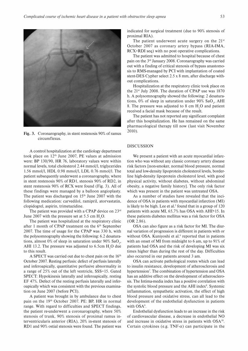
A control hospitalization at the cardiology department took place on 12th June 2007, PE values at admission were: BP 130/90, HR 76, laboratory values were within normal levels, total cholesterol 2.44 mmol/l, triglycerides 1.56 mmol/l, HDL 0.98 mmol/l, LDL 0.76 mmol/l. The patient subsequently underwent a coronarography, where in stent restenosis 90% of RD1, stenosis 90% of RD2, in stent restenosis 90% of RCX were found (Fig. 3). All of these findings were managed by a balloon angioplasty. The patient was discharged on 15th June 2007 with the following medication: carvedilol, ramipril, atorvastatin, clopidogrel, aspirin, trimetazidine.
The patient was provided with a CPAP device on 23rd June 2007 with the pressure set at 5.5 cm H2O.
The patient was hospitalized at the respiratory clinic after 1 month of CPAP treatment on the 6th September 2007. The time of usage for the CPAP was 330 h, with the polysomnography showing the following: 6.2 desatura-tions, almost 0% of sleep in saturation under 90% SaO2, AHI 13.2. The pressure was adjusted to 6.5cm H2O due to this result.
A SPECT was carried out due to chest pain on the 16th October 2007. Resting perfusis: defect of perfusis laterally and inferoapically, quantitative perfusive abnormality in a range of 25% out of the left ventricle, SSS=15. Gated SPECT: Hypokinesis laterally and inferoapically, resting EF 47%. Defect of the resting perfusis laterally and infer-oapically which was consistent with the previous examina-tion on June 2007 (before PCI).
A patient was brought in by ambulance due to chest pain on the 19th October 2007. PE: BP, HR in normal range. With regard to difficulties and SPECT findings, the patient re-underwent a coronarography, where 50% stenosis of trunk, 90% stenosis of proximal ramus in-terventricularis anterior (RIA), 20% in-stent stenosis of RD1 and 90% ostial stenosis were found. The patient was
indicated for surgical treatment (due to 90% stenosis of proximal RIA).
The patient underwent acute surgery on the 21st October 2007 as coronary artery bypass (RIA-IMA, RCX+RDI seq) with no post operative complications.
The patient was admitted to hospital because of chest pain on the 3rd January 2008. Coronarography was carried out with a finding of critical stenosis of bypass anastomo-sis to RMS-managed by PCI with implantation of coated stent-DES Cypher select 2.5 x 8 mm, after discharge with-out complications.
Hospitalization at the respiratory clinic took place on the 21st July 2008. The duration of CPAP use was 1870 h. A polysomnography showed the following: 2 desatura-tions, 0% of sleep in saturation under 90% SaO2, AHI 8. The pressure was adjusted to 8 cm H2O and patient received a facial mask because of the result.
The patient has not reported any significant complaint after this hospitalization. He has remained on the same pharmacological therapy till now (last visit November 2010).
DISCUSSION
We present a patient with an acute myocardial infarc-tion who was without any classic coronary artery disease risk factors [non-smoker, normal blood pressure, normal total and low-density lipoprotein cholesterol levels, border-line high-density lipoprotein cholesterol level, with good physical activity, without diabetes, without abdominal obesity, a negative family history]. The only risk factor which was present in the patient was untreated OSA.
As a number of studies have revealed that the inci-dence of OSA in patients with myocardial infarction (MI) is likely to be high. Lee et al.5 found that in a group of 120 patients with acute MI, 65,7% has OSA with AHI>15. In these patients diabetes mellitus was a risk factor for OSA (OR 2.86).
OSA can also figure as a risk factor for MI. The diur-nal variation of progression is different in patients with or without OSA. Kuniyoshi et al.6 reported that in patients with an onset of MI from midnight to 6 am, up to 91% of patients had OSA and the risk of developing MI was six times higher than during the rest of the day. Difficulties also occurred in our patients around 3 am.
OSA can activate pathological routes which can lead to insulin resistance, development of atherosclerosis and hypertension7. The combination of hypertension and OSA has an additive effect on the development of atherosclero-sis. The Intima-media index has a positive correlation with the systolic blood pressure and the AHI index8. Systemic inflammation, sympathetic activation, the effect of high blood pressure and oxidative stress, can all lead to the development of the endothelial dysfunction in patients with OSA9.
Endothelial dysfunction leads to an increase in the risk of cardiovascular disease, a decrease in endothelial NO and increase in oxidative stress in patients with OSA10. Certain cytokines (e.g. TNF-α) can participate in the
Fig. 3. Coronarography, in stent restenosis 90% of ramus circumflexus.

54 E. Sovova, M. Sova, M. Hobzova, M. Kaminek, V. Kolek, M. Taborsky, J. Ostransky
development of the endothelial dysfunction11. Although classical risk factors were absent in the case of the patient described, the findings of advanced atherosclerosis were substantial, with significant progressions in the period without adequate treatment for OSA.
A potential mechanism for the development of acute MI in our patient could have been higher trombocyte agregability of thrombocytes, which has been described in patients with OSA12.
The clinical status of our patient had not been sta-bilized over maximal cardiological treatment until one year of adequate use of CPAP. The improved prognosis of patients with heart disease after CPAP treatment has been described by several authors.
Buchner et al.13 for example, showed that CPAP treat-ment of patients with OSA had reduced risk of cardio-vascular event (non-fatal MI, acute coronary syndrome with revascularization, death because of MI ) by 64% in a group of 449 patients independent of age and coinci-dent co-morbidities. In a similar fashion, Marin et al.14 described an increase in fatal events (OD 2.87) and non-fatal events in patients with untreated OSA. In the same manner, Doherty et al.15 have confirmed reduced cardio-vascular mortality in patients with treated OSA in com-parison with the control group. CPAP treatment leads to a decrease in thrombocyte activation and this may explain the stabilization of the health status of our patient follow-ing long-term use of the CPAP device16.
Cassar et al.17 showed that patients with OSA who are treated with CPAP have a lower incidence of post PCI car-diac death in comparison with patients without treatment.
One year of continuous positive airway pressure treat-ment was needed to stabilize health condition of our pa-tient, which is now stable for up two years.
CONCLUSION
Given the complicated course of ischemic heart dis-ease in patients with OSA, we believe that OSA diagnosis would be advisable each time these patients with symp-toms of myocardial infarction, ischemic heart disease and OSA are examined. Even more important, however, is proper treatment of the OSA when it is present.
REFERENCES
1. Shahar E, Whitney CW, Renine S, Lee ET, Newman AB, Nieto FJ, O'Connor GT, Boland LL, Schwartz JE, Samet JM. Sleep disordered breathing and cardiovascular disease. Cross sectional results of the Sleep Heart Health Study. Am J Respir Crit Care Med 2001;163(1):19–25.
2. Newman AB, Nieto FJ, Guidry U, Lind BK, Redline S, Pickering TG, Quan SF. Relation of sleep- disordered breathing to cardio-
vascular disease risk factors: the Sleep Heart Health Study. Am J Epidemiol 2001;154(1):50–9.
3. Mooe T, Franklin KA, Holmstrom K, Rabben T, Wiklund U. Sleep-disordered breathing and coronary artery disease. Am J Respir Care Med 2001;164(10):1910–1913.
4. Gottlieb DJ, Yenokyan G, Newmann AB, O´Connor GT, Punjabi NM, Quan SF, Redline S, Resnick HE, Tong EK, Diener-West M, Shahar E. Prospective study of obstructive sleep apnea and incident coronary heart disease and heart failure. The Sleep Heart Health Study. Circulation 2010;122:352–360.
5. Lee CH, Khoo SM, Tai BC, Chong EY, Lau C, Than Y, Shi DX, Lee LC, Kailasam A, Low AF, Teo SG, Tan HC. Obstructive sleep apnea in patients admitted for acute myocardial infarction. Prevalence, predictors, and effect on microvascular perfusion. Chest 2009;135(6):1488–95.
6. Kuniyoshi FH, Garcia-Touchard A, Gami AS, Romero-Corral A, van der Walt C, Pusalavidyasagar S, Kara T, Caples SM, Pressman GS, Vasquez EC, Lopez-Jimenez F, Somers VK. Day night varia-tion of acute myocardial infarction in obstructive sleep apnea. J Am Coll Cardiol 2008;52(5):343–6.
7. Pack AI, Gislason T. Obstructive sleep apnea and cardiovascular disease: a perspective and future directions. Prog Cardiovasc Dis 2009;51(5):434–51.
8. Sin DD, Fitzgerald F, Parker JD, Newton G, Floras JS, Bradley TD. Risk factors for central and obstructive sleep apnea in 450 men and women with congestive heart failure. Am J Respir Crit Care Med 1999;160:1101–1106.
9. Somers VK, White DP, Amin R, Abraham WT, Costa F, Culebras A, Daniels S, Floras JS, Hunt CE, Olson LJ, Pickering TG, Russell R, Woo M, Young T. Sleep apnea and cardiovascular disease: An American Heart Association/American College of Cardiology Foundation Scientific Statement from the American Heart Association Council for High Blood Pressure Research Professional Education Committee, Council on Clinical Cardiology, Stroke Council, and Council on Cardiovascular Nursing In Collaboration With the National Heart, Lung, and Blood Institute National Center on Sleep Disorders Research (National Institute of Health). J Am Coll Cardiol 2008;52:686–717.
10. Jelic S, Le Jemtel TH. Inflammation, oxidative stress, and the vas-cular endothelium in obstructive sleep apnea. Trends Cardiovasc Med 2008;18(7):253–60.
11. Lavie L, Polotsky V. Cardiovascular aspects in obstructive sleep apnea syndrome- molecular issues, hypoxia and cytokine profiles. Respiration 2009;78(4):361–370.
12. Cox D, Bradford A. Continuous positive airway pressure and plate-let activation in obstructive sleep apnoea. Respiration 2009;77:18–20.
13. Buchner NJ, Sanner BM, Borgel J, Rump LC. Continuous positive airway pressure treatment of mild to moderate obstructive sleep apnea reduces cardiovascular risk. Am J Respir Crit Care Med 2007;176(12):1274–80.
14. Marin JM, Carrizo SJ, Vincente E, Agusti AGN. Long term car-diovascular outcomes in men with obstructive sleep apnoea-hypop-noea with or without treatment with continuous positive airway pressure: an observational study. Lancet 2005;365:1046–1053.
15. Doherty LS, Kiely JL, Swan V, McNicolas WT. Long term effects of nasal continuous positive airway pressure therapy on cardiovas-cular outcomes in sleep apnea. Chest 2005;127(6):2076–2084.
16. Akinnusi ME, Paasch LL, Szarpa KR, Wallace PK, El Solh AA. Impact of nasal continuous positive airway pressure therapy on markers of platelet activation in patients with obstructive sleep apnea. Respiration 2009;77:25–31.
17. Cassar A, Morgenthaler T, Lennon R, Rihal CS, Lerman A. Treatment of obstructive sleep apnea is associated with decreased cardiac death after percutaneous coronary intervention. Journal of the American College of Cardiology 2007;50(14):1310–1314.