Embed Size (px)
Citation preview

Comparison of the Effects of Surface Treatmentson Roughness of Two Ceramic Systems
Erhan Dilber, D.D.S.,1 Tevfik Yavuz, D.D.S.,1 Haluk Baris Kara, D.D.S., Ph.D.,2
and A. Nilgun Ozturk, D.D.S., Ph.D.1
Abstract
Objective: The aim of this study was to evaluate the effects of different surface treatments on the surfaceroughness of lithium disilicate-based core (IPS Empress 2, shade 210, Ivoclar Vivadent, Schaan, Liechtenstein)and feldspathic ceramics (Vita VM9, VITA Zahnfabrik H. Rauter GmbH & Co. KG, Bad Sackingen, Germany).Background data: Er:YAG laser irradiation is expected to be an alternative surface treatment, thus enhancessurface roughness of procelains and produces morphological changes. Methods: Fifty lithium disilicate-basedcore ceramic discs and 50 feldspathic ceramic discs were prepared (diameter, 10 mm; thickness, 1 mm) accordingto the manufacturers’ instructions. All-ceramic discs were polished to standardize, and surface roughness of thediscs was evaluated before treatment and serving as controls. Both of two ceramic groups were divided into fivegroups (n = 10), and the following treatments were applied: (1) sandblasting with aluminum oxide (Al2O3; GroupSB); (2) Al2O3 + Er:YAG laser (Group SB-L); (3) Er:YAG laser irradiation (distance, 1 mm; 500 mJ; 20 Hz; 10W;manually, contact handpiece [R 14]) (Group L); (4) 5% hydrofluoric acid etching (Group HF); and (5) Er:YAGlaser + 5% hydrofluoric acid (Group HF-L). Surface roughness was evaluated by profilometry, and specimenswere then examined with atomic force microscopy. Results: Data were analyzed by Wilcoxon signed rank,Mann–Whitney U, and Kruskal–Wallis tests (a = 0.05). The Wilcoxon signed rank test results indicated thatsurface roughness after sandblasting was significantly different from the surface roughness after laser irradiationand acid etching (p < 0.001). Mann–Whitney U and Kruskal–Wallis test results indicated that groups SB and SB-Lhad significantly higher mean roughness values ( p < 0.05) than those in the other groups. Conclusions: GroupsSB and SB-L had rougher surfaces than the groups subjected to the other surface treatment methods. There wasno significant difference in surface roughness between the HF acid etching, Er:YAG laser irradiation, and HF andEr:YAG ( p < 0 .05).
Introduction
All-ceramic restorations are highly aesthetic restor-ative materials that better simulate the appearance of
natural dentition and have been accepted by clinicians andpatients because of their aesthetic properties.1 Adhesion be-tween all-ceramic restorations and resin cement has manybenefits, including improved retention, better marginal ad-aptation, and fracture resistance.2,3 Current understanding ofthe adhesion of dental restorative materials is based ontwo fundamental theories: chemical adhesion and micro-mechanical retention. The first theory is based on chemicaladhesion, namely, the molecular connections made at the in-terface, and the second is based on micromechanical retention,where adhesion occurs as a result of interpenetration of thecomponents of two surfaces.4
This information indicates that the inner surface of theceramic restoration must be roughened to optimize micro-mechanical retention of cement. Treatment of porcelain sur-faces enhances the surface area and creates microporositieson the porcelain surface, increasing the potential for me-chanical retention of cement.1,5 Various techniques have beenreported to mechanically facilitate resin–ceramic bonding inorder to increase the bonding strength of the resin cement tothe inner surface of the ceramic. Abrading the inner surfaceof a restoration with air abrasion or etching with aluminumoxide (Al2O3) particles or hydrofluoric acid (HF) and appli-cation of a silane coupling agent is a well-known and re-commended procedure for enhancing bond strength.6
Ceramics are characterized by an amorphous glassy ma-trix that consists of a haphazard network of cross-linkedsilica in a tetrahedral arrangement, embedded in varying
1Department of Prosthodontics, Faculty of Dentistry, Selcuk University, Konya, Turkey.2Department of Prosthodontics, Faculty of Dentistry, Medipol University, Istanbul, Turkey.
Photomedicine and Laser SurgeryVolume 30, Number 6, 2012ª Mary Ann Liebert, Inc.Pp. 308–314DOI: 10.1089/pho.2011.3153
308
amounts of undissolved feldspar and leucite crystals. HFreacts with the glassy matrix and forms hexafluorosilicates;once glassy matrix has been partially removed, the crystal-line structure is exposed, creating a surface relief with tun-nel-like undercuts. Consequently, the surface of the ceramicis decontaminated and roughened, aiding micromechanicalretention on its surface.7,8 Sandblasting produces a roughirregular surface and facilitates micromechanical retention byincreasing the surface area and energy available for the ad-hesion of resin cements. Fine alumina oxides under pressure(which are used in this method) decrease surface tension,enabling optimal wetting of silane-coupling agents.3,9
Laser treatments for dental materials have recently beeninvestigated, especially for fusing ceramics to tooth surfaces,and laser treatment of dental ceramics has been performed ina few studies.1,3,9–16 However, little information is availableregarding the effects of laser irradiation on the surfaceroughness and shear bond strength of ceramics. Er:YAG,Nd:YAG, and CO2 lasers have been used for this purpose.10
The Er:YAG laser can be used on the inner surface of ce-ramics because of its good interaction with dental structures,and is therefore a potential alternative method for repairingceramics.12,15
For evaluation of dental microstructures by scanningelectron microscopy (SEM), samples should be dehydrated,covered with gold sputtering, and placed in a vacuum en-vironment. On an SEM image, information on topographiccharacteristics is restricted because interpretations can onlybe made by comparison.17 In addition, SEM imaging doesnot provide three-dimensional quantitative measurements ofsurface morphology.18 In contrast, atomic force microscopy(AFM) characterizes the surface morphology of many dif-ferent materials with near-atomic resolution, requires mini-mal preparation of specimens, and is capable of evaluatingthe three-dimensional surface using a very small probe thatfollows the profile of the surface.18 AFM provides not onlyimages but also quantitative information (dimensions, pro-file, roughness, periodicity) related to the surface. Because ofits mechanism of image formation, there is no need forstaining, dehydration, thin film covering, or a vacuumenvironment.17
The purpose of this study was to compare the surfaceroughness (Ra in lm) of two different ceramics treated with:(1) sandblasting (Group SB); (2) sandblasting + Er:YAGlaser (Group SB-L); (3) Er:YAG laser sandblasting (GroupL); (4) 5% hydrofluoric acid (Group HF); and (5) 5% hy-drofluoric acid + Er:YAG laser (Group HF-L). The nullhypothesis was that sandblasting, acid etching, and Er:YAGlaser irradiation would increase surface roughness com-pared with untreated surfaces.
Methods
Specimen preparation
To obtain 50 lithium disilicate-based core ceramic discs(diameter, 10 mm; thickness, 1 mm), IPS Empress 2 waxpatterns were prepared and invested in IPS Empress 2 Speedinvestment. The wax was eliminated in a burnout furnacepre-heated to 850�C with an alumina plunger for 90 min. TheIPS Empress 2 ingots were softened at 920�C and were au-tomatically pressed into the mold in a furnace (EP 600;Ivoclar-Vivadent).
After pressing and cooling to room temperature, thespecimens were divested with 125-lm glass beads at 4-barpressure, ultrasonically cleaned in a special liquid (Invexliquid; Ivoclar-Vivadent) for 10 min, washed in runningwater, and dried. They were then treated with airborneparticle abrasion with 50-lm Al2O3 at 2-bar pressure.
To obtain 50 feldspathic ceramic discs (diameter, 10 mm;thickness, 1 mm) metal molds with disc-shaped holes wereused, and an impression of the metal mold was made withSilicone Putty (Virtual vinylpolysiloxane impression mate-rial, Ivoclar, Schaan, Liechtenstein). The refractory die ma-terial (Vitadur Vest Rovetman; Vita Zahnfabrik H RauterGmbH & Co. KG, Bad Sackingen Germany) was then pouredinto the Silicone Putty. Veneering porcelain powder (VitaVM9 Powder; VITA Zahnfabrik H. Rauter GmbH & Co.) wasmixed with the manufacturer-supplied condensing liquidand condensed using the vibration blotting technique. Theslurry obtained was blotted with tissue to eliminate excesswater and then condensed into the mold. The prepared diskswere fired in a programmable vacuum porcelain furnace(Vita Vacumat 4000 Premium T; Vita Zahnfabrik) in accor-dance with the firing programs provided by the manufac-turer. No glaze was applied to the ceramic surface of thediscs.
The bonding surfaces of all 100 porcelain discs were po-lished using silicon carbide paper (800 grit) under watercooling, and then polished with OptraFine Assortment(Ivoclar, Schaan, Liechtenstein) to standardize them. Thesurfaces were cleaned with ethanol and dried carefully in airbefore surface treatment. After the finishing procedures, thediscs were subjected to ultrasonic treatment (Biosonic JR;Coltene Whaledent) in 99.5% acetone to remove any surfaceresidues and dried. After these procedures, the ceramic discswere randomly divided into five groups (n = 10), based onthe surface treatments to be applied.
Group C (untreated control). All untreated polisheddiscs served as controls and the surface roughness of theceramic discs was evaluated before surface treatments wereapplied.
Group SB (sandblasting). Ceramic surfaces were abra-ded with 50-lm Al2O3 particles (Korox; Bego, Bremen, Ger-many) at a pressure of 2.8 bar, from a distance of 10 mmperpendicular to the treated surface for 20 sec.
Group SB-L (sandblasting + Er:YAG laser). Ceramicsurfaces were abraded using the same parameters as forgroup SB. After sandblasting, an Er:YAG laser (Fotona; AtFidelis, Ljubljana, Slovenia) was used to irradiate the ce-ramics. A contact hand piece (R14) (1.3 mm in diameter) withan integrated spray nozzle was placed perpendicular to theceramic surface at a 1-mm distance, and the entire ceramicarea was manually scanned with water cooling. The laserparameters were as follows: 500 mJ (pulse energy); 10 W(power); MSP mode (100 ls pulse length); 20 Hz (pulses persecond), 37,68 J/cm2 (energy density).
Group HF-L (acid etching + Er:YAG laser). Ceramicdiscs were etched with 5% HF acid (IPS Ceramic Etching Gel;Ivoclar Vivadent, Schaan, Liechtenstein) for 20 sec; the gelwas rinsed off with water for 20 sec and then dried with oil-
EFFECTS OF SURFACE TREATMENTS ON THE ROUGHNESS OF CERAMICS 309
free compressed air for 20 sec. Similar procedures were per-formed for feldspathic ceramic discs at 60 sec for each pro-cedure. Er:YAG laser irradiation was performed using thesame parameters as for Group SB-L.
Group L (Er:YAG laser). Ceramic surfaces were irradi-ated with an Er:YAG laser using the same parameters as forGroup SB-L.
Group HF (Acid etching). Ceramic discs were etchedwith 5% HF acid using the same procedure as for Group HF-L with two different ceramics.
Evaluation of surface roughness
Surface roughness of the porcelain discs was evaluatedusing a profilometer (Mitotoyo Surf Test SJ 201 P/M; Mitu-toyo Corp, Takatsu-ku, Japan) before and after surfacetreatment. To measure the roughness profile value in mi-crometers, a diamond stylus (tip radius, 5 lm) was movedacross the surface under a constant load of 0.75 mN with aspeed of 0.5 mm/sec and a range of 350 lm. The instrument
was calibrated using a standard precision reference speci-men. Three traces were recorded for each specimen at threedifferent locations in different positions (parallel, perpen-dicular, and oblique) giving nine tracings per sample. Theaverage of these nine mean surface roughness measurementswas used as the score for each sample. The scores were en-tered into a spreadsheet (Excel; Microsoft, Seattle, WA) forcalculating descriptive statistics.
AFM evaluation
One additional specimen from each group was evaluatedunder AFM (NTEGRA Solaris, NTMDT, Russia). Digitalimages were obtained in air. A 0.01- to 0.025-O cm gold-doped (Au-doped) silicon tip (40 lm) was used in non-contact mode. Changes in vertical position provided theheight of the images and were registered as bright anddark regions. A constant tip-sample ‘‘tap’’ was main-tained through a constant oscillation amplitude (set-pointamplitude). Five 25 · 25 lm digital images were ac-quired for each surface and recorded with a slow scan rate(1 Hz).
Table 1. Surface Roughness Values (Ra_in lm) of Ceramic Groups According
to Surface Treatments (Mean + SD)
Surface treatments
Ceramicgroups
Untreated(control)
SB(sandblasting)
SB-L (sandblasting+ Er:YAG)
HF-L (acid etching+ Er:YAG)
L(Er:YAG)
HF (acidetching)
Empress 2 0.32 – 0.11 1.29 – 0.2 0.79 – 0.26 0.31 – 0.05 0.35 – 0.20 0.37 – .0.14Vita VM9 0.92 – 0.23 2.31 – 0.41 1.83 – 0.27 1.21 – 0.15 1.05 – 0.13 1.16 – 0.24
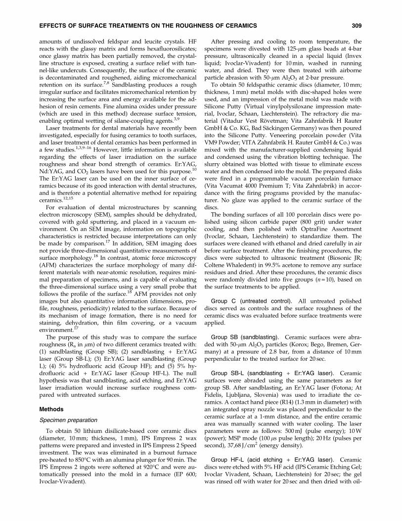
FIG. 1. Box-plot diagrams ofthe surface roughness (Ra inlm) of Empress 2 ceramicsaccording to the surface treat-ments applied. The median isshown by a horizontal linewithin the box. The minimumand maximum values are il-lustrated by the upper andlower strokes. (O) Marks out-liers.
310 DILBER ET AL.

Statistical analyses
Surface roughness data did not follow a normal distribu-tion; therefore, a non-parametric statistical analysis wasperformed for data comparisons. Surface roughness (Ra)values were analyzed using a Wilcoxon signed rank test tocompare the surface treatments applied on lithium disilicate-based and feldspathic ceramics. Mann–Whitney U andKruskal–Wallis tests were used for pairwise comparisonsamong the ceramic groups (a = 0.05).
Results
The surface roughness values and box-plot diagrams ofthe ceramic groups for each of the surface treatments arepresented in Table 1 and Figs. 1 and 2. Wilcoxon signed ranktest results showed that all groups except for Groups L andHF-L (Empress 2) had rougher surfaces than the untreatedgroups ( p < .05) (Table 2). Among the Empress 2 ceramicgroups, groups SB and SB-L had higher values and groupHF-L had lower values. A Mann–Whitney U test showedthat there was no significant difference among groups HF-L,
L, and HF ( p > 0.05). Group SB had the roughest surface forboth lithium disilicate-based core and feldspathic ceramicdiscs (p < 0.05).
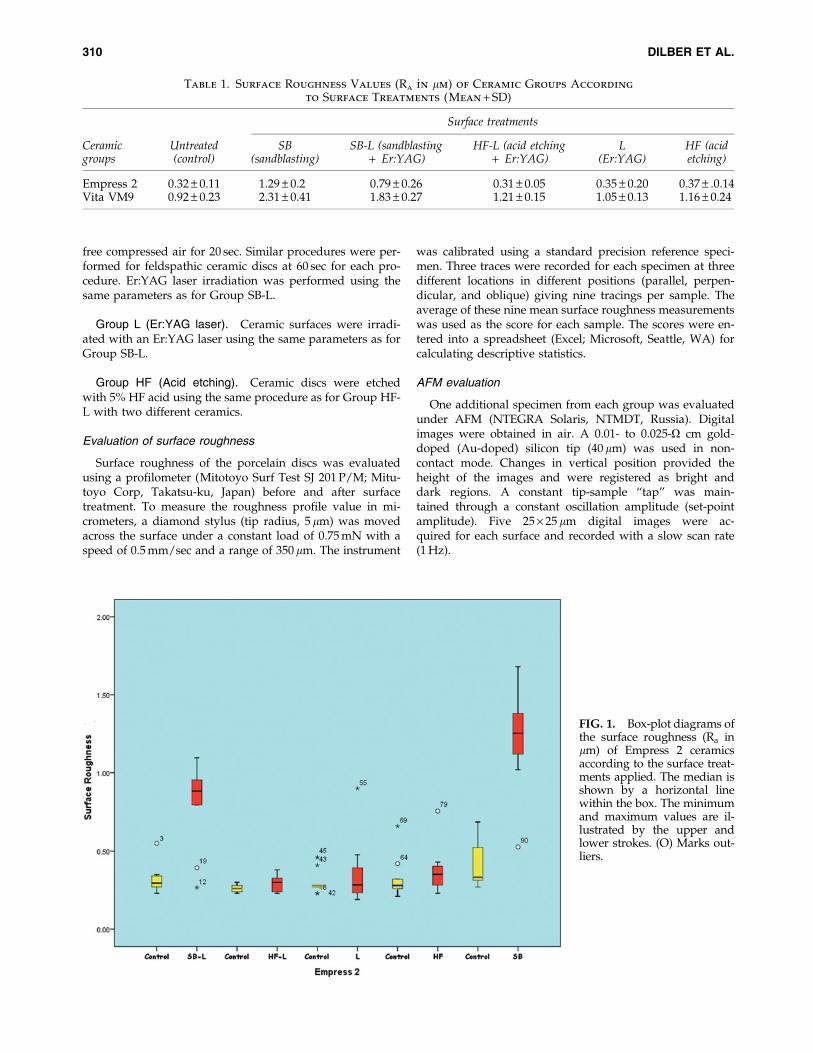
Representative AFM images of Empress 2 and Vita VM9porcelain surfaces treated with the different techniques arepresented in Figs. 3, 4, and 5. The surface treatment pro-cedures generated similar topographies, except for sur-faces treated with the Er:YAG laser (Group L). Group Lsurfaces showed moderate irregularity with peaks and val-leys, and less roughness than was achieved with sandblast-ing and HF acid (Figs. 4c and 5c). Group SB had the mostdistinct sharp peaks compared with those in the other groups(Fig. 3).
Discussion
Micromechanical interlocking and chemical bonding roughenand clean the surface for adequate activation and hence play amajor role in the bond strength between resin cement and theinner surface of a ceramic restoration.1,3,10,13,15,19,20 Various sur-face treatment procedures for achieving a micromechanicallyretentive porcelain surface have been used clinically.9–11,14,15
FIG. 2. Box-plot diagrams ofthe surface roughness (Ra inlm) of feldspathic ceramicsaccording to the surface treat-ments applied. The median isshown by a horizontal linewithin the box. The minimumand maximum values are il-lustrated by the upper andlower strokes. (O) Marks out-liers.
Table 2. Results of W_ilcoxon S_igned Rank Test ( p Values) Compar_ing the Roughness
Values Between the Treated and Untreated Groups
Ceramicgroups
SBsandblasting
SB-L (sandblasting+ Er:YAG)
HF-L (acid etching+ Er:YAG) L (Er:YAG)
HF(acid etching)
Empress 2 0.00 0.00 0.12 0.28 0.02Vita VM9 0.00 0.00 0.02 0.11 0.00
Asymptotic significance (2-tailed)
p < 0.05.
EFFECTS OF SURFACE TREATMENTS ON THE ROUGHNESS OF CERAMICS 311

This study presented an alternative combination of anEr:YAG laser with Al2O3 sandblasting and HF acid etching.Although many studies have investigated the effects of laserson hard tissues,21–29, their effect on porcelain surfaces has notbeen clear. In this study, AFM images of surfaces treated withlaser irradiation revealed a shallow irregular surface. Wehypothesized that these irregularities would enhance me-chanical retention between the resin composite and porcelainsurface. The null hypothesis was partially accepted: sand-blasting and HF acid etching increased the surface roughnesscompared to untreated surfaces, but laser irradiation alonedid not.
Sandblasting produces a rough irregular surface with anincreased surface area, and enhances the wettability of theceramic and the composite resin. However, excessive sand-blasting induces chipping or a significant loss of porcelainmaterial and is not recommended for cementation of silica-based and feldspathic ceramic restorations.20,30,31 Among thesurface treatments available for ceramic surfaces, sandblast-ing with Al2O3 has been widely used to provide micro-mechanical retention in several types of ceramics.1,9,32–34 Karaet al.3,14 showed that air abrasion provides the highest surfaceroughness in low-fusing and lithium disilicate-based core all-ceramic materials. AFM images of air-abraded lithium dis-ilicate-based core specimens showed a non-uniform surfacewith distinct sharp projections dotted with pores. The studyby Kara et al. also indicated that on SEM images, laser irra-diated surfaces appeared to be relatively smoother thansandblasted surfaces.14 In the current study, sandblasting was
applied before laser irradiation to simulate clinical circum-stances, as it is a conventional procedure performed in com-mercial laboratories. In the present study, sandblastedporcelain specimens showed the highest surface roughness,and AFM images had more distinct sharp peaks than those ofthe other groups.
Er:YAG laser irradiation of a ceramic surface can removethe glass phase of the ceramic and create a rough surface.35
Furthermore, Er:YAG laser irradiation increases the micro-mechanical retention of resin.10,35 However, Subasxı andInan36 used 400 mJ pulse energy and found significantlylower surface roughness values than air abrasion. Similarly,in this study, Er:YAG laser-irradiated porcelain specimensshowed lower surface roughness values even though higherlaser energy parameters were used (500 mJ, 10 W). On theother hand, Ersu and colleagues13 reported that surfaceroughness and shear bond strength values were higher inCO2 laser-irradiated In-Ceram� SPINELL, In-Ceram ALU-MINA, and In-Ceram ZIRCONIA ceramics than in thosetreated with sandblasting and HF acid etching, but SEManalyses of the surfaces irradiated with the CO2 laser re-vealed microcracks. Gokce et al.35 reported that the shearbond strength of Empress 2 specimens after Er:YAG laserirradiation at 300 mJ was higher than that of surfaces irra-diated with 600 and 900 mJ. The differing compositions andreflectance of ceramic materials might have affected theseresults. A hydroxyapatite powder could be applied to stainthe ceramic surface to enhance energy absorption and createa favorable surface.15
FIG. 3. Atomic force mi-croscopy (AFM) images ofsandblasted ceramics (SB).(a) Empress 2; (b) Vita.
FIG. 4. Atomic force microscopy(AFM) images of Empress 2 ceram-ics. (a) SB-L; (b) HF-L; (c) L; (d) HF.
312 DILBER ET AL.
HF acid selectively dissolves glassy or crystalline compo-nents of ceramics and creates a microporous irregular surface,thereby roughening the surface area and facilitating pene-tration of the resin into the etched ceramic surface.6,37 Karaet al.14 reported that treatment of low fusing ceramics with 5%HF acid etching produced same roughness values withEr:YAG laser. IPS Empress 2 specimens have a high crystal-line content and exhibit significantly higher bond strengthsthan those of IPS Empress specimens, independent of surfaceconditioning.3,20,38 Akyil et al.10 determined that the bondstrengths obtained from groups exposed to HF acid etchingafter each laser irradiation were lower than those in groupsexposed to HF acid etching alone. This might be because HFacid etching was not effective on ceramic surfaces, as laserirradiation might have led to the development of a heat-damaged layer on the ceramic surface.10,35 Given this risk, thereverse procedure (HF + laser) was used in this study. Despitethis, the surface roughness values of Empress 2 ceramics ex-posed to Er:YAG laser irradiation after HF acid etching werelower than those of ceramics exposed to Er:YAG laser irra-diation alone. A heat-damaged layer on the ceramic surfacemight also have been responsible for the reduction in thevalues of Group SB-L compared with Group SB.
Differences in the composition and microstructure of allceramic restorations might affect the surface texture and bondstrength between the ceramics and resin cement. Furtherstudies are required to evaluate the effects of different powersettings and different laser applications on ceramic surfaces toobtain optimum bond strength and roughness values.
Conclusions
Based on the results obtained and within the limitations ofthis in vitro study, the following conclusions can be drawn.
Sandblasting created a rougher surface than the othersurface treatment methods (p < 0.05). There were no signifi-cant differences in surface roughness after HF acid etching,
Er:YAG laser irradiation, or the combination of HF and Er:-YAG irradiation (p < 0.05). AFM images of these laser-irradiated surfaces showed superficial irregularities withpeaks and valleys, and less roughness than was visible aftersandblasting and HF acid etching. Groups subjected tosandblasting had more distinct sharp peaks than the othergroups.
Author Disclosure Statement
No competing financial interests exist.
References
1. Borges, G.A., Sophr, A.M., de Goes, M.F., Sobrinho, L.C.,and Chan, D.C. (2003). Effect of etching and airborne particleabrasion on the microstructure of different dental ceramics.J. Prosthet. Dent. 89, 479–488.
2. Atsu, S.S., Kilicarslan, M.A., Kucukesmen, H.C., and Aka,P.S. (2006). Effect of zirconium-oxide ceramic surface treat-ments on the bond strength to adhesive resin. J. Prosthet.Dent. 95, 430–436.
3. Kara, H.B., Dilber, E., Koc, O., Ozturk, A.N., and Bulbul, M.(2012). Effect of different surface treatments on roughness ofIPS Empress 2 ceramic. Lasers Med. Sci. 27, 262–272.
4. Martinez–Insua, A., Da Silva Dominguez, L., Rivera, F.G.,and Santana–Penin, U.A. (2000). Differences in bonding toacid-etched or Er:YAG-laser-treated enamel and dentinsurfaces. J. Prosthet. Dent. 84, 280–288.
5. Osorio, E., Toledano, M., da Silveira, B.L., and Osorio, R.(2010). Effect of different surface treatments on In-CeramAlumina roughness. An AFM study. J. Dent. 38, 118–122.
6. Ozcan, M., and Vallittu, P.K. (2003). Effect of surface con-ditioning methods on the bond strength of luting cement toceramics. Dent. Mater. 19, 725–731.
7. Chaiyabutr, Y., McGowan, S., Phillips, K.M., Kois, J.C., andGiordano, R.A. (2008). The effect of hydrofluoric acid surface
FIG. 5. Atomic force mi-croscopy (AFM) images ofVita ceramics. (a) SB-L; (b)HF-L; (c) L; (d) HF.
EFFECTS OF SURFACE TREATMENTS ON THE ROUGHNESS OF CERAMICS 313

treatment and bond strength of a zirconia veneering ce-ramic. J. Prosthet. Dent. 100, 194–202.
8. Soderholm, K.J., and Shang, S.W. (1993). Molecular orien-tation of silane at the surface of colloidal silica. J. Dent. Res.72, 1050–1054.
9. Panah, F.G., Rezai, S.M., and Ahmadian, L. (2008). The in-fluence of ceramic surface treatments on the micro-shearbond strength of composite resin to IPS Empress 2. J. Pros-thodont. 17, 409–414.
10. Akyil, M.S., Yilmaz, A., Bayindir, F. and Duymus, Z.Y.(2011). Microtensile bond strength of resin cement to afeldspathic ceramic. Photomed. Laser Surg. 29, 197–203.
11. Akyil, M.S., Yilmaz, A., Karaalioglu, O.F. and Duymus, Z.Y.(2010). Shear bond strength of repair composite resin to anacid-etched and a laser-irradiated feldspathic ceramic sur-face. Photomed. Laser Surg. 28, 539–545.
12. da Silva Ferreira, S., Hanashiro, F.S., de Souza–Zaroni, W.C.,Turbino, M.L., and Youssef, M. N. (2010). Influence of alu-minum oxide sandblasting associated with Nd, YAG orEr:YAG lasers on shear bond strength of a feldspathic ce-ramic to resin cements. Photomed. Laser Surg. 28, 471–475.
13. Ersu, B., Yuzugullu, B., Ruya Yazici, A., and Canay, S.(2009). Surface roughness and bond strengths of glass-infil-trated alumina-ceramics prepared using various surfacetreatments. J. Dent. 37, 848–856.
14. Kara, H.B., Ozturk, A.N., Aykent, F., Koc, O., and Ozturk, B.(2010). The effect of different surface treatments on rough-ness and bond strength in low fusing ceramics. Lasers Med.Sci. 26, 599–604.
15. Shiu, P., De Souza–Zaroni, W.C., Eduardo Cde, P., andYoussef, M.N. (2007). Effect of feldspathic ceramic surfacetreatments on bond strength to resin cement. Photomed.Laser Surg. 25, 291–296.
16. Ayad, M.F., Fahmy, N.Z., and Rosenstiel, S.F. (2008). Effectof surface treatment on roughness and bond strength of aheat-pressed ceramic. J. Prosthet. Dent. 99, 123–130.
17. Sanches, R.P., Otani, C., Damiao, A.J. and Miyakawa, W.(2009). AFM characterization of bovine enamel and dentineafter acid-etching. Micron 40, 502–506.
18. Matos, A.B., de Freitas, A.C.P., Espejo, L.C., et al. (2010).AFM analysis of bleaching effects on dental enamel micro-topography. Appl. Surf. Sci. 256, 2915–2919.
19. Chen, J.H., Matsumura, H., and Atsuta, M. (1998). Effect ofetchant, etching period, and silane priming on bond strengthto porcelain of composite resin. Oper. Dent. 23, 250–257.
20. Blatz, M.B., Sadan, A., and Kern, M. (2003). Resin–ceramicbonding: a review of the literature. J. Prosthet. Dent. 89, 268–274.
21. Bader, C., and Krejci, I. (2006). Marginal quality in enameland dentin after preparation and finishing with an Er:YAGlaser. Am. J. Dent. 19, 337–342.
22. Dunn, W.J., Davis, J.T., and Bush, A.C. (2005). Shear bondstrength and SEM evaluation of composite bonded toEr:YAG laser-prepared dentin and enamel. Dent. Mater. 21,616–624.
23. Keller, U., and Hibst, R. (1989). Ablative effect of an Er:YAGlaser on enamel and dentin [in German]. Dtsch. Zahnarztl.Z. 44, 600–602.
24. Hossain, M., Nakamura, Y., Yamada, Y., Kimura, Y., Na-kamura, G., and Matsumoto, K. (1999). Ablation depths andmorphological changes in human enamel and dentin afterEr:YAG laser irradiation with or without water mist. J. Clin.Laser Med. Surg. 17, 105–109.
25. Firat, E., Gurgan, S., and Gutknecht, N. (2012). Microtensilebond strength of an etch-and-rinse adhesive to enamel anddentin after Er:YAG laser pretreatment with different pulsedurations. Lasers Med. Sci. 27, 15–21.
26. Armengol, V., Laboux, O., Weiss, P., Jean, A., and Hamel, H.(2003). Effects of Er:YAG and Nd:YAP laser irradiation onthe surface roughness and free surface energy of enamel anddentin: an in vitro study. Oper. Dent. 28, 67–74.
27. Delme, K.I., and De Moor, R.J. (2007). Scanning electronmicroscopic evaluation of enamel and dentin surfaces afterEr:YAG laser preparation and laser conditioning. Photomed.Laser Surg. 25, 393–401.
28. Usumez, S., Orhan, M., and Usumez, A. (2002). Laser etch-ing of enamel for direct bonding with an Er,Cr:YSGG hy-drokinetic laser system. Am. J. Orthod. Dentofacial Orthop.122, 649–656.
29. Rodriguez–Vilchis, L.E., Contreras–Bulnes, R., Olea–Mejia,O.F., Sanchez–Flores, I., and Centeno–Pedraza, C. (2011).Morphological and structural changes on human dentalenamel after Er:YAG laser irradiation: AFM, SEM, and EDSevaluation. Photomed. Laser Surg. 29, 493–500.
30. Calamia, J.R. (1985). Etched porcelain veneers: the currentstate of the art. Quintessence Int. 16, 5–12.
31. Kern, M., and Thompson, V.P. (1994). Sandblasting andsilica coating of a glass-infiltrated alumina ceramic: volumeloss, morphology, and changes in the surface composition.J. Prosthet. Dent. 71, 453–461.
32. Guler, A.U., Yilmaz, F., Ural, C., and Guler, E. (2005). Eva-luation of 24–hour shear bond strength of resin composite toporcelain according to surface treatment. Int. J. Prosthodont.18, 156–160.
33. Della Bona, A., and van Noort, R. (1995). Shear vs. tensilebond strength of resin composite bonded to ceramic. J. Dent.Res. 74, 1591–1596.
34. Kim, B.K., Bae, H.E., Shim, J.S., and Lee, K.W. (2005). Theinfluence of ceramic surface treatments on the tensile bondstrength of composite resin to all-ceramic coping materials.J. Prosthet. Dent. 94, 357–362.
35. Gokce, B., Ozpinar, B., Dundar, M., Comlekoglu, E., Sen,B.H., and Gungor, M.A. (2007). Bond strengths of all-ceramics: acid vs laser etching. Oper. Dent. 32, 173–178.
36. Subasxı, M.G., and Inan, O. (2011). Evaluation of the topo-graphical surface changes and roughness of zirconia afterdifferent surface treatments. Lasers Med. Sci. Jul 24. [Epubahead of print].
37. de Melo, R.M., Valandro, L.F., and Bottino, M.A. (2007).Microtensile bond strength of a repair composite to leucite-reinforced feldspathic ceramic. Braz. Dent. J. 18, 314–319.
38. Della Bona, A., Anusavice, K.J., and Shen, C. (2000). Mi-crotensile strength of composite bonded to hot-pressed ce-ramics. J. Adhes. Dent. 2, 305–313.
Address correspondence to:Erhan Dilber
Selcuk UniversityFaculty of Dentistry
Department of Prosthodontics42075, Konya
Turkey
E-mail: [email protected]
314 DILBER ET AL.