Embed Size (px)
Citation preview

www.wjpps.com Vol 5, Issue 4, 2016.
438
Azeez et al. World Journal of Pharmacy and Pharmaceutical Sciences
COMPARISON BETWEEN TUMOR MARKER CEA, CA19-9 AND
SOME MARKER ENZYMES (ALKALINE SPHINGOMYELINASE,
CYCLOOXYGENASE-2, THYMIDYLATE SYNTHASE AND
ARGINASE) IN SERUM OF COLON CANCER PATIENTS.
M.Sc Noor Abdulaali Azeez*, Assist. Prof. Dr. Sarab Daoud Sulayman Alshamaa,
Assist. Prof. Dr. Iman Adel Hadi
Ministry of Higher Education and Scientific Research/ University of Mosul/ College of
Science, 2016.
ABSTRACT
Carcinoma of the colon was ranked as the sixth cancer among the top
ten cancers in Iraq. Different markers are used for different purposes –
namely, some of them are more appropriate for the follow-up of the
disease and the others for the early detection of the disease recurrence.
In many studies, an increase in CA 19-9 has been found to indicate a
poor prognosis and high serum levels of either CEA or CA 19-9 in
patients with colorectal cancer are significant, independent prognostic
factors. Method: 104 patients diagnosed with colon cancer, period
March 2013 to April 2014 from the patients treated in Mosul Oncology
and Nuclear Medicine Hospital, Ten milliliters of venous blood was
taken from each patient, then serum were separated for quantitative
measurement of CEA and CA19-9 levels on mini VIDAS
(Biomerieux, UK), Alk-SMase, COX2 and THYMS was determined
by using ELISA. Arginase enzyme activity was determined according to (kocna et al., 1996)
procedure. Results: The statistical analysis of data listed in table (18)show that there was
significant difference (P˂0.05) of enzymatic activity for serum (Cyclooxygenase-2, and
Arginase) which significantly increase (26.77 ±1.91 U/L, 23.3±2.50 ng/ml) respectively for
patients with early stage A colon cancer compared with controls (12.57+1.65 U/L, 4.18±0.34
ng/ml) where as statistical analysis shows non significant increase (P˃0.05) with the levels of
CEA and C19–9 tumor marker (4.04±1.6 ng/mL, 10.9±2.1 U/ml) respectively compared with
controls (2.8±1.06 ng/mL, 7.2±1.7 U/ml) measured in stage A revealed that
WORLD JOURNAL OF PHARMACY AND PHARMACEUTICAL SCIENCES
SJIF Impact Factor 6.041
Volume 5, Issue 4, 438-460 Research Article ISSN 2278 – 4357
*Correspondence for
Author
M.Sc Noor Abdulaali
Azeez
Ministry of Higher
Education and Scientific
Research/ University of
Mosul/ College of
Science, 2016.
Article Received on
07 Feb 2016,
Revised on 29 Feb 2016,
Accepted on 21 March 2016
DOI: 10.20959/wjpps20164-6542

www.wjpps.com Vol 5, Issue 4, 2016.
439
Azeez et al. World Journal of Pharmacy and Pharmaceutical Sciences
(Cyclooxygenase-2 and Arginase) may plays a key role in the early stage of intestinal polyp-
formation. This study also include the measurement of the enzymatic activity of serum
alkaline sphingomyelinase which was significantly decreased (P˂0.05) in colon cancer
patients (82.21±6.95 U/L) in stage A compared with controls (117.10±6.25 U/L) and this
decrease was independent of Dukes stage, thus strengthening the hypothesized validity of this
assay to be used as serum test for the early detection of colonic neoplasia. As table (18) show
that serum TYMS activities in early stages A, (54.93±25.60 U/L), were non significant
increased compared with healthy control (44.22±18.26 U/L). Conclusions: this study shows
that these antigenic tumor markers are not sensitive for early colon cancer detection. But
these parameters may be used as a prognostic indicator to predict the aggressiveness of the
malignant tumor in colon cancer.
KEYWORD: CEA, CA19-9, Serum Alkaline Sphingomyelinase (Alk-SMase), Human
Cyclooxygenase-2(COX2), Serum thymidylate synthase (THYMS) and Arginase, colon
Cancer.
INTRODUCTION
Cancer is a term used for diseases in which abnormal cells divide without control and are able
to invade other tissues (Hasan et al., 2013; Dzivenu et al., 2003). Large intestine performs
wide variety of functions, ranging from breakage of large molecules to nutrients and water
absorption (Rathore et al., 2013) Colon is one major constituent of large intestine, and its
cancer is a major reason of deaths in western and industrialized world (Kaur et al., 2013; AL-
Hasnawi et al, 2009) . Colorectal cancer is the second most common cause of cancer related
death in western countries after lung cancer in men and breast cancer in women (Mihajlovic-
Bozic, 2004). Carcinoma of the colon was ranked as the sixth cancer among the top ten
cancers in Iraq (M. O. H.; 2009). Colorectal cancer can be classified by a system called
Dukes’ staging ranging from stage A to stage D, In 1929 Cuthbert Dukes proposed a
classification designed to represent a step-wise progression of local-regional invasion by
rectal cancers. The classification has been modified on many occasions in an attempt to
increase its prognostic value. The most commonly employed modification of Dukes' system
is that of Astler and Coller.(Adachi et al.,1994). The Dukes’ stage describes the extent of
invasion or spread of a tumor and correlates with overall survival, i.e. patients have an 83%
survival chance with a Dukes stage A tumor versus 3% chance of survival if diagnosed with
Dukes’ stage D, both over five years (Campbell et al., 2001; Brünner et al., 2006) Tumour
www.wjpps.com Vol 5, Issue 4, 2016.
440
Azeez et al. World Journal of Pharmacy and Pharmaceutical Sciences
markers are substances, usually proteins that are produced by the body in response to cancer
growth or by the cancer tissue itself. These substances may be detected in blood, urine and
tissue samples. Some tumor markers are specific for a particular type of cancer, while others
are seen in several cancer types (Hamed et al., 2011). The qualitative and quantitative
evaluation of these markers is possible through modern techniques of sensitive
immunoassays using monoclonal or polyclonal antibodies or polymerase chain reaction
techniques (Tietz, 2006; Selvam, 2011). Different markers are used for different purposes –
namely, some of them are more appropriate for the follow-up of the disease and the others for
the early detection of the disease recurrence (Bast et al., 2001). In body fluids, tumor markers
are found in low concentrations and for their determination highly sensitive technology is
needed. The techniques that are being used are more or less based on the same principle – i.e.
the determination of antigen-antibody complexes. Most widely used techniques are the radio-
immune assay, the enzyme-immune assay, and the luminometric-immune assay, which differ
in the compound bound to the detection antibodies, and in the method of detection of the
formed complexes (Novaković, 2004). Carcinoembryonic antigen (CEA) is a high molecular
weight glycoprotein belonging to the immunoglobulin super family. The carboxy-terminal of
CEA contains a hydrophobic region which is modified to provide a glycosyl phosphatidyl
inositol link to the cell membrane (
Tanaka et al., 2010). While the presence can be
determined in biopsy samples, it is usually identified in serum. This protein has been used for
many years as a biomarker of CRC as well as cancers developing in other tissues. (CEA) was
first described in 1965 by Gold and Freedman and it is most widely used tumor markers
worldwide (Al-Saadi et al., 2014). CA 19-9 is synthesized by normal human pancreatic and
bilaryductular cell and by gastric, colon, endometeial and salivary epithelia(Pavai, 2003) .in
serum, it exists as a mucin, a high molecular mass (200 to 1000 kDa) glycoprotein complex,
Elevations in CA 19-9 level correlate with the degree of tumor differentiation as well as the
extent of tumor mass(Abdallah et al.,2013) Carbonhydrate antigen 19-9 (CA 19-9) is a ligand
for e-selectin that plays an important role in the adhesion of cancer cells to endothelial cells.
It has been used as a tumor marker in gastrointestinal cancers. It may also be increase in
several benign diseases. In many studies, an increase in CA 19-9 has been found to indicate a
poor prognosis and high serum levels of either CEA or CA 19-9 in patients with colorectal
cancer are significant, independent prognostic factors(Sisik et al.,2013). American Society of
Clinical Oncology guidelines suggested that serum testing for CA19-9 is an integral part of
the diagnosis and management of colorectal carcinomas. Sphingomyelinase is enzyme was
first discovered in the intestinal tract by Nilsson in 1969 (Duan, 2006). but got real attention

www.wjpps.com Vol 5, Issue 4, 2016.
441
Azeez et al. World Journal of Pharmacy and Pharmaceutical Sciences
only in the last decade, when dietary sphingomyelin was found to inhibit colonic
tumorigenesis in animals. and the first mutation of the enzyme was discovered in 2004 in one
human colon cancer cell line(Wu et al., 2004). Alkaline sphingomyelinase is present in the
intestinal tract andadditionally human bile. It hydrolyses sphingomyelin in both intestinal
lumen and themucosal membrane in a specific bile salt dependent manner. Several isoforms
of alk-SMases have been identified and classified by their pH optima: alk-SMase has been
located specifically to the intestinal tract, where high levels are found in the small intestine
and lower in the colon with a gradualdecline towards rectum, acid SMase and neutral SMases
have been found in many tissues and are considered as common cellular enzymes (Wu et
al.2010; Kurek et al., 2013). The enzyme sharesno structural similarity with other SMases
(Duan et al., 2003). The enzyme is of specific properties, such asbile salt dependency, high
stability, and tissue specific expression (Cheng et al., 2002; Wu et al., 2004). In the colon, the
enzyme may play antiproliferative and antiinflammatory roles through generating ceramide
(Duan et al., 2007; Wu et al., 2006). COX-1 and COX-2 are the two isoforms of
cyclooxygenase, which convert arachidonic acid (AA) into several eicosanoids such as
prostaglandin, thromboxanes and prostacyclin, which participate in several normal
physiologicprocesses and inflammation (Urade, 2008). COX-1 and COX-2 share the same
substrates, generate the same products and catalyze the same reaction using identical catalytic
mechanisms (Zha et al., 2004). Whereas COX-1 isconstitutively expressed in most tissues,
COX-2 is an inducible enzyme, stimulated by cytokines, growth factors, oncogenes or tumor
promoters during inflammation or malignancy(Gallegoa et al., 2007). In recent years,
overexpression of COX-2 has been reported in a variety of cancers (Urade, 2008). COX-2 is
induced by stimuli such as mitogens, cytokines, growth factors and tumor promoters, and has
been elucidated to be involved in cancer development and pathogenesis (Rakesh et al.,2014).
COX-2-expressing interstitial cells accelerates colon carcinogenesis (Ota et al.,2002).
Thymidylate synthase (TS) plays a central role in the biosynthesis of thymidylate,an essential
precursor for DNA synthesis see -(voeller et al., 2004;Bruni et al. 2002)., the inhibition of TS
result in the cessation of cellular proliferation and growth(Rahman et al., 2004). Several
clinical studies have shown that TS protein and mRNA levels are higher in cervical, breast,
kidney, bladder, lung, and gastrointestinal tumor tissues than in their normal counterparts and
that high TS level have been associated with poor clinical outcome in these cancer (Nomura
et al., 2002; Mizutani et al., 2003). Tumors with elevated TS levels are thought to undergo
more progresactive cellular proliferation, which in turn is associated with tumor invasiveness
and metastasis(Mizutani et al., 2003;Edler et al., 2000). Due to the importance of arginase
www.wjpps.com Vol 5, Issue 4, 2016.
442
Azeez et al. World Journal of Pharmacy and Pharmaceutical Sciences
enzyme in different malignant disorders Arginase (L-arginine amidinohydrolase, EC 3.5.3.1)
is homotrimeric binuclear manganese metalloenzyme that catalyzes the hydrolysis of L-
arginine, rendering urea for ammonia elimination, mainly in urotelic animals, and L-ornithine
(a non-protein amino acid) for biosynthetic pathways. There are atleast two forms of
arginase. Arginase cytosolic and is most abundant in the liver, primarily responsible for
ammonia detoxification as urea (Mahmoud et al., 2009).A second isoenzyme, arginase II, is
involved in the production of ornithine as a precursor to proline, glutamate orpolyamines,
such as spermine and putresine, essential for cellular growth(Ash, 2004) Polyamines are vital
for cell proliferation and it is possible thatthe increased level of ornithine, due to theelevated
arginase activity, may be linked to thedevelopment of carcinogenesis(Soda, 2011). The
enzyme also is present in other human tissues, such as kidney, brain, lung, stomach, bowel,
prostate lactating mammary gland and activated macrophages, where its physiologic role is
still poorly understood (Munder, 2009). Arginase is a very stable enzyme and it does not lose
its activity when it is stored at low temperatures for long periods of time. In a previous report,
we showed that two extrahepatic arginases are present in human colon carcinoma and CRC,
and only one of them appears in blood serum. Its activity is low in healthy individuals and
rises significantly in the serum from patients with primary colorectal cancer (Porembska et
al., 2002). Arginase activity is markedly elevated in prostatic carcinoma, gastric cancer,
colorectal cancer, chronic lymphocytic leukemia (Konarska et al., 1993). Non-small cell lung
carcinoma and breast cancer. Increased arginase activity has also been reported in cancer
metastases (Gökmen et al., 2010).
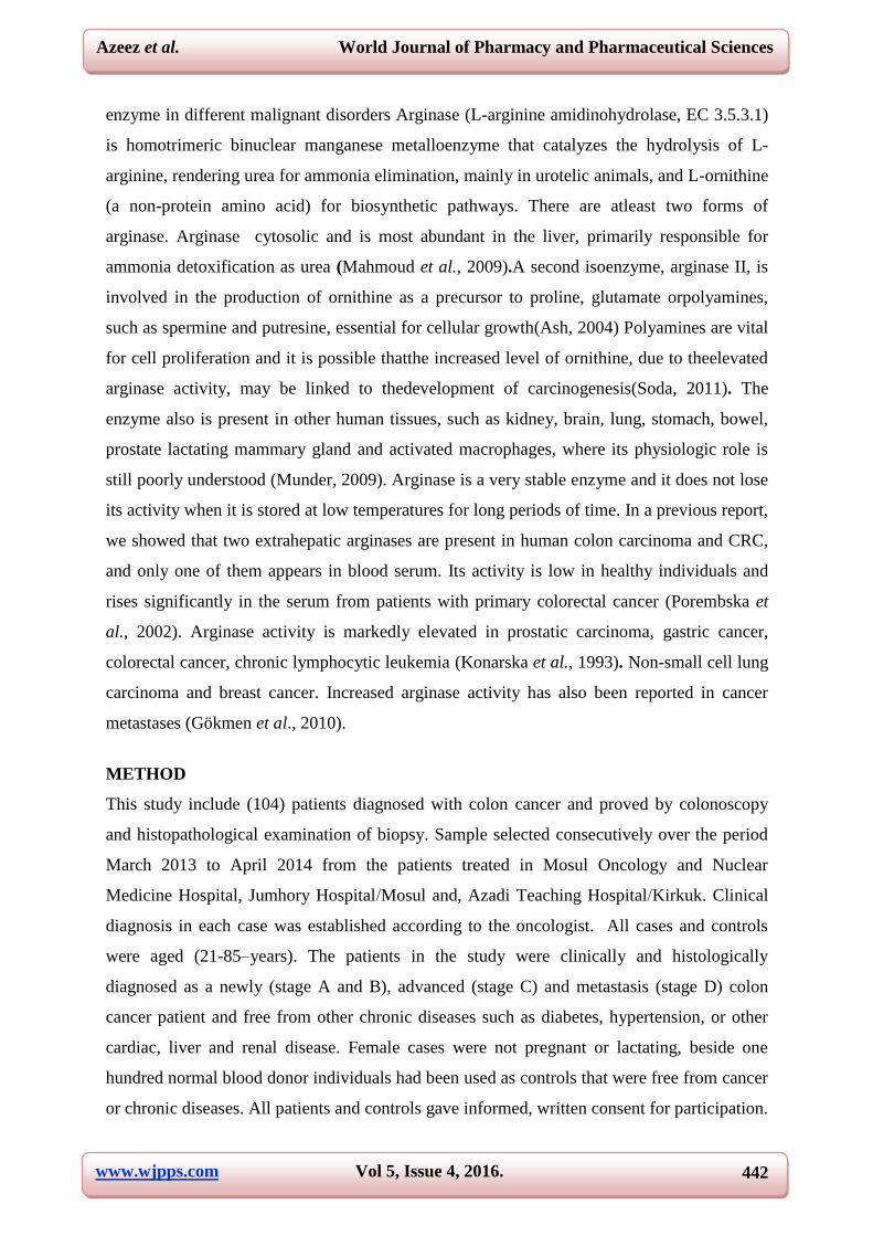
METHOD
This study include (104) patients diagnosed with colon cancer and proved by colonoscopy
and histopathological examination of biopsy. Sample selected consecutively over the period
March 2013 to April 2014 from the patients treated in Mosul Oncology and Nuclear
Medicine Hospital, Jumhory Hospital/Mosul and, Azadi Teaching Hospital/Kirkuk. Clinical
diagnosis in each case was established according to the oncologist. All cases and controls
were aged (21-85–years). The patients in the study were clinically and histologically
diagnosed as a newly (stage A and B), advanced (stage C) and metastasis (stage D) colon
cancer patient and free from other chronic diseases such as diabetes, hypertension, or other
cardiac, liver and renal disease. Female cases were not pregnant or lactating, beside one
hundred normal blood donor individuals had been used as controls that were free from cancer
or chronic diseases. All patients and controls gave informed, written consent for participation.

www.wjpps.com Vol 5, Issue 4, 2016.
443
Azeez et al. World Journal of Pharmacy and Pharmaceutical Sciences
Serum from these patients obtained before surgery. Ten milliliters of venous blood was taken
from each patient and left for (15) minutes at room temperature for coagulation, then serum
were separated by centrifugation at (3000 xg)for 10 minutes and divided in aliquot and kept
frozen at (-20°C) for the assays, CEA and CA19-9 kits (Biomerieux, France) was assayed
using ELFA technique (Enzyme Linked Fluorescent Assay). for quantitative measurement of
CEA and CA19-9 levels on mini VIDAS (Biomerieux, UK) instruments using serum
specimens. Serum Alkaline Sphingomyelinase (Biosource, Cat. No.MBS039905, USA),
Human Cyclooxygenase-2(Biosource, Cat. No.MBS164164, USA) and Serum thymidylate
synthase (Cusabio Biotech, Cat. No. MBS032123, USA) was determined by using ELISA
(FLx800 Fluorescence Microplate Reader and FLx800 Microplate Washer, BioTek, USA) kit
assayed according to the manufactured procedure. Create a standard curve by reducing the
data using computer software (Excel/ office 2007).
Figure (1) ALK-SMASE standard curve.

www.wjpps.com Vol 5, Issue 4, 2016.
444
Azeez et al. World Journal of Pharmacy and Pharmaceutical Sciences
Figure (2) Human Cyclooxygenase-2 standard curve.
Figure (3) TYMS Standard curve.
Arginase, (L-arginine amidinohydrolase, EC3.5.3.1.) The enzyme activity was determined
according to (kocna et al., 1996) procedure. Principle. Arginase, (L-
arginineamidinohydrolase, EC3.5.3.1) converts L-arginine in to L-ornithine and urea. The
elevated activity of arginase has been reported in serum as well as in colorectal, gastric and
mammary carcinoma tissues. Arginase activity in the serum is determined by a two-step
method. The concentration of ornithine (as a product of the first reaction) is measured by a
colorimetric method (UV./VIS. Spectrophotometer, Cecil Instruments Ltd.GE, England).
Figure (4) The arginase reaction(Ash, 2004).
Reaction conditions consist: 500 μΐ of assay solutions (35 mmol/1 Tris-HCl buffer pH 9.5 -
20, arginine 0.348, 0.009 gm MnCl2), 25 μΐ of serum, 25 μΐ of 5 mmol/1 Tris-HCl buffer.
The incubation of samples was carried out in a water-bath at 37°C for 120 minutes and
stopped by immersing tubes in a boiling water-bath for 5 minutes concentration was
determined by an end-point ninhydrin. Ninhydrin reagent (0.5 ml) and acetic acid (1.5 ml)
were added to the incubation medium reaction carried out in a boiling water bath for 60
minutes. This reaction was stopped by cooling to room-temperature and the colored reaction
product was evaluated by using a spectrophotometer at 515 nm in a 1 cm glass cuvette

www.wjpps.com Vol 5, Issue 4, 2016.
445
Azeez et al. World Journal of Pharmacy and Pharmaceutical Sciences
ornithine concentration was calculated from calibration standards curve as shown in figure
(5).
Figure (5) Ornithine - standard curve.
RESULTS AND DISCUSSION
Serum concentration of CEA in colon cancer patients.
Table (1) describes the results of the serum CEA concentrations in colon cancer patients
with different Dukes Stage of the disease compared with Control.
Table (1): Serum CEA concentrations in colon cancer patients with different Dukes
stage of the disease compared with control.
(*):Statistically significant differences at (p< 0.05) with control.
The results of the serum CEA concentrations in colon cancer patients with different Dukes
stage of the disease compared with control as shown in table (1) reveled a significant increase
at probability (p<0.05) in CEA Level in stages C and D which were, (29.45±12.6) ng/ml,
(27.9±11.7) ng/ml respectively compared with control group (2.8±1.06) ng/ml. while there
were no significant difference in serum CEA Level in stages A and B (4.04±1.6) ng/ml,
(5.07±1.90) ng/ml respectively compared with control groups. These results had been
accepted with several other researches such as (Polat et al., 2014) who indicate that serum
CEA
(ng/mL)
Dukes Stage M±SD Control
M±SD Dukes A Dukes B Dukes C Dukes D
Male 4.1±1.64 5.23±1.8 30.5±12.7
28.6±12.65 2.84±1.09
Female 3.8±1.8 4.8±2.2 28.2±12.6 27±11.2 2.76±1.09
Total 4.04±1.6 5.07±1.90 29.45±12.6* 27.9±11.7* 2.8±1.06
www.wjpps.com Vol 5, Issue 4, 2016.
446
Azeez et al. World Journal of Pharmacy and Pharmaceutical Sciences
CEA levels were significantly higher in stages C and D than in stage A. (Ding et al., 2014)
reported that the mean of serum's CEA level were higher in dukes stage C of tumor than stage
B or A. (Eleftheriadis et al., 2009) found a low sensitivity associated with serum CEA assays
in the early stage detection of colorectal cancer's. While (Ding et al., 2014) showed that CEA
level can be different between Duke stage A and C, C and B, but not between A and B. (Al-
bayatti, 2009) indicate that the elevation of CEA level occur only in patients with advanced
stages in colon cancer. Serum carcino embryonic antigen (CEA) is a 201 kDa highly
glycosylated antigen expressed on the apical surface of colonic epithelial cells and excreted
via the colonic lumen (Elias et al., 2012). With the disruption of normal tissue architecture in
malignancy and loss of polarization of neoplastic cells located deep inside the tumor
glandular tissue, CEA may be expressed on the whole cell surface and is eventually shed to
the bloodstream leading to serum CEA level's rise (Tibbetts et al, 1993; Hammarstrom,
1999).
Results in table (12) also shows that there is no significant differences in serum CEA level
between males and females patients for each stage which was agree with (Mohammadi et al.,
2013)results who reported that no differences were observed between male and female
patients and between patients aged <50 and >50 years regarding the distribution of serum
CEA levels and CEA levels may rise in patients without recurrence, and may even be below
the cut-off point of 5 μg/l in patients with disseminated disease. Therefore, additional
biological markers are needed to improve the identification of patients at risk of recurrent
disease after intended curative resection and to monitor such patients during and after
adjuvant therapy (AL-Dulamy et al., 2010).
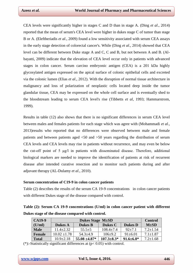
Serum concentration of C19-9 in colon cancer patients
Table (2) describes the results of the serum CA 19-9 concentrations in colon cancer patients
with different Dukes stage of the disease compared with control.
Table (2): Serum CA 19-9 concentrations (U/ml) in colon cancer patient with different
Dukes stage of the disease compared with control.
CA19-9
(U/ml)
Dukes Stage M±SD Control
M±SD Dukes A Dukes B Dukes C Dukes D
Male 11.4±2.32 55.5±5 108.4±7.4 92±7.1 7.2±1.54
Female 10.02 ±1.78 54.3±4.9 106±9.2 91±6.01 7.1±1.87
Total 10.9±2.18 55.08 ±4.87* 107.3±8.3* 91.6±6.8* 7.2±1.68
(*):-Statistically significant differences at (p< 0.05) with control.

www.wjpps.com Vol 5, Issue 4, 2016.
447
Azeez et al. World Journal of Pharmacy and Pharmaceutical Sciences
Results in table (2) revealed statistically significant increase at (P<0.05) in the mean values of
the serum's CA19-9 Level in patients with stages (B,C) and (D) (55.08 ±4.8)
U/ml,(107.3±8.3) U/ml and (91.6±6.8) U/ml respectively compared with controls (7.2±1.68)
U/ml, where as no significant variation in patients with stage (A) serum's CA19-9
(10.9±2.18) U/ml compared with control group (7.2±1.7) U/ml. These results indicate that
CA19-9 concentration is significantly elevated in patients with metastatic disease and with
increasing the degree of dysplasia or with the size of the lesion. This will agrees with the
reports of (Pavai and Yap., 2003) who observed that CA19-9 was very high only in patients
with advanced stages of colorectal carcinoma, and (Partyka, 2014) that reported increased
serum CA19-9 Level in 21-67% of advanced colon cancer patients (Hyeon et al., 2013),
indicates that metastases showed a stronger expression of CA 19-9 than primary tumors and
showed a lower expression in Dukes' A and B tumors than in more advanced stages. Beside
(Narimatsu et al., 1998) showed that the increasing of serum CA19-9 concentration is
associated with a modification of the antigen expression. The values of CA19-9 in tissues are
highest than values in the patients sera with the same type of cancer (Kajiwara et al., 2005).
This is an indicator that the cancer cells produce the antigen CA19-9 and release it to the
blood (Al-dujaili et al., 2009).
The activity of some marker enzymes in serum of colon cancer patients.
Serum Alkaline Sphingomyelinase (alk-SMase). Activity in colon cancer patients.
Table (3): Serum sphingomyelinase activity in different Dukes stages of colon cancer
patients compared with control.
(*):-Statistically significant differences at (p< 0.05) with control.
Serum alk-SMase activities related to Dukes stage of colon cancer were shown to be
significantly lower in colon cancer patients compared with healthy control, as table (3)
revealed that. Serum, alk-SMase activities was found to be significantly reduced in the early
stages A, B stages which were (82.21+6.95) U/L and (79.66+5.81) U/L respectively
compared with healthy (117.10+6.25) U/L). In addition reduced serum, alk-SMase activities
was shown in advanced and metastasis stages C, D which were (75.44±5.19) and
(71.44±5.19) U/L respectively compared with healthy (117.10+6.25) U/L), this agrees with
Alk-SMase
(U/L)
Dukes Stage M±SD Control
M±SD Dukes A Dukes B Dukes C Dukes D
Male 82.52±7.2 79.73±6.2 75.33±4.91 71.25±5.02 117.97±6.88
Female 81.6±7.1 79.5±5.5 75.56±3.34 71.66±5.64 116.24±5.64
Total 82.21±6.95* 79.66±5.81* 75.44±4.22* 71.44±5.19* 117.10±6.25

www.wjpps.com Vol 5, Issue 4, 2016.
448
Azeez et al. World Journal of Pharmacy and Pharmaceutical Sciences
the reports of (Hertervig et al., 1997), who found that, alk-SMase activity preferentially
decreases in human colorectal carcinoma, suggesting a regulatory role of the enzyme in colon
mucosa cell proliferation. In addition (Hertervig1 et al., 1999) showed that the analysis of,
alk-SMase activity in intestinal biopsies from control and colorectal adenocarcinoma patients
showed a significant decrease between the enzyme activity in tumor samples and controls
with a mean reduction of 90%.
It is important to recognize that sphingomyelin pathway is considered as one of the most
important intracellular mechanism in regulating cell-growth, differentiation and apoptosis
(Condorelli et al., 1999). Alk-SMase (no EC number assigned), is specifically expressed in
the intestinal tract and human liver.
In the intestinal tract, alk-SMase may prevent colonic tumorigenesis. First, the enzyme
generates antiprolife-rative and proapoptotic lipid messenger ceramide (Duan et al., 2003).
Second, the enzyme inhibits proliferation of human colon cancer cells (Hertervig et al.,
2003). Third, the enzyme activity is reduced in both long standing ulcerative colitis and
colonic tumorigenesis (Sjöqvist et al., 2002).
The results in table(3) also shows that no statistically significant difference was found
between Dukes’ stages B, C, and D, this agrees with (Duan et al., 1997) who found that In
relation to Dukes’ stage of adenocarcinoma and tumor cell differentiation, the, alk-SMase
activity in tumor samples was shown to be always significantly reduced when compared with
normal mucosa, whereas no significant difference was observed when tumor samples, at all
Dukes stages or differentiation grade, were compared. In addition (Marzio et al., 2013)
showed that, alk-SMase was significantly decreased in tumor line intestinal mucosa of
patients compared with controls in dependently of Dukes’ stage and tumor differentiation
grade. These results suggest that the decreasing activity gradient from ascending colon to
rectum could be totally abrogated in adenocarcinoma patients. Markedly reduced mucosal,
alk-SMase activity has been associated with colorectal carcinoma (Hertervig et al., 1997),
colorectal adenomas (Duan, 2006).

www.wjpps.com Vol 5, Issue 4, 2016.
449
Azeez et al. World Journal of Pharmacy and Pharmaceutical Sciences
Serum Cyclooxygenase-2 (COX2) (EC1.14.99.1), activity in colon cancer patients.
Table (4): Serum COX2 activity in different Dukes stages of colon cancer patients
compared with control.
COX2
(U/L)
Dukes Stage M±SD Control
M±SD Dukes A Dukes B Dukes C Dukes D
Male 27.24±1.6 28.6±1.3 30.1±2.7 27.04±1.6 12.07±1.09
Female 25.9±2.2 27.3±1.8 30.2±2.04 27.5±1.3 12.78±2.02
Total 26.77±1.91* 28.05±1.60* 30.18±2.42* 27.26±1.56* 12.43+1.65
(*):-statistically significant differences at (p< 0.05) with control.
The mean of COX2 activity was found to be significantly increased in serum of malignant
colon cancer in comparison to healthy control serum. as table (4) revealed. Serum COX2
activities in early stages A, B, (26.77±1.91) U/L, (28.05±1.60) U/L respectively were
significantly increased compared with healthy control (12.43+1.65) U/L. In addition
significantly increased in mean activity of COX2 was shown in advanced and metastasis
stages C, D which were (30.18±2.42) U/L and (27.26±1.56) U/L respectively compared with
healthy (12.57+1.68) U/L, the results was similar to (Singh et al., 2011) who reported that the
serum COX-2 was found to be significantly (P>0.0001) elevate in breast and oral cancer
patients compare to the normal control. However, the results in table (4) shows the
relationship between COX2 activity levels and tumor stages in patients with advanced stage
(C ) showed higher COX2 activity compared to those with early stage (A, B) which in
agreement with (Feng, 2007) who reported that gastric cancer and colorectal cancer in
patients with advanced serum COX-2 levels were significantly higher. In addition the
differences are also further in patients with stage D showed lower activity levels when
compared with stage B and C. These findings support the evidence that Aspirin use was
associated with a risk reduction in patients whose colon tumors expressed higher levels of
COX-2 and this agreement with (Chan et al., 2007) Figure (29). In addition to prevention,
regular aspirin use after the diagnosis of CRC at stage A, B and C improves overall survival,
especially among individuals with tumors that over express COX-2 (Wang and Bois., 2010),
suggesting the potential therapeutic use of non-steroidal anti-inflammatory drugs (NSAID),
such as aspirin and ibuprofen in advanced CRC.

www.wjpps.com Vol 5, Issue 4, 2016.
450
Azeez et al. World Journal of Pharmacy and Pharmaceutical Sciences
Figure (6): Metabolism of arachidonic acid by cyclooxygenase-2.
Thymidylate Synthase: (TYMS) (5,10-methylenetetrahydrofol-ate, dUMPC
methyltransferase (EC2.1.1.148).
Table (5): Serum thymidylate synthase activity in different Dukes stages of colon cancer
patients compared with control.
TYMS
(U/L)
Dukes Stage M±SD Control
M±SD Dukes A Dukes B Dukes C Dukes D
Male 48.58±18.59 51.69±22.47 139.59±30.77 135.86±16.83 49.06±22.06
Female 48.8±18.94 53.89±19.94 120.18±24.41 128.08±19.87 39.52±13.79
Total 54.93±25.60 52.57±20.78 130.64±29.43* 132.32±18.26* 44.22±18.81
(*):-statistically significant differences at (p< 0.05) with control.
Serum Thymidylate Synthase TYMS Activity was found to be significantly increased in
serum of colon cancer patients in comparison to healthy control. As table (5) show that serum
TYMS activities in early stages A, B,(54.93±25.60 U/L), (52.57±20.78 U/L) were
nonsignificant increased compared with healthy control (44.22±18.26U/L). In addition
significantly increased in serum TYMS activities in advanced and metastasis stages C, D
which were (130±29.43U/L) and (132.32 ±18.26U/L) respectively compared with healthy
control (44.3±17.5U/L), the results was agree with (Patla and Pawlga, 2005) observed that
TYMS expression in the metastasis would be different than in the primary tumor, which
could determine the response to 5-FU based chemotherapy.
Serum concentration of Arginase (L-arginine amidino hydr-olase, EC3.5.3.1.(, in colon
cancer patients.

www.wjpps.com Vol 5, Issue 4, 2016.
451
Azeez et al. World Journal of Pharmacy and Pharmaceutical Sciences
Table (6): Serum arginase concentrations in different Dukes stages of colon cancer
patients compared with control.
(*):-statistically significant differences at (p< 0.05) with control.
In the present study, the activity levels of arginase were determined in colon cancer patients
serum that revealed significantly increased levels of arginase in colon cancer patients
compared to normal subjects (P < 0.05). The mean activity of arginase was found to be
significantly higher in the early stages A (23.3±2.50), (P < 0.05), and in the advanced stages
B, C, and D which was(9.9±0.9), (8.3±1.4), (11.1±1.1 ) respectively for colon cancer patients
in comparison with those of the normal subjects. (4.18±0.35). table(17) shows that there is no
significant differences in mean activity of arginase between males and females patients for
each stage and control. Investigators observe either enzymes that are native to normal tissue
or those that could be associated with changes in metabolism and that are unique to cancer
tissue. One of these enzymes is arginase. The enzyme has at least two forms. (Arginase I) is
cytosolic and is most abundant in the liver, primarily responsible for ammonia detoxication as
urea. A second isoenzyme, (arginase II), is involved in the production of ornithine which acts
as aprecursor to proline, glutamate or polyamines, such as spermine and putresine, essential
forcellular growth (Cederbaum, 2000). Polyamines are vital for cell proliferation and it is
possible that the increased level of ornithine due to the elevated arginase activity may be
linked to the development of carcinogenesis (Porembska et al., 2003). Leu and Wang 1991
determined that the serum arginase activity had been 2 times higher in the colorectal cancer
tissues in colorectal cancer patients when compared to normal mucosal tissues.
The present data are in agreement with previous studies (Leu and Wang, 1991; Porembska et
al., 2003) who reported that serum arginase activity levels in patients with colon cancer
significantly higher than those found in control subjects. The same researchers also
investigated the relationship between serum arginase activity and the stage of gastric cancer.
The mean serum arginase activity in patients with early stage gastric cancer was significantly
higher than the control group. Moreover, serum arginase activity was higher in the advanced
stage of gastric cancer than in the early-stage gastric cancer and in the control group.
Previous studies, determined that arginase activity was found to have a close association with
Arginase
(ng/ml)
Dukes Stage M±SD Control M±SD
Dukes A Dukes B Dukes C Dukes D
Male 23.8±2.7 9. 9±0.96 8.3±1.4 11.2±1.27 4.15 ±0.34
Female 22.4±1.9 9.8 ±0.94 8.24±1.8 11±0.97 4.22±0.36
Total 23.3±2.50* 9.9±0.9* 8.3±1.4* 11.1±1.1* 4.18±0.35

www.wjpps.com Vol 5, Issue 4, 2016.
452
Azeez et al. World Journal of Pharmacy and Pharmaceutical Sciences
various types of cancer, especially colorectal, prostate, pancreas and stomach cancer, and
increased activity has been also demonstrated. The pattern of enzymatic alterations may be
linked with the malignant state and the progression of cancerous cells in the tumor
(Jamshidzadeh et al., 2001). Differences in the activities or concentration of certain enzymes
between cancer cells and their normal counterparts might be useful as biological markers of
malignancy and/or aggressiveness in particular tumors (Tietz, 2006).
Comparison between patients in stage A colon cancer and control for measured (CEA,
CA19-9, Alk-SMase, COX2, THYMS and Arginase)
The slow and progressive nature of colon cancer presents an opportunity to implement
screening programs and diagnostic tools for the early detection of the disease that have the
attempt to reduce incidence and to detect the disease in its early stages before symptoms are
evident (Burt et al., 2013). Due to the heterogeneous nature of CRC, a single biomarker is
unlikely to have sufficient sensitivity or specificity for use as a stand-alone diagnostic
screening test and a panel of markers may be more effective (Fung et al., 2015).
Table (7) Comparison between patients in stage A colon cancer and control.
Parameters Mean ±SD
Duke stage A Control
CEA ng/mL 4.04±1.6 2.8±1.06
CA19-9 ng/mL 10.9±2.18 7.2±1.68
Alk-SMase U/L 82.21±6.95* 117.10±6.25
COX2 U/L 26.77±1.91* 12.43+1.65
THYMS U/L 54.93±25.60 44.22±18.81
Arginase ng/ml 23.3±2.50* 4.18±0.35
(*):-statistically significant differences at (P< 0.05) with control.
The statistical analysis of data listed in table (7)show that there was significant difference
(P˂0.05) of enzymatic activity for serum (Cyclooxygenase-2, and Arginase) which
significantly increase (26.77 ±1.91 U/L, 23.3±2.50 ng/ml) respectively for patients with early
stage A colon cancer compared with controls (12.57+1.65 U/L, 4.18±0.34 ng/ml) where as
statistical analysis shows non significant increase (P˃0.05) with the levels of CEA and C19–9
tumor marker (4.04±1.6 ng/mL, 10.9±2.1 U/ml) respectively compared with controls
(2.8±1.06 ng/mL, 7.2±1.7 U/ml) measured in stage A revealed that (Cyclooxygenase-2 and
Arginase) may plays a key role in the early stage of intestinal polyp-formation. This study
also include the measurement of the enzymatic activity of serum alkaline sphingomyelinase
which was significantly decreased (P˂0.05) in colon cancer patients (82.21±6.95 U/L) in
www.wjpps.com Vol 5, Issue 4, 2016.
453
Azeez et al. World Journal of Pharmacy and Pharmaceutical Sciences
stage A compared with controls (117.10±6.25 U/L) and this decrease was independent of
Dukes stage, thus strengthening the hypothesized validity of this assay to be used as serum
test for the early detection of colonic neoplasia. As table (18) show that serum TYMS
activities in early stages A, (54.93±25.60 U/L), were non significant increased compared with
healthy control (44.22±18.26 U/L).
CONCLUSIONS
Results of this study conclude
1. Serum's CEA and CA19-9 concentrations revealed a significant increase at p<0.05 in colon
cancer patients in stages C and D in comparison with its concentration in control group.
While no significant difference had been observed in stages A and B, there for this study
shows that these antigenic tumor markers are not sensitive for early colon cancer detection.
But these parameters may be used as a prognostic indicator to predict the aggressiveness of
the malignant tumor in colon cancer.
2. Serum's enzymes such as thymidylate synthase, alkaline sphingomyelinase,
cyclooxygenase-2 and arginase reflect the human intestinal mucosal enzyme level and could
represent a new marker for human colorectal adenocarcinoma. Mainly taking into account its
early appearance in intestinal neoplasms and may be useful for the early diagnosis of patients
who develop recurrent colon cancer and colorectal liver metastases.
3. A significant incerase was also noticed in additional enzymetic markers such as (Alkaline
phosphatase, lactate dehydrogenases and α-amylases) compared with control grope. This
could be explained by the fact that the presence of the enzyme in high concentration inside
the cell and the changes in the membrane permeability of the tumor cells would lead to the
release of the enzyme into the systemic circulation. and it could be good indicators for early
detection of colon cancer.
REFERENCE
1. Abdallah, A.; Belal, M.; El Bastawisy, A. and Gaafar, R. (2013)."Plasma CA19-9 in
Advanced Non-Small Cell Lung Cancer".Journal of Cancer and Clinical Oncology, 2(2):
11-18.
2. Adachi, Y.; Mori, M.; Maehara, Y. and Sugimachi, K. (1994). "Dukes's classification: a
valid prognostic indicator for gastric cancer". Journal of. Gut., 35: 1368–1371.

www.wjpps.com Vol 5, Issue 4, 2016.
454
Azeez et al. World Journal of Pharmacy and Pharmaceutical Sciences
3. Al byatti, R. K.; Zainal, I. G. and AL-Garawi, Z. S. (2009)." Study of Total Protein and
Protein Profile in Patients With (Breast, Ovary and Uterus) Cancer". Journal of Diala.,
39(4): 1-8.
4. Al Hasnawi, S. M.; Al Mosawi, A. J.; Al khzaie, A.; Unan, O. F.; Fadhil, H. M. and Sami,
S. (2009). "Cancer in Iraq: Distribution by primary tumor site" New Iraqi Journal of
Medicine., 5(1): 5-8.
5. Al-dujaili, A. H.; Al-Taei; W. F.; Turky, K. M. and Al-Ubaidi, G. H. (2009)."
Comparative study of CA19-9 levels as tumor marker in sera andtissues of patients with
stomach, colon and rectum cancers". Journal of Fac Med Baghdad., 51(2): 223-226.
6. Al-dulaymi, B.; Christensen, I.; Soletormos, G.; Jess, P.; Nielsen, S. E.; Brunner, N. and
Nielsen, H. J. (2010)." Changes in Soluble CEA and TIMP-1 Levels during Adjuvant
Chemotherapy for Stage III Colon Cancer". Journal of Anticancer research., 30: 233-238.
7. Al-Saadi, N. H.; Al-Daami, N. M. and Hussain, A. (2014)." Biochemical Bone Markers
in Prostate Cancer Patients with Advanced Bone Metastasis". Journal of Kerbala
University., 12(4): 143-149.
8. Ash, D. E. (2004)."Arginine Metabolism: Enzymology, Nutrition, and Clinical
Significance". American Society for Nutritional Sciences., 4: 22-3166.
9. Bast, R. C.; Ravdin, P.; Hayes, D. F.; Bates, S.; Fritsche, H.; Jessup, J. M.; Kemeny, N.;
Locker, G. Y.; Mennel, R. G. and Mark R. S. (2001)."2000 Update of Recommendations
for the Use of Tumor Markers in Breast and Colorectal Cancer: Clinical Practice
Guidelines of the American Society of Clinical Oncology". Journal of Clinical Oncology,
19(6): 1865-1878.
10. Bruni, P.; Minopoli, G.; Brancaccio, T.; Napolitano, M.; Faraonio, R.; Zambrano, N.;
Hansen, U. and Russo, T. (2002)." Fe65, a Ligand of the Alzheimer’s-Amyloid Precursor
Protein, Blocks Cell Cycle Progression by Down-regulating Thymidylate Synthase
Expression". The Journal of Biological chemistry, 277(38): 35481–35488.
11. Brünner, N.; Haglund, C.; Andersen, M.; Nielsen, H. and Duffy, M. (2006)."National
Academy of Clinical Biochemistry Guidelines for the Use of Tumor Markers in
Colorectal Cancer". Journal of NACB: Practice Guidelines and Recommendations For
Use Of Tumor Markers In The Clinic Colorectal Cancer, 3: 2-18.
12. Burt, R.W.; Cannon, J. A.; David, D. S.; Early, D. S.; Ford, J. M. and Giardiello, F. M.
(2013)."Colorectal cancer screening". Journal of Natl Compr Canc., 11: 1538–1575.

www.wjpps.com Vol 5, Issue 4, 2016.
455
Azeez et al. World Journal of Pharmacy and Pharmaceutical Sciences
13. Campbell, N. C.; Elliott, A. M.; Sharp, L.; Ritchie, L. D.; Cassidy, J. and Little, J.
(2001)."Rural and urban differences in stage at diagnosis of colorectal and lung cancers".
Journal of Cancer, 84(7): 910-914.
14. Cederbaum, S. D.; Yu, H.; Grody, W. W.; Kern, R. M.; Yoo, P. and Iyer, R. K. (2004)."
Arginases I and II: do their functions overlap? ".Mol. Genet. Metab., 81(1): 38-44.
15. Chan, A. T.; Ogino, S. and Fuchs, C. S. (2007). "Aspirin and the Risk of Colorectal
Cancerin Relation to the Expression of COX-2". Journal of NEngl. Med., 356: 2131-42.
16. Cheng, y.; Nilsson, A.; Tömquist, E. and Duan, R. (2002)." Purification, characterization,
and expression of rat intestinal alkaline sphingomyelinase". Journal of Lipid Research.,
43: 316–324.
17. Condorelli, F.; Canonico, P. L. and Sortino, M.A. (1999)."Distinct effects of ceramide-
generating pathways in prostate adenocarcinoma cells" British Journal of Pharmacology,
127: 75-84.
18. Ding, Y.; Xuan, W.; Chen, C.; Chen, Z.; Yang, Z.; Zuo, Y. and Ren, S. (2014).
"Differences in carcinoembryonic antigen levels between colon and rectal
cancer".Journal of Molecular and clinical oncology, 2: 618-622.
19. Duan, R. and Nilsson, A. (1997)."Purification of a Newly Identified Alkaline
Sphingomyelinase in Human Bile and Effects of Bile Salts and Phosphatidylcholine on
Enzyme Activity " Journal of Hepatology, 26(4): 823-830.
20. Duan, R. D. (2006)."Alkaline sphingomyelinase An old enzyme with novel implications"
Journal of Biochimica et Biophysica Acta., 1761(3): 91-281.
21. Duan, R.; Bergman, T.; Xu, N.; Wu, J.; Cheng, Y.; Duan, J.; Nelander, S.; Palmberg, C.
and Nilsson, A. (2003)."Identification of Human Intestinal Alkaline Sphingomyelinase as
aNovel Ecto-enzyme Related to the Nucleotide Phosphodiesterase Family". The Journal
of biological chemistry, 278(40): 38528–38536.
22. Duan, R.; Cheng, Y.; Jo¨nsson, B.; Ohlsson, L.; Herbst, A.; Hellstro¨m-Westas, L. and
Nilsson, A. (2007). "Human Meconium Contains Significant Amounts of Alkaline
Sphingomyelinase, Neutral Ceramidase, and Sphingolipid Metabolites". Journal of
International Pediatric Research, 61(1): 61-66.
23. Dzivenu, O. K.; Phil, D. and Tormey, J. (2003)."Cancer and the immune system". Journal
of Cancer Research Institute, 1: 3-6.
24. Edler, D.; Hallstro¨m, M.; Johnston, P. G.; Magnusson, I.; Ragnhammar, P. and
Blomgren, H. (2000). "Thymidylate Synthase Expression An Independent Prognostic

www.wjpps.com Vol 5, Issue 4, 2016.
456
Azeez et al. World Journal of Pharmacy and Pharmaceutical Sciences
Factor for Local Recurrence, Distant Metastasis, Disease-free and Overall Survival in
Rectal Cancer". Journal of Clinical Cancer Research, 6: 1378–1384.
25. Eleftheriadis, N.; Papaloukas, C. and Pistevou-Gompaki, K. (2009). "Diagnostic value of
serum tumor markers in asymptomatic individuals". J BUON., 14(4): 707-10.
26. Elias, E.; Mukherji, D.; Faraj, W.; Khalife, M.; Dimassi, H.; Eloubeidi, M.; Hattoum, H.;
Abou-Alfa, G.; Saleh, A. and Shamseddine, A. (2012)." Lymph-node ratio is an
independent prognosticfactor in patients with stage III colorectal cancer aretrospective
study from the Middle East". World Journal of Surgical Oncology, 10(63): 1-7.
27. Feng, Z. X. (2007). "The Clinical Research on the Expression of COX-2 in the Serum of
Gastroenteric Malignant Tumor Patients". Journal of Oncology research, 6(2): 51-56.
28. Fung, K. C.; Tabor, B.; Buckley, M. J.; Priebe, I. K.; Purins, L.; Pompeia, C.; Brierley, G.
V.; Lockett, T. K.; Gibbs, P. I.; Tie, J.; McMurrick, P.; Moore, J.; Ruszkiewicz, A.; Nice,
E.; Adams, T. E.; Burgess, A., and Cosgrove, L. J. (2015). "Blood-Based Protein
Biomarker Panel for the Detection of Colorectal Cancer" PLos. ONE Journal, 10(3):
1-11.
29. Gallegoa, G. A.; Pradoa, S. D.;Fonsecab, P. J.; Campeloc, R. G.; Espinosad, J. C. and
Aparicioc, L. M. (2007)."Cyclooxygenase-2 (COX-2).a molecular target in prostate
cancer" Journal. of Clin Transl Oncol, 9: 694-702.
30. Gökmen, S. S.; Kazezoğlu, C.; Aygit, C. A.; Yildiz, A.; Çakir, A. Türe, M. and Gülen, S.
(2010). "Arginase and Ornithine in Human Benign and Malignant Skin Tumors". Turk J
Biochem.com., 35(4): 319–324.
31. Hamed, M. A.; Ahmed, S. A. and Khaled, H. M. (2011)."Efficiency of diagnostic
biomarkers among colonic schistosomiasis Egyptian patients".Journal of Mem Inst
Oswaldo Cruz., 106(3): 322-329.
32. Hammarstrom, S. (1999)." The carcinoembryonic antigen (CEA) family for prevention
and therapy of oral cancer". Japanese Dental Science Review, 44: 57-65.
33. Hasan, F. F.; Saydoka, K. M. and Zaki S. M. (2013) "The effect of radiotherapy treatment
on some blood elements in patient with some types of cancer". Tikrit Medical Journal,
19(1): 30 -36.
34. Hertervig, E.; Nilsson, A.; Björk, J.; Hultkrantz, H. and Duan, R. D. (1999)." Familial
adenomatous polyposis is associated with a marked decrease in alkaline
sphingomyelinase activity a key factor to the unrestrained cell proliferation?". British
Journal of Cancer, 81(2): 232–236.

www.wjpps.com Vol 5, Issue 4, 2016.
457
Azeez et al. World Journal of Pharmacy and Pharmaceutical Sciences
35. Hyeon, Y; Son, G. and Joh, Y.(2013)."The clinical significance of preoperative serum
levels ofcarbohydrate antigen 19-9 in colorectal cancer". Journal of Koreansurg Soc., 84:
231-237.
36. Jamshidzadeh, A.; Aminlari, M. and Rasekh, H. (2001). "Rhodanese and arginase activity
in normal and cancerous tissues of human breast, esophagus, stomach and lung". Journal
of Arch Irn Med., 4(2): 88-92.
37. Kajiwara, H.; Yasuda, M.; Kumaki, N.; Shibayama, T. and Osamura, Y. (2005)."
Expression of carbohydrate antigens (SSEA-1, sialyl-Lewis X, DU-PAN-2 and CA19-9)
and E-selectin in urothelial carcinoma of the renal pelvis, ureter and urinary bladder".
Tokai. J. Exp. Clin. Med., 30(3): 177-82.
38. Kaur, J.; Kaur, G.; Kaur, S. and Mahajan, A. (2013)."Preparation and Characterization of
Colon Targeted Beads of Indomethacin Using Chitosan and Chondroitin
Sulphate".International Journal of Scientific and Research Publications, 3(1): 2250-3153.
39. Kocna, P.; Fric, P.; Zavoral, M. and Pelech, T. (1996). "Arginase Activity Determination
A Marker of Large Bowel Mucosa Proliferation". Eur. J. CHn. Chem. Clin. Biochem., 34:
619-623.
40. Konarska, L.; Widzynska, I.; Zienkiewicz, H. and Sutek, K. (1993). "Arginase activity
alterations in peripheral blood lymphocytic leukemia". Journal of Acta Biochimica
Polonica, 40(1): 160-163.
41. Kurek, K.; Aukaszuk, B.; Piotrowska, D. M.; Wiesio Bek, P.; Chabowska A. and
gendzian-Piotrowska, M. (2013)." Metab-olism, Physiological Role and Clinical
Implications of Sphingolipids in Gastrointestinal Tract". Journal of BioMed Research
International, 12(3): 1-10.
42. Leu, S. and Wang, S. (1991)."Clinical Significance of Arginase in Colorectal cancer".
Journal of Cancer, 70(4): 733-736.
43. M. O. H. (2009). "Iraqi Cancer Registry 2009". Iraqi Cancer Board iraqi Cancer Registry
Center: [email protected] Baghdad– Iraq 2012.
44. Mahmoud, A. A.; El-Said, S. E.; Mandour, M. A.; Zakhary, M. M. and Maximous D. W.
(2009)." Arginase activity in brest cancer : is it significant biomarker?". Juornal for Bull.
Pharm. Sci., Assiut University, 32(2): 241-247.
45. Marzio, L.; Leo, A.; Cinque, B.; Fanini, D.; Agnifili, A.; Berloco, P.; Linsalata, M.;
Lorusso, D.; Barone, M.; Simone, C. and Cifone, M. (2013)." Detection of Alkaline
Sphingomyelinase Activity in Human Stool: Proposed Role as a New Diagnosticand

www.wjpps.com Vol 5, Issue 4, 2016.
458
Azeez et al. World Journal of Pharmacy and Pharmaceutical Sciences
Prognostic Marker of Colorectal Cancer". Juornal for American Association for Cancer
Research, 14: 856-862.
46. Mihajlovic-Bozic, V. (2004). "Risk factors for colorectal cancer". Institute of Oncology
Sremska Kamenica, Serbia and Montenegro, 12(1): 45-49.
47. Mizutani, Y.; Wada, H.; Yoshida, O.; Fukushima, M.; Nonomura, M.; Nakao, M. and
Miki, T. (2003)."Carcinoma Significance of Thymidylate Synthase Activity in Renal
Cell". Journal of Clin Cancer Res., 9: 1453-1460.
48. Mohammadi, M.; Ahmadi, R.; Abbasy, N. and Farara, A. (2013). "Evaluation of Serum
Levels of CEA and CA19-9 Tumor Markers Healthy Individuals in Northwestern Iran".
International Conference on Medical, Biological and Pharmaceutical Sciences, 4:
104-105.
49. Munder, M. (2009). "Arginase:an emerging key player in the mammalian immune
system". British Journal of Pharmacology, 158: 638–651.
50. Narimatsu, H.; Iwasaki, H.; Nakayama, F.; Ikehara, Y.; Kudo, T.; Sugano, N.; Okura, H.;
Fujita, S. and Hirohashi, S. (1998)." Lewis and Secretor Gene Dosages Affect CA19-9
and DU-PAN-2 Serum Levels in Normal Individuals and Colorectal Cancer Patients
".Juornal for Cancer research, 58: 512-518.
51. Nomura, T.; Nakagawa, M.; Fujita, Y.; Hanada, T.; Mimata, H. and Nomura, Y. (2002).
"Clinical significance of thymidylate synthase expression in bladder cancer".
International Journal of Urology, 9: 368–376.
52. Novaković, S. (2004). "Tumor markers in clinical oncology". Journal of Radiol Oncol,
38(2): 73-83.
53. Ota, S.; Bamba, H.; Kato, A.; Kawamoto, C.; Yoshida, Y. and Fujiwara, K. (2002)."
Review article COX-2, prostanoids and colon cancer". Journal of Aliment Pharmacol
Ther., 16(2): 102–106.
54. Partyka, R. (2014)."Role of Tumour Markers in Diagnosis and Follow up of Colorectal
Cancer—Potential for Future Research". Journal of Anticancer research, 29: 4202-4208.
55. Patla, A. and Pawl´ga, J. (2005). "Prognostic value of thymidylate synthase expression in
colorectal cancer". Nowotwory Journal of Oncology, 2: 130–136.
56. Pavai, S. and Yap, S.V. (2003)."The Clinical Significance of Elevated Levels of Serum
CA 19·9". Med. J. Malaysia., 58(5): 667-672.
57. Pavai, S. and Yap, S.V. (2003). "The Clinical Significance of Elevated Levels of Serum
CA 19·9". Med. J. Malaysia., 58(5): 667-672.

www.wjpps.com Vol 5, Issue 4, 2016.
459
Azeez et al. World Journal of Pharmacy and Pharmaceutical Sciences
58. Polat, E.; Duman, U.; Duman, M.; Atici, A.; Reyhan, E.; Dalgic, T. Bostanci, E. and Yol,
S. (2014). "Diagnostic value of preoperative serum carcinoembryonic antigen and
carbohydrate antigen 19-9 in colorectal cancer". Journal of Current Oncology, 21(1): 1-7.
59. Porembska, Z.; Skwarek, A.; Mielczarek, M.; czyk-Kuz´ma A. (2002). "Serum Arginase
Activity in Postsurgical Monitoring of Patients with Colorectal Carcinoma". Journal of
Cancer, 94(11): 2930-2934.
60. Porembska, Z.; Skwarek, A.; Mielczarek, M.; czyk-Kuz´ma A. (2002)."Serum Arginase
Activity in Postsurgical Monitoring of Patients with Colorectal Carcinoma". Journal of
Cancer, 94(11): 2930-2934.
61. Rahman, L.; Voeller, D.; Rahman, M.; Lipkowitz, S.; Allegra, C.; Barrett, J. C.; Kaye, F.
J. and Zajac-Kaye, M. (2004). "Thymidylate synthase as an oncogene: A novel role for an
essential DNA synthesis enzyme".Journal of Cancer cell., 5: 341-351.
62. Rakesh, N.; Nagesh, K. S.; Iyengar, A. R. and Patil, D. B. (2014)."Inflammation in Oral
Cancer and Role of COX-2: A Review" Journal of Pharmaceutical, Biological and
Chemical Sciences, 5(2): 319-328.
63. Rathore, S.; Hussain, M.; Ali, A. and Khan, A. (2013)."A Recent Survey on Colon
Cancer Detection Techniques" Transactions on computational biology and
bioinformatics, 10(3): 4-10.
64. Selvam, N. T.; Elumamai, P.; Venkatakrishnan, V. and Damodarkumar, S.
(2011)."Molecular Markers in Cancer Diagnosis and Management A Review" Journal of
Applied Biological Sciences, 5(3): 69-74.
65. Singh, A. K.; Pandey, A.; Tewari, M.; Kumar, R.; Sharma, A.; Singh, K. A.; Pandey, H.
P. and Shukla, H. S. (2013). "Advanced stage of breast cancer hoist alkaline phosphatase
activity: risk factor for females in India". Biotech., 3: 517–520.
66. Sisik, A.; Kaya, M.; Bas, G.; Basak, F. and Alimoglu, O. (2013)."CEA and CA 19-9 are
Still Valuable Markers for the Prognosis of Colorectal and Gastric Cancer Patients".
Asian Pac J Cancer Pre., 14(7): 4289-4294.
67. Sjöqvist, U.; Hertervig, E.; Nilsson, A.; Duan, R. D.; Ost, A.; Tribukait, B. and Löfberg,
R. (2002)."Chronic colitis is associated with a reduction of mucosal alkaline
sphingomyelinase activity" Journal of Inflamm Bowel Dis., 8(4): 258-63.
68. Soda, K. (2011)."The mechanisms by which polyamines accelerate tumor spread". Soda
Journal of Experimental & Clinical Cancer Research, 30: 95.2-4.
69. Tanaka, T.; Tanaka, M.; Tanaka, T. and Ishigamori, R. (2010). "Biomarkers for
Colorectal Cancer". International Journal of Molecular Sciences, 11: 3209-3225.

www.wjpps.com Vol 5, Issue 4, 2016.
460
Azeez et al. World Journal of Pharmacy and Pharmaceutical Sciences
70. Tibbetts, L. M.; Doremus, C. M.; Tzanakakis, G. N. and Vezeridis, M. P. (1993)."Liver
Metastases with 10 Human Colon Carcinoma Cell Lines in Nude Mice andAssociation
with Carcinoembryonic Antigen Production". Journal of. Cancer, 71(2): 315-321.
71. Tietz by Burtis, C. A. and Ashwood, E. R. (2006). Tietz textbook of clinical chemistry.
4rd
ed., Saunders company, USA., 203-318.
72. Urade, M. (2008)."Cyclooxygenase (COX)-2 as a potent molecular target for prevention
and therapy of oral cancer". Japanese Dental Science Review, 44: 57-65.
73. Voeller, D.; Rahman, L. and Zajac-Kaye, M. (2004)."Elevated levels of Thymidylate
Synthase Linked to Neoplastic Transformation of Mammalian Cells".Cell Cycle., 3(8):
1005-1007.
74. Wang, D. and Du Bois, R. (2010). "The role of COX-2 in intestinal inflammation and
colorectal cancer". Oncogene, 29: 781–788.2.
75. Wu, B. X.; Clarke, C. J. and Hannun, Y. S. (2010). "Mammalian Neutral
Sphingomyelinases: Regulation and Roles in Cell Signaling Responses". Neuromolecular
Med., 12(4): 320–330.
76. Wu, J.; Liu, F.; Nilsson, A. and Duan, R. (2004). "Pancreatic trypsin cleaves intestinal
alkaline sphingomyelinase from mucosa and enhances the sphingomyelinase activity".
American Journal of Physiol Gastrointest Liver Physiol., 287: 967–973.
77. Wu, J.; Liu, F.; Nilsson, A. and Duan, R. (2004)."Pancreatic trypsin cleaves intestinal
alkaline sphingomyelinase from mucosa and enhances the sphingomyelinase activity".
American Journal of Physiol Gastrointest Liver Physiol., 287: 967–973.
78. Wu, J.; Nilsson, A.; J ¨onsson, B.; Stenstad, H.; Agace, W.; Cheng Y. and Duanr, R.
(2006)."Intestinal alkaline sphingomyelinase hydrolyses and inactivates platelet-
activating factor by a phospholipase C activity". Biochem. Journal, 394: 299–308.
79. Zha, S.; Yegnasubramanianc, V.; Nelson W. G.; Isaacs, W. B. and Marzo, A. M. (2004).
"Cyclooxygenases in cancer: progress and perspective". Cancer Letters, 215: 1–20.


![53. Rundversuch „TUMOR - qmimed.oequasta.atqmimed.oequasta.at/GAWOUT/11/Soll_11_053.pdf · Rundversuch „TUMOR – MARKER“ Probe 1 = A Probe 2 = B CEA [µg/l] AFP [µg/l]](https://img.dokumen.tips/doc/110x75/5d65e24c88c993bc348bb0a1/53-rundversuch-tumor-rundversuch-tumor-marker-probe-1-a-probe.jpg)