Embed Size (px)
Citation preview

Strahlenschutzkommission
Geschäftsstelle der Strahlenschutzkommission
Postfach 12 06 29 D-53048 Bonn
http://www.ssk.de
Comparative Assessment of the Evidence of Cancer Risks from Electromagnetic Fields and Radiation
Statement by the German Commission on Radiological Protection with Scientific Reasoning
Adopted at the 248th meeting of the German Commission on Radiological Protection on 14/15 April 2011

2 Comparative Assessment of the Evidence of Cancer Risks from Electromagnetic Fields and Radiation
Contents
1 Introduction .................................................................................................... 3
2 Bases ............................................................................................................... 3
3 Evaluation ....................................................................................................... 6
4 Results ............................................................................................................ 7
5 References .................................................................................................... 10

Comparative Assessment of the Evidence of Cancer Risk from Electromagnetic Fields and Radiation 3
1 Introduction
Health risks are omnipresent and unavoidable in our environment. We are all exposed to them, either consciously or unconsciously. However, individual behaviour to protect against risk is not only determined by the objective level of a risk; it is also substantially influenced by the subjective perception of its severity. This is why some risk factors which have fairly low significance from a scientific perspective are nonetheless viewed as threatening and may even come to dominate the public debate, while other risk factors which would justify greater awareness remain underestimated.
In order to make an objective contribution to the public debate, the German Commission on Radiological Protection (SSK) was commissioned by the Federal Environment Minister to carry out a clear and transparent comparison of the risks posed by electric and magnetic fields, by electromagnetic (EM) waves and EM radiation for the entire frequency range (from static fields up to and including ionising radiation), based on objective criteria1.
2 Bases
One difficulty, when undertaking a risk comparison, relates to the varying qualities of potential adverse health effects from exposure (SSK 2011). These can include randomly occurring stochastic effects (e.g. carcinogenesis) as well as acute or delayed deterministic effects (e.g. sunburn or skin ageing). In order to establish a common basis of comparison, the Commission has decided to make cancer risk the basis for the present study. No further differentiation was made of cancer malignancy. The comparison is based on typical every-day non-occupational and occupational exposure and also takes account of common sources such as x-ray imagers for diagnostic purposes, solariums (sunbeds and tanning beds), infrared cabins, mobile telephony, high-voltage power lines, and magnetic resonance imagers (MRI).
A particular challenge was posed by the strong variations in the evidence2 for a causal association with cancer, including leukaemia, across the wide frequency range, as well as by the differences in data availability and quality (= the data situation) (see annexed Scientific Reasoning). In some cases, there is a lack of or insufficient data of exposure and/or biological studies. In others, data are inconsistent and unreliable. In others again, sufficient and largely validated data are available. In some frequency ranges there is convincing evidence for cancer risks, while in others, the evidence is merely weak. In a third group, it is unclear whether a cancer risk can be assumed at all, or there may even be evidence of non-causality.
In order to differentiate evidence, the SSK had introduced a three-category evidence classification system already in 2001 (with the categories scientific proof / suspicion / indication) (SSK 2001). Actually, this scheme was developed further, resulting in an evidence differentiation system with five classes. Evidence of a causal relationship with cancer is categorised as follows: “convincing (E3)”, “incomplete (E2)”, “weak (E1)”, “lack of or insufficient evidence (E0)” and “evidence for non-causality (EN)”. In addition, three categories were introduced to rank the body of data which may be inadequate and therefore may not permit any classification of evidence: these categories are “inconsistent (D2)”, “unreliable (D1)” and “lack of or insufficient data (D0)”. 1 In the interests of brevity, the different physical phenomena in the various frequency ranges are summarised as
“electromagnetic fields and radiation” in the title of this Statement and are abbreviated to “EMF” in the text. 2 In this Statement, the term “evidence” is used synonymously with the meaning of “evidence” as well as
“proof” and, hence, also as a measure of the “soundness of the data”.
4 Comparative Assessment of the Evidence of Cancer Risks from Electromagnetic Fields and Radiation
This improved classification scheme has not only increased differentiability. It has also avoided didactic problems such as those of the IARC’s classification scheme, which indicates increasing evidence with decreasing figures, places the category “not classifiable” in the midst of the ordinal evidence scale, and assigns the highest numeral to the category “probably not carcinogenic”.
In addition, the category “no evidence for causality” was introduced to take account of the fact that in the case of negative results of an insufficient number of studies, or of studies which are not sufficiently comprehensive, it is not justified to conclude a lack of any causality. The observation that there is no evidence of causality does not categorically exclude the possibility of causality. The category “evidence for non-causality” therefore makes a stronger statement, for it is based on a more reliable conclusion based on sufficient data. However, evidence for non-causality cannot be classified in the same way as evidence of causality, for as a matter of scientific principle, the non-existence of an effect can never be proved entirely, because even one single item of reliable counter-evidence can overturn the assumption of non-causality.
The following evidence categories have thus been introduced (for the reasoning behind the differences to the IARC classification system, please refer to Section 5.2):
Convincing evidence (E3): This applies if a sufficient number of the available studies consistently report a statistically significant association between exposure and carcinogenicity. The studies must be of a sufficient size with a sufficient number of different endpoints and must have been performed with sufficient methodical quality. The results must also have been reproduced by independent groups. Bias and confounding can be excluded with sufficient certainty, and the results must be convincingly supported by established theoretical knowledge.
Incomplete evidence (E2): This applies if only a limited number of studies is available, but these predominantly report a statistically significant association between exposure and carcinogenicity. The studies may be of limited size with an insufficient number of different endpoints, but must have been performed with sufficient methodical quality. The results must also have been reproduced, at least in part, by independent groups. Bias and confounding should be low. It must be possible to explain the results by established theoretical knowledge.
Weak evidence (E1): This applies if an insufficient number of studies is available, with an insufficient number of endpoints studied. The methodical quality and size of the studies are often limited. The results have hardly been reproduced by independent groups and, predominantly, do not report any statistically significant association between exposure and carcinogenicity. Bias and confounding cannot be excluded. A causal connection is not based on proven mechanisms but can be supported by hypotheses which are not in conflict with established theoretical knowledge.
Lack of or insufficient evidence (E0) for the existence or non-existence of causality: This applies if only a limited number of studies is available, but they predominantly report a lack of a statistically significant association between exposure and carcinogenicity. The studies may be of limited size with an insufficient number of different endpoints but must have been performed with sufficient methodical quality. Furthermore, the results must have been reproduced, at least in part, by independent groups. Bias and confounding should be low. It must be possible to explain the results in terms of established theoretical knowledge.
Evidence for non-causality (EN): This applies if a sufficient number of the studies available consistently report no statistically significant association between exposure and

Comparative Assessment of the Evidence of Cancer Risk from Electromagnetic Fields and Radiation 5
carcinogenicity. The studies must be of sufficient size with a sufficient number of different endpoints and must have been performed with sufficient methodical quality. The results must also have been reproduced by independent groups. Bias and confounding can be excluded with sufficient certainty, and the results must be convincingly supported by established theoretical knowledge.
In addition, for those cases in which the data do not permit any evaluation of evidence, the following three categories (“inconsistent data”, “unreliable data” and “lack of or insufficient data”) were introduced for the purpose of more sophisticated differentiation of the body of data:
Inconsistent data (D2): This applies if studies report conflicting or inconsistent results relating to an association between exposure and carcinogenicity. These studies have not been reproduced by independent groups, and bias and confounding cannot be excluded.
Unreliable data (D1): This applies if available studies are of insufficient size and were performed with insufficient methodical quality, with an insufficient number of different endpoints. Bias and confounding are probable.
Lack of or insufficient data (D0): No studies exist, or the number of studies is inadequate.
As a general point, it should be noted that the available experimental data were not collected systematically across the entire frequency range. In the majority of cases, available studies were performed in response to concern arising from emerging technical applications. Therefore, they are restricted to selected frequencies such as of railways, power lines or mobile phones. Consequently, statements could only be extended to the entire frequency range based on the established theoretical physical and biological knowledge.
Table 1 summarises the criteria for the classification of evidence. It should be borne in mind that these criteria do not carry the same weight for every evidence category, in particular not all criteria need to be fulfilled simultaneously. The consistency of results and their confirmation by independent reproduction are particularly important. For example, even if a large number of studies are available, the evidence may be weak if the results are not sufficiently consistent, if only few results report an association with carcinogenicity, and no reproduction has taken place.

6 Comparative Assessment of the Evidence of Cancer Risks from Electromagnetic Fields and Radiation
Table 1: Criteria to classify evidence
Evidence No. of studies
Study size (statist. power)
Method. quality
Bias, confound-
ers
Reprod-uced
No. of endpoints
Relation to cancer
Support by basic
knowledge
E3
Convincing sufficient sufficient sufficient no yes sufficient
consistent
YES convincing
E2
Incomplete limited limited sufficient possible partly insufficient
predomin-antly
YES
possible
E1
Weak
insufficient
insufficient
limited possible hardly insufficient partly
YES
hypothe-tical
E0
Lack of or insufficient evidence
limited limited sufficient possible partly insufficient
predominantly
NO
possible
EN
Evidence for non-causality
sufficient sufficient sufficient no yes sufficient consistent
NO convincing
D2
Inconsistent data
- - - possible no -
inconsistent,
unclassifiable
-
D1
Unreliable data - insufficient insufficient probable - insufficient
unclassifiable
-
D0
Lack of or insufficient data
insufficient - - - - - unclassifiab
le -
- No criterion
3 Evaluation
In standards and regulations, the individual risk of a person being exposed to a specific hazard within a given reference period (e.g. per year or lifetime) is calculated by multiplying the magnitude of the harm (e.g. the severity of the health impairment) by the probability of the occurrence of such harm (EN 14971).
The effects of electromagnetic fields, waves and radiation (EMF) may relate to biological endpoints which vary substantially in terms of their health relevance and can range, for example, from acute effects, i.e. immediate but temporary effects of exposure, such as excitation of nerve cells or impairment of wellbeing (especially in persons who describe

Comparative Assessment of the Evidence of Cancer Risk from Electromagnetic Fields and Radiation 7
themselves as electrosensitive3) to stochastic effects, when there is just a statistical probability for an occurrence of the effect, and whose onset is delayed (such as lethal cancer). Deciding which types of health damage should be assessed is therefore extremely difficult. Whereas these endpoints can each be compared individually, their cumulative assessment with weights given according to their health relevance poses a problem which cannot be adequately resolved by a pure scientific approach. This is because the process of determining the weighting factors accounting for health relevance would inevitably need weighing up and evaluating events which are highly disparate. A joint evaluation of all these various aspects is therefore a task which involves subjective, ethical and social value judgements, which fall outside the scope of this statement.
For that reason, the present statement is limited to a comparison of the occurrence of cancer (including leukaemia)4
because cancer, together with cardiovascular diseases, is one of the most frequent causes of death.
However, various types of cancer may pose different levels of threat and may be treatable with different degrees of success. The purpose of this Statement is therefore to carry out a basic review of the evidence for an association between exposure and cancer, without any additional weighting for malignancy. By identifying the biological endpoint, i.e. the “harm”, the cancer risk associated with electromagnetic fields and radiation (EMF) is described solely in terms of the increase of probability – i.e. beyond the spontaneous rate – of developing cancer (cancer induction) or promoting its growth or malignancy (cancer promotion).
4 Results
The evidence for a potential association between exposure to electromagnetic fields and radiation (EMF), on the one hand, and cancer, on the other, is based on diverse scientific approaches, namely
- established theoretical knowledge about the physical nature of the factor (i.e. the substance or agent) and its possible cancer-relevant physical interaction mechanisms,
- cancer-relevant biological interaction mechanisms,
- knowledge of the cancer-relevant exposure level and the significance of exposure duration (e.g. whether cumulative effects over time, i.e. dose-dependent effects, must be anticipated),
- in vitro studies on individual cells or tissue,
- in vivo animal studies, up to and including lifelong exposure,
- epidemiological studies based on comparison of different exposure groups.
The evidence for potential carcinogenicity is thus based on the contributions made by these various scientific approaches. However, the question which then arises is how much weight should be given to their findings in the overall assessment. The Commission does not support disproportionate weighting of individual approaches, such as epidemiological findings. From 3 Numerous scientific studies, including those performed within the the German Mobile Telecommunication Research Programme (DMF), have investigated whether some people’s subjective conviction of being “electro-sensitive” can be confirmed objectively. To date, however, the electrosensitivity hypothesis could not be confirmed. The studies are consistent in finding no causal link between symptoms and EMF exposure (WHO 2007). 4 In this Statement, the terms “cancer” and “carcinogenesis” generally refer to malignant neoplasms and
leukaemia. Benign neoplasms are only considered in exceptional cases (e.g. benign brain tumors in the context of mobile phone usage).

8 Comparative Assessment of the Evidence of Cancer Risks from Electromagnetic Fields and Radiation
the Commission’s perspective, it is essential to incorporate established and sound theoretical knowledge into the assessment. For this reason, the Commission does not consider the epidemiological findings about a statistical association between magnetic field exposure and childhood leukaemia, which are still not supported by other investigative approaches, as providing convincing evidence for causality. That is not to say that results must be available from all the investigative approaches if the overall picture is sufficiently consistent. For example, an assessment can be made for static electric fields because the theoretical knowledge is consistent and convincing, despite a lack of data from epidemiological studies.
The assessments of the evidence in the various frequency ranges are summarised in Table 2. The evidence for cancer risk resulting from EMF exposure varies considerably in the different frequency ranges. This follows from the number and quality of available scientific studies and from the consistency of the overall scientific picture derived from the various investigative approaches.
Whereas there is convincing evidence for a cancer risk from ionising radiation and UV radiation, based on consistent results from various investigative approaches, the evidence steadily weakens with decreasing EMF frequency. For visible light, there is still some weak evidence, based on the possible carcinogenicity of the proportion of blue light and the influence of night-time light exposure (light-at-night) on melantonin secretion. In the frequency range below optical radiation, the evidence for an association with cancer steadily decreases. In the mobile telephony range, the review of all the various scientific approaches provides insufficient evidence of causality. Despite what is, in essence, incomplete evidence from epidemiological studies (E2), for alternating magnetic fields, the evidence for an association with childhood leukaemia can only be classified as weak (E1), which is in line with its classification under the IARC system. No evidence of causality was found for alternating electric and static magnetic fields. Indeed, for static electric fields, the assessment provides evidence that there is no connection between exposure and carcinogenesis (evidence for non-causality).
In sum, the comparison shows that there is a discrepancy between the scientific evidence for cancer risk and the public’s risk perception. Table 2 provides an overview of SSK’s evidence assessment in the various frequency ranges5.
5 Although two columns do not contain any entries, they are required for the assessments of the individual
scientific approaches (contained in the annexed Scientific Reasoning).

Table 2: Evidence for cancer as the endpoint for various EMF frequency ranges, based on typical exposure of the general population and
occupationally exposed groups
Frequency range Examples of EMF
applications
Evidence for
non-causality
Unclassifiable evidence Evidence for causality
Lack of or insufficient
data Unreliable data
Inconsistent data
Lack of or insufficient evidence
Weak evidence Incomplete evidence
Convincing evidence
EN D0 D1 D2 E0 E1 E2 E3
Ionising radiation Diagnostic X-ray devices X
UV Solaria X
Visible light
Illuminated signs, fluorescent tubes
X1)
Incandescent lamps X2)
Infrared Infrared cabins X
Terahertz Body scanners X
Microwaves Mobile telephony X
HF-MF Broadcasting X
LF-MF High-voltage power lines, X3)
electrical appliances X4)
LF-EF High-voltage power lines X
Static MF Magnetic fasteners/locks X5)
Magnetic resonance imagers (MRI)
X6)
Static EF Static electric discharges X
E3: convincing evidence 1) relates to blue light and general night-time light exposure (light-at-night)
E2: incomplete evidence 2) relates to other light exposure
E1: weak evidence 3) relates to childhood leukaemia
E0: lack of or insufficient evidence 4) relates to other types of cancer affecting children and adults
EN: evidence for non-causality 5) relates to static ambient fields
D2: inconsistent data 6) relates to exposure of patients and personnel during magnetic resonance imaging
D1: unreliable data
Vergleichende Bewertu
ng der E
videnz von Krebsrisiken durch elektro
magnetischer F
elder u
nd Stra
hlungen
9
10 Comparative Assessment of the Evidence of Cancer Risk from Electromagnetic Fields and Radiation
5 References
EN 14971 DIN EN ISO 14971: Medizinprodukte - Anwendung des Risiko-managements auf Medizinprodukte, 2009
SSK 2001 Strahlenschutzkommission (SSK): Grenzwerte und Vorsorgemaß-nahmen zum Schutz der Bevölkerung vor elektromagnetischen Feldern. Empfehlung der Strahlenschutzkommission, verabschiedet in der 173. Sitzung der SSK am 04.07.2001, BAnz Nr. 224 vom 30.10.2001
SSK 2011 Strahlenschutzkommission (SSK): Risiken ionisierender und nichtionisierender Strahlung; Zusammenfassung und Bewertung der Klausurtagung der Strahlenschutzkommission am 05./06.11.2009, Veröffentlichungen der Strahlenschutzkommission Band 66, 2011
WHO 2007 WHO, Environmental Health Criteria Monograph No. 238: Extremely Low Frequency Fields, 2007

Comparative Assessment of the Evidence of Cancer Risk from Electromagnetic Fields and Radiation 11
Scientific Reasoning
Contents
1 Introduction .................................................................................................... 3
2 Bases ............................................................................................................... 3
3 Evaluation ....................................................................................................... 6
4 Results ............................................................................................................ 7
5 References .................................................................................................... 10
1 Aims and Observed Frequency Ranges ....................................................... 13
2 Risk ............................................................................................................... 14
2.1 Risk perception .......................................................................................................... 14
2.2 Terminology ............................................................................................................... 15
2.2.1 Hazard ............................................................................................................... 15
2.2.2 Risk ................................................................................................................... 16
3 Exposure Scenario ........................................................................................ 17
4 Exposure Limit and Dose ............................................................................. 17
5 Evidence ....................................................................................................... 17
5.1 Classification of uncertainty ...................................................................................... 17
5.2 Evidence categories ................................................................................................... 19
6 Frequency ranges .......................................................................................... 24
6.1 Ionising radiation ....................................................................................................... 24
6.1.1 Physical interaction mechanisms ...................................................................... 24
6.1.2 Biological interaction mechanisms ................................................................... 24
6.1.3 Dose-effect relationship .................................................................................... 25
6.1.4 Evidence ........................................................................................................... 26
6.1.5 Exposure ........................................................................................................... 27
6.1.6 Overall assessment of the evidence .................................................................. 27
6.2 UV radiation .............................................................................................................. 28
6.2.1 Physical interaction mechanisms ...................................................................... 28
6.2.2 Biological interaction mechanisms ................................................................... 28
6.2.3 Dose-effect relationship .................................................................................... 29
6.2.4 Evidence ........................................................................................................... 30
6.2.5 Exposure ........................................................................................................... 32
6.2.6 Overall assessment of the evidence .................................................................. 33
6.3 Visible light ................................................................................................................ 33
6.3.1 Physical interaction mechanisms ...................................................................... 33
6.3.2 Biological interaction mechanisms ................................................................... 33
6.3.3 Dose-effect relationship .................................................................................... 34
6.3.4 Evidence ........................................................................................................... 34
6.3.5 Exposure ........................................................................................................... 35
6.3.6 Overall assessment of the evidence .................................................................. 35
6.4 Infrared radiation ....................................................................................................... 36
6.4.1 Physical interaction mechanisms ...................................................................... 36
6.4.2 Biological interaction mechanisms ................................................................... 36
6.4.3 Dose-effect relationship .................................................................................... 36
6.4.4 Evidence ........................................................................................................... 36
6.4.5 Exposure ........................................................................................................... 37
6.4.6 Overall assessment of the evidence .................................................................. 37

Comparative Assessment of the Evidence of Cancer Risk from Electromagnetic Fields and Radiation 12
6.5 Terahertz radiation ..................................................................................................... 37
6.5.1 Physical interaction mechanisms ...................................................................... 38
6.5.2 Biological interaction mechanisms ................................................................... 38
6.5.3 Dose-effect relationship .................................................................................... 38
6.5.4 Evidence ........................................................................................................... 38
6.5.5 Exposure ........................................................................................................... 38
6.5.6 Overall assessment of the evidence .................................................................. 38
6.6 Microwaves ................................................................................................................ 39
6.6.1 Physical interaction mechanisms ...................................................................... 39
6.6.2 Biological interaction mechanisms ................................................................... 39
6.6.3 Dose-effect relationship .................................................................................... 39
6.6.4 Evidence ........................................................................................................... 39
6.6.5 Exposure ........................................................................................................... 43
6.6.6 Overall assessment of the evidence .................................................................. 43
6.7 High-frequency electromagnetic waves ..................................................................... 44
6.7.1 Physical interaction mechanisms ...................................................................... 44
6.7.2 Biological interaction mechanisms ................................................................... 44
6.7.3 Dose-effect relationship .................................................................................... 44
6.7.4 Evidence ........................................................................................................... 44
6.7.5 Exposure ........................................................................................................... 44
6.7.6 Overall assessment of the evidence .................................................................. 45
6.8 Low-frequency magnetic fields ................................................................................. 45
6.8.1 Physical interaction mechanisms ...................................................................... 45
6.8.2 Biological interaction mechanisms ................................................................... 46
6.8.3 Dose-effect relationship .................................................................................... 46
6.8.4 Evidence ........................................................................................................... 47
6.8.5 Exposure ........................................................................................................... 49
6.8.6 Overall assessment of the evidence .................................................................. 49
6.9 Low-frequency electric fields .................................................................................... 50
6.9.1 Physical interaction mechanisms ...................................................................... 50
6.9.2 Biological interaction mechanisms ................................................................... 51
6.9.3 Dose-effect relationship .................................................................................... 51
6.9.4 Evidence ........................................................................................................... 51
6.9.5 Exposure ........................................................................................................... 52
6.9.6 Overall assessment of the evidence .................................................................. 52
6.10 Static magnetic fields ............................................................................................. 53
6.10.1 Physical interaction mechanisms ...................................................................... 53
6.10.2 Biological interaction mechanisms ................................................................... 53
6.10.3 Dose-effect relationship .................................................................................... 53
6.10.4 Evidence ........................................................................................................... 53
6.10.5 Exposure ........................................................................................................... 54
6.10.6 Overall assessment of the evidence .................................................................. 54
6.11 Static electric fields ................................................................................................ 55
6.11.1 Physical interaction mechanisms ...................................................................... 55
6.11.2 Biological interaction mechanisms ................................................................... 55
6.11.3 Dose-effect relationship .................................................................................... 55
6.11.4 Evidence ........................................................................................................... 55
6.11.5 Exposure ........................................................................................................... 55
6.11.6 Overall assessment of the evidence .................................................................. 56
7 Conclusion .................................................................................................... 56
8 References .................................................................................................... 60

Comparative Assessment of the Evidence of Cancer Risk from Electromagnetic Fields and Radiation 13
1 Objectives and Considered Frequency Ranges
The aim of the present Statement is to undertake a comparison of health risks across the entire frequency spectrum of electromagnetic fields (EMF), from static electric and magnetic fields and alternating electromagnetic fields up to and including ionising radiation. Across this large frequency range, the distance to the source, measured in wavelengths, and hence the physical behaviour of EMF, vary considerably (Leitgeb 1991). In the static and low-frequency range, the field character dominates: electric and magnetic fields can be treated separately and remain bound to their source6 . At radio frequencies, electric and magnetic fields remain inseparably coupled to each other and manifest as electromagnetic waves which separate from their source and propagate in space. In even higher frequency ranges, where wavelengths are small enough for propagation described in terms of optical laws, the term used is “electromagnetic radiation”.
The frequency range is therefore subdivided into the following categories, based on their physical properties and biological interaction mechanisms, which change with frequency, with, firstly, frequency and then wavelength being used for characterisation (Table 3):
Table 3: Classification of the electromagnetic (EMF) spectrum
Frequency range Type of field Examples of technical field
sources
0 Hz Static electric fields Static electric charges
Static magnetic fields Magnetic fasteners, MRI
>0 Hz - 30 kHz Low-frequency electric fields High-voltage power lines
Low-frequency magnetic fields electric power supply, electrical
appliances
30 kHz - 300 MHz Radio-frequency electromagnetic waves Broadcasting
300 MHz - 300 GHz Microwaves Mobile telephony, radar
100 GHz - 10 THz
Terahertz radiation:
transitional range between microwave and infrared
range
Body scanners
1 mm - 780 nm
Infrared (heat) radiation(with subranges):
IRC: 3 µm - 1000 µm
IRB: 1.44 µm - 3 µm,
IRA: 0.78 µm - 1.44 µm
IR cabins
780 nm - 380 nm Visible light Energy-saving light bulbs
380 nm - 100 nm
UV radiation: (with subranges):
UVA: 380 nm - 315 nm,
UVB: 315 nm - 280 nm,
UVC: 280 nm - 100 nm)
Solariums
<100 nm Ionising radiation: X-rays, gamma radiation Radiology,
nuclear medicine
6 For that reason, there is no “irradiation” from electric and magnetic fields from high-voltage power lines, for
example.

Comparative Assessment of the Evidence of Cancer Risk from Electromagnetic Fields and Radiation 14
2 Risk
2.1 Risk perception
Not only the risk itself but also perception, assessment and evaluation of a risk may vary. The differences arise not only among the various groups involved in risk assessment (e.g. scientists, technical experts, economists, insurers); they also relate to personal assessments of risk. Many differently individually weighted factors can play a part in shaping personal perceptions of the risk associated with a particular factor. This explains why there may be differences in the perception of a given risk among individuals within a population, and likewise in the scientific assessment of risk.
Scientific studies on the personal risk perception (such as the risks associated with technologies, substances or activities) and the factors influencing risk can be classed in three categories: a) studies which focus on information processing, b) psychometric studies and c) survey-based research.
Individual processing of information which forms the basis of risk perception has been investigated with a particular focus on errors and pitfalls in individual assessment of probability. Furthermore, a number of studies on the understanding of risk, knowledge of cause-effect relationships in the context of risk, and the influence of motivation and emotion on individual risk evaluation have also been carried out.
Psychometric studies look at the individual aspects which are significant for the forming of intuitive judgements about risk. These factors include, for example, the severity or gravity of the risk, whether it is taken voluntarily, the level of controllability, and the awareness of the inherent disaster potential.
Survey techniques need no further explanation. To date, no systematic comparative surveys have been carried out on risk perception across the entire electromagnetic spectrum. A number of studies carried out on risk perception in subranges – low-frequency fields (ELF), radio-frequency fields (RF), microwaves (MW) and ultraviolet light (UV) – and on risk perception in relation to ionising radiation produce the following picture (INFAS 2006, Eurobarometer 2007, Wiedemann et al. 2002, BMU 2008):
The study commissioned by the BMU (2008) reveals wide variations in the perception of environmental risk factors (Table 4). For example, 40 % of respondents reported that they regarded the effects of the hole in the ozone layer as a very serious or serious problem, due to the increase in UV radiation. This risk was thus ranked higher than the risk posed by nuclear power plants and radioactive waste, which was perceived by 31% of respondents as being a very serious or serious problem. Of the respondents 25% regarded microwaves from mobile phones and mobile phone masts as a very serious or serious problem while 22% of respondents were concerned about the risk which they perceived as being posed by low-frequency magnetic fields from high-voltage power lines and electrical appliances. Other studies (Börner et al. 2009, Eurobarometer 2007, Infas 2006) reported similar results. No data are available on risk perceptions for other EMF frequency ranges such as visible light, IR, or static magnetic or static electric fields.

Comparative Assessment of the Evidence of Cancer Risk from Electromagnetic Fields and Radiation 15
Table 4: Results of the survey on perceptions of environmental health risks (BMU 2008)
Question: How strongly do you feel that
your own or your family’s health is at risk
from ...?
Question: To what extent do these factors
pose a problem for the general population?
% Very serious
/ serious Moderate
Not at all / to some extent
Very serious / serious
Moderate Not at all / to a minor extent
Solar ultraviolet radiation (ozone hole)
22 28 51 40 33 26
Harmful substances in foods 17 30 53 36 37 27
Fine particulate matter 15 28 57 37 37 26
Genetically modified foods 15 27 58 33 32 34
Car exhaust fumes 13 23 64 45 38 17
Nuclear power stations, radioactive waste
12 16 72 31 34 35
Radiation from mobile phones, cordless phones, wireless local area networks (WLAN), etc.
11 20 69 25 34 41
Harmful substances in consumer products, e.g. textiles, toys
11 25 64 26 40 34
Magnetic fields from electrical appliances and high-voltage power lines
10 17 73 22 37 41
Radiation from mobile phone base stations
10 17 73 24 34 42
Water pollution 9 18 73 29 35 37
Noise 9 18 73 30 41 29
Tobacco smoke 8 13 78 23 31 46
2.2 Terminology
The concept of risk varies according to viewpoint and field of application, e.g. science and technology, insurance industry and banking, but it also varies according to a person’s individual risk perception. Additional confusion arises because often, “risk”, i.e. the possible adverse effect of a risk factor, is equated with hazard, i.e. the property of a risk factor.
Very few studies investigate the question whether laypersons make any distinction between the concepts of hazard and risk. It is important to note, however, that the awareness that a hazard exists does not, in itself, provide sufficient information for an accurate assessment of the individual risk.
2.2.1 Hazard
In English-language publications in the field of toxicology, the term “hazard” is often used in reference to the inherent property of an agent having the potential to cause adverse health effects. However, in publications on the modelling of health risks from exposure to ionising radiation, the term “hazard rate” is often used as a synonym for incidence or mortality rate. Therefore in order to avoid confusion, the term “hazard” is used throughout this Statement in the meaning as used in toxicology.

Comparative Assessment of the Evidence of Cancer Risk from Electromagnetic Fields and Radiation 16
The International Programme on Chemical Safety (IPCS) defines a hazard as follows: “Inherent property of an agent or situation having the potential to cause adverse effects when
an organism, system, or (sub) population is exposed to that agent.” (IPCS 2004)
By contrast, a “risk factor” is an influencing factor, such as a physical variable, substance, characteristic or situation, which has the potential to cause harm; in other words, it is associated with an increased risk that an adverse health effect will occur.
2.2.2 Risk
There are many different definitions and interpretations of what constitutes a “risk”. In a technical context, the individual risk that a person will be affected by a harmful event within a reference period (e.g. per year or per average lifetime) is defined as the multiplication of the probability of occurrence of harm by the severity of that harm (EN 14971).
For the area of cancer research, Williams and Paustenbach (2002) state that “risk is a unitless probability of an individual developing cancer”. The Commission on Hazardous Incidents (SFK 2004) defines the risk for technical processes in accordance with the German Industry Standard (DIN) VDE 31000, Section 2 as follows: “The risk relating to a certain technical process or condition can be summed up as statement of probability comprising the probable frequency and probable magnitude of future damage.”
Whether a risk exists can be determined objectively by comparing exposed and non-exposed groups, provided that it is sufficiently great. If in addition the frequency and level of exposure are also known, it is possible to estimate the relationship between the risk and the exposure level and, if appropriate, accumulation over time (“dose”) if this exists.
Whereas each of various health-relevant endpoints could be evaluated separately, a cumulative assessment for the purpose of defining an overall health risk poses a problem which cannot be adequately resolved by a purely scientific approach. This is because the process of determining the weighting factors to be applied to health relevance would inevitably necessitate the weighing up and evaluation of events which are highly disparate in qualitative terms, such as fatality or impairment of wellbeing. Furthermore, these effects may be acute, i.e. directly linked to exposure (e.g. excitation of nerve cells), or there may be only a statistical probability of the effects occurring with the onset being even delayed (e.g. carcinogenesis). A joint evaluation of all these various aspects is therefore a task which involves subjective, ethical and social value judgements, which fall outside the scope of this Statement.
For that reason, the risk comparison in the present Statement is confined to the development of cancers. However, various types of cancer may pose different levels of threat and may be treatable with different degrees of success. However, as cancer, together with cardiovascular diseases, is one of the most frequent causes of death, a risk comparison which focuses on cancer is justified, and this Statement therefore compares the risk for all types of cancer. To ensure that the investigation focuses on a common endpoint and the “harm” is identical for all risk factors (across the EMF spectrum), “risk” is, for practical purposes, defined in this Statement as the probability of any cancer to occur.
In cancer studies, the term background risk is often used to denote the incidence rate in the absence of exposure to the type of radiation under study. The background risk refers to the likelihood of developing or dying from cancer. Cancer risks and therefore also the background risk are generally strongly age-dependent.
The reference period for the determination of risk may be a selected age range or whole life-time (including all ages of life). The lifetime risk is calculated as an average over all ages/age groups. In Germany, for example, the average background lifetime risk of developing cancer

Comparative Assessment of the Evidence of Cancer Risk from Electromagnetic Fields and Radiation 17
is approximately 0.43 (incidence risk), and the risk of dying from cancer is around 0.23 (mortality risk); these figures do not include non-melanocytic skin cancers (RKI 2010).
3 Exposure Scenario
An important prerequisite for estimating risk is to determine the exposure scenario. This could be based, for example, on the (unrealistic) assumption that the entire population was continuously exposed at the level of the related permissible limit value for the entire duration of the reference period. For the purposes of the present Statement, however, this option was rejected, the reasons being that this hypothetical assumption is too far away from the real-world situation and, moreover, because limit values for the general population are not available for the entire spectrum of electromagnetic fields and radiation. No such limit values are available for optical radiation, for example.
The risk assessment for the various frequency ranges was therefore based on estimated average environmental exposure.
4 Exposure Limit and Dose
To evaluate exposure, knowledge of the relevant exposure limit and the relevance of the duration of exposure is required. For ionising radiation, for example, there is consensus that the occurrence of cancer depends on the total amount of radiation energy absorbed during a time period, i.e. the dose rate. For non-ionising radiation, however, even the exposure metric cannot be clearly determined. In the case of alternating magnetic fields, for example, there is still a lack of consensus, and in some cases a lack of knowledge, as to whether the mean time value, the mean value of the dose above a certain value, or transients should be taken into account. What is certainly unclear is whether accumulated exposure over time and hence a “dose” metric exists at all. As a consequence, even in the scientific literature, the term “dose” is often used without being clearly defined, and may even be used incorrectly.
To estimate the effect of long-term exposure, however, it is necessary to know whether there is a correlation between the level and duration of exposure; in other words, whether there is a “dose” metric and, furthermore, whether the dose rate (i.e. the dosage absorbed per unit of time) – the period of exposure – is relevant and how these variables are linked to the biological effect (in other words, whether a threshold for the induction and/or promotion of cancer exists, or whether intermittent exposure should be assessed differently from continuous exposure, or whether the cumulative period of exposures above a threshold value is decisive).
5 Evidence
The term “evidence” is used with the meaning both of “evidence” and “proof”, and therefore also as a measure of the soundness of data. The evidence for a link between carcinogenicity and EMF exposure varies considerably across the entire EMF spectrum, and a differentiated assessment is therefore required.
5.1 Classification of uncertainty
In the literature, the question of classifying evidence for the existence of a causal relationship between exposure to an assumed hazard and a health-relevant endpoint is resolved in different ways (van der Sluijs et al. 2004, Schütz et al. 2008).

Comparative Assessment of the Evidence of Cancer Risk from Electromagnetic Fields and Radiation 18
Most methods used to determine whether a relationship between exposure to a factor and an effect may be causal are based on the Bradford-Hill criteria (Schütz et al. 2008, BAFU 2009). The real problem, however, concerns the characterisation of the strength of evidence. There is a lack of consensus on this issue within the international scientific community, for several reasons:
- The necessity to differenciate types of evidence for risk is not yet generally accepted.
- A universally accepted “weight of evidence” approach (Weed 2005) does not yet exist.
- The strength of evidence is characterised using many different descriptive formats which are also interpreted in highly diverse ways (SSK 2001, EPA 2005, IARC 2006).
There are legal and economic reasons to support the view that protective measures are only justified once there is convincing scientific proof of a hazard and only once a specific level of risk is reached. In the interests of prevention, it is also important to provide adequate protection against risks for which the evidence is still inconclusive (CEC 2000).
To identify the hazard, various investigative approaches are used, such as theoretical studies about possible interaction mechanisms, in vitro studies conducted on tissues and cells, in vivo studies on animals and human subjects, and epidemiological studies.
With regard to the weighting of evidence, it is important to determine how studies of different size, diligence and quality, and utilising diverse scientific assays should be incorporated into the assessment (Leitgeb 2008, Röösli 2008). The quality of a study also depends on the avoidance of possible sources of error, such as bias, confounding, and random influences. It is also important to consider that uncertainty about the actual existence of a hazard may have diverse causes, i.e.:
1. the absence of reliable (i.e. methodologically acceptable) studies as the basis for judgement,
2. inconsistency or ambiguity of the results of a specific type of study,
3. diversity of evidence produced by various types of study, and
4. relevance of the investigated biological endpoint to risk assessment: this may be a subject of controversy even if evidence is clear.
In order to differentiate the evidence sufficiently, the SSK introduced an evidence classification system already in 2001. However, since then other divergent schemes have also been proposed7.
A standardised classification scheme must meet two requirements. Firstly, it must distinguish between a sufficient number of strengths of evidence and thus facilitate fair and accurate rating of uncertainties; secondly, the description categories must be chosen in such a way that serious misunderstandings are avoided in risk communication (Thalmann 2005). Risk communication research shows that semantic classification of uncertainties and risk magnitudes may be interpreted in quite different ways by different individuals and groups, depending on context (Lipkus 2007, Fox and Irwin 1998). This may be due to the recipient’s preconceived ideas, but it may also relate to the specific topic, knowledge about the basic incidence rate of the events concerned,8 and the framing and semantic description of the uncertainty (Wiedemann and Schütz 2010).
7 The IARC and the US Environmental Protection Agency (EPA) schemes, for example, differ in their
assessment of carcinogenicity, e.g. as regards the weighting of animal experiments. 8 The meanings of the terms “rare”, “occasional” and “frequent” are interpreted in different ways according to
context.

Comparative Assessment of the Evidence of Cancer Risk from Electromagnetic Fields and Radiation 19
Studies also show that in the characterisation of a hazard, information about uncertainties may be misinterpreted, especially when there is a lack of expert knowledge on the part of the recipient (Johnson 2003, Wiedemann et al. 2010).
Based on these considerations, the following criteria must be fulfilled by a system to rate the strength of scientific evidence:
- It should be rule-based and show how, within a study type, e.g. in epidemiology, the evidence can be assessed on a summarised basis.
- It should be evaluated on the basis of the rules to determine how the evidence from the various study types can be summarised to provide an overall scientific picture.
- Classification schemes should be structured in a way which permits fair and differentiated categorisation. This applies especially to the rating of the overall scientific picture.
- The verbal messages sent should be as simple as possible and must be presented clearly in a progressive scheme.
5.2 Evidence categories
In this Statement, carcinogenicity – in other words, the ability to cause cancer – is defined as the endpoint for the identification of the hazard.
The International Agency for Research on Cancer (IARC 2002) takes account of uncertainty by allocating substances or physical exposure to one of five categories of carcinogenicity to humans:
Group1 Carcinogenic to humans: there is sufficient evidence of carcinogenicity,
Group 2A Probably carcinogenic to humans; this means that there is limited evidence of carcinogenicity,
Group 2B Possibly carcinogenic to humans,
Group 3 Not classifiable as to its carcinogenicity to humans, due to insufficient evidence of carcinogenicity
Group 4 Probably not carcinogenic to humans.
Based on the available evidence for carcinogenicity, the IARC has classified a total of 935 agents and factors, including ionising radiation (Group 1), UV radiation (Group 1), low-frequency magnetic fields (Group 2B) and low-frequency electric fields (Group 3) according to this scheme (Table 5).

Comparative Assessment of the Evidence of Cancer Risk from Electromagnetic Fields and Radiation 20
Table 5: Carcinogenicity of agents (IARC, 2 April 2009,
http://monographs.iarc.fr/ENG/Classification/ClassificationsCASOrder.pdf)
Classification of carcinogenicity to humans Number of agents
classified
Group 1: Carcinogenic 108
Group 2A: Probably carcinogenic 63
Group 2B: Possibly carcinogenic 248
Group 3: Not classifiable as to its carcinogenicity 515
Group 4: Probably not carcinogenic 1
In order to differentiate the data and evidence within the entire frequency range of electromagnetic fields and radiation sufficiently, SSK considered it necessary to improve its three-category evidence grading system introduced in 2001 (proof / suspicion / indication) (SSK 2001) and also to avoid semantic ambiguities more effectively9. This resulted in a new SSK evidence differentiation system comprising five classes (Table 6), of evidence of a causal relationship with cancer categorised as follows: “convincing (E3)”, “incomplete (E2)”, “weak (E1)”, “lack of or insufficient evidence (E0)” and “evidence for non-causality (EN)”. In addition, three categories were introduced to characterise data which are inadequate and therefore do not allow any classification of evidence: these data categories are “inconsistent (D2)”, “unreliable (D1)” and “lack of or insufficient data (D0)”. 9 As studies have shown, the three categories (proof / suspicion / indication) were perceived to be insufficiently
differentiated. This applies especially to the distinction between suspicion and indication, whose ranking is regarded as insufficiently clear.
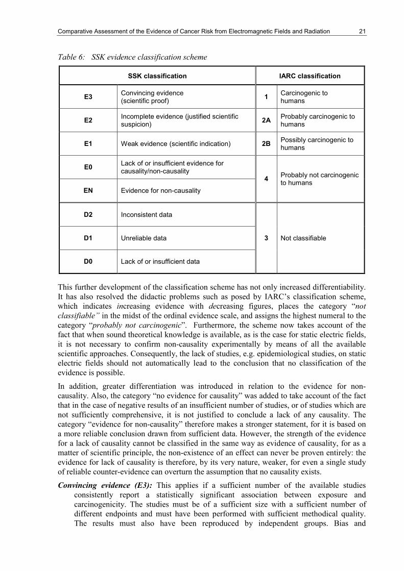
Comparative Assessment of the Evidence of Cancer Risk from Electromagnetic Fields and Radiation 21
Table 6: SSK evidence classification scheme
SSK classification IARC classification
E3 Convincing evidence (scientific proof)
1 Carcinogenic to humans
E2 Incomplete evidence (justified scientific suspicion)
2A Probably carcinogenic to humans
E1 Weak evidence (scientific indication) 2B Possibly carcinogenic to humans
E0 Lack of or insufficient evidence for causality/non-causality
4 Probably not carcinogenic to humans
EN Evidence for non-causality
D2 Inconsistent data
D1 Unreliable data 3 Not classifiable
D0 Lack of or insufficient data
This further development of the classification scheme has not only increased differentiability. It has also resolved the didactic problems such as posed by IARC’s classification scheme, which indicates increasing evidence with decreasing figures, places the category “not classifiable” in the midst of the ordinal evidence scale, and assigns the highest numeral to the category “probably not carcinogenic”. Furthermore, the scheme now takes account of the fact that when sound theoretical knowledge is available, as is the case for static electric fields, it is not necessary to confirm non-causality experimentally by means of all the available scientific approaches. Consequently, the lack of studies, e.g. epidemiological studies, on static electric fields should not automatically lead to the conclusion that no classification of the evidence is possible.
In addition, greater differentiation was introduced in relation to the evidence for non-causality. Also, the category “no evidence for causality” was added to take account of the fact that in the case of negative results of an insufficient number of studies, or of studies which are not sufficiently comprehensive, it is not justified to conclude a lack of any causality. The category “evidence for non-causality” therefore makes a stronger statement, for it is based on a more reliable conclusion drawn from sufficient data. However, the strength of the evidence for a lack of causality cannot be classified in the same way as evidence of causality, for as a matter of scientific principle, the non-existence of an effect can never be proven entirely: the evidence for lack of causality is therefore, by its very nature, weaker, for even a single study of reliable counter-evidence can overturn the assumption that no causality exists.
Convincing evidence (E3): This applies if a sufficient number of the available studies consistently report a statistically significant association between exposure and carcinogenicity. The studies must be of a sufficient size with a sufficient number of different endpoints and must have been performed with sufficient methodical quality. The results must also have been reproduced by independent groups. Bias and

Comparative Assessment of the Evidence of Cancer Risk from Electromagnetic Fields and Radiation 22
confounding can be excluded with sufficient certainty, and the results must be convincingly supported by established theoretical knowledge.
Incomplete evidence (E2): This applies if only a limited number of studies is available, but these predominantly report a statistically significant association between exposure and carcinogenicity. The studies may be of limited size with an insufficient number of different endpoints, but must have been performed with sufficient methodical quality. The results must also have been reproduced, at least in part, by independent groups. Bias and confounding should be low. It must be possible to explain the results in terms of established theoretical knowledge.
Weak evidence (E1): This applies if an insufficient number of studies is available, with an insufficient number of endpoints studied. The methodical quality and size of the studies are often limited. The results have hardly been reproduced by independent groups and, predominantly, do not report any statistically significant association between exposure and carcinogenicity. Bias and confounding cannot be excluded. A causal connection is not based on proven mechanisms but can be supported by hypotheses which are not in conflict with established theoretical knowledge.
Lack of or insufficient evidence (E0) for the existence or non-existence of causality: This applies if only a limited number of studies is available, but they predominantly report a lack of a statistically significant association between exposure and carcinogenicity. The studies may be of limited size with an insufficient number of different endpoints but must have been performed with sufficient methodical quality. Furthermore, the results must have been reproduced, at least in part, by independent groups. Bias and confounding should be low. It must be possible to explain the results in terms of established theoretical knowledge.
Evidence for non-causality (EN): This applies if a sufficient number of the studies available consistently report no statistically significant association between exposure and carcinogenicity. The studies must be of a sufficient size with a sufficient number of different endpoints and must have been performed with sufficient methodical quality. The results must also have been reproduced by independent groups. Bias and confounding can be excluded with sufficient certainty, and the results must be convincingly supported by established theoretical knowledge.
In addition, for those cases in which the existing data do not permit any evaluation of the evidence, the following three categories (“inconsistent data”, “unreliable data” and “lack of or insufficient data”) were introduced for the purpose of more sophisticated differentiation of the data situation:
Inconsistent data (D2): This applies if studies report conflicting or inconsistent results relating to an association between exposure and carcinogenicity. These studies have not been reproduced by independent groups, and bias and confounding cannot be excluded.
Unreliable data (D1): This applies if available studies are of an insufficient size and were performed with insufficient methodical quality, with an insufficient number of different endpoints. Bias and confounding are probable.
Lack of or insufficient data (D0): No studies exist, or the number of studies is inadequate.
Table 7 summarises the criteria for the classification of evidence. It should be borne in mind that these criteria do not carry the same weight for every evidence category, so not all criteria need to be fulfilled simultaneously. The consistency of results and their confirmation by reproduction are particularly important. For example, even if a large number of studies is
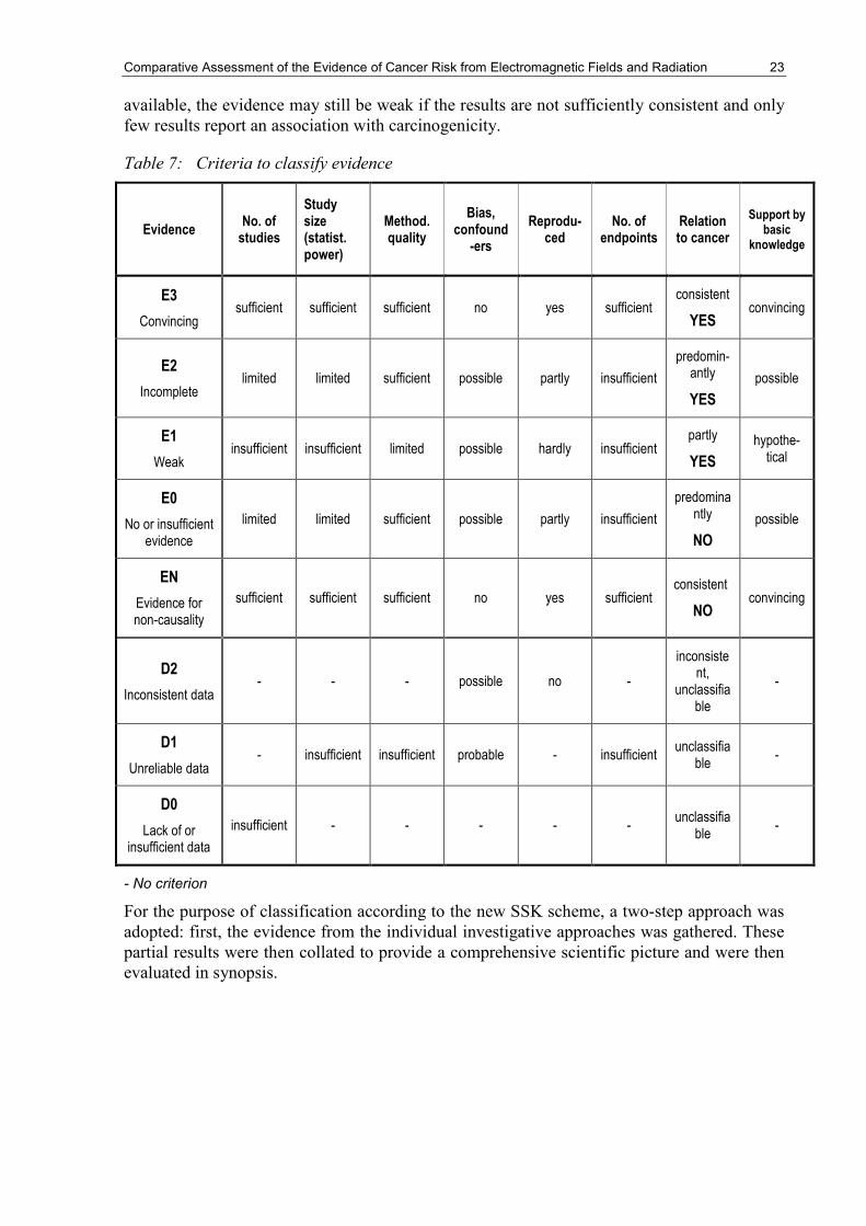
Comparative Assessment of the Evidence of Cancer Risk from Electromagnetic Fields and Radiation 23
available, the evidence may still be weak if the results are not sufficiently consistent and only few results report an association with carcinogenicity.
Table 7: Criteria to classify evidence
Evidence No. of studies
Study size (statist. power)
Method. quality
Bias, confound-ers
Reprodu-ced
No. of endpoints
Relation to cancer
Support by basic
knowledge
E3
Convincing sufficient sufficient sufficient no yes sufficient
consistent
YES convincing
E2
Incomplete limited limited sufficient possible partly insufficient
predomin-antly
YES
possible
E1
Weak insufficient insufficient limited possible hardly insufficient
partly
YES
hypothe-tical
E0
No or insufficient evidence
limited limited sufficient possible partly insufficient
predominantly
NO
possible
EN
Evidence for non-causality
sufficient sufficient sufficient no yes sufficient consistent
NO convincing
D2
Inconsistent data - - - possible no -
inconsistent,
unclassifiable
-
D1
Unreliable data - insufficient insufficient probable - insufficient
unclassifiable
-
D0
Lack of or insufficient data
insufficient - - - - - unclassifia
ble -
- No criterion
For the purpose of classification according to the new SSK scheme, a two-step approach was adopted: first, the evidence from the individual investigative approaches was gathered. These partial results were then collated to provide a comprehensive scientific picture and were then evaluated in synopsis.

Comparative Assessment of the Evidence of Cancer Risk from Electromagnetic Fields and Radiation 24
6 Frequency ranges
6.1 Ionising radiation
6.1.1 Physical interaction mechanisms
The principle governing the primary physical action and the ensuing chemical action of ionising radiation is generally understood (Kraft and Krämer 1993; Nüsslin and Kneschaurek 2009). As the name suggests, the main property of ionising radiation is that it consists of radiation quanta (photons) which carry sufficient quantum energy to release an electron from an atom or molecule (= ionisation).
Often, ionisation of a molecule is followed by the release of a positively charged particle (often an H+ ion), making the molecule now a free radical, i.e. an yet uncharged molecule which contains a single unpaired electron in its electron shell. Molecules with unpaired electrons are highly chemically reactive and generally form chemical bonds with other molecules within fractions of a second. These chemical reactions are particularly critical if they affect genetic information (DNA). Other molecules (e.g. membrane molecules) can also be damaged by free radical attack, which contributes to the radiation risk. The described pathway, involving free radical mechanisms, plays a particularly important role after exposure to sparsely ionising radiation, i.e. radiation with relatively low linear energy transfer (LET) such as gamma radiation or X-rays. A proportion of the radiation effect also comes from direct energy deposition in DNA, but this physical mechanism occurs far more frequently in the context of particle radiation (i.e. through exposure to neutrons or alpha particles), which is not considered here.
6.1.2 Biological interaction mechanisms
DNA damage caused at the physical/chemical level is considered to be primarily responsible for carcinogenesis. However, whether cancer can be caused by a changing the DNA in just one single cell (“monoclonal”) or whether several cells must be altered (“polyclonal”) remains a contentious issue (Parsons 2008; Tanooka 2004). Recent research indicates that carcinogenesis is a multi-step process, and that cancer is not caused by one single change to DNA (Coleman and Tsongalis 2006, Karakosta et al. 2005).
As cancer cells show a strong tendency to uncontrolled cell proliferation, it is not surprising that oncogenes (= mutated forms of proto-oncogenes) and mutated tumor suppressor genes play a key role. Proto-oncogenes encode proteins which stimulate cell division, whereas the proteins encoded in tumor suppressor genes inhibit cell division. When the proto-oncogenes and the tumor suppressor genes operate normally, the cell cycle is perfectly controlled and the number of cells in the body is held in check. If mutations occur in these genes, however, this balance can be disrupted, resulting in overproduction of cells and consequently tumor formation.
A large number of more or less detailed models of carcinogenesis have been proposed (Cox and Huber 2007; Jacob et al. 2010). Using computer simulations, these models are intended to address very specific questions relating to the underlying mechanisms. For example, some studies investigate the role of genetic predisposition, genomic instability or immune defence in cancer initiation, promotion and progression. “Epigenetic” mechanisms of carcinogenesis are also the subject of discussion.

Comparative Assessment of the Evidence of Cancer Risk from Electromagnetic Fields and Radiation 25
6.1.3 Dose-effect relationship
For ionising radiation, the primary dose quantity is the “energy dose”. This is the radiation energy absorbed per (tissue)mass. The SI unit for absorbed dose is gray (Gy); 1 Gy = 1 J/kg. However, different types of radiation (e.g. X-rays or particle beams such as neutron or alpha rays) can cause different biological effects even with the same energy dose. In an attempt to account for this diversity in a dose quantity, the energy dose is additionally weighted. For the “operational” dose equivalent quantities, the weighting takes place via a quality factor (sometimes referred to as a weighting factor), producing a “dose equivalent”. In order to obtain “protection values”, weighting takes place using radiation weighting factors, resulting in an organ dose. For sparsely ionising EMF (photon radiation), both the quality factor and the radiation weighting factor have the value 1, so that energy dose, dose equivalent quantity and organ dose are numerically equal. In order to indicate that this is a weighted dose quantity, both the dose equivalent and the organ dose are measured in units of sievert (Sv).
In the dose range from around 100 mSv to approx. 2 000 mSv, a linear association with cancer is well-documented. Above around 2 000 mSv, the dose-effect relationship flattens out. At doses below approx. 100 mSv, any potentially existing radiation-induced cancer risk in adults can no longer be significantly distinguished from the spontaneous cancer frequency. Within certain limits, therefore, there is certainly scope for differing opinions on how the risk observed in a dose range above 100 mSv continues in the dose range below that value (Figure 1).
The lifetime risk (i.e. the probability of developing or dying from cancer in the course of a lifespan) for a radiation-exposed cohort depends primarily on the time-integral of the dose rates (dose) and thus exhibits a dose-effect relationship. However, exposure distribution over time may also have a bearing. The excess absolute lifetime risk of a harmful event (e.g. cancer incidence or mortality) resulting from exposure is calculated as the difference between the lifetime risk and the background lifetime risk. The excess absolute lifetime risk is thus the term used to describe the additional lifetime risk of developing or dying from radiation-induced cancer compared with the normal background cancer risk.
For ionising radiation, the excess absolute lifetime risk increases with dose.
In radiological protection, the linear no-threshold (LNT) model is a method for describing the effects in the low dose region. At present, it is not possible to verify or refute this model. Furthermore, there are certainly arguments in favour of the other curves depicted in Figure 1, e.g. supra-linear, threshold dose (in the single-digit to double-digit mSv range) and hormesis (i.e. the hypothesis that low doses of ionising radiation are beneficial, stimulating repair mechanisms and thus protecting against disease).

Comparative Assessment of the Evidence of Cancer Risk from Electromagnetic Fields and Radiation 26
D
R
mSv100
0
verringertes Risiko
erhöhtes Risiko
0
a
b
c
LNT d
Fig. 1: Depiction of excess risk R, depending on radiation dose D, with various
hypotheses, extrapolated from the range above approx. 100 mSv and applied to the
low radiation dose range.
Area above the x-axis: increase in excess risk resulting from radiation exposure;
area below the x-axis: risk reduced by exposure (preventive effect), LNT: linear no-
threshold extrapolation, a: overproportional, b: threshold dose, c: protective effect
in the low dose range (hormesis), d: linear-quadratic
It must be noted that for short-term (acute) exposure sufficiently exceeding the level of natural background radiation, a clear dose dependency can be observed, with an increase in cancer incidence caused by ionising radiation10.
6.1.4 Evidence
In vitro
As described above (Section 6.1.2), DNA damage plays a key role in tumor formation. DNA damage can take the form of point mutations, small deletions and chromosome damage (such as fragments, dicentric chromosomes, or translocations). All these effects of exposure to ionising radiation on cells have been conclusively demonstrated in vitro (Kiefer et al. 1999; Obe and Vijayalaxmi 2007). It has also been demonstrated that there is a close relationship between these mutations and tumorigenesis (Hagmar et al. 1998).
In vitro analyses on transformation reveal an even closer relationship with cancer. Transformed cells are distinguished from normal cells by many different properties. One example is loss of contact inhibition, which means that unlike normal cells, there is no longer any cessation of cellular growth and division even when transformed cells are completely surrounded by neighbouring cells. Transplantation of transformed cells into immuno-compromised animals was found to result in tumor formation in these animals. In vitro studies have provided evidence of the transformations caused by ionising radiation (Redpath 2004).
In vivo
Ionising radiation can cause tumors also in animals (Broerse et al. 1985, 1989). Studies have therefore been carried out on various species of animal, primarily in order to obtain 10 A detailed discussion of dose-effect relationships in the context of radiation-induced cancer and leukaemia
cases can be found in UNSCEAR 2006 and SSK 2007.
increased risk
reduced risk

Comparative Assessment of the Evidence of Cancer Risk from Electromagnetic Fields and Radiation 27
information about the mechanisms underlying radiation-induced carcinogenesis (UNSCEAR 1993).
One difficulty which arises in this context is that the spectrum of tumor types induced by ionising radiation is extremely diverse. For example, different strains of mice show different responses when exposed to radiation, developing different types of tumor and displaying different dose-effect relationships. This makes extrapolation to humans difficult. However, the fact that ionising radiation can cause tumors in animals is undisputed.
Epidemiology
Epidemiological studies clearly show that ionising radiation can cause cancer, including leukaemia (UNSCEAR 2006). The following description relates primarily to electromagnetic ionising radiation (X-ray and gamma radiation), but also makes reference to particle radiation in some cases (such as data from Hiroshima and Nagasaki). Studies which primarily report on results obtained from subjects exposed to particle radiation (such as those carried out among uranium miners) are not considered here. There is epidemiological evidence that exposure to an acute dose of ionising radiation from above approx. 100 mSv to approx. 2,000 mSv in adults (Preston et al. 1994; Preston et al. 2007) and above 10 mSv in the foetus (Wakeford and Little 2002) increases tumor frequency proportionally to dose. This observation underlines the foetus’s greater sensitivity to radiation. However, this does not necessarily mean that lower doses are also carcinogenic, but there is a lack of reliable data relating to chronic exposure in the 0.1 µSv/h – 1 µSv/h range (= single-digit mSv/a).
In adults, with acute exposures exceeding approx. 100 mSv, there is a statistically significant linear association between ionising radiation and tumor frequency. The analyses of data from Hiroshima and Nagasaki show that mortality rates for cancer generally increase by around 7 % following an acute dose of 1 Sv to a population, adjusted for age and time after exposure (UNSCEAR 2006). If the conservative assumption made in radiological protection is correct – namely that this risk can be linearly extrapolated with no threshold dose until zero dose– the spontaneous frequency of dying from cancer due to a dose of 1 mSv would increase from around 25 % to 25.007 %. Because of the large variability in the spontaneous frequency of cancer, it is apparent that this minimal increase cannot be verified by means of epidemiological studies. Only if medical exposure which already occurs with relevant frequency, such as X-ray/CT applications ranging from a few mSv to around 20 mSv per procedure, would have been included as well convincing epidemiological evidence (E3) of an association could be provided.
6.1.5 Exposure
Public exposure to ionising radiation comes from a number of sources: from soil, food and air, and from extraterrestrial sources, including solar and cosmic radiation, with the level of exposure amounting to a few mSv/a, depending on place of residence and lifestyle. Over the
years, this adds up to the lifetime dose. Radiation exposure of a similar order of magnitude also occurs during medical procedures, primarily X-ray diagnosis.
6.1.6 Overall assessment of the evidence
Taking account of lifetime dose, and despite the uncertainty regarding the LNT hypothesis, there is convincing evidence (E3) for an association between cancer risk and exposure to ionising radiation (Table 8).

Comparative Assessment of the Evidence of Cancer Risk from Electromagnetic Fields and Radiation 28
Table 8: Overall assessment of the evidence for ionising radiation (IS)
Physical
interaction
mechanisms
Biological
interaction
mechanisms
Dose-
effect
In vitro
studies
In vivo
studies
Epidem.
studies
Total
evidence
IS E3 E3 E3 E3 E3 E3 E3
6.2 UV radiation
Ultraviolet (UV) radiation is electromagnetic radiation with wavelengths from 100 nm to 380 nm, the border to visible (blue) light. Its propagation follows the laws of geometrical optics. Due to the frequency dependence of its effects, UV radiation is further subdivided into the wavebands UVC (100 nm - 280 nm), UVB (280 nm - 315 nm) and UVA (315 nm - 380 nm).
6.2.1 Physical interaction mechanisms
The principle governing the primary physical action and the subsequent chemical effects of UV radiation are generally understood. Two reaction pathways can be distinguished:
a) Direct reaction pathway (mainly UVC and UVB radiation)
In this reaction pathway, UV photons are directly absorbed by the DNA molecule, and the absorbed energy induces a photodimerisation reaction of adjacent pyrimidine bases. This generally produces two UV-specific photoproducts: the cyclobutane pyrimidine dimer (CPD) and the pyrimidine(6-4)pyrimidone photoproduct (6-4PP). In accordance with the UV action spectrum, UVB radiation is around 1,000 times more effective in the induction of these DNA lesions than UVA radiation (Rosenstein and Mitchell 1987). However, the level of (6-4) photoproduct production is about three-fold lower than dimer induction (Mitchell et al. 1990). In irradiation of cell cultures, a dose of just 300 J/m2 UVB (broadly equivalent to a minimal erythemal dose (MED) in vivo in individuals with skin type II) was sufficient to produce several 100,000 CPDs in the genome of human keratinocytes in vitro (Greinert et al. 2000).
b) Indirect reaction pathway (mainly UVA radiation)
UVA radiation mainly damages the DNA molecule along indirect reaction pathways. The energy of UVA photons is absorbed by photosensitive endogenous cellular chromophores (photosensitisers) such as riboflavin and NADH. In the electron’s excited (triplet) state, via Type I and Type II photoreactions in interaction with molecular oxygen (whose ground state is a triplet), these can then form reactive oxygen species (ROS), such as superoxide anions, H2O2, OH radicals or singlet oxygen. These reactive oxygen species (ROS) can then cause single-strand breaks (SSB) in the DNA, DNA-protein crosslinks, alkali-labile sites or base modifications, such as the UVA-specific 8-oxoguanine (Matsumura and Ananthaswamy 2002). Hence the spectrum of UVA-induced, DNA-damaging molecular species and the DNA damage profile induced by them are very similar to the indirect effect of ionising radiation (Lehnert and Iyer 2002; von Deutsch et al. 2005).
6.2.2 Biological interaction mechanisms
If CPDs in the cell’s genome are not removed by nucleotide excision repair (NER), they can lead to C→T or CC→TT transition mutations, known as “UV signature mutations”, for in the

Comparative Assessment of the Evidence of Cancer Risk from Electromagnetic Fields and Radiation 29
broad spectrum of all the possible DNA mutations, these are, in the main, unique to UV radiation (Matsumura and Ananthaswamy 2002). These mutations have been identified in skin tumors in a range of tumor suppressor genes and proto-oncogenes (e. g. patched, p16, ras, p53) which play a key role in the etiology of basal cell carcinoma (BCC), squamous cell carcinoma (SCC) and malignant melanoma (MM) (Wikonkal and Brash 1999).
UVA-induced premutagenic DNA lesions can contribute to the occurrence of mutations (e.g. T→G transversions caused by 8-oxoguanine) which may be responsible for the development of skin cancer (Matsumura and Ananthaswamy 2002, Dumaz et al. 1997, de Gruijl et al. 1993).
Furthermore, new studies show that UV-induced DNA lesions such as the generation of cyclobutane pyrimidine dimers (CPDs) and single-strand breaks (SSBs) can be converted into DNA double-strand breaks dependently of replication (Limoli et al. 2002) and that UVA is even able to produce DSBs independently of replication (Greinert et al. 2004). The underlying mechanisms are not yet fully understood, but it is clear that UVA radiation is able to induce phosphorylation of the histone protein H2AX (γ-H2AX) and contribute to the formation of γ-H2AX foci in the cell nucleus, which are characteristic for DNA DSBs (Thompson and Limoli 2003). DSBs may be responsible for radiation-induced genomic instability which can manifest at chromosomal level in the form of aberrations and deletions. UV radiation therefore plays an important role in these processes, which are closely associated with skin cancer.
New studies show, furthermore, that UVA radiation is also able to produce CPDs, albeit with less significant yield than UVB radiation. The mechanisms of CPD formation following exposure to UVA radiation are not yet fully understood. It is significant, however, that CPDs are the predominant premutagenic DNA lesions in human skin exposed to UVA radiation (Mouret et al. 2006). Given that 95 % of solar UV radiation falls within the UVA range, this finding is particularly significant when assessing the risks posed by solar (but also artificial) UV radiation in relation to skin carcinogenesis.
It is becoming increasingly apparent that UV-induced mutations are of particular significance for skin carcinogenesis when they occur in adult epidermal stem cells (Mitchell et al 2001a, Mitchell et al 2001b, Gambardella and Barrandon 2003, Bickenbach and Holbrook 1987, Braun and Watt 2004, Cairns 1975, Cairns 2002, Potten et al. 2002).
The mutagenic and carcinogenic effect of UV radiation has been shown in a very large number of publications (see also (SSK 2008b)). The wealth of findings in the fields of molecular biology, cell biology and epidemiology prompted IARC, in 2009, to classify UV radiation in Group 1 (“carcinogenic to humans”) (El Ghissassi et al. 2009).
6.2.3 Dose-effect relationship
The fact that UV radiation causes skin cancer – both non-melanocytic types such as basal cell carcinoma (BCC) and squamous cell carcinoma (SCC), and malignant melanoma (MM) – is now undisputed. There is a substantial body of molecular biological and epidemiological evidence for this, and compared with the cause-effect relationships for other cancer entities, the evidence is sound. However, there is still a lack of well-designed studies which document the link between UV dose and the incidence of skin cancer (and thus determine the excess risks that are important for radiological protection). This poses a problem from a radiation hygiene perspective.

Comparative Assessment of the Evidence of Cancer Risk from Electromagnetic Fields and Radiation 30
Epidemiological studies, and especially environmental studies11, point to a link between UV exposure and skin cancer, but with clear differences between BCC, SCC and MM. To date, no targeted studies which specifically investigate the dose dependence of skin cancer incidence for the various skin cancer entities have been carried out. Individual findings provide indications but do not yet appear to be sufficient (Armstrong and Kricker 2001). Other studies on dose-effect relationships refer to latitude dependences, but it is often impossible to ascertain, to an adequate extent, the UV dose (erythema-weighted, physical dose, UVB, UVA, UVB + UVA, etc.) on which the results are based.
Some years ago, De Gruijl et al., using a mouse model, described a non-linear relationship between UV dose, time after exposure, and skin cancer incidence (de Gruijl et al. 1993). However, the applicability of these findings to humans is still uncertain.
New data from satellite-based measurements of weighted wavelength-dependent UV irradiance levels with high spatial resolution (cf. www.EUROSUN.org), combined with valid data for skin cancer incidence (e.g. from the German Skin Cancer Screening Programme) should contribute to considerably improve the data situation.
It may be assumed that cumulative UV dose is responsible for the induction of SCC, whereas intermittent strong UV exposure (e.g. sunburn) is clearly the primary cause for MM. BCC has intermediate status in regard to UV exposure patterns (Armstrong and Kricker 2001).
6.2.4 Evidence
In vitro
The in vitro evidence for an association between UV radiation and skin carcinogenesis is convincing (SSK 2008b), as the following examples show:
- UV-specific “signature mutations” (C→T, transition mutations) were identified in the p53, PTCH and smoothened genes (Aszterbaum et al. 1999a, Aszterbaum et al. 1999b, Daya-Grosjean and Sarasin 2000, Evans et al. 2000, Ratner et al. 2001, Couve-Privat et al. 2002, Kim et al. 2002). This finding must be seen as a further important indicator of the significance of UV exposure for the development of BCC. UV-induced p53 mutations in skin cells accumulate in “hot spots”, which differ from those found in internal tumors. There are indications that UV-specific mutations of the p53 gene could be specific to BCC (SSK 2008b).
- For the etiology of SCC, a relatively well-described model exists, in which early UV-specific mutations in the p53 gene during the phase of tumor initiation are conducive to the development of actinic keratoses (AK), i.e. pre-cancerous skin lesions with the potential to develop into SCC. It is assumed that in AK, only one allele of the p53 gene is initially mutated. This impedes p53-dependent apoptosis in UV-damaged cells (“sunburn cells”). As neighbouring cells undergo normal apoptosis at the same time, p53-mutated cells have a selection advantage, which may lead to clonal expansion of these cells into AK. If, in these cells, the second p53 allele is mutated during the phase of tumor promotion, the p53-dependent cell cycle checkpoint function is disabled. Uncontrolled cell growth occurs, with the induction of further (possibly UV-induced) mutations in other genes (e.g. ras) during the phase of tumor progression and the formation of invasive SCC (Cleaver and Crowley 2002, Ziegler et al. 1994, Brash 1997).
11 Studies in the field of environmental epidemiology collect data relating to location-specific, rather than
individual, factors that affect health.

Comparative Assessment of the Evidence of Cancer Risk from Electromagnetic Fields and Radiation 31
- It is now accepted that 50 % – 60 % of all melanomas exhibit BRAF mutations, of which 90 % lead to a valine-to-glutamate mutation in codon 600 (BRAFV600E). These BRAF mutations cause kinase activation in the constitutive MAPK pathway (Sondak and Smalley 2009). Phosphorylation of tumor suppressor LKB1 (a serine/threonine protein kinase) initiates its negative regulation, contributing to proliferation of melanoma cells and a weakening of the apoptotic response to metabolic stress (Lopez-Bergami 2009, Zheng et al. 2009, Esteve-Puig et al. 2009).
In contrast to SCC and BCC, UV-induced mutations in the p53 gene appear to be less significant in melanomas. Only around 20 % of malignant melanomas exhibit p53 mutations (Zerp et al. 1999). There are indications that the role of p53 in the etiology of malignant melanoma is complex (Whiteman et al. 1998) and requires more detailed investigation. Other mechanisms, such as the induction of genetic instability, may possibly play a more important role.
The etiology of MM is characterised by a high degree of UV-induced genomic instability, which increases throughout MM development until metastasis. Genomic instability manifests as a gain or loss of chromosomes (or portions of chromosome), the occurrence of chromosomal aberrations and loss of heterozygosity (LOH). Depending on the location – the eye or skin – two genetically distinct subtypes can be identified. Losses of chromosome 3 and 1p and gains at 8q are often observed in melanomas of the eye, whereas gains of chromosome 6p and losses of chromosome 6q appear to be specific to skin melanomas (Hoglund et al. 2004). Studies of melanoma metastasis cell lines using spectral karyotyping (SKY) have revealed that genomic instability at chromosomal level in late stage metastatis can be so marked that almost every chromosome is affected by numerical or partially complex structural aberrations (Greinert et al. 2004).
Recent studies provide firm evidence of the association between UV exposure and the development of malignant melanoma. In 2010, Pleasance et al. 2010 produced the first comprehensive catalogue of somatic mutations in the genomes of a malignant melanoma (Pleasance et al. 2010). This showed that the majority (approx. 70 %) of the detected single base substitutions were type C→T and approx. 70 % of dinucleotide substitutions were type CC→TT. As it is known that these are signature mutations induced by UV radiation, this finding provides very significant evidence for the association between malignant melanoma and UV exposure.
In vivo
Many experimental studies focus on the association between UV radiation and skin carcinogenesis (NRPB 2002). However, there is still no recognised animal model for UV-induced skin carcinogenesis that is specific to malignant melanoma (Noonan et al. 2003). However, for squamous cell carcinoma (SCC), an action spectrum was established for a nude mouse model which shows that alongside UVB radiation as the main factor, UVA radiation also contributes to the induction of SCC (de Gruijl et al 1993, de Gruijl 1995 ).
Older studies support the finding that there is UVA involvement in the development of malignant melanoma (MM), both in a fish model (Xiphophorus) and in opossum (Monodelphis domestica) (Setlow et al. 1993, Ley 1997, Wang 2008, Schartl et al. 1997, Robinson et al. 2000).
In transgenic mice, neonatal UV irradiation was found to induce the development of malignant melanoma (Noonan et al. 2003). It was found that UVB rather than UVA radiation is responsible for the induction of MM in this animal model (De Fabo et al. 2004, van Schanke et al. 2005).

Comparative Assessment of the Evidence of Cancer Risk from Electromagnetic Fields and Radiation 32
Transgenic mice (hupki mice) in which the DNA binding domain of the p53 gene is replaced by the homologous human sequence have been used for mutation analysis following chronic UVB irradiation and are regarded as a new experimental tool in the fields of molecular epidemiology and biomedical sciences (Luo et al. 2001).
Older experiments using the mouse model provided the initial bases for developing an understanding of the cellular mechanisms responsible for UV-induced immunosuppression (Kripke 1974, Ullrich 2005, Ullrich 2007).
A number of studies were also carried out on “knockout” (KO) mice which lack nucleotide excision repair genes, such as those which are significant in Cockayne syndrome or Xeroderma Pigmentosum (CSB-/-, CSB+/-, XPA-/-, XPC-/-) (van der Horst et al. 1997, Berg et al. 1998), in order to gain a better understanding of their role in skin carcinogenesis. This was also studied in KO mice in which genes which encode for specific reaction pathways of relevance to skin carcinogenesis have been inactivated; e.g. in p53-/- mice (for SCC) (Li et al. 1996), ptch -/- mice (for BCC) (Aszterbaum et al. 1999b, Aszterbaum et al. 1999a) and Ink4a -/- mice for malignant melanoma (Serrano et al. 1996).
Based on the findings of these and many other (also older) studies, the International Agency for Research on Cancer (IARC) stated as early as 1992 that there is convincing evidence that broad-spectrum UV radiation and the individual wavelength ranges UVC, UVB and UVA are carcinogenic in the animal model (IARC 1992).
Epidemiology
Skin cancer – comprising non-melanocytic types (basal cell carcinoma (BCC) and squamous cell carcinoma (SCC)) and malignant melanoma (MM) – is the most common type of cancer in white populations worldwide, and its incidence has been steadily increasing in recent decades. Incidence rates of malignant melanoma have increased more rapidly than any other form of cancer, with rates doubling every 10-15 years. This also applies to Germany. According to new data from the Schleswig-Holstein Cancer Registry (2007), every year in Germany, around 195,000 people are newly diagnosed with skin cancer (approx. 24,000 people are diagnosed with MM, and more than 170,000 people are diagnosed with BCC and SCC).
The epidemiological evidence that both natural (solar) and artificial UV radiation (solariums) can cause skin cancer is unequivocal (SSK 2008b). For both the non-melanocytic types of skin cancer (NMSC) and malignant melanoma, constitutive risk factors (groups) and risk exposure factors for the effects of UV exposure have been clearly identified (IARC 1992, Gandini et al. 2005a, Gandini et al. 2005b). These are:
- for NMSC: skin type, already chronically UV-damaged skin, actinic keratosis, personal history of non-melanocytic skin cancer, immunosuppression, cumulative damage following exposure to X-rays, and individual (epidermal) stem cell pool.
- for MM: skin type, personal or family history of melanoma, number of (UV-induced) nevi, clinically atypical nevi, congenital nevi.
Epidemiological studies show that UV radiation is the dominant external risk factor for the development of all types of skin cancer. There are differences, however, in the UV exposure pattern in terms of its responsibility for the various types of skin carcinogenesis.
6.2.5 Exposure
The general public is exposed to UV radiation at levels which are relevant to carcinogenesis, sometimes at sufficiently high levels that active protection is required to prevent acute effects. The main source of exposure is solar radiation. Regular visits to solariums lead to additional

Comparative Assessment of the Evidence of Cancer Risk from Electromagnetic Fields and Radiation 33
relevant contributions. Individuals who mainly work outdoors have higher levels of occupational exposure to UV radiation.
6.2.6 Overall assessment of the evidence
Overall, there is convincing evidence for an association between exposure to UV radiation and skin carcinogenesis (Table 9).
Table 9: Overall assessment of the evidence for UV radiation
Physical
interaction
mechanisms
Biological
interaction
mechanisms
Dose-
effect
In vitro
studies
In vivo
studies
Epidem.
studies
Total
Evidence
UV E3 E31)
E2 E3 E31)
E3 E3
E3: convincing evidence 1) for UVA the evidence is incomplete (E2)
E2: incomplete evidence
6.3 Visible light
Visible light is the narrow band of the electromagnetic spectrum between 380 nm and 780 nm, in which photons can be perceived by sensory cells in the human retina.
6.3.1 Physical interaction mechanisms
Photons of visible light can be absorbed by many different biologically important molecules (e.g. in the photoreceptors in the retina, and in the skin cells) such as protoporphyrins or porphyrins in certain proteins or other cellular “photosensitisers”, some of which have not yet been precisely characterised (Hoffmann-Dörr et al. 2005, Kielbassa et al. 1997). Through the induced excitation and via Type I / Type II photoreactions or Förster-type energy transfer, reactive oxygen species (ROS) may be generated (Hoffmann-Dörr et al. 2005, Kielbassa et al. 1997), whose carcinogenic effect is well-known. Here, the wavelength range of blue light between 400 nm and 450 nm appears to be of particular significance (Kielbassa et al. 1997).
6.3.2 Biological interaction mechanisms
Recent research shows very clearly that skin carcinogenesis is a multi-stage process and that it takes more than a single mutation in the DNA to cause cancer (Coleman and Tsongalis 2006, Karakosta et al. 2005). Reactive oxygen species (ROS) which can also be generated by visible light (especially within 400 nm - 450 nm) are a recognised risk factor for carcinogenesis (Mena et al. 2009). At present, however, there are no reliable data showing that ROS induced by visible light would, via a specific cancer-associated reaction pathway, lead to mutations which might be associated with carcinogenesis.
Initial studies indicate that on the one hand, the action spectrum for carcinogenesis ranges from the UV band to the shortwave band of visible light, and that on the other hand, the action spectrum for infrared radiation could extend into the longwave band of visible light (ICNIRP 2004). The extension of the action spectra from the UV into the blue light range exhibits a disproportionate reduction in biological effectiveness, amounting to more than 4 orders of magnitude compared with the maximum in the UV range, but in view of the omnipresent exposure to visible light, the number of exposed persons and exposure duration are greater than with UV exposure.

Comparative Assessment of the Evidence of Cancer Risk from Electromagnetic Fields and Radiation 34
The photoreceptors responsible for vision (rods and cones) contain specific photopigments (11-cis-retinal and certain proteins), with absorption maxima in the range of green light (rods, adapted to light and dark), and in the ranges of red, green and blue light (cones, colour perception). The high density of the receptors, especially in the fovea centralis, results in high spatial resolution of optical radiation. Besides vision, light also plays a central role in maintaining circadian rhythms (“internal clock”). Melatonin synthesis is suppressed by light, resulting in characteristic fluctuations of melatonin concentrations in the blood, with high levels at night and barely measurable levels by day. These melatonin rhythms, in turn, control other biological processes: they are important, for example, for regulating body temperature and sleep. The maximal spectral sensitivity for melatonin suppression lies in the range of blue light and is quite separate from the maximum spectral sensitivity for light perception, such that the existence of a further photoreceptor, independent of the classic photoreceptor for vision, was postulated some time ago. And indeed, it has been shown, in recent years, that neuronal cells (ganglion cells) in the retina can also absorb light. The photopigment of photoreceptive ganglion cells is melanopsin, which absorbs light mainly in the range of blue light. Unlike cones and rods, these ganglion cells and their dendrites exhibit excessive branches and do not play a role in the perception of objects (vision) but act as light sensors for circadian rhythms.
As melatonin is a very effective scavenger of free radicals, neutralising the highly aggressive hydroxyl (OH-) radicals in particular, the possibility that melatonin suppression is associated with increased cancer incidence has been under discussion for some time and generally accords with the findings of epidemiological studies and animal experimental data (Mediavilla et al. 2010).
6.3.3 Dose-effect relationship
The dependence of melatonin suppression on light at various wavelengths and intensities is well-documented (e.g. Flynn-Evans et al. 2009).
The dose quantity for evaluation of long-term exposure in relation to carcinogenesis is still unclear, however, although a dose-proportional increase in oxidative base modifications and DNA single-strand breaks in human skin cells could be demonstrated.
6.3.4 Evidence
In vitro
There are no in vitro studies which focus on chronodisruption, nor can any such studies be anticipated, as cells or cell systems are not suitable models for this type of study. To date, very few experiments have shown a possible association between exposure to (blue) light and cancer induction (Di Cesare et al. 2009). No evidence is available for the remaining visible light spectrum. The in vitro evidence must therefore be classified as insufficient.
With regard to photochemical effects, the in vitro data indicate that there is a dose-proportional increase in oxidative DNA base modifications and DNA single-strand breaks in human skin cells (fibroblasts) and melanoma cells following exposure to visible light (Hoffmann-Dörr et al. 2005, Kielbassa et al. 1997).
In vivo
Very few studies currently available have been able to demonstrate an association between disruptions in circadian rhythms and carcinogenesis in an animal model (e.g. Filipski et al. 2004).
For skin carcinogenesis, no results of in vivo studies are available.

Comparative Assessment of the Evidence of Cancer Risk from Electromagnetic Fields and Radiation 35
Epidemiology
No results of epidemiological studies on skin carcinogenesis induced by light exposure are available.
Recent findings suggest that night-time light exposure (light-at-night – LAN) – especially among shift workers and flight attendants – is associated with increased breast cancer incidence in women. It is suspected that the disruption of circadian rhythms and hence the reduction in melatonin levels are responsible for increasing breast cancer incidence (the “melatonin hypothesis”, see overview in Stevens 2009). This is also implied by studies which found a significantly lower breast cancer incidence in blind women than in sighted controls (Flynn-Evans et al. 2009). This has resulted in an International Agency for Research on Cancer (IARC) classification of shift work as probably carcinogenic to humans (2A) if it causes disruption of circadian rhythms (IARC 2007). To what extent these findings should be attributed to the disruption in circadian periodicity or to direct effects of light, and how these findings relate to the general population, is still unclear.
Further studies are needed to clarify whether light-at-night, which causes suppression of melatonin synthesis and hence disruption of circadian rhythms (“chronodisruption”), is a risk factor for carcinogenesis in general, not only for specific occupations (shift workers, flight attendants). At present, no firm conclusion can be made as to whether also light-at-night must be regarded as a potential cancer risk factor for the general population.
6.3.5 Exposure
Exposure to light-at-night for the general population is determined on the one hand by lifestyle and habits and, on the other, by external light sources. Exposure to light-at-night is also relatively common due to a fear of darkness (e.g. in children), known as achluophobia. Exposure to light-at-night is associated with disruption of circadian rhythms in night and shift workers.
6.3.6 Overall assessment of the evidence
Overall, for visible light, the evidence for an association between exposure to light-at-night or blue light and cancer is weak. The available data for assessing a potential cancer risk associated with other types of light exposure are unreliable.
Table 10: Overall assessment of the evidence for visible light (VL)
Visible light Physical
interaction
mechanisms
Biological
interaction
mechanisms
Dose-
effect
In vitro
studies
In vivo
studies
Epidem.
studies
Total
evidence
Blue light,
light-at-night E2 E1 D0 E1 E1 E1 E1
General light
exposure E2 D1 D0 D0 D0 D0 D0
E2: incomplete evidence D1: unreliable data E1: weak evidence D0: lack of or insufficient data

Comparative Assessment of the Evidence of Cancer Risk from Electromagnetic Fields and Radiation 36
6.4 Infrared radiation
Infrared radiation is emitted by every object with a temperature above absolute zero (-273 °C). In the electromagnetic spectrum, it comprises the range right below visible light, from 0.78 µm - 1 mm and, depending on wavelength, is divided into three further segments: IRA = 0.78 µm – 1.44 µm, IRB = 1.44 µm -3 µm and IRC = 3 µm -1000 µm.
6.4.1 Physical interaction mechanisms
It can be assumed that infrared radiation may possibly contribute to carcinogenesis indirectly, first and foremost, through its thermal effects. A sufficiently prolonged and high elevation of skin temperature resulting from exposure to IR radiation is considered to be a co-carcinogen in skin carcinogenesis when combined with UV radiation, although there is no assumption that heat on its own is capable of causing relevant DNA damage (Dewhirst et al. 2003a, Dewhirst et al. 2003b).
Furthermore, infrared radiation could also act on cellular processes or DNA through non-thermal effects following IR photon absorbtion by cellular chromophores (Karu 1999).
6.4.2 Biological interaction mechanisms
In model calculations, van der Leun and de Gruijl have shown that the carcinogenic effectiveness of UV radiation on averageincreased by 3-7% per ˚C rise in temperature (van der Leun and de Gruijl 2002). The temperature increase which may be significant here could also be attributed to heat sources other than solar IR radiation, however. Due to the potentially reinforcing effect of heat, ICNIRP, for example, warns that it is important to avoid the use of warming cabins for at least 24 hours following substantial UV exposure (ICNIRP 2006).
As cellular chromophores for IRA radiation, components of the mitochondrial respiratory chain, e.g. cytochrome C oxidase (with the Cu(I), Cu(II) and Cu(III) atoms in the active centre) have been proposed (Karu 1999), which due to the absorbed radiation energy can have effects on intracellular signalling cascades, for example (Butow and Avadhani 2004). In addition, some studies show that IRA in particular is capable of inducing non-thermal effects associated with mechanisms which could be significant for carcinogenesis, namely IRA-induced gene expressions which are of significance for carcinogesis and which could possibly induce modifications which play a role in the development of cancer.
6.4.3 Dose-effect relationship
Based on the mechanisms described above, cumulative effects and hence a dose-effect relationship are possible. However, not enough experimental or epidemiological data are available about dose-effect relationships for the IR radiation spectrum and carcinogenesis.
6.4.4 Evidence
In vitro
Studies show that continuous exposure of immortal human HaCaT skin keratinocytes – which are used as in vitro models for skin carcinogenesis worldwide – to 40°C reproducibly resulted in tumorigenic conversion and tumorigenicity such as chromosomal aberrations (Boukamp et al. 1999). Furthermore, there is increasing evidence that mitochondria play a critical role in intracellular signalling cascades which are activated by IRA radiation. Retrograde mitochondrial signalling, which is a pathway of communication from mitochondria to the nucleus, appears to be significant in this context (Butow and Avadhani 2004). IRA-induced expression of matrix metalloproteinase-1 (MMP-1), which involves a mitogen-activated protein kinase (MAKP) signalling pathway, was found to be mediated by the formation of

Comparative Assessment of the Evidence of Cancer Risk from Electromagnetic Fields and Radiation 37
intracellular reactive oxygen species (ROS) and is distinct from a UVB/UVA-activated pathway (Schieke et al. 2002; Schieke et al. 2003; Schroeder et al. 2007; Schroeder et al. 2008; Schroeder et al. 2009). The role of MMP in skin ageing is well-known. The involvement of the MAPK pathway suggests that IRA-induced modifications may play a role in carcinogenesis.
The available in-vitro evidence of a possible carcinogenic potential of IR(A) radiation must therefore be classed as weak.
In vivo
There is no evidence for direct carcinogenic effects of IR from animal experiments. Early studies (Bain and Rusch 1943; Bain et al. 1943), however, demonstrated the carcinogenic role of elevated temperature. Irradiation of mice with UV radiation in the range 280 nm - 340 nm (mercury arc lamp) resulted in a higher incidence of skin cancer at temperatures of 35 °C -38 °C than at approx. 23 °C. This was later confirmed by Freeman et al. in a comparison of 32 °C with 24 °C (Freeman and Knox 1964). At higher temperatures (32 °C - 40 °C), a reduced repair rate of UV-induced CPDs in human fibroblast and melanoma cell cultures was reported, which may induce a higher mutation rate and skin cancer incidence (Goss and Parsons 1976).
Epidemiology
No reliable epidemiological studies are available.
6.4.5 Exposure
There is a high proportion of IRA radiation in the solar spectrum, amounting to around 30% of emitted total solar energy. Every-day environmental exposure to IR radiation, especially in combination with solar or artificial UV radiation, can therefore reach levels which could be relevant in promoting or accelerating skin cancer formation (ICNIRP 2006).
6.4.6 Overall assessment of the evidence
Based on physical and biological mechanisms and supported by in vitro studies, the evidence for an association between exposure to IR radiation and carcinogenesis is weak. However, due to the lack of data from in vivo studies, the applicability of these findings to humans is still uncertain and the data are not sufficiently reliable to allow an overall assessment of the evidence.
Table 11: Overall assessment of the evidence for infrared (IR) radiation
Physical
interaction
mechanisms
Biological
interaction
mechanisms
Dose-
effect
In vitro
studies
In vivo
studies
Epidem.
studies
Total
evidence
IR E1 E1 D0 E1 D0 D0 D1
E1: weak evidence D1: unreliable data D0: lack of or insufficient data
6.5 Terahertz radiation
Terahertz (THz) radiation is defined as the region of the electromagnetic spectrum which lies between 100 GHz and 10 THz (3 mm to 30 µm) between infrared and microwaves. It

Comparative Assessment of the Evidence of Cancer Risk from Electromagnetic Fields and Radiation 38
comprises radiation emitted by warm bodies at the long-wave end of the spectrum of thermal emission. Interest in THz radiation has increased in recent years due to its proposed use for body scanners in security checks (Choi et al. 2004). This has been made possible as a result of the development of efficient generators.
6.5.1 Physical interaction mechanisms
The dominant interaction mechanism of THz radiation is absorption of radiation energy, mainly by water molecules. The radiation can therefore penetrate clothing. Through the absorption, the electromagnetic energy is converted into mechanical movement of particles which is manifested as heat. As the penetration depth is very small – less than 1 mm – only surface effects in the skin need to be considered. The extent of the heating depends on radiation intensity, the duration of exposure, and local temperature regulation mechanisms. Biological macromolecules such as proteins and nucleic acids also show characteristic absorption spectra in the THz range (Applied Research and Photonics 2010).
6.5.2 Biological interaction mechanisms
Elevated temperatures at sufficiently prolonged and high levels (in the epidermis) have been suggested as a co-carcinogen, in association with simultaneous exposure to carcinogens, in IR-induced skin carcinogenesis. The extent to which absorption by nucleic acids or proteins could lead to biologically relevant effects has yet to be clarified.
6.5.3 Dose-effect relationship
The data are not sufficiently reliable to infer a possible dose-effect relationship.
6.5.4 Evidence
In vitro
Very few studies have examined the effects at cellular level. In vitro experiments have not identified genotoxic or cytotoxic effects (Smye et al. 2001, Zeni et al. 2007, Gallenaro et al. 2008), the only exception being a study by Korenstein et al. (2008) which reports an increase in genomic instability.
In vivo
There are no in vivo studies available.
Epidemiology
There are no epidemiological studies available.
6.5.5 Exposure
In daily life exposure to THz radiation is minimal. Its main sources are radiant heaters which emitted THz radiation at very low intensity. Selective exposure of the general public to THz radiation, even taking account of the wider diffusion of new technologies such as THz scanners (“naked” body scanners) which is anticipated in future, must be classed as comparatively rare and of short duration. Exposure times are therefore too short to cause relevant heating.
6.5.6 Overall assessment of the evidence
The available data are insufficiently reliable to allow any assessment of whether there is evidence for an association between exposure to THz radiation and carcinogenesis. A judgement would therefore have to be based on extrapolation of the results for the IR and

Comparative Assessment of the Evidence of Cancer Risk from Electromagnetic Fields and Radiation 39
microwave ranges. However, due to the lack of specific data, no firm classification of the evidence is undertaken in this Statement.
Table 12: Overall assessment of the evidence for terahertz radiation
Physical
interaction
mechanisms
Biological
interaction
mechanisms
Dose-
effect
In vitro
studies
In vivo
studies
Epidem.
studies
Total
evidence
THz E0 D1 D0 D1 D0 D0 D0
E0: lack of or insufficient evidence of causality D1: unreliable data D0: lack of or insufficient data
6.6 Microwaves
Microwaves are electromagnetic waves whose frequency range (300 GHz - 300 MHz) follows that of IR radiation and extends down to the range of radio waves.
6.6.1 Physical interaction mechanisms
The physical interaction mechanisms of electromagnetic microwaves are well-known. Due to the relatively low quantum energy of microwaves, quantum effects are no longer relevant. Microwave radiation cannot ionise or change atomic or molecular structures. The dominant primary effect is described through well-established laws of physics and is based on forces on electric charges and nuclear magnetic moments. In this way, electromagnetic field energy is converted into kinetic energy, which manifests as heat (Brownian motion). The penetration depth of microwaves rapidly decreases with increasing frequency, so that localised part-body exposure predominates. In mobile telephony, penetration depth amounts to only a few centimetres, for example. Resonance effects demonstrated experimentally with free-movable water molecules at around 21 GHz, are not relevant due to the prevailing binding forces within biological tissue.
6.6.2 Biological interaction mechanisms
A reliable biological interaction mechanism for the development and/or promotion of cancer is not available. The data on possible carcinogenicity of microwaves (SSK 2007) are mainly derived from studies on cancer resulting from the use of mobile handsets (i.e. the mobile telephony range) (SSK 2008a).
6.6.3 Dose-effect relationship
No reliable information is available about the existence of a dose-effect relationship between microwave exposure and carcinogenesis (SSK 2008a).
6.6.4 Evidence
In vitro
If genotoxic effects could be demonstrated in vitro, this would be an indication that mobile phone fields may cause cancer in humans. A great many studies have been performed in this field which have been critiqued in a number of recent review papers (Heynick et al. 2003, Meltz 2003, Vijayalaxmi and Obe 2004, Vijayalaxmi and Prihoda 2008, Verschaeve 2009, Rüdiger 2009). With few exceptions such as of Rüdiger (2009), the authors conclude that

Comparative Assessment of the Evidence of Cancer Risk from Electromagnetic Fields and Radiation 40
there is no sufficiently conclusive proof of a genotoxic effect, although attention is drawn to the inconsistencies in many of the results. The SSK (2007) has also addressed this issue in a comprehensive Statement and arrived at the same conclusion. When reviewing the literature, it is noticeable that not all the experimental procedures available for assessing genotoxicity are used; instead, there is a reliance on relatively simply but often incorrectly applied methodologies such as comet and micronucleus assay. An investigation of colony-forming ability of exposed cells, which is standard with ionising or ultraviolet radiation, is absent from all studies. Mutation induction and neoplastic transformations have been identified, but only in a very small number of recent studies, with negative results reported at least at field intensies around current recommended limits (Hirose et al. 2008, Koyama et al. 2007, Ono et al. 2004).
The European Union (EU) has commissioned two comprehensive studies on this issue, known as PERFORM B and REFLEX12. In the former, meticulously performed parallel studies of various groups were able to exclude genotoxicity in the human lymphocytes analysed (Stronati et al. 2006). However, the REFLEX study produced inconsistent results. Two out of 12 participating laboratories observed statistically significant alterations in DNA in the various cell types studied using comet and micronucleus assays. To date, only one of these studies has been published (Diem et al. 2005). However, efforts to independently repeat the results using the same methodology were unsuccessful (Speit et al. 2007). The findings of Diem et al. 2005 have been the challenged on the grounds of alleged data manipulation (Lerchl and Wilhelm 2010).
Overall, the majority of the available studies report negative results. However, as there is a lack of comprehensive studies involving systematic utilisation of all the procedures which are already mandatory for the assessment of medicinal or cosmetic products and are routinely performed in this context, the data situation is still classed conservatively as inconsistent (D2).
In vivo
In animal experiments, good irradiation apparatus and reliable dosimetry are required to determine exposure. Some earlier studies failed to meet this criterion. Dasenbrock (2005) reviewed the results reported to 2005 and concludes: “Under the described experimental circumstances and with the shortcomings listed below, the animal cancer studies reviewed and published until now did not show a significant tumor-promoting or co-carcinogenic effect due to mobile phone-relevant RF radiation. The only exception was Repacholi’s study of 1997”. This latter study (Repacholi et al. 1997) prompted a number of follow-up experiments in which it proved impossible to replicate the original results (Utteridge et al. 2002, 2003, Oberto et al. 2007). Similar studies in which mice were subjected to long-term exposure showed no tumor-initiating or tumor-promoting effect (Sommer et al. 2004, 2007). After administration of carcinogenic DMBA in rats, too, no tumor-promoting effects were reported (Yu et al. 2006, Hruby et al. 2008). The results from various groups participating in a multicentric study (PERFORM A) provided no evidence of radio frequency radiation from mobile telephony possessing a carcinogenic or co-carcinogenic potential in animal experiments (Tillmann et al. 2007, Smith et al. 2007, Oberto et al. 2007, Hruby et al. 2008). Overall, then, there is a lack of or insufficient evidence for an association with cancer (E0).
Epidemiology
There are numerous epidemiological studies investigating a possible association between radio frequency radiation from mobile telephony and cancer but also with benign neoplasms. 12 The final report is available on the Internet at: www.verum-foundation.de.admin. excellent-ms.net/
www2004/html/pdf/euprojekte01/ REFLEX_final%20report.pdf

Comparative Assessment of the Evidence of Cancer Risk from Electromagnetic Fields and Radiation 41
Most of them are case-control studies. As it was generally not possible to determine individual exposure, an estimate using “proxies” was undertaken; for example, questions were asked about the duration and frequency of mobile phone use and individual exposure was estimated individually based on the information provided. However, this procedure offers much potential for systematic bias on the part of both the interviewer and especially the respondent. As it is mainly the head which is exposed during mobile phone use, brain tumors (meningioma, glioma) and tumors of the hearing organ and salivary glands were studied. Most studies found no statistically significant association. However, the conclusion that no association exists has been criticised by some authors (Khurana et al. 2009, Kundi 2009a, Hardell and Carlberg. 2009, Hardell et al. 2009). The papers published up to 2009 have been analysed in a detailed review by Ahlbom et al. (2009). Their main findings are summarised in Table 13. This shows that positive associations have been reported for glioma and meningioma only, but not for neurinoma and tumors of the salivary glands. The large majority of the 31 studies found no association at all. The studies which report positive results (two on glioma) and one on meningioma) come from a single working group (Hardell et al 2009). In the meta-analysis, these studies did not produce a statistically significant association, nor did they confirm the individual findings.
Table 13: Main findings of the review by Ahlbom et al. (2009). (For original references,
please refer to the review).
Tumor type
Number of individual studies
Number of studies with significant positive
association
Total number of cases
“Odds ratios” of all pooled studies (95% CI)
“Odds ratios” of pooled
INTERPHONE studies (95% CI)
Glioma 12 2 360 1.1 (0.8 -1.4) 1.0
(0.7 - 1.2)
Meningioma 6 1 87 1.2 (0.7 - 2.2) 0.9
(0.7 - 1.3)
Neurinoma 8 0 46 1.4 (0.7 - 2.5) 1.0
(0.7 - 1.5)
Salivary gland 5 0 38 0.9 (0.5 - 1.4) -
The largest multinational study on the association betwen mobile phone use and cancer, the Interphone study, was coordinated by the International Agency for Research on Cancer (IARC) and comprises 16 case-control studies carried out in 13 countries. Most of the published results have been are included in the review by Ahlbom et. al. (2009). Full results for the endpoints meningioma and glioma have been published (INTERPHONE 2010). The main findings are shown in Figures 2 and 3.

Comparative Assessment of the Evidence of Cancer Risk from Electromagnetic Fields and Radiation 42
0
1
2
<5 5-12.9 13-30.9 31-60.9 61-114.9 115-199.9 200-359.9 360-734.9 735-1639.9 >1640
OR
Figure 2: Odds ratios (OR) and 95% confidence intervals for meningioma in dependence on cumulative call time in hours (based on INTERPHONE 2010)
0
1
2
<5 5-12.9 13-30.9 31-60.9 61-114.9 115-199.9 200-359.9 360-734.9 735-
1639.9
>1640
OR
Figure 3: Odds ratios (OR) and 95% confidence intervals for glioma ion dependence on
cumulative call time in hours (based on INTERPHONE 2010)
It is striking that a statistically significant, reduced odds ratio (OR) related to “ever having been a regular mobile phone” user was observed for both glioma and meningioma. The only statistically significant increased risk was found for glioma at the longest exposure level (Figure 3). However, this singular finding is not supported by the results for other cumulative call times. The authors do not exclude the possibility that methodological errors occurred in data collection.
The results of the available epidemiological studies are overwhelmingly negative; these studies therefore provide no evidence for a link between tumor formation in the head region and mobile phone use.

Comparative Assessment of the Evidence of Cancer Risk from Electromagnetic Fields and Radiation 43
The difficulties posed by epidemiological investigation of long-term mobile phone use and a potential induction of tumors with a long latency period continue do exist. They are exacerbated by the dramatic and ongoing changes in technologies, exposure conditions and user behaviour over the years.
Assuming that a relevant cancer risk exists, this would presumably be reflected in the health data for the general public with increasing mobile phone use over time. This was investigated in a study by Deltour et al. (2009) in the Scandinavian countries. No change in brain tumor incidence trends was observed for the period when prevalence of mobile phone use was increasing. A similar earlier study, which was confined to health conditions other than cancer, produced similar results (zur Nieden et al. 2009). The cited studies focused mainly on adults. There is currently no scientific proof for the hypothesis that children and young people could potentially exhibit greater vulnerability (Kundi 2009b) (see also the SSK Statement on mobile phones and children (SSK 2006)).
An epidemiological case-control study of around 7,000 children aged 0-4 years, which included 1,397 cases of leukaemia in children and 5,588 healthy controls, concluded that there is no association between risk of early childhood cancers and proximity to mobile phone base stations, their total power output, or estimated modelled exposure to radiofrequency (Elliott et al. 2010). As a point of criticism, however, it must be stressed that distance to the base station is not a suitable surrogate for exposure. Overall, epidemiological studies provide a lack of or insufficient evidence (E0) for a link with cancer.
6.6.5 Exposure
The main source of microwave exposure is the use of mobile devices such as mobile phones, laptops, cordless (DECT) phones, baby monitors, etc. Large area immissions but with much lower intensities come from radar applications (including automotive short-range radar (SRR) systems) and mobile phone base stations.
6.6.6 Overall assessment of the evidence
The assessment of the evidence for a link between exposure to microwaves and cancer is based primarily on studies on radio frequency radiation from mobile telephony. Also including multinational studies, there is still no evidence for any link between mobile phone use and cancer. Some few epidemiological studies with inaccurate exposure data, memory bias and changes in mobile phone technologies during the study period reporting on possible brain cancer risk after more than 10 years of mobile phone use are not sufficiently reliable to justify changing this evidence classification.
Table 14: Overall assessment of the evidence for microwaves (MW)
Physical
interaction
mechanisms
Biological
interaction
mechanisms
Dose-
effect
In vitro
studies
In vivo
studies
Epidem.
studies
Total
evidence
MW E0 D1 E0 D2 E0 E0 E0
E0: lack of or insufficient evidence D2: inconsistent data D1: unreliable data

Comparative Assessment of the Evidence of Cancer Risk from Electromagnetic Fields and Radiation 44
6.7 Radio-frequency electromagnetic waves
Radio-frequency (RF) electromagnetic waves range from microwaves to the border with low-frequency electric and magnetic fields, i.e. from 300 MHz to 30 kHz.
6.7.1 Physical interaction mechanisms
The physical interaction mechanisms of RF electromagnetic waves are well-known. Due to their low quantum energy, which is even lower than that of microwaves, quantum effects are no longer relevant. They cannot ionise or change atomic or molecular structures. The dominant primary effect is described through well-established laws of physics and is based on forces acting upon electric charges and nuclear magnetic moments. In this way, electromagnetic field energy is converted into kinetic energy, which manifests as heat (Brownian motion). Unlike mobile telephony, however, the penetration depth of the waves is high enough to justify assuming whole-body exposure.
6.7.2 Biological interaction mechanisms
A biological interaction mechanism for the development and/or promotion of cancer does not exist.
6.7.3 Dose-effect relationship
Based on the physical interaction mechanisms, no dose-effects from long-term exposure are anticipated. Biological and epidemiological studies also do not provide any reliable indications of the existence of a dose-effect relationship for exposure to RF electromagnetic waves.
6.7.4 Evidence
In vitro
No evidence for a carcinogenic potential of RF electromagnetic waves is available from in vitro experiments (ICNIRP 2009b).
In vivo
No evidence for a carcinogenic potential of RF electromagnetic waves is available from experiments on animals (ICNIRP 2009b).
Epidemiology
The data of relevance to potential carcinogenicity are mainly derived from epidemiological studies on cancer around radio transmitters. The epidemiological indications of a possible association between RF electromagnetic waves and cancer are not convincing, however (SSK 2008a). Initial, methodologically inadequate studies which investigated a possible association between proximity to transmitters and leukaemia could not be reproduced. Despite a substantially improved experimental design and greatly increased scope of the study, the comprehensive investigation carried out under the German Mobile Telecommunication Research Programme (DMF) did not find any link between RF electromagnetic waves and cancer (including childhood leukaemia) (Schüz and Ahlbom 2008).
6.7.5 Exposure
Long-term exposure to RF electromagnetic waves is mainly caused by radio and TV transmitters and radio communication services, while short-term exposure is caused by applications such as anti-theft systems, walk-through (airport-style) metal detectors, and RFID gates.
Comparative Assessment of the Evidence of Cancer Risk from Electromagnetic Fields and Radiation 45
6.7.6 Overall assessment of the evidence
Overall, there is no evidence for a link between RF electromagnetic waves and cancer (including leukaemia).
Table 15: Overall assessment of the evidence for radio-frequency electromagnetic waves (RF-
EMF)
Physical
interaction
mechanisms
Biological
interaction
mechanisms
Dose-
effect
In vitro
studies
In vivo
studies
Epidem.
studies
Total
evidence
RF-EMF E0 E0 E0 D1 D1 E0 E0
E0: lack of or insufficient evidence D1: unreliable data
6.8 Low-frequency magnetic fields
In the low-frequency range, which in the field of radiological protection is defined ranging from 30 kHz to 1 Hz, electric and magnetic fields are uncoupled and can be discussed and evaluated separately. The fields do no longer “radiate” away from the source, but the strength of the field decreases rapidly with increasing distance.
6.8.1 Physical interaction mechanisms
With exposure to low-frequency magnetic fields (LF-MF), the magnetic flux density in the body and outside is almost identical. The body does not cause any distortion of the magnetic field because the magnetic permeability of tissue and air is virtually the same. Magnetic fields can interact, however, with endogenous ferromagnetic material and magnetic dipoles in the body.
The predominant physical interaction between LF-MF and the human body is induction of electric field strengths and resulting electric (eddy) currents. Their strength depends on:
- external factors, such as magnetic flux density and its rate of change,
- geometry: direction and position of the body in the magnetic field, size of interactive area,
- internal factors: electrical conductivity of tissue, anatomical inhomogeneities.
In Germany, limit values have been set in the 26th Ordinance Implementing the Federal Immission Control Act (26. BimSchV) at a level which ensures that excitation of nerve cells in the central nervous system can be reliably prevented. The extent of changes in the endogenous electric fields at cellular level through induced electric fields was estimated. Model calculations using anatomically realistic voxel models of the human body show that the induced electric field strengths in the central nervous system, even with exposure to the reference level 100 µT, remain below 9 mV/m, i.e. well below the exitation threshold (Dimbylow 2005).
Hypothetically, the following physical interactions are also possible (Polk and Postow 1995, WHO 2007):
- forces on particles with increased permeability (e.g. biogenic magnetite particles)
- forces on atomic or molecular magnetic dipoles.

Comparative Assessment of the Evidence of Cancer Risk from Electromagnetic Fields and Radiation 46
The relevance of these mechanisms under real conditions and a causal link with carcinogenesis could not be proven, however.
6.8.2 Biological interaction mechanisms
An intracorporal electric current density caused by induced electric fields can only directly excite nerve and muscle tissue if the current density exceeds the cell-specific limit values. The limit values for neuronal and neuromuscular stimulation range from 100 mA/m2 to several hundred mA/m2 at frequencies between approx. 10 Hz and 1 kHz and are thus several times higher than the permissible exposure level for the general population.
In order to explain, in theoretical terms, how magnetic alternating fields could potentially influence carcinogenesis processes even below the limit values, the following hypotheses have been proposed (IARC 2002, ICNIRP 2003, WHO 2007):
- Induced electric fields could influence endogenous signal transmission both across membranes and in the intra- and intercellular space. These changes could, in turn, affect the function and growth of cells associated with tumor formation. Here, it must be assumed that these signalling changes are only effective if the induced electric field strength exceeds the physical background level and the response exceeds the biological background level. In a 50 Hz field, depending on cell type and signalling function, this amounts to approx. 10 V/m - 100 V/m (corresponding to an induced current density of about 2 mA/m2 - 20 mA/m2). It is assumed that this level decreases with frequency.
- Static and low-frequency magnetic fields could change the lifespan of free radicals in cells and thus affect chemical reactions.
The hypothetical models would also suggest that LF-MF do not cause direct DNA damage. They could, however, be considered as a possible factor influencing tumor development (as a promoter or co-factor). This hypothesis then would have the following implications:
- Effects occur only once a threshold has been reached, with different frequency dependencies for different types of tissue and functions.
- The effects could, either promote or induce cancer.
- If the effects appear to be more likely to promote cancer, it may be anticipated that these effects are stronger if LF-MF exposure takes place in combination with or following the application of a carcinogenic substance.
- A review of in vitro studies (Hug et al. 2009) suggests that the effects are dependent on cell type and that a limit value may be assumed to exist which, on the one hand, is cell-specific and, on the other, could be dependent on the biological endpoint studied (e.g. genotoxic effects were observed only from 1 mT, whereas a change in gene expression was reported already at 1.2 µT).
6.8.3 Dose-effect relationship
Based on the physical interaction mechanisms, no dose-effects from long-term exposure are expected.
A time-dependent dose quantity cannot be deduced from biological and epidemiological studies. The studies were performed with highly disparate exposure patterns over time, meaning that the results are not comparable and may even be inconsistent. Certain questions – whether intermittent exposure is more effective than continuous exposure, and which exposure ,metrics could provide the best dose approximation – are unresolved (some

Comparative Assessment of the Evidence of Cancer Risk from Electromagnetic Fields and Radiation 47
epidemiological studies suggest, for example, that the most accurate dose-effect relationship is based on the accumulated time with exposures above a threshold value ).
Based on the established interaction mechanisms, no evidence for an existing dose-effect relationship exists, nor can a plausible dose-effect relationship be deduced from the proposed hypotheses about biophysical interaction mechanisms or from the results of in vitro and epidemiological studies.
6.8.4 Evidence
In vitro
In vitro studies have investigated a number of endpoints as indicators for possible carcinogenic effects, e.g. mutation rate (genotoxicity, mutagenicity), growth control, gene expression and immunocompetence.
Genotoxicity
In 10 studies on genotoxicity, published between 2005 and 2008 (Hug et al. 2009), 6 studies found a significant effect and 4 studies found no effect of LF-MF on genotoxicity. Intermittent (but not continuous) exposure to a 50 Hz MF at a flux density of 1 mT induced a slight but significant increase of DNA fragmentation; this can be explained by minor disturbances in S-phase of the cell cycle and occasional triggering of apoptosis rather than by the generation of DNA damage (Ivancsits et al. 2005, Focke et al. 2010). In the 4 studies with negative results, the magnetic flux density was well below 1 mT, whereas in all studies in which an effect was observed, the magnetic flux density was equal or above1 mT. In the majority of studies in which an effect was observed, co-exposure to known carcinogenic substances such as chemical mutagens, gamma radiation or antibiotics in combination with static magnetic fields was investigated. In most studies, an increase in DNA strand breaks was observed, indicating a cancer-promoting action. However, in one study (Villarini et al. 2006) in which leukocytes were used to investigate in vitro the possible genotoxic and/or co-genotoxic activity of extremely low frequency magnetic fields (ELF-MF) at 3 mT intensity and the possible interaction between ELF-MF and MNNG13, an increase in DNA strand breaks was observed, whereas with simultaneous exposure to 4-NQO14, the number of DNA strand breaks was reduced. The authors hypothesise that an influence of ELF-MF on the activity of the enzyme involved in the synthesis of GSH leading to different activation/deactivation of the model mutagens used may explain the different trends observed in MNNG and 4NQO genotoxic activity in the presence of an applied ELF-MF.
Growth control: cell proliferation and apoptosis
In 14 studies on genotoxicity, published between 2005 and 2008 (Hug et al. 2009), human cancer cells were generally used as models, with exposure to magnetic fields of 50 Hz and 1.2 µT to 20 mT for time periods ranging from one hour to several days. Practically all studies showed statistically significant but contradictory effects. Based on the hypotheses, low-frequency magnetic fields could have both a promoting and an inhibiting effect on cell proliferation and apoptosis.
Gene expression
In 5 studies on gene expression, published between 2005 and 2008 (Hug et al. 2009), the 3 studies carried out using human cancer cells show significant changes in gene expression. It is important to note that in these studies, the effects could already be observed at 1.2 µT. A change in gene expression in human glioma cells has been described (Kanitz et al. 2007); 13 N-methyl-N'-nitro-N-nitrosoguanidine 14 4-nitroquinoline N-oxide

Comparative Assessment of the Evidence of Cancer Risk from Electromagnetic Fields and Radiation 48
activation of heat shock protein 72 (Hsp72) in human leukaemia cells is interpreted by some authors as the cell’s stress response induced by magnetic fields (Gottwald et al. 2007). It is also hypothesised that ELF-MF exposure could contribute to tamoxifen resistance and hence to increased cell proliferation in breast cancer cells (Girgert et al. 2008). The two other studies, in which no effects were observed, were carried out using animal cells.
Although the change in gene expression cannot be regarded as the only cause of a carcinogenic effect, it is suitable for the framing of hypotheses about possible interaction mechanisms.
Immune competence
One study investigated immunological parameters in mouse macrophages after exposure to 1 mT/50 Hz magnetic fields. The authors conclude that exposure to 50 Hz magnetic fields may stimulate physiological functions of immune-relevant cells.
Overall, the results of the studies are inconsistent. They could be interpreted as showing that LF- MF could potentially influence processes of relevance to carcinogenesis at the cellular level. Whether the observed effects are reversible or not and whether they could promote or inhibit the cancer risk is unclear, however.
In vivo
Transgenic mice or rats with an increased incidence of cancer have often been used in in vivo studies to investigate the influence of low-frequency MF on tumor development. Only co-carcinogenic or tumor-promoting effects, but not tumor-initiating effects, of LF- MF could be studied in this way.
An review of studies published between 2005 and 2008 (Hug et al. 2009) showed that low- frequency magnetic fields have no effects on lymphoma in mice and brain tumors in rats. In the study by Fedrowitz and Löscher (2008), exposure to a flux density of 100 µT facilitates mammary tumorigenesis in the 7,12-dimethylbenz[a]anthracene (DMBA) model of breast cancer in Fischer 344 rats, DMBA being a chemical carcinogen. Another study reports enhanced cell proliferative activity in Sprague-Dawley (SD) rats, but only in the substrain SD1, not SD2 (Fedrowitz and Löscher 2005). This would indicate that genetic background plays a significant role and would explain the conflicting results in some studies.
Overall, the studies exhibit inconsistent results regarding tumor-promoting effect in combination with other tumor-initiating noxae, especially for breast cancer (at 100 µT, 50 Hz), but not for every-day exposure which is generally well below 1 µT on average. Due to the different sensitivities and the significance of the strain-specific results in the animal model, the applicability of these findings to humans is still uncertain.
Epidemiology
Childhood leukaemia
Since the first study in 1979, more than 20 epidemiological studies, including 2 meta-analyses, have investigated whether there is an association between exposure to low-frequency MF and the risk of childhood leukaemia. The results of the studies are reasonably consistent and, taken together, yield a relative risk (odds ratio) of 2.04 (95% CI: 1.33-3.15) for exposures >0.4 µT compared with the reference group <0.1 µT (Schüz and Ahlbom 2008). Recent studies in particular have determined historical exposure on an individual basis, with meticulous efforts being made to identify possible exposure errors, false classifications, selection bias and disturbance variables. None of these artifacts can explain the results. One weakness of all these studies, however, is the very small number of cases (especially in the high-exposure category) and the insufficient variability of exposure. Although a dependence

Comparative Assessment of the Evidence of Cancer Risk from Electromagnetic Fields and Radiation 49
on the level of night-time exposure could be found in the studies with measured magnetic field exposure, the authors cannot exclude the possibility that a combination of selection bias, disturbance variables and chance could explain the results. No link between increased leukaemia risks in children and parental/maternal exposure preconceptionally or during pregnancy was established (Hug et al. 2010). Overall, there is incomplete evidence for a link between magnetic field exposure and leukaemia, and a lack of or insufficient evidence for a link with other types of cancer in children.
Carcinogenesis in adults
The studies on cancer risks in adults have mainly focused on individuals with occupational magnetic field exposure, with leukaemia, brain tumors and breast cancer being the main types of cancer studied. The major weakness of almost all these studies is the way in which exposure is determined. Very few studies measured workplace exposure to MF. Classification of individuals into “exposed” and “non-exposed” groups took place either en bloc based on job title (e.g. electricians, sewing machine operators) or was based on job categories with a rough estimate of MF exposure. Analyses were also carried out for a “cumulative dose” in µT years. The categories and cut-off points varied widely, making it difficult to compare the studies. Exposures were generally very low and showed only minimal variability with very few individuals being subjected to a high level of exposure. As a result, the reliability of the results is extremely limited.
One exception is the Cohort Study of Swiss Railway Employees (Röösli 2007). The best study in methodological terms (with measured magnetic fields), this Cohort Study found a significantly increased hazard ratio for myeloid leukaemia of 4.74 (95% CI: 1.04 to 21.60) (median cumulative lifetime exposure 120 µT‐years) compared with station masters (6 µT-years), as well as a dose-effect relationship. Lymphoid leukaemia and non-Hodgkin’s disease were not associated with magnetic field exposure. In contrast to an earlier study, overall mortality from leukaemia was not found to be statistically significant. The trend towards a lower hazard ratio in more recent studies has been observed in other studies as well, e.g. (Kheifets et al. 2008).
In a recent meta-analysis on breast cancer and LF- MF, 15 case-control studies published between 2000 and 2009 were analysed (Chen et al. 2010). No link between breast cancer and LF- MF was established (OR = 0.988, 95% CI: 0.898-1.088). Overall, there is a lack of or insufficient evidence for a link with carcinogenesis in adults.
6.8.5 Exposure
The average level of exposure to low-frequency magnetic fields in daily life is low. The median is 30 nT during daytime and 22 nT during night-time (Schüz et al. 2000). Long-term exposure mainly comes from the electricity supply systems (internal and external), while short-term exposure is linked to the use of electrical appliances. The fields are mainly inhomogeneous and can substantially exceed the existing reference value (Leitgeb et al. 2008a).
6.8.6 Overall assessment of the evidence
Epidemiological studies offer incomplete evidence for a link between exposure to ELF magnetic fields and the risk of developing childhood leukaemia, but this is not supported by action models or other investigative approaches. Overall, therefore, there is only weak evidence for a link with childhood leukaemia; this conclusion is in agreement with IARC‘s classification (Table 6). There is a lack of or insufficient evidence for a link with other types of cancer in adolescents and with cancer, including leukaemia, in adults.
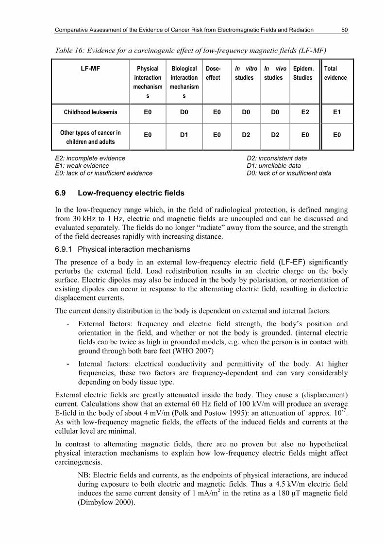
Comparative Assessment of the Evidence of Cancer Risk from Electromagnetic Fields and Radiation 50
Table 16: Evidence for a carcinogenic effect of low-frequency magnetic fields (LF-MF)
LF-MF Physical
interaction
mechanism
s
Biological
interaction
mechanism
s
Dose-
effect
In vitro
studies
In vivo
studies
Epidem.
Studies
Total
evidence
Childhood leukaemia E0 D0 E0 D0 D0 E2 E1
Other types of cancer in
children and adults E0 D1 E0 D2 D2 E0 E0
E2: incomplete evidence D2: inconsistent data E1: weak evidence D1: unreliable data E0: lack of or insufficient evidence D0: lack of or insufficient data
6.9 Low-frequency electric fields
In the low-frequency range which, in the field of radiological protection, is defined ranging from 30 kHz to 1 Hz, electric and magnetic fields are uncoupled and can be discussed and evaluated separately. The fields do no longer “radiate” away from the source, and the strength of the field decreases rapidly with increasing distance.
6.9.1 Physical interaction mechanisms
The presence of a body in an external low-frequency electric field (LF-EF) significantly perturbs the external field. Load redistribution results in an electric charge on the body surface. Electric dipoles may also be induced in the body by polarisation, or reorientation of existing dipoles can occur in response to the alternating electric field, resulting in dielectric displacement currents.
The current density distribution in the body is dependent on external and internal factors.
- External factors: frequency and electric field strength, the body’s position and orientation in the field, and whether or not the body is grounded. (internal electric fields can be twice as high in grounded models, e.g. when the person is in contact with ground through both bare feet (WHO 2007)
- Internal factors: electrical conductivity and permittivity of the body. At higher frequencies, these two factors are frequency-dependent and can vary considerably depending on body tissue type.
External electric fields are greatly attenuated inside the body. They cause a (displacement) current. Calculations show that an external 60 Hz field of 100 kV/m will produce an average E-field in the body of about 4 mV/m (Polk and Postow 1995): an attenuation of approx. 10-7. As with low-frequency magnetic fields, the effects of the induced fields and currents at the cellular level are minimal.
In contrast to alternating magnetic fields, there are no proven but also no hypothetical physical interaction mechanisms to explain how low-frequency electric fields might affect carcinogenesis.
NB: Electric fields and currents, as the endpoints of physical interactions, are induced during exposure to both electric and magnetic fields. Thus a 4.5 kV/m electric field induces the same current density of 1 mA/m2 in the retina as a 180 µT magnetic field (Dimbylow 2000).

Comparative Assessment of the Evidence of Cancer Risk from Electromagnetic Fields and Radiation 51
6.9.2 Biological interaction mechanisms
Current density distributions in an anatomically realistic voxel model of the human body for uniform, low-frequency vertically aligned electric fields for a body grounded and isolated from 50 Hz and 1 kV/m were calculated with maximum electric field strengths of 4.9 mV/m in the spinal cord and 6.1 mV/m in the brain (Dimbylow 2005). Intracorporal electric field strengths can only stimulate myelinated nerve fibres above several V/m. The question which arises in relation to carcinogenesis, however, is whether electric field strengths and currents could induce processes or changes at the cellular level which, with long-term exposure, could influence tumor formation. The hypothetical models proposed here focus on the possible effects on cell membranes. However, as cell membranes are in the order of 10-5 to 10-6 less conductive than the surrounding extracellular fluids, they act as a barrier to the flow of electricity.
Some studies have suggested that the electrical properties of cell membranes may be altered in response to induced electric fields and currents in the extracellular field, leading to biochemical responses that in turn involve changes in cellular functional and proliferative states (ICNIRP 2002).
The question which then arises is how strong these electric fields and currents would need to be in order to exceed the level of endogenous physical and biological noise in cellular membranes. This noise level, which has been modelled in a number of studies, has various causes. For thermally induced changes, the electric field for elongated cells such as fibroblast, nerve and muscle cells should be approximately 100 mV/m, and approximately 1 V/m for smaller spherical cells such as lymphocytes (ICNIRP 2003). To induce fields of this magnitude, however, extremely high external electric fields would be required, which cannot be produced due to the limited dielectric strength in air. It is postulated, however, that one possibility is that in a cell agglomeration of electrically bound cells, due to the “signal amplification”, the signal could be much lower than in an isolated cell to produce the same signal-to-noise ratio, i.e. 1 (ICNIRP 2002, Veyret 2003). The proposed hypothetical action model would only function, however, if the induced low-frequency electric fields behave semi-statically. This means that their frequency must be lower than biological relaxation frequencies. These are dependent on cell size – the larger the cell, the greater the relaxation time / the smaller the relaxation frequency (IARC 2002, ICNIRP 2003, WHO 2007). The hypothetical models are not confirmed by the small number of heterogeneous in vivo and in vitro studies available.
6.9.3 Dose-effect relationship
Based on the proven interaction mechanisms, no evidence for a dose-effect relationship exists, nor can a plausible dose-effect be deduced from the proposed hypotheses about biophysical interaction mechanisms and the results of in vitro and epidemiological studies.
6.9.4 Evidence
In vitro
There are very few in vitro studies on the possible influence of LF-EF fields on carcinogenesis. The small number of studies on electric fields have investigated not only genotoxic but also possible non-genotoxic effects which may influence carcinogenesis, such as changes in signal transmission, influence on the immune system, changes in endocrine functions, and oxidative stress. The results of the studies provide no evidence that LF-EF induce biological effects which could in turn potentially induce cancer (IARC 2002, ICNIRP 2003, WHO 2007, Turkozer et al. 2008).

Comparative Assessment of the Evidence of Cancer Risk from Electromagnetic Fields and Radiation 52
In vivo
There are very few in vivo studies on the possible influence of LF-EF on carcinogenesis. The studies on electric fields have investigated not only genotoxic but also possible non-genotoxic effects which may influence carcinogenesis, such as changes in signal transmission, influence on the immune system, and changes in endocrine functions. The results of the studies provide no evidence that LF-EF induce biological effects which could in turn potentially induce cancer (IARC 2002, ICNIRP 2003, WHO 2007).
Epidemiology
In a review by Kheifets et al. (2010) which assesses the evidence for a link between LF-EF and cancer, the authors conclude their analysis with the comment: “...there seems little basis
to suppose there might be a risk for electric fields, and with a possible exception of
occupational studies, there seems little basis for continued research on electric fields”.
Eight studies on childhood leukaemia and low-frequency magnetic fields also investigated a possible association with electric fields. The results are divergent and the risk assessment ranges from “no effect” in 3 studies to one study with a significant increase in risk, by a factor of 4.7, for mean night-time field exposure above 20 V/m. Exposure was determined in a wide variety of ways. Average field strengths ranged from approx. 3 V/m to above 20 V/m.
Of 4 studies on occupational low-frequency electric field exposure and cancer, one study on train drivers found no effects, and a study based on dose register data found no significant effects. In the first study, exposures were estimated through job exposure matrices; in the second study, periodic measurements were also performed, with the criterion “above 20 V/m” defining the group with the highest exposure. In two studies conducted among electric utility workers in France and Canada, exposures were estimated through job exposure matrices and also individual measurements. For leukaemia, a Canadian study by Miller et al. (1996) reveals a significantly increased risk, by a factor of approx. 4.5 (odds ratio), for cumulative exposure above 345 V/m-year compared with exposures below 172 V/m-year. By contrast, the French study (Guenel 1996) does not show any increased risk for leukaemia, but finds a significantly increased risk for brain tumors (odds ratio 3.1) for exposure above the 90th percentile (> or = 387 V/m-year), compared with exposure below 277 V/m-year. In the report on the Canadian study (Villeneuve 2000), an increased association with leukaemia risk was only observed if the dose was defined in terms of exposure duration above a specific threshold value.
6.9.5 Exposure
Due to the shielding effect of buildings, good protection is provided against exposure to LF-EF emissions from external field sources such as high-voltage power lines. Indoors, field sources such as the internal power supply, lamps and electrical appliances determine the level of exposure. As LF-EF are distorted by the electrical conductive body, exposure varies with body position and distance to the field source. Relatively high exposures occur as a result of the physical proximity to electrical appliances during use (Leitgeb et al. 2008b), when field strength can exceed electric reference values.
6.9.6 Overall assessment of the evidence
Given the lack of interaction mechanisms and the absence of any evidence for a dose-effect, and in view of the effective shielding of the body from external electric fields, it can be concluded, overall, that despite the inconsistencies in the data from epidemiological studies, there is no evidence for an association between low-frequency electric fields and cancer, including childhood leukaemia.

Comparative Assessment of the Evidence of Cancer Risk from Electromagnetic Fields and Radiation 53
Table 17: Evidence for a carcinogenic effect of low-frequency electric fields (LF-EF)
Physical
interaction
mechanisms
Biological
interaction
mechanisms
Dose-
effect
In vitro
studies
In vivo
studies
Epidem.
Studies
Total
evidence
LF-EF E0 E0 E0 E0 E0 D2 E0
E0: lack of or insufficient evidence D2: inconsistent data
6.10 Static magnetic fields
Static magnetic fields (SMF) are generated wherever direct (DC) currents are used, or by materials with inherent magnetic properties (e.g. permanent magnets).
6.10.1 Physical interaction mechanisms
The physical interaction mechanisms of static magnetic fields are well-known. They are mainly the result of Lorentz forces exerted on moving charges. The force exerted oncharges with different polarity moving in the same direction causes a separation of charges and the generation of electric voltage / field strengths. Another mechanism is based on the force of the static magnetic field acting on magnetic dipoles (magnetomechanical effect) such as ferromagnetic particles (e.g. magnetite) or atoms with an inherent magnetic moment (e.g. hydrogen). A further mechanism exists at atomic level: a change in the energy level of shell electrons could, in sufficiently strong fields, manifest as an alteration of chemical reaction equilibria, lengthening the lifespan of (already existing) radicals.
6.10.2 Biological interaction mechanisms
Movements of charges which could potentially be separated in the static magnetic field are generated (also in the resting body) by physiological actions (e.g. heart beat and blood flow) or by movements of the body or body parts. In strong fields, hypothetical biological interaction mechanisms could be based primarily on changes in enzyme activity, in a longer lifespan of radicals and in their ensuing effects; these may include both cancer-promoting and cancer-inhibiting effects. Indications of such effects result from studies with SMF above 100 mT (Strelczyk et al. 2009).
6.10.3 Dose-effect relationship
In contrast to force effects which are acutely bound to the existence of the magnetic field, with regard to changes in energy level of shell electrons, accumulative effects cannot be excluded on principle. However, a dose-effect relationship cannot be deduced from the available data.
6.10.4 Evidence
In vitro
There is a number of in vitro studies with various endpoints (e.g. cell orientation, cell metabolic activity, cell growth, gene expression and genotoxicity) with a wide range of field strengths, in every case above the strength of the Earth’s geomagnetic field. There are some indications that static magnetic fields can affect several biologic endpoints in the mT range (ICNIRP 2009a), or at inductions above 1 T. Overall, however, there is a lack of or insufficient evidence for a link between SMF and adverse health effects/cancer.

Comparative Assessment of the Evidence of Cancer Risk from Electromagnetic Fields and Radiation 54
In vivo
Available animal studies have mainly concentrated on inductions exceeding 1 T and have investigated the potential effects of magnetic resonance imaging (MRI) in medical applications. Neurophysiological effects were commonly observed, indicating that exposure to static magnetic fields greater than 4 T may be unpleasant, inducing aversive responses. Despite distortions induced by the magnetic field on electrocardiograms (ECG), due to magnetohydrodynamic effects in the aorta, several hours of exposure to flux densities of up to 8 T did not result in any cardiovascular effect. Several studies with inconsistent results suggest that SMF exposure may affect the endocrine system, but other studies have been unable to reproduce these findings. Studies on male BALB/cByJ mice found an attenuated mortality rate from disseminated intravascular coagulation (DIC) following exposure to 250 mT (Lin et al. 2009). The available data do not allow any firm conclusions to be drawn about genotoxicity and carcinogenicity.
Epidemiology
Epidemiological studies have been carried out mainly on groups of workers occupationally exposed to static magnetic fields (e.g. welders, aluminium smelters, etc.) and pregnant female MRI operators (ICNIRP 2003, 2009, WHO 2006). Increased risks of cancer were reported for some endpoints, but the results were not consistent across studies. In light of other co-factors which adversely affect health and the methodological limitations, the data situation must be classified as inadequate.
6.10.5 Exposure
Exposure to static magnetic fields is determined by the Earth’s geomagnetic field of approx. 45 µT, with considerable variations caused by ferromagnetic elements (e.g. iron reinforcements in buildings) but generally remaining well below 1 mT. Occupational exposure to static magnetic fields in workplace environments generated by equipment operated by large DC currents (e.g. electrolytic processes or electric welding) is as much as two orders of magnitude greater. In addition, magnetic resonance imaging (MRI) is already a standard medical procedure with increasing prevalence. As a result, exposure to high static magnetic fields amounting to several tesla can no longer be regarded as uncommon, with not only patients but also medical and technical personnel being exposed.
6.10.6 Overall assessment of the evidence
Although there are various hypothesised physical and biological models of carcinogenicity in the range above several 100 mT and the influence of chemical reactions and enzyme activity has been demonstrated, overall, there is no evidence for a link between every-day exposure at lower static magnetic inductions and cancer. However, data are not yet sufficient to estimate a potential cancer risk of higher static magnetic fields.

Comparative Assessment of the Evidence of Cancer Risk from Electromagnetic Fields and Radiation 55
Table 18: Evidence for a carcinogenic effect of static magnetic fields (static MF)
Static MF
Physical
interaction
mechanisms
Biological
interaction
mechanisms
Dose-
effect
In vitro
studies
In vivo
studies
Epidem.
studies
Total
evidence
General E0 E0 E0 E0 E0 D1 E0
MRI E1 E1 D0 D1 D1 D0 D1
E1: weak evidence D1: unreliable data E0: lack of or insufficient evidence D0: lack of or insufficient data
6.11 Static electric fields
a Static electric fields (SEF)are generated by electric charges. Conductive objects, including living beings, distort the fields depending on the shape and position of the body and the distance from the field source.
6.11.1 Physical interaction mechanisms
The physical interaction mechanisms of static electric fields are well-known. In accordance with the principle of Coulomb attraction, SEF cause a redistribution of charge in the body such as electric charges are encountered on the surface adjacent to the field. Due to repulsive forces (since similar charges repel each other), a force will be exerted on body hair, and microdischarges may also be caused on the body surface. At the same time, however, the inner body is almost completely shielded from the external electric field.
6.11.2 Biological interaction mechanisms
With sufficient field strength, the charge can cause the hair to erect, which may be perceived. Microdischarges may be unpleasant, and with sufficient field strength, they may even cause microshocks. However, there are no indications of interaction mechanisms which could potentially cause cancer.
6.11.3 Dose-effect relationship
Neither physical nor biological interaction mechanisms indicate cumulative effects or the existence of a dose-dependent effect. Experimental and epidemiological studies provide no evidence to suggest a dose-effect relationship.
6.11.4 Evidence
Due to the lack of any plausible physical and biological interaction mechanisms and the fact that the inner body is shielded almost completely from external static electric fields, there is no plausible evidence for a link with cancer. For that reason, no studies, applying various scientific methodologies and focusing on various biological endpoints, have been conducted to systematically evaluate a potential cancer link.
6.11.5 Exposure
Exposure to static electric fields in the open air mainly results from the natural electric field existing between the surface of the earth and the ionosphere; every-day exposure mainly results from static charges caused by friction and exposure to direct current sources such as
Comparative Assessment of the Evidence of Cancer Risk from Electromagnetic Fields and Radiation 56
electric rail/tram systems, for example. Occupational exposure to stronger static electric fields occurs during the use of high direct currents in applications such as electric arc welding, galvanising and electrolytic processes.
6.11.6 Overall assessment of the evidence
Overall, the evidence suggests that there is no link between exposure to static electric fields and cancer.
Table 19: Evidence for a carcinogenic effect of static electric fields (static EF)
Physical
interaction
mechanisms
Biological
interaction
mechanisms
Dose-
effect
In vitro
studies
In vivo
studies
Epidem.
studies
Total
evidence
Static EF EN E0 EN E0 E0 D1 EN
E0: lack of or insufficient evidence EN: evidence for non-causality D1: unreliable data
7 Conclusion
Summarising the evidence for a causal link between exposure to electric and magnetic fields, electromagnetic waves and electromagnetic radiation in the various frequency ranges, taking account of the various scientific approaches, it is immediately apparent that the evidence for an association between energy-rich radiation and cancer is convincing, becoming steadily weaker with decreasing frequency, i.e. with decreasing quantum energy (Table 20). Similarly, the evidence for a dose-effect relationship becomes weaker with decreasing frequency. Whereas in the energy-rich radiation range, the dose metric, i.e. the temporal sum of absorbed radiation intensity, is well-established, a dose-effect becomes less likely with decreasing frequency. This is not only apparent from the fact that certain questions remain unanswered, such as which basic physical parameter is relevant (e.g. the spatial mean value for magnetic fields, the mean value of the excess above a limit value, night-time exposure, temporal field gradients, etc.) and whether a temporal summation is indeed necessary to assess long-term exposure, and if so, how it is to be defined (e.g. as a time-integral, mean values of limit value exceedances multiplied by exposure years, daily mean value, night-time mean value etc.). Despite proposals made in a few individual studies, the available studies do not provide sufficient evidence of a dose-effect relationship. Even the increase in the odds ratio with increasing mean induction value of LF-MF, calculated in a number of epidemiological studies on childhood leukaemia, does not take account of the duration of exposure.
In assessing the evidence, the question which arises is how much weight should be fiven to the various scientific approaches in the overall assessment and whether elements of individual approaches could be dispensed. This also applies to the weight attached to established theoretical knowledge about physical and biological interaction mechanisms. From the SSK’s perspective, it is essential to incorporate establishedtheoretical knowledge into the assessment. However, the Commission does not support disproportionate weighting of epidemiological findings. For this reason, in the Commission’s view, the epidemiological findings about a statistical association between magnetic field exposure and childhood leukaemia, which are still not supported by other investigative approaches but which per se must be regarded as providing incomplete evidence (E2), should in the overall assessment of the results be classed

Comparative Assessment of the Evidence of Cancer Risk from Electromagnetic Fields and Radiation 57
as providing only weak evidence (E1) for a causal association between exposure to alternating magnetic fields and childhood leukaemia.
In the range of visible light, by contrast, there is weak evidence that light-at-night could be associated with cancer. Given the prevalence of this type of exposure, this deserves more attention. The overall assessments of the evidence in the various frequency ranges are summarised in Table 21.

Comparative Assessment of the Evidence of Cancer Risk from Electromagnetic Fields and Radiation 58
Table 20: Evidence for cancer as the endpoint for various EMF frequency ranges for every-
day exposure, based on various scientific approaches
Frequency range
Physical
interaction
mechanisms
Biological
interaction
mechanisms
Dose-
effect
In vitro
studies
In vivo
studies
Epidem.
studies
Ionising radiation E3 E3 E3 E3 E3 E3
UV E3 E31) E2 E3 E31) E3
Visible light 2) E2 E1 D0 E1 E1 E1
Visible light 3) E2 D1 D0 D0 D0 D0
IR E1 E1 D0 E1 D0 D0
Terahertz E0 D1 D0 D1 D0 D0
Microwaves E0 D1 E0 D2 E0 E0
HF-MF E0 E0 E0 D1 D1 E0
LF-MF4) E0 D0 E0 D0 D0 E2
LF-MF5) E0 D1 E0 D2 D2 E0
LF-EF E0 E0 E0 E0 E0 D2
Static MF (general) E0 E0 E0 E0 E0 D1
Static MF (MRI) E1 E1 D0 D1 D1 D0
Static EF EN E0 EN E0 E0 D1
E3: convincing evidence E2: incomplete evidence E1: weak evidence E0: lack of or insufficient evidence EN: evidence for non-causality D2: inconsistent data D1: unreliable data D0: lack of or insufficient data 1) for UVA, the evidence is incomplete (E2)
2) relates to blue light and general night-time light exposure (light-at-night)
3) relates to other light exposure
4) relates to childhood leukaemia
5) relates to other types of cancer affecting children and adults

Comparative Assessment of the Evidence of Cancer Risk from Electromagnetic Fields and Radiation 59
Table 21: Evidence for cancer as the endpoint for various EMF frequency ranges (related to
every-day exposure)
Frequency range
Evidence for non-causality
Evidence unclassifiable Evidence for causality
Lack of or insufficient
data
Unreliable data
Inconsistent data
Lack of or insufficient evidence
Weak evidence
Incomplete evidence
Convincing evidence
EN D0 D1 D2 E0 E1 E2 E3
Ionising radiation X
UV X
Visible light
X1)
X2)
IR X
Terahertz X
Microwaves X
HF-MF X
LF-MF
X3)
X4)
LF-EF X
Static MF (general)
X
Static MF (MRI) X
Static EF X
E3: convincing evidence E2: incomplete evidence E1: weak evidence E0: lack of or insufficient evidence EN: evidence for non-causality D2: inconsistent data D1: unreliable data D0: lack of or insufficient data 1) relates to blue light and general night-time light exposure (light-at-night)
2) relates to general day-time light exposure
3) relates to childhood leukaemia
4) relates to other types of cancer in children and adults

Comparative Assessment of the Evidence of Cancer Risk from Electromagnetic Fields and Radiation 60
8 References
Ahlbom et al. 2009
Ahlbom, A.; Feychting, M.; Green, A.; Kheifets, L.; Savitz, D. A.; Swerdlow, A. J.: ICNIRP (International Commission for Non-Ionizing Radiation Protection) Standing Committee on Epidemiology. Epidemiologic evidence on mobile phones and tumor risk: a review. Epidemiology 20(5):639-652, 2009
Applied Research and Photonics 2010
Applied Research and Photonics. White Paper February 04, 2010 Application of ARP’s terahertz spectrometer with exemplary data http://www.arphotonics.net/
Armstrong and Kricker 2001
Armstrong, B. K.; Kricker, A.: The epidemiology of UV induced skin cancer. J. Photochem. Photobiol. B; 63(1-3):8-18, 2001
Aszterbaum et al. 1999a
Aszterbaum, M.; Epstein, J.; Oro, A.; Douglas, V.; LeBoit, P. E.; Scott, M. P. et al.: Ultraviolet and ionizing radiation enhance the growth of BCCs and trichoblastomas in patched heterozygous knockout mice. Nat. Med. 5(11):1285-1291, 1999
Aszterbaum et al. 1999b
Aszterbaum, M.; Beech, J.; Epstein, E. H. Jr.: Ultraviolet radiation mutagenesis of hedgehog pathway genes in basal cell carcinomas. J. Investig. Dermatol. Symp. Proc. 4(1):41-45, 1999
BAFU 2009 BAFU: Niederfrequente Magnetfelder und Krebs. Bewertung von wissenschaftlichen Studien im Niedrigdosisbereich. Stand: August 2008. Basel, 2009 http://www.bafu.admin.ch/publikationen/Publikation/01511/index.html?lang=de
Bain et al. 1943
Bain J. A.; Rusch H. P.; Kline, B. E.. The effect of temperature upon ultraviolet carcinogenesis with wavelength of 2,800-3,400 A. Cancer Res. 1943; 3:610-612.
Bain and Rusch 1943
Bain, J. A.; Rusch, H. P.: Carcinogenesis with ultraviolet radiation of wavelength 2,800-3,400 A. Cancer Res. 3:425-430, 1943
Berg et al. 1998
Berg, R. J.; Ruven, H. J.; Sands, A. T.; de Gruijl, F. R.; Mullenders, L. H.: Defective global genome repair in XPC mice is associated with skin cancer susceptibility but not with sensitivity to UVB induced erythema and edema. J. Invest. Dermatol. 110(4):405-409, 1998
Bickenbach and Holbrook 1987
Bickenbach, J. R.; Holbrook, K. A.: Label-retaining cells in human embryonic and fetal epidermis. J. Invest. Dermatol. 88(1):42-46, 1987
BMU 2008 Bundesministerium für Umwelt, Naturschutz und Reaktorsicherheit (BMU) Hrsg.: Umweltbewusstsein in Deutschland 2008 – Ergebnisse einer Repräsentativen Bevölkerungsumfrage. Forschungsprojekt Förderkennzeichen 370717101, 2008

Comparative Assessment of the Evidence of Cancer Risk from Electromagnetic Fields and Radiation 61
Börner et al. 2009
Börner, F.; Schütz, H.; Greinert, R.; Wiedemann, P. M.: UV-Risikowahrnehmung in der Bevölkerung: Ergebnisse einer repräsentativen Umfrage in Deutschland. Gesundheitswesen. 71:1-9, 2009
Boukamp et al. 1999
Boukamp, P.; Popp, S.; Bleuel, K.; Tomakidi, E.; Burkle, A.; Fusenig, N. E.: Tumorigenic conversion of immortal human skin keratinocytes (HaCaT) by elevated temperature. Oncogene 18(41):5638-5645, 1999
Brash 1997 Brash, D. E.: Sunlight and the onset of skin cancer. Trends Genet. 13(10):410-414, 1997
Braun and Watt 2004
Braun, K. M.; Watt, F. M.: Epidermal label-retaining cells: background and recent applications. J. Investig. Dermatol. Symp. Proc. 9(3):196-201, 2004
Broerse et al. 1985
Broerse, J. J.; Hennen, L. A.; van Zwieten, M. J.: Radiation carcinogenesis in experimental animals and its implications for radiation protection. Int. J. Radiat. Biol. Relat. Stud. Phys. Chem. Med. 48:167-187, 1985
Broerse et al. 1989
Broerse, J. J.; van Bekkum, D. W.; Zurcher, C.: Radiation carcinogenesis in experimental animals. Experientia 45:60-69, 1989
Butow and Avadhani 2004
Butow, R. A.; Avadhani, N. G.: Mitochondrial signaling: the retrograde response. Mol Cell. 14(1):1-15, 2004
Cairns 1975 Cairns, J.: Mutation selection and the natural history of cancer. Nature. 255(5505):197-200, 1975
Cairns 2002 Cairns, J.: Somatic stem cells and the kinetics of mutagenesis and carcinogenesis. Proc. Natl. Acad. Sci. U S A. 99(16):10567-10570, 2002
CEC 2000 CEC (Kommission der Europäischen Gemeinschaften): Mitteilung der Kommission - die Anwendbarkeit des Vorsorgeprinzips. Brüssel, 2000 http://eur-lex.europa.eu/LexUriServ/site/de/com/2000/ com2000_0001de01.pdf
Chen et al. 2010
Chen, C.; Ma, X.; Zhong, M.; Yu, Z.: Extremely low-frequency electromagnetic fields exposure and female breast cancer risk: a meta-analysis based on 24,338 cases and 60,628 controls. Breast Cancer Res. Treat. 123(2):569-576, 2010
Choi et al. 2004
Choi, M. K.; Bettermann, A.; van der Weide, D. W.: Potential for detection of explosive and biological hazards with electronic terahertz systems. Philos. Transact A Math. Phys. Eng. Sci. 362:337-347, 2004
Cleaver and Crowley 2002
Cleaver, J. E.; Crowley, E.: UV damage, DNA repair and skin carcinogenesis. Front Biosci 7:d1024-d1043, 2002
Coleman and Tsongalis 2006
Coleman, W. B.; Tsongalis, G. J.: Molecular mechanisms of human carcinogenesis. EXS (96):321-349, 2006

Comparative Assessment of the Evidence of Cancer Risk from Electromagnetic Fields and Radiation 62
Couve-Privat et al. 2002
Couve-Privat, S.; Bouadjar, B.; Avril, M. F.; Sarasin, A.; Daya-Grosjean, L.: Significantly high levels of ultraviolet-specific mutations in the smoothened gene in basal cell carcinomas from DNA repair-deficient xeroderma pigmentosum patients. Cancer Res. 62(24):7186-7189, 2002
Cox and Huber 2007
Cox, L. A. Jr.; Huber, W. A.: Symmetry, identifiability, and prediction uncertainties in multistage clonal expansion (MSCE) models of carcinogenesis. Risk Anal. 27(6):1441-1453, 2007
Dasenbrock 2005
Dasenbrock, C.: Animal carcinogenicity studies on radiofrequency fields related to mobile phones and base stations. Toxicol. Appl. Pharmacol. 207(2 Suppl):342-346, 2005
Daya-Grosjean and Sarasin 2000
Daya-Grosjean, L.; Sarasin, A.: UV-specific mutations of the human patched gene in basal cell carcinomas from normal individuals and xeroderma pigmentosum patients. Mutat. Res. 450(1-2):193-199, 2000
De Fabo et al. 2004
De Fabo, E. C.; Noonan, F. P.; Fears, T.; Merlino, G.: Ultraviolet B but not ultraviolet A radiation initiates melanoma. Cancer Res. 64(18) :6372-6376, 2004
de Gruijl 1995 de Gruijl, F. R.: Action spectrum for photocarcinogenesis. Recent Results Cancer Res. 139:21-30, 1995
de Gruijl et al. 1993
de Gruijl, F. R.; Sterenborg, H. J.; Forbes, P. D.; Davies, R. E.; Cole, C.; Kelfkens, G. et al.: Wavelength dependence of skin cancer induction by ultraviolet irradiation of albino hairless mice. Cancer Res. 53(1):53-60, 1993
Deltour et al. 2009
Deltour, I.; Johansen, C.; Auvinen, A.; Feychting, M.; Klaeboe, L.; Schüz, J.: Time trends in brain tumor incidence rates in Denmark, Finland, Norway, and Sweden, 1974-2003. J. Natl. Cancer Inst. 101(24):1721-4, 2009 Dec 16
Dewhirst et al. 2003a
Dewhirst, M. W.; Lora-Michiels, M.; Viglianti, B. L.; Dewey, W. C.; Repacholi, M.: Carcinogenic effects of hyperthermia. Int. J. Hyperthermia 19(3):236-251, 2003
Dewhirst et al. 2003b
Dewhirst, M. W.; Viglianti, B. L.; Lora-Michiels, M.; Hanson, M.; Hoopes, P. J.: Basic principles of thermal dosimetry and thermal thresholds for tissue damage from hyperthermia. Int. J. Hyperthermia 19(3):267-294, 2003
Di Cesare et al. 2009
Di Cesare, S.; Maloney, S.; Fernandes, B. F.; Martins, C.; Marshall, J. C.; Antecka, E.; Odashiro, A. N.; Dawson, W. W.; Burnier, M. N. Jr.: The effect of blue light exposure in an ocular melanoma animal model. J. Exp. Clin.Cancer Res. 28:48, 2009
Diem et al. 2005
Diem, E.; Schwarz, C.; Adlkofer, F.; Jahn, O.; Rüdiger, H.: Non-thermal DNA breakage by mobile-phone radiation (1800 MHz) in human fibroblasts and in transformed GFSH-R17 rat granulosa cells in vitro. Mutat. Res. 583 :178-83, 2005

Comparative Assessment of the Evidence of Cancer Risk from Electromagnetic Fields and Radiation 63
Dimbylow 2000
Dimbylow, P. J.: Current densities in a 2mmm resolution anatomically realistic model of the body induced by extra low frequency electric fields. Phys.Med.Biol. 45:1013-1022, 2000
Dimbylow 2005
Dimbylow, P. J.: Development of the female voxel phantom, NAOMI, and its application to calculations of induced current densities and electric fields from applied low frequency magnetic and electric fields. Phys.Med.Biol. 50(6):1047-1070, 2005
DIN VDE 31000 Teil 2
DIN VDE 31000, Teil 2 (1987): Allgemeine Leitsätze für das sicherheitsgerechte Gestalten technischer Erzeugnisse – Begriffe der Sicherheitstechnik Grundbegriffe
Dumaz et al. 1997
Dumaz, N.; van Kranen, H. J.; de Vries, A.; Berg, R. J.; Wester, P. W.; van Kreijl, C. F. et al.: The role of UV-B light in skin carcinogenesis through the analysis of p53 mutations in squamous cell carcinomas of hairless mice. Carcinogenesis 18(5):897-904, 1997
El Ghissassi et al. 2009
El Ghissassi, F.; Baan, R.; Straif, K.; Grosse, Y.; Secretan, B.; Bouvard, V. et al. : A review of human carcinogens-part D: radiation. Lancet Oncol. 10(8):751-752, 2009
Elliott et al. 2010
Elliott, P.; Toldeano, M. B.; Bennett, J.; Beale, L.; de Hoogh, K.; Best, N.; Briggs, D. J.: Mobile phone base stations and early childhood cancers: case-control study, BMJ 340:c3077, 2010
EN 14971 DIN EN ISO 14971: 2009-10, Medizinprodukte - Anwendung des Risikomanagements auf Medizinprodukte (ISO 14971:2007, korrigierte Fassung 1. Oktober 2007); Deutsche Fassung EN ISO 14971, 2009
EPA 2005 EPA: Guidelines for Carcinogen Risk Assessment. Risk Assessment Forum U.S. Environmental Protection Agency Washington, DC, 2005
Esteve-Puig et al. 2009
Esteve-Puig, R.; Canals, F.; Colome, N.; Merlino, G.; Recio, J. A.: Uncoupling of the LKB1-AMPKalpha energy sensor pathway by growth factors and oncogenic BRAF. PLoS One 4(3):e4771, 2009
Eurobarometer 2007
Special Eurobarometer 272a Electromagnetic Fields, 2007 http://ec.europa.eu/public_opinion/archives/ebs/ebs_272a_en.pdf
Evans et al. 2000
Evans, T.; Boonchai, W.; Shanley, S.; Smyth, I.; Gillies, S.; Georgas, K. et al.: The spectrum of patched mutations in a collection of Australian basal cell carcinomas. Hum. Mutat. 16(1):43-48, 2000
Fedrowitz and Löscher 2005
Fedrowitz, M.; Löscher, W.: Power frequency magnetic fields increase cell proliferation in the mammary gland of female Fischer 344 rats but not various other rat strains or substrains. Oncology 69 (6) :486-498, 2005
Fedrowitz and Löscher 2008
Fedrowitz, M.; Löscher, W.: Exposure of Fischer 344 rats to a weak power frequency magnetic field facilitates mammary tumorigenesis in the DMBA model of breast cancer. Carcinogenesis 29 (1):186-193, 2008

Comparative Assessment of the Evidence of Cancer Risk from Electromagnetic Fields and Radiation 64
Filipski et al. 2004
Filipski, E.; Delaunay, F.; King, V. M.; Wu, M. W.; Claustrat, B.; Gréchez-Cassiau, A.; Guettier, C.; Hastings, M. H.; Francis, L.: Effects of chronic jet lag on tumor progression in mice. Cancer Res. 64:7879-85, 2004;
Flynn-Evans et al. 2009
Flynn-Evans, E. E.; Stevens, R. G.; Tabandeh, H.; Schernhammer, E. S.; Lockley, S. W.: Total visual blindness is protective against breast cancer. Cancer Causes Control. 20 :1753-6, 2009
Focke et al. 2010
Focke, F.; Schuermann, D.; Kuster, N.; Schär, P.: DNA fragmentation in human fibroblasts under extremely low frequency electromagnetic field exposure. Mutat. Res. 683(1-2). 74-83, 2010
Fox and Irwin 1998
Fox, C. R.; Irwin, J. I.: The role of context in the communication of uncertain beliefs. Basic and Applied Social Psychology 20(1):57-70, 1998
Frahm et al. 2006
Frahm, J.; Lantow, M.; Lupke, M.; Weiss, D. G.; Simko, M.: Alteration in cellular functions in mouse macrophages after exposure to 50 Hz magnetic fields. J. Cell Biochem. 99(1):168-177, 2006
Freeman and Knox 1964
Freeman, R. G.; Knox, J. M.: Influence of temperature on ultraviolet injury. Arch Dermatol. 89:858-864, 1964;
Gallenaro et al. 2008
Tera-Hertz radiation in Biological Research. Investigations on Diagnostics and study on potential Genotoxic Effects. Final Report THz BRIDGE, http://www.frascati.enea.it/THz-BRIDGE/reports/
Gambardella and Barrandon 2003
Gambardella, L.; Barrandon, Y.: The multifaceted adult epidermal stem cell. Curr Opin Cell Biol. 15(6):771-777, 2003
Gandini et al. 2005a
Gandini, S.; Sera, F.; Cattaruzza, M. S.; Pasquini, P.; Picconi, O.; Boyle, P. et al.: Meta-analysis of risk factors for cutaneous melanoma: II. Sun exposure. Eur. J. Cancer 41(1):45-60, 2005
Gandini et al. 2005b
Gandini, S.; Sera, F.; Cattaruzza, M. S.; Pasquini, P.; Zanetti, R.; Masini, C. et al.: Meta-analysis of risk factors for cutaneous melanoma: III. Family history, actinic damage and phenotypic factors. Eur. J. Cancer 41(14): 2040-2059, 2005
Girgert et al. 2008
Girgert, R.; Gründker, C.; Emons, G.; Hanf, V.: Electromagnetic fields alter the expression of estrogen receptor cofactors in breast cancer. Bioelectromagnetics 29 (3):169-176, 2008
Goss and Parsons 1976
Goss, P.; Parsons, P. G.: Temperature-sensitive DNA repair of ultraviolet damage in human cell lines. Int. J. Cancer 17(3):296-303, 1976
Gottwald et al. 2007
Gottwald, E.; Sonntag, W.; Lahni, B.; Weibezahn, K.: Expression of HSP72 after ELF-EMF exposure in three cell lines. Bioelectromagnetics 28 (7):509-518, 2007
Comparative Assessment of the Evidence of Cancer Risk from Electromagnetic Fields and Radiation 65
Greinert et al. 2000
Greinert, R.; Boguhn, O.; Harder, D.; Breitbart, E. W.; Mitchell, D. L.; Volkmer, B.: The dose dependence of cyclobutane dimer induction and repair in UVB-irradiated human keratinocytes. Photochem. Photobiol. 72(5):701-708, 2000
Greinert et al. 2004
Greinert, R.; Breitbart, E. W.; Volkmer, B.: UV-radiation biology as part of cancer research. In: Kiefer J, editor. Life sciences and radiation. Berlin. Springer. 139-155, 2004
Guenel et al. 1996
Guenel, P.; Nicolau, J.; Imbernon, E.; Chevalier, A.; Goldberg, M.: Exposure to 50-Hz electric field and incidence of leukemia, brain tumors, and other cancers among French electric utility workers. Am. J. Epidemiol, 144(12):1107-1121, 1996
Hagmar et al. 1998
Hagmar, L.; Bonassi, S.; Stromberg, U.; Brogger, A.; Knudsen, L. E.; Norppa, H.; Reuterwall, C.: Chromosomal aberrations in lymphocytes predict human cancer: a report from the European Study Group on Cytogenetic Biomarkers and Health (ESCH). Cancer Res. 58:4117-4121, 1998
Hardell et al. 2009
Hardell, L.; Carlberg, M.; Hansson, Mild, K.: Epidemiological evidence for an association between use of wireless phones and tumor diseases. Pathophysiology 16:113-122, 2009
Hardell and Carlberg 2009
Hardell, L.; Carlberg, M.: Mobile phones, cordless phones and the risk for brain tumours. Int. J. Oncol. 35 :5-17, 2009
Heynick et al. 2003
Heynick, L. N.; Johnston, S. A.; Mason, P. A.: Radio frequency electromagnetic fields:cancer, mutagenesis, and genotoxicity. Bioelectromagnetics Suppl. 6. S74-100, 2003
Hirose et al. 2008
Hirose, H.; Suhara, T.; Kaji, N.; Sakuma, N.; Sekijima, M.; Nojima, T.; Miyakoshi, J.: Mobile phone base station radiation does not affect neoplastic transformation in BALB/3T3 cells. Bioelectromagnetics 29:55-64, 2008
Hoffmann-Dörr et al. 2005
Hoffmann-Dörr, S.; Greinert, R.; Volkmer, B.; Epe, B.: Visible light (>395 nm) causes micronuclei formation in mammalian cells without generation of cyclobutane pyrimidine dimers. Mutat. Res. 572(1-2):142-149, 2005
Hoglund et al. 2004
Hoglund, M.; Gisselsson, D.; Hansen, G. B.; White, V. A.; Sall, T.; Mitelman, F. et al.: Dissecting karyotypic patterns in malignant melanomas: temporal clustering of losses and gains in melanoma karyotypic evolution. Int. J. Cancer 108(1):57-65, 2004
Hruby et al. 2008
Hruby, R.; Neubauer, G.; Küster, N.; Frauscher, M.: Study on potential effects of “902-MHz GSM-type Wireless Communication Signals” on DMBA-induced mammary tumours in Sprague- Dawley rats. Mutat. Res. 649:34-44, 2008

Comparative Assessment of the Evidence of Cancer Risk from Electromagnetic Fields and Radiation 66
Hug et al. 2009 Hug, K.; Schär, P.; Rapp, R.; Taschner, N.: Niederfrequente Magnetfelder und Krebs. Bewertung von wissenschaftlichen Studien im Niedrigdosisbereich. Stand: August 2008. Umwelt-Wissen Nr. 0934. Bundesamt für Umwelt, Bern. 118 S., 2009
Hug et al. 2010 Hug, K.; Grize, L.; Seidler, A.; Kaatsch, P.; Schüz, J.: Parental occupational exposure to extremely low frequency magnetic fields and childhood cancer: a German case-control study. Am. J. Epidemiol. 171(1):27-35, 2010
IARC 1992 IARC: IARC Monographs on the Evaluation of Carcinogenic Risk to Humans, Volume 55: Solar and Ultraviolet Radiation, 1992
IARC 2002 IARC: IARC Monographs on the Evaluation of Carcinogenic Risks to Humans, Volume 80: Non-Ionizing Radiation, Part 1: Static and Extremely Low-Frequency (ELF) Electric and Magnetic Fields, 2002
IARC 2006 IARC: IARC Monographs on the Evaluation of Carcinogenic Risks to Humans. Preamble, 2006 http://monographs.iarc.fr/ENG/Preamble/CurrentPreamble.pdf
IARC 2007 IARC: Press Release N. 180, IARC Monographs Programme finds cancer hazards associated with shiftwork, painting and firefighting, 5. Dezember 2007, http://www.iarc.fr/en/media-centre/pr/2007/pr180.html
ICNIRP 2002 International Commission on Non-Ionizing Radiation Protection: General Approach to Protection against Non Ionizing Radiation. Health Phys. 82 (4):540-548, 2002
ICNIRP 2003 International Commission on Non-Ionizing Radiation Protection: Exposure to Static and Low Frequency Electromagnetic Fields, Biological Effects and Health Consequences (0-100 kHz). ICNIRP 13/2003
ICNIRP 2004 International Commission on Non-Ionizing Radiation Protection: Guidelines on Limits of Exposure to Ultraviolet Radiation of Wavelengths Between 1800 nm and 400 nm (Incoherent Optical Radiation). Health Phys. 87(2):171-186, 2004
ICNIRP 2006 International Commission on Non-Ionizing Radiation Protection: Statement on far infrared radiation exposure. Health Phys. 91(6):630-645, 2006
ICNIRP 2009a International Commission on Non-Ionizing Radiation Protection: Guidelines on limits of exposure to static magnetic fields. Health Phys. 96(4):504-514, 2009
ICNIRP 2009b International Commission on Non-Ionizing Radiation Protection: Exposure to high frequency electromagnetic fields, biological effects and health consequences (100 kHz - 300 GHz) - Review of the Scientific Evidence and Health Consequences. Munich: International Commission on Non-Ionizing Radiation Protection; 2009

Comparative Assessment of the Evidence of Cancer Risk from Electromagnetic Fields and Radiation 67
INFAS 2006 INFAS (Institut für angewandte Sozialwissenschaft): Ermittlung der Befürchtungen und Ängste der breiten Öffentlichkeit hinsichtlich möglicher Gefahren der hochfrequenten elektromagnetischen Felder des Mobilfunks – jährliche Umfragen. Abschlussbericht über die Befragung im Jahr 2006, 2006
INTERPHONE 2010
The INTERPHONE study group: Brain tumour risk in relation to mobile telephone use: results of the INTERPHONE international case-control study. Int J Epidemiol. 39(3):675-94, 2010 (Epub 2010 May 17)
IPCS 2004 International Programme on Chemical Safety/IPCS: Risk assessment terminology - Part 1 and Part 2. Geneva, Switzerland: World Health Organization, 2004
Ivancsits et al. 2005
Ivancsits, S.; Pilger, A.; Jahn, O.; Rudiger, H. W.: Cell type-specific genotoxic effects of intermittent extremely low-frequency electromagnetic fields. Mutat. Res: 583(2):184-188, 2005
Jacob et al. 2010
Jacob, P.; Meckbach, R.; Kaiser, J. C.; Sokolnikov, M.: Possible expressions of radiation-induced genomic instability, bystander effects or low-dose hypersensitivity in cancer epidemiology. Mutat. Res. 687:34-39, 2010
Johnson 2003 Johnson, B.: Further notes on public response to uncertainty in risks and science. Risk Analysis, 23(4):781-790, 2003
Kanitz et al. 2007
Kanitz, M. H.; Witzmann, F. A.; Lotz, W. G.; Conover, R. E.; Savager Jr.: Investigation of protein expression in magnetic field-treated human glioma cells. Bioelectromagnetics: 28(7):546-552, 2007
Karakosta et al. 2005
Karakosta, A.; Golias, Ch.; Charalabopoulos, A.; Peschos, D.; Batistatou, A.; Charalabopoulos, K.: Genetic modesl of human cancer as a multistep process. Paradigm models of coloractal cancer, breast cancer, ans chronic myelogenous and acute lymphoblastic leukemia. J. Exp. Clin. Cancer, 24:505-514, 2005
Karu 1999 Karu, T.: Primary and secondary mechanisms of action of visible to near-IR radiation on cells. J. Photochem. Photobiol. B 49(1):1-17, 1999
Kheifets et al. 2008
Kheifets, L.; Monroe, J.; Vergara, X.; Mezei, G.; Afifi, A. A.: Occupational electromagnetic fields and leukemia and brain cancer: an update to two meta-analyses. J. Occup. Environ. Med: 50(6):677-688, 2008
Kheifets et al. 2010
Kheifets, L.; Renew, D.; Sias, G.; Swanson, J.: Extremely low frequency electric fields and cancer: assessing the evidence, Bioelectromagnetics. 31(2):102-3, 2010
Khurana et al. 2009
Khurana, V. G.; Teo, C.; Kundi, M.; Hardell, L.; Carlberg, M.: Cell phones and brain tumors: a review including the long-term epidemiologic data. Surg. Neurol. 72:205-214, 2009

Comparative Assessment of the Evidence of Cancer Risk from Electromagnetic Fields and Radiation 68
Kiefer et al. 1999
Kiefer, J.; Schreiber, A.; Gutermuth, F.; Koch, S.; Schmidt, P.: Mutation induction by different types of radiation at the Hprt locus. Mutat. Res. 431:429-448, 1999
Kielbassa et al. 1997
Kielbassa, C.; Roza, L.; Epe, B.: Wavelength dependence of oxidative DNA damage induced by UV and visible light. Carcinogenesis 18(4):811-816, 1997
Kim et al. 2002 Kim, M. Y.; Park, H. J.; Baek, S. C.; Byun, D. G.; Houh, D.: Mutations of the p53 and PTCH gene in basal cell carcinomas: UV mutation signature and strand bias. J. Dermatol. Sci. 29(1):1-9, 2002
Korenstein et al. 2008
Korenstein-Ilan, A.; Barbul, A.; Hasin, P.; Eliran, A.; Gover, A.; Korenstein, R.: Terahertz radiation increases genomic instability in human lymphocytes. Radiat. Res. 170:224-234, 2008
Koyama et al. 2007
Koyama, S.; Takashima, Y.; Sakurai, T; Suzuki, Y.; Taki, M.; Miyakoshi, J.: Effects of2.45 GHz electromagnetic fields with a wide range of SARs on bacterial and HPRT gene mutations. J. Radiat. Res. 48:69-75, 2007
Kraft and Krämer 1993
Kraft, G.; Krämer, M.: Linear energy transfer and track structure. Adv Radiat. Biol. 17:1-52, 1993
Kripke 1974 Kripke, M. L.: Antigenicity of murine skin tumors induced by ultraviolet light. J. Natl. Cancer Inst. 53(5):1333-1336, 1974
Kundi 2009a Kundi, M.: The controversy about a possible relationship between mobile phone use and cancer. Environ. Health Perspect. 117:316-324, 2009
Kundi 2009b Kundi, M. Kindergesundheit und Mobilfunk. Pädiatrie Pädologie 4:22-27, 2009
Lehnert and Iyer 2002
Lehnert, B. E.; Iyer, R.: Exposure to low-level chemicals and ionizing radiation: reactive oxygen species and cellular pathways. Hum. Exp. Toxicol. 21(2):65-69, 2002
Leitgeb 2008 Leitgeb, N.: Procedures for Characterizing Evidence: German Commission on Radiation. In P. M. Wiedemann & H. Schütz (Eds.), The Role of Evidence in Risk Characterization. Making Sense of Conflicting Data. Weinheim: Wiley-VCH. 121-128, 2008
Leitgeb et al. 2008a
Leitgeb, N.; Cech, R.; Schröttner, J.; Lehofer, P.; Schmidpeter, U.; Rampetsreiter, M.: Magnetic emission ranking of electric appliances. A comprehensive market survey. Radiat. Prot. Dosim 129:439-445, 2008
Leitgeb et al. 2008b
Leitgeb, N.; Cech, R.; Schröttner, J.: Electric emissions of electric appliances. Radiat. Prot. Dosim. 129:446-455, 2008
Lerchl and Wilhelm 2010
Lerchl, A.; Wilhelm, A. F.: Critical comments on DNA breakage by mobile-phone electromagnetic fields [Diem et al., Mutat. Res. 583 (2005) 178-183]. Mutat. Res. 697:60-65, 2010

Comparative Assessment of the Evidence of Cancer Risk from Electromagnetic Fields and Radiation 69
Ley 1997 Ley, R. D.: Ultraviolet radiation A-induced precursors of cutaneous melanoma in Monodelphis domestica. Cancer Res. 57(17):3682-3684, 1997
Li et al. 1996 Li, G.; Mitchell, D. L.; Ho, V. C.; Reed, J. C.; Tron, V. A.: Decreased DNA repair but normal apoptosis in ultraviolet-irradiated skin of p53-transgenic mice. Am. J. Pathol. 148(4):1113-1123, 1996
Limoli et al. 2002
Limoli, C. L.; Giedzinski, E.; Bonner, W. M.; Cleaver, J. E.: UV-induced replication arrest in the xeroderma pigmentosum variant leads to DNA double-strand breaks, gamma -H2AX formation, and Mre11 relocalization. Proc. Natl. Acad. Sci. U S A 99(1):233-238, 2002
Lin et al. 2009 Lin, S. L.; Chang, W. J.; Lin, Y. S.; Ou, K. L.; Lin, C. T.; Lin, C. P.; Huang, H. M.: Static magnetic field attenuates mortality rate of mice by increasing the production of IL-1 receptor antagonist. Int. J. Radiat. Biol. 85(7):633-640, Jul 2009
Lipkus 2007 Lipkus, I.: Numeric, Verbal, and Visual Formats of Conveying Health Risks: Suggested Best Practices and Future Recommendations. Medical Decision Making 27(5):696-713, 2007
Lopez-Bergami 2009
Lopez-Bergami, P.: The long arm of BRAF V600E gets to mTORC1. Pigment Cell Melanoma Res. 22(3):244-245, 2009
Luo et al. 2001 Luo, J. L.; Tong, W. M.; Yoon, J. H.; Hergenhahn, M.; Koomagi, R.; Yang, Q. et al.: UV-induced DNA damage and mutations in Hupki (human p53 knock-in) mice recapitulate p53 hotspot alterations in sun-exposed human skin. Cancer Res. 61(22):8158-8163, 2001
Matsumura and Ananthaswamy 2002
Matsumura, Y.; Ananthaswamy, H. N.: Molecular mechanisms of photocarcinogenesis. Front Biosci. 7:d765-d783, 2002
Mediavilla et al. 2010
Mediavilla, M. D.; Sanchez-Barcelo, E. J.; Tan, D. X.; Manchester, L.; Reiter, R. J.: Basic mechanisms involved in the anti-cancer effects of melatonin. Curr. Med. Chem. 17:4462-81, 2010
Meltz 2003 Meltz, M. L.: Radiofrequency exposure and mammalian cell toxicity, genotoxicity, and transformation. Bioelectromagnetics; Suppl 6:196-213, 2003
Mena et al. 2009
Mena, S.; Ortega, A.; Estrela, J. M.: Oxidative stress in environmental-induced carcinogenesis. Mutat. Res. 674(1-2):36-44, 2009
Miller et al. 1996
Miller, A. B.; To, T.; Agnew, D. A. et al.: Leukemia following occupational exposure to 60-Hz electric and magnetic fields among Ontario electric utility workers. Am. J. Epidemiol. 144:150-60, 1996

Comparative Assessment of the Evidence of Cancer Risk from Electromagnetic Fields and Radiation 70
Mitchell et al. 1990
Mitchell, D. L.; Brash, D. E.; Nairn, R. S.: Rapid repair kinetics of pyrimidine(6-4)pyrimidone photoproducts in human cells are due to excision rather than conformational change. Nucleic Acids Res 18(4):963-971, 1990
Mitchell et al. 2001a
Mitchell, D. L.; Byrom, M.; Chiarello, S.; Lowery, M. G.: Effects of chronic exposure to ultraviolet B radiation on DNA repair in the dermis and epidermis of the hairless mouse. J. Invest. Dermatol. 116(2):209-215, 2001
Mitchell et al. 2001b
Mitchell, D. L.; Volkmer, B.; Breitbart, E. W.; Byrom, M.; Lowery, M. G.; Greinert, R.: Identification of a non-dividing subpopulation of mouse and human epidermal cells exhibiting high levels of persistent ultraviolet photodamage. J. Invest. Dermatol. 117(3):590-595, 2001
Mouret et al. 2006
Mouret, S.; Baudouin, C.; Charveron, M.; Favier, A.; Cadet, J.; Douki, T.: Cyclobutane pyrimidine dimers are predominant DNA lesions in whole human skin exposed to UVA radiation. Proc. Natl. Acad. Sci. U S A 103(37):13765-13770, 2006
Noonan et al. 2003
Noonan, F. P.; Dudek, J.; Merlino, G.; De Fabo, E. C.: Animal models of melanoma: an HGF/SF transgenic mouse model may facilitate experimental access to UV initiating events. Pigment Cell Res. 16(1):16-25, 2003
NRPB 2002 NRPB: Health Effects from Ultraviolet Radiation. National radiaological Protection Board, UK, 2002
Nüsslin and Kneschaurek 2009
Nüsslin, F.; Kneschaurek, P.: Allgemeine Strahlungsphysik und Dosimetrie. In: Bamberg M, Molls M, Sack H, editors. Radioonkologie Grundlagen. München: W. Zuckschwerdt. p 9-21, 2009
Obe and Vijayalaxmi 2007
Obe, G.; Vijayalaxmi (Hrsg.): Chromosomal Alterations. Berlin, Heidelberg: Springer. 515 p, 2007
Oberto et al. 2007
Oberto, G.; Rolfo, K.; Yu, P.; Carbonatto, M.; Peano, S.; Kuster, N.; Ebert, S.; Tofani, S.: Carcinogenicity study of 217 Hz pulsed 900 MHz electromagnetic fields in Pim1 transgenic mice. Radiat. Res. 168:316-326, 2007
Ono et al. 2004 Ono, T.; Saito, Y.; Komura, J.; Ikehata, H.; Tarusawa, Y.; Nojima, T.; Goukon, K.; Ohba, Y.; Wang, J.; Fujiwara, O.; Sato, R.: Absence of mutagenic effects of 2.45 GHz radiofrequency exposure in spleen, liver, brain, and testis of lacZ-transgenic mouse exposed in utero. Tohoku J. Exp. Med. 202:93-103, 2004
Parsons 2008 Parsons, B. L.: Many different tumor types have polyclonal tumor origin: evidence and implications. Mutat. Res. 659:232-247, 2008:
Comparative Assessment of the Evidence of Cancer Risk from Electromagnetic Fields and Radiation 71
Pleasance et al. 2010
Pleasance, E. D.; Cheetham, R. K.; Stephens, P. J.; McBride, D. J.; Humphray, S. J.; Greenman, C. D. et al.: A comprehensive catalogue of somatic mutations from a human cancer genome. Nature 463(7278):191-196, 2010
Polk and Postow 1995
Polk, Ch.; Postow, E.: Handbook of Biological Effects of Electromagnetic Fields. CRC Press, 1995
Potten et al. 2002
Potten, C. S.; Owen, G.; Booth, D.: Intestinal stem cells protect their genome by selective segregation of template DNA strands. J. Cell Sci. 115(Pt 11):2381-2388, 2002
Preston et al. 1994
Preston, D. L.; Kusumi, S.; Tomonaga, M.; Izumi, S.; Ron, E.; Kuramoto, A.; Kamada, N.; Dohy, H.; Matsui, T.; Nonaka, H.; Thompson, D. E.; Soda, M.; Mabuchi, K.: Cancer incidence in atomic bomb survivors. Part III: Leukemia, lymphoma and multiple myeloma, 1958-1987. Radiat. Res. 137:S68-S97, 1994
Preston et al. 2007
Preston, D. L.; Ron, E.; Tokuoka, S.; Funamoto, S.; Nishi, N.; Soda, M.; Mabuchi, K.; Kodama, K.: Solid cancer incidence in atomic bomb survivors: 1958-1998. Radiat. Res. 168:1-64, 2007
Ratner et al. 2001
Ratner, D.; Peacocke, M.; Zhang, H.; Ping, X. L.; Tsou, H. C.: UV-specific p53 and PTCH mutations in sporadic basal cell carcinoma of sun-exposed skin. J. Am. Acad. Dermatol. 44(2):293-297, 2001
Redpath 2004 Redpath, J. L.: Radiation-induced neoplastic transformation in vitro: evidence for a protective effect at low doses of low LET radiation. Cancer Metastasis Rev. 23:333-339, 2004
Repacholi et al. 1997
Repacholi, M. H.; Basten, A.; Gebski, V.; Noonan, D.; Finnie, J.; Harris, A. W.: Lymphomas in EA-Pim1 transgenic mice exposed to pulsed 900 MHz electromagnetic fields. Radiat. Res. 147:631-640, 1997
RKI 2010 Robert-Koch-Institut: Beiträge zur Gesundheitsberichterstattung des Bundes: Krebs in Deutschland 2005/2006, Häufigkeiten und Trends, RKI, Berlin, 2010
Robinson et al. 2000
Robinson, E. S.; Hill, R. H. Jr.; Kripke, M. L.; Setlow, R. B.: The Monodelphis melanoma model: initial report on large ultraviolet A exposures of suckling young. Photochem. Photobiol. 71(6):743-746, 2000
Röösli et al. 2007
Röösli, M.; Lörtscher, M.; Egger, M.; Pfluger, D.; Schreier, N.; Lörtscher, E.; Locher, P.; Spoerri, A.; Minder, C.: Leukaemia, Brain Tumours and Exposure to Extremely Low Frequency Magnetic Fields: Cohort Study of Swiss Railway Employees. Occup. Environ. Med. 64(8):553-559, 2007
Röösli 2008 Röösli, M.: The Swiss Health Risk Approach. In P. M. Wiedemann & H. Schütz (Eds.), The Role of Evidence in Risk Characterization. Making Sense of Conflicting Data. Weinheim: Wiley-VCH. 111-120, 2008

Comparative Assessment of the Evidence of Cancer Risk from Electromagnetic Fields and Radiation 72
Rosenstein and Mitchell 1987
Rosenstein, B. S.; Mitchell, D. L.: Action spectra for the induction of pyrimidine(6-4)pyrimidone photoproducts and cyclobutane pyrimidine dimers in normal human skin fibroblasts. Photochem. Photobiol. 45(6):775-780, 1987
Rüdiger 2009 Rüdiger, H. W.: Genotoxic effects of radiofrequency electromagnetic fields. Pathophysiology 16:89-102, 2009
Schartl et al. 1997
Schartl, A.; Pagany, M.; Engler, M.; Schartl, M.: Analysis of genetic factors and molecular mechanisms in the development of hereditary and carcinogen-induced tumors of Xiphophorus. Recent Results Cancer Res. 143:225-235, 1997
Schieke et al. 2002
Schieke, S.; Stege, H.; Kurten, V.; Grether-Beck, S.; Sies, H.; Krutmann, J.:Infrared-A radiation-induced matrix metalloproteinase 1 expression is mediated through extracellular signal-regulated kinase 1/2 activation in human dermal fibroblasts. J. Invest. Dermatol. 119(6):1323-1329, 2002
Schieke et al. 2003
Schieke, S. M.; Schroeder, P.; Krutmann, J.: Cutaneous effects of infrared radiation: from clinical observations to molecular response mechanisms. Photodermatol. Photoimmunol. Photomed. 19(5):228-234, 2003
Schroeder et al. 2007
Schroeder, P.; Pohl, C.; Calles, C.; Marks, C.; Wild, S.; Krutmann, J.: Cellular response to infrared radiation involves retrograde mitochondrial signaling. Free Radic. Biol. Med. 43(1):128-135, 2007
Schroeder et al. 2008
Schroeder, P.; Lademann, J.; Darvin, M. E.; Stege, H.; Marks, C.; Bruhnke, S. et al.: Infrared radiation-induced matrix metalloproteinase in human skin: implications for protection. J. Invest. Dermatol. 128(10):2491-2497, 2008
Schroeder et al. 2009
Schroeder, P.; Calles, C.; Krutmann, J.: Prevention of infrared-A radiation mediated detrimental effects in human skin. Skin Therapy Lett. 14(5):4-5, 2009
Schütz et al. 2008
Schütz, H.; Wiedemann, P. M.; Spangenberg, A.: Evidence Maps - A Tool for Summarizing and Communicating Evidence in Risk Assessment. In P. M. Wiedemann & H. Schütz (Eds.), The Role of Evidence in Risk Characterization. Making Sense of Conflicting Data. Weinheim: Wiley-VCH. 151-160, 2008
Schüz et al. 2000
Schüz, J.; Grigant, J.-P.; Störmer, B.; Rippin, G.; Brinkmann, K.; Michaelis, J.: Extremely low freqeuency magnetic fields in residence s in Germany. Distzribution of measurements, comparoison of two methods for assessing exposure, and predictors for the occurence of magnetic fields above background level. Rad. Environ. Biophys. 39:233-240, 2000
Schüz and Ahlbom 2008
Schüz, J.; Ahlbom, A.: Exposure to electromagnetic fields and the risk of childhood leukaemia: a review. Radiation Protection Dosimetry 132(2):202-211, 2008
Comparative Assessment of the Evidence of Cancer Risk from Electromagnetic Fields and Radiation 73
Serrano et al. 1996
Serrano, M.; Lee, H.; Chin, L.; Cordon-Cardo, C.; Beach, D.; DePinho, R. A.: Role of the INK4a locus in tumor suppression and cell mortality. Cell 85(1):27-37, 1996
Setlow et al. 1993
Setlow, R. B.; Grist, E.; Thompson, K.; Woodhead, A. D.: Wavelengths effective in induction of malignant melanoma. Proc. Natl. Acad. Sci. U S A 90(14):6666-6670, 1993
SFK 2004 Störfallkommission (SFK) beim Bundesministerium für Umwelt, Naturschutz und Reaktorsicherheit: Risikomanagement im Rahmen der Störfall-Verordnung, Bericht des Arbeitskreises Technische Systeme, Risiko und Verständigungsprozesse SFK-GS-41, 2004
Smith et al. 2007
Smith, P.; Kuster, N.; Ebert, S.; Chevalier, H. J.: GSM and DCS wireless communication signals: combined chronic toxicity/carcinogenicity study in the Wistar rat. Radiat. Res. 168(4):480-492, 2007
Smye et al. 2001
Smye, S. W.; Chamberlain, J. M.; Fitzgerald, A. J.; Berry, E.: The interaction between Terahertz radiation and biological tissue. Phys. Med. Biol. 46:R101-R112, 2001
Sommer et al. 2004
Sommer, A. M.; Streckert, J.; Bitz, A. K.; Hansen, V. W.; Lerchl, A.: No effects of GSM-modulated 900 MHz electromagnetic fields on survival rate and spontaneous development of lymphoma in female AKR/J mice. BMC Cancer. 4:77, 2004
Sommer et al. 2007
Sommer, A. M.; Bitz, A. K.; Streckert, J.; Hansen, V. W.; Lerchl, A.: Lymphoma development in mice chronically exposed to UMTS-modulated radiofrequency electromagnetic fields. Radiat. Res. 168:72-80, 2007
Sondak and Smalley 2009
Sondak, V. K.; Smalley, K.: Targeting mutant BRAF and KIT in metastatic melanoma: ASCO 2009 meeting report. Pigment Cell Melanoma Res.22(4):386-387, 2009
Speit et al. 2007
Speit, G.; Schütz, P.; Hoffmann, H.: Genotoxic effects of exposure to radiofrequency electromagnetic fields (RF-EMF) in cultured mammalian cells are not independently reproducible. Mutat. Res. 626:42-47, 2007
SSK 2001 Strahlenschutzkommission: Grenzwerte und Vorsorgemaßnahmen zum Schutz der Bevölkerung vor Elektromagnetischen Feldern. Empfehlung der Strahlenschutzkommission, verabschiedet in der 173. Sitzung der SSK am 04.07.2001, veröffentlicht im BAnz Nr. 224 vom 30.10.2001
SSK 2006 Strahlenschutzkommission: Mobilfunk und Kinder. Stellungnahme der Strahlenschutzkommission und wissenschaftliche Begründung, verabschiedet in der 213. Sitzung der SSK am 05./06.12.2006, http://www.ssk.de/de/werke/2006/volltext/ssk0619.pdf
SSK 2007 Strahlenschutzkommission: Wirkung hochfrequenter Felder auf das Genom: Genotoxizität und Genregulation. Stellungnahme der Strahlenschutzkommission, verabschiedet in der 213. Sitzung der SSK am 05./06.12.2006, veröffentlicht im BAnz Nr. 135a vom 24.07.2007

Comparative Assessment of the Evidence of Cancer Risk from Electromagnetic Fields and Radiation 74
SSK 2008a Strahlenschutzkommission: Deutsches Mobilfunk-Forschungsprogramm. Stellungnahme der Strahlenschutzkommission, verabschiedet in der 223. Sitzung der SSK am 13.05.2008, veröffentlicht im BAnz Nr. 179 vom 19.11.2008
SSK 2008b Strahlenschutzkommission: Einfluss der natürlichen Strahlenexposition auf die Krebsentstehung in Deutschland. Stellungnahme der Strahlenschutzkommission und wissenschaftliche Begründung, verabschiedet in der 220. Sitzung der SSK am 05./06. Dezember 2007, Veröffentlichungen der Strahlenschutzkommission Band 62, S. 216-313, H. Hoffmann GmbH – Fachverlag, Berlin, 2008
Stevens 2009 Stevens, R. G.: Light-at-night, circadian disruption and breast cancer: assessment of existing evidence. Int. J. Epidemiol. 38(4):963-970, 2009
Strelczyk et al. 2009
Strelczyk, D.; Eichhorn, M. E.; Luedemann, S.; Brix, G.; Dellian, M.; Berghaus, A.; Strieth, S.: Static magnetic fields impair angiogenesis and growth of solid tumors in vivo. Cancer Biol. Ther. 8(18):1756-1762, 2009
Stronati et al. 2006
Stronati, L.; Testa, A.; Moquet, J.; Edwards, A.; Cordelli, E.; Villani, P.; Marino, C.; Fresegna, A. M.; Appolloni, M.; Lloyd, D.: 935 MHz cellular phone radiation. An in vitro study of genotoxicity in human lymphocytes. Int. J. Radiat. Biol. 82:339-346, 2006
Tanooka 2004 Tanooka, H.: X chromosome inactivation-mediated cellular mosaicism for the study of the monoclonal origin and recurrence of mouse tumors: a review. Cytogenet Genome Res. 104:320-324, 2004
Thalmann 2005
Thalmann, A.: Risiko Elektrosmog - Wie ist das Wissen in der Grauzone zu kommunizieren? Weinheim: Beltz PVU, 2005
Thompson and Limoli 2003
Thompson, L. H.; Limoli, C. L.: Origin, Recognition, Signaling and Repair of DNA Double-Strand Breaks in Mammalian Cells. In: Caldecott KW, editor. Eukaryotic DNA Damage Surveillance and Repair. Eurekah.com and Kluwer Academic/Plenum Publishers. 107-145, 2003
Tillmann et al. 2007
Tillmann, T.; Ernst, H.; Ebert, S.; Kuster, N.; Behnke, W.; Rittinghausen, S.; Dasenbrock, C.: Carcinogenicity study of GSM and DCS wireless communication signals in B6C3F1 mice. Bioelectromagnetics 28:173-187, 2007
Turkozer et al. 2008
Turkozer, Z.; Güler, G.; Seyhan, N.: Effects of exposure to 50 Hz electric field at different strengths on oxidative stress and antioxidant enzyme activities in the brain tissue of guinea pigs. Int. J. Radiat. Biol. 2008; 84(7):581-590, 2008
Ullrich 2005 Ullrich, S. E.: Mechanisms underlying UV-induced immune suppression. Mutat. Res. 571(1-2):185-205, 2005
Ullrich 2007 Ullrich, S. E.: Sunlight and skin cancer: lessons from the immune system. Mol. Carcinog. 46(8):629-633, 2007

Comparative Assessment of the Evidence of Cancer Risk from Electromagnetic Fields and Radiation 75
UNSCEAR 1993
UNSCEAR: Annex E: Mechanisms of radiation oncogenesis. Sources and Effects of Ionizing Radiation. New York: United Nations Publication, Sales No. E.94.IX.2. p 551-618, 1993
UNSCEAR 2006
UNSCEAR: Annex A: Epidemiological studies of radiation and cancer. Effects of ionizing radiation. New York: United Nations. p 13-322, 2006
Utteridge et al. 2002
Utteridge, T. D.; Gebski, V.; Finnie, J. W.; Vernon-Roberts, B.; Kuchel, T. R.: Long-term exposure of E-mu-Pim1 transgenic mice to 898.4 MHz microwaves does not increase lymphoma incidence. Radiat. Res. 158:357-364, 2002
Utteridge et al. 2003
Utteridge, T. D.; Gebski, V.; Finnie, J. W.; Vernon-Roberts, B.; Kuchel, T. R.: Response to the letters to the Editor sent by (1) Kundi, (2) Goldstein/Kheifets/van Deventer/Repacholi, and (3) Lerchl. Radiat. Res. 159:276-278, 2003
van der Horst et al. 1997
van der Horst, G. T.; van Steeg, H.; Berg, R. J.; van Gool, A. J.; de Wit, J.; Weeda, G. et al.: Defective transcription-coupled repair in Cockayne syndrome B mice is associated with skin cancer predisposition. Cell 89(3):425-435, 1997
van der Leun and de Gruijl 2002
van der Leun, J. C.; de Gruijl, F. R. : Climate change and skin cancer. Photochem. Photobiol. Sci. 1(5):324-326, 2002
van der Sluijs et al. 2004
van der Sluijs, J. P.; Janssen, P. H. M.; Petersen, A. C.; Kloprogge, P.; Risbey, J. S.; Tuinstra, W.; van Asselt, M. B. A.; Ravetz, J. R.: RIVM/MNP Guidance for Uncertainty Assessment and Communication: Tool Catalogue for Uncertainty Assessment Department of Science, Technology and Society. Report NWS-E-2004-37 (Utrecht: Copernicus Institute, Utrecht, 2004
van Schanke et al. 2005
van Schanke, A.; Jongsma, M. J.; Bisschop, R.; van Venrooij, G. M.; Rebel, H.; de Gruijl, F. R.: Single UVB overexposure stimulates melanocyte proliferation in murine skin, in contrast to fractionated or UVA-1 exposure. J. Invest. Dermatol. 124(1):241-247, 2005
Verschaeve 2009
Verschaeve, L.: Genetic damage in subjects exposed to radiofrequency radiation. Mutat. Res. 681:259-270, 2009
Veyert 2003 Veyert, B.: Rapporteur report: Interaction mechanisms. Radiat. Prot. Dosim. 106(4):317-319, 2003
Vijayalaxmi and Obe 2004
Vijayalaxmi, Obe, G.: Controversial cytogenetic observations in mammalian somatic cells exposed to radiofrequency radiation. Radiat. Res. 162:481-496, 2004
Vijayalaxmi and Prihoda 2008
Vijayalaxmi; Prihoda, T. J.: Genetic damage in mammalian somatic cells exposed to radiofrequency radiation: a meta-analysis of data from 63 publications (1990-2005). Radiat. Res. 169:561-574, 2008

Comparative Assessment of the Evidence of Cancer Risk from Electromagnetic Fields and Radiation 76
Villarini et al. 2006
Villarini, M.; Moretti, M.; Scassellati-Sforzolini, G.; Boccioli, B.; Pasquini, R.: Effects of co-exposure to extremely low frequency (50 Hz) magnetic fields and xenobiotics determined in vitro by the alkaline comet assay. Sci. Total Environ. 361 (1-3):208-219, 2006
Villeneuve 2000
Villeneuve, Paul J.: Leukemia in electric utility workers: The evaluation of alternative indices of exposure to 60 Hz electric and magnetic fields, Am. J. Ind. Med. 37:607-617, 2000
von Deutsch et al. 2005
von Deutsch, A. W.; Mitchell, C. D.; Williams, C. E.; Dutt, K.; Silvestrov, N. A.; Klement, B. J. et al.: Polyamines protect against radiation-induced oxidative stress. Gravit Space Biol. Bull 18(2):109-110, 2005
Wakeford and Little 2002
Wakeford, R.; Little, M. P.: Childhood cancer after low-level intrauterine exposure to radiation. J. Radiol. Prot. 22. A123-A127, 2002
Wang 2008 Wang, Y.: Bulky DNA lesions induced by reactive oxygen species. Chem. Res. Toxicol. 21(2):276-281, 2008
Weed 2005 Weed, D. L.: Weight of Evidence: A Review of Concept and Methods. Risk Analysis, 25(6):1545-1557, 2005
Whiteman et al. 1998
Whiteman, D. C.; Parsons, P. G.; Green, A. C.: p53 expression and risk factors for cutaneous melanoma: a case-control study. Int. J. Cancer 77(6):843-848, 1998
WHO 2006 WHO: Environmental Health Criteria No 232: Static fields, 2006
WHO 2007 WHO: Environmental Health Criteria No 238: Extremely Low Frequency Fields, 2007
Wiedemann et al. 2002
Wiedemann, P. M.; Schütz, H.; Thalmann, A. T.: Mobilfunk und Gesundheit. Risikobewertung im wissenschaftlichen Dialog. Programmgruppe Mensch, Umwelt, Technik. Forschungszentrum Jülich, 2002
Wiedemann et al. 2010
Wiedemann, P. M.; Löchtefeld, St.; Claus, F.; Markstahler, St.; Peters, I.: Laiengerechte Kommunikation wissenschaftlicher Unsicherheiten im Bereich EMF. Abschlussbericht zum BfS Forschungsprojekt StSch 3608S03016, 2010
Wiedemann and Schütz 2010
Wiedemann, P; Schütz, H.: Risikokommunikation als Aufklärung: Informieren über und Erklären von Risiken. In: V. Linneweber und E.-D. Lantermann, Enzyklopädie der Psychologie (Vol 2), Umweltpsychologie, Göttingen 2010
Wikonkal and Brash 1999
Ultraviolet radiation induced signature mutations in photocarcinogenesis. J. Investig. Dermatol. Symp. Proc. 4(1):6-10, 1999

Comparative Assessment of the Evidence of Cancer Risk from Electromagnetic Fields and Radiation 77
Williams and Paustenbach 2002
Williams, P. R.; Paustenbach D. J.: Risk characterization. principles and practice. J. Toxicol. Environ. Health B. Crit. Rev. 5(4):337-406, 2002
Yu et al. 2006 Effects of 900 MHz GSM Wireless Communication Signals on DMBA-Induced Mammary Tumors in Rats Radiat. Research 165(2):174-180, 2006
Zeni et al. 2007 Zeni, O.; Gallerano, G. P.; Perrotta, A.; Romanò, M.; Sannino, A.; Sarti, M.; D'Arienzo, M.; Doria, A.; Giovenale, E.; Lai, A.; Messina, G.; Scarfì, M. R.: Cytogenetic observations in human peripheral blood leukocytes following in vitro exposure to THz radiation: a pilot study. Health Phys. 92:349-357, 2007
Zerp et al. 1999
Zerp, S. F.; van Elsas, A.; Peltenburg, L. T.; Schrier, P. I.: p53 mutations in human cutaneous melanoma correlate with sun exposure but are not always involved in melanomagenesis. Br. J. Cancer 79(5-6):921-926, 1999
Zheng et al. 2009
Zheng, B.; Jeong, J. H.; Asara, J. M.; Yuan, Y. Y.; Granter, S. R.; Chin, L. et al.: Oncogenic B-RAF negatively regulates the tumor suppressor LKB1 to promote melanoma cell proliferation. Mol Cell 33(2):237-247, 2009
Ziegler et al. 1994
Ziegler, A.; Jonason, A. S.; Leffell, D. J.; Simon, J. A.; Sharma, H. W.; Kimmelman, J. et al.: Sunburn and p53 in the onset of skin cancer. Nature 372(6508):773-776, 1994
Zur Nieden et al. 2009
Zur Nieden, A.; Dietz, C.; Eikmann, T.; Kiefer, J.; Herr, C. E.: Physicians appeals on the dangers of mobile communication-what is the evidence? Assessment of public health data. Int. J. Hyg. Environ. Health. 212(6):576-587, 2009