Embed Size (px)
Citation preview

Common foot and ankle
ballet injuries
The instrument through which dance
speaks is also the instrument
through which life is lived…...
the human body
Martha Graham, 1979

How Can We Help You?
Physical therapy assists you in
accelerating recovery from common foot
and ankle ballet injuries

Prevalence Of Ballet Injuries
Dance is a high risk activity with a high
incidence of musculoskeletal injuries
Annual injury rates in ballet companies
average from 67% to 95% (1)
Foot and ankle injuries account for 34%-62%
of all ballet injuries (1)
(1) Shaw Bronner, Sheyi Ojofeitimi, and Donald Rose, “Injuries in a Modern Dance Company: Effect of
Comprehensive Management on Injury Incidence and Time Loss,” The American Journal of Sports Medicine 31,
no. 3 (June 2003): 365-373.

We will focus on common foot and ankle injuries
in ballet
The foot and ankle are particularly loaded while performing ballet. Below are some examples:
o When a dancer stands “en pointes” the total plantar pressure under the toe box is 1.5 MPa (1)
o Ideally, a ballet dancer is able to externally rotate their hips 90 degrees when standing in the turnout position, allowing the feet to be placed in a 180 degrees position on the floor. In case a dancer is unable to reach such angles of hip external rotation, he/she will compensate by pronating with both feet.
(1) C C Teitz, R M Harrington, and H Wiley, “Pressures on the Foot in Pointe Shoes
,” Foot & Ankle 5, no. 5 (April 1985): 216-221.

Common Foot And Ankle Ballet Injuries (the first 4 will be discussed in further detail)
Cuboid subluxations
Hallux rigidus
Ankle sprains
Posterior ankle impingements
Anterior ankle impingements
Achilles tendinopathy
Flexor hallucis longus tenosynovitis
Sesamoid injuries
Stress fractures
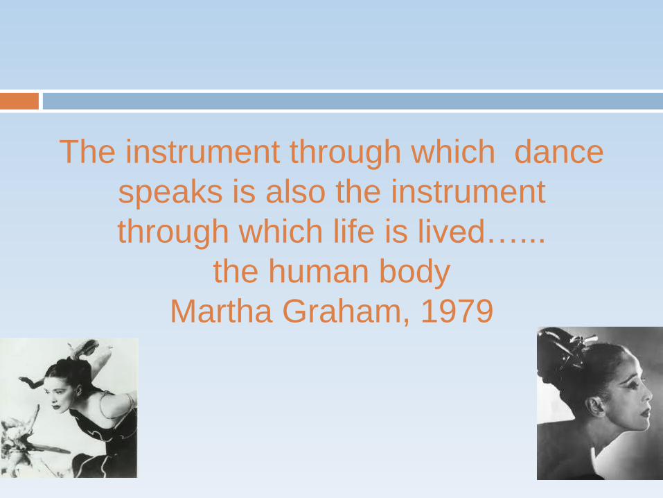
How can physical therapy assist
you in accelerating recovery from
those common foot and ankle
ballet injuries?
Below are 4 examples

Cuboid Syndrome
This syndrome is caused by a small subluxation (misalignment) of
one of the mid foot bones
On inspection, we often find an indentation on the dorsal side and a
fullness on the plantar side

When moving the surrounding joints (accessory movements) we often find a decreased joint play
Stabilizing hand on navicluar and lateral cuneiform
moving hand on cuboid stabilizing hand on calcaneal trumpet
Moving hand on cuboid

Cuboid Syndrome
Cuboid syndrome accounts for 17% of all foot and ankle injuries sustained by ballet dancers (1)
A dancer with cuboid sub-luxation will complain of lateral foot pain and weakness when attempting to move from foot flat to demi pointe and full pointe
(1) P Marshall and W G Hamilton, “Cuboid Subluxation in Ballet Dancers,” The American Journal of Sports Medicine 20, no. 2 (April 1992): 169-175.

Cuboid Syndrome
Treatment
Manipulation and mobilization to reduce the subluxation
Followed by taping to maintain the reduction
Jennings et al 2005 (1) performed a case series of 7 patients that developed cuboid syndrome after sustaining an ankle sprain. All 7 patients were able to return to competitive activities after 1 or 2 treatments with cuboid manipulations. Up to this point, no randomized clinical trials have been performed on cuboid syndromes.
Jason Jennings and George J Davies, “Treatment of Cuboid Syndrome Secondary to Lateral Ankle Sprains: a Case Series,” The Journal of Orthopaedic and Sports Physical Therapy 35, no. 7 (July 2005): 409–415.

Hallux Rigidus
Any restriction in big toe extension at the MTP joint will
prevent a dancer from performing “relevé ” (demi-pointe)
A dancer will compensate for this by rolling on the lateral
foot (“sickling”). This in turn can lead to other foot and
ankle injuries. Therefore, early intervention are
imperative.

Hallux Rigidus
Presents In 3 Stages (1)
Stage 1: the joint is preserved and there are marginal osteophytes
Stage 2: the joint is involved with some joint space narrowing on X-rays. There are marginal oteophytes.
Stage 3: the joint is involved with clear osteoarthritis with dorsal and lateral osteophytes
S J Hattrup and K A Johnson, “Subjective Results of Hallux Rigidus Following Treatment with Cheilectomy,” Clinical Orthopaedics and Related Research, no. 226 (January 1988): 182–191.

Hallux Rigidus
Treatment
Stages 1 and 2 can be treated by manual therapy of the MTP 1 joint including mobilization and manipulation to break down capsular adhesions and restore range of motion, followed by exercises (1).
Even better outcomes can be achieved by adding sesamoid joint mobilization, flexor hallucis longus strengthening and gait training (2)
In addition, we treat all identified impairments (ROM and strength) in the proximal chain.
James W. Brantingham et al., “Manipulative Therapy for Lower Extremity Conditions: Update of a Literature Review,” Journal of Manipulative and Physiological Therapeutics 35, no. 2 (February 2012): 127–166.
Jennifer Shamus et al., “The Effect of Sesamoid Mobilization, Flexor Hallucis Strengthening, and Gait Training on Reducing Pain and Restoring Function in Individuals with Hallux Limitus: a Clinical Trial,” The Journal of Orthopaedic and Sports Physical Therapy 34, no. 7 (July 2004): 368–376
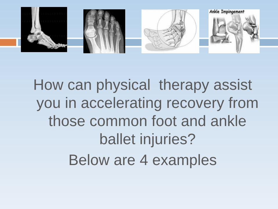
Acute And Recurrent Ankle Sprains
Ankle sprains are the most common traumatic injury in dancers. They represent 13.8% of all reported dancing injuries (1)
Dancers are particularly vulnerable for inversion traumas given the frequency in which they plantar flex their ankles. Mechanisms of injury include improper jump landings and rolling over the lateral aspect of the foot while on demi pointe (2).
The risk for ankle inversion trauma increases in case a dancer has sustained previous sprains without appropriate treatment and rehabilitation (1).
Ballet training alone without concurrent additional coordinative training does not lead to improvements in ankle joint position sense or improved measures of balance (3).
(1) E R Wiesler et al., “Ankle Flexibility and Injury Patterns in Dancers,” The American Journal of Sports Medicine 24, no. 6 (December 1996): 754–757.
(2) N Kadel, “Foot and Ankle Injuries in Dance,” Physical Medicine and Rehabilitation Clinics of North America 17, no. 4 (November 2006): 813–826
(3) Holger Schmitt, Benita Kuni, and Desiderius Sabo, “Influence of Professional Dance Training on Peak Torque and Proprioception at the Ankle,” Clinical Journal of Sport Medicine: Official Journal of the Canadian Academy of Sport Medicine 15, no. 5 (September 2005): 331–339.

Acute Ankle Sprain
Treatment
Grade 1 injuries: this represents a strain of the anterior talo fibular ligament. This type of sprain requires applying RICE principles for the first 48 hours, followed by early ROM with the use of a tape or brace. Manual therapy can assist in regaining ROM early on in the rehabilitation (1). This is followed by proprioception training, balance training and peroneal strengthening (2). In addition, we treat all identified impairments (ROM and strength) in the proximal chain.
(1) Philip J van der Wees et al., “Effectiveness of Exercise Therapy and Manual Mobilisation in Ankle Sprain and Functional Instability: a Systematic Review,” The Australian Journal of Physiotherapy 52, no. 1 (2006): 27–37.
(2) John G Kennedy and Christopher W Hodgkins, “Foot and Ankle Injuries in Dance. Preface,” Clinics in Sports Medicine 27, no. 2 (April 2008): xi–xii.

Acute Ankle Sprain
Treatment
Grade 2 injuries: this represents a tear of the anterior talo fibular ligament + sprain of the calaneo fibular ligament. There is a positive anterior drawer sign but a negative talar tilt test. This injury has to be protected by a brace (6 weeks) in combination with physical therapy that focuses on early mobilization including manual therapy, proprioceptive training, balance training and strengthening. Again additionally, we treat all identified impairments (ROM and strength) in the proximal chain.
(1)John G Kennedy and Christopher W Hodgkins, “Foot and Ankle Injuries in Dance. Preface,” Clinics in Sports Medicine 27, no. 2 (April
2008): xi–xii.

Acute Ankle Sprain
Treatment
Grade 3 injuries: this injury represents an unstable ankle. There is a positive anterior drawer test and a positive talar tilt test, secondary to tears of the anterior talofibular ligament and calcaneofibular ligament. This type of sprain is typically treated with 6 weeks of a more rigid protection (walking boot or short leg cast), with weight bearing as tolerated, in combination with physical therapy to gradually restore full ROM and to establish a functionally stable joint. Early functional treatment produces a faster recovery of ankle mobility and earlier return to activity as compared to surgical treatment (1).
Syndesmosis injuries: take longer to recover than lateral ankle sprains, and no clear guidelines exist to assist the clinician in assessing the severity of the injury, making a decision in terms of operative versus non operative management or deciding when the athlete may return to sport more difficult (2).
(1) KerkhoffsGM, Handoll HH,de BieR,etal. Surgicalversusconservativetreatmentforacuteinjuries of the lateral ligament complex of the ankle in adults. Cochrane Database Syst Rev2007;(2):CD000380
(2) Glenn N Williams, Morgan H Jones, and Annunziato Amendola, “Syndesmotic Ankle Sprains in Athletes,” The American Journal of Sports Medicine 35, no. 7 (July 2007): 1197-1207.

Chronic Recurrent Ankle Sprain
Chronic ankle instability (CAI) can be caused by mechanical instability or by functional instability. CAI is characterized by an impaired neuro-muscular control and proprioception (1).
Treatment consists of proprioceptive training and neuromuscular reeducation addressing the whole kinetic chain (2)…..
(1) Alison Holmes and Eamonn Delahunt, “Treatment of Common Deficits Associated with Chronic Ankle Instability,” Sports Medicine 39, no. 3 (2009): 207-224.
(2) JoEllen M. Sefton, “Six Weeks of Balance Training Improves Sensorimotor Function in Individuals With Chronic Ankle Instability,” Journal of Orthopaedic and Sports Physical Therapy (February 2011), http://www.jospt.org/issues/id.2513/article_detail.asp.

Chronic Recurrent Ankle Sprain
…..and of manual therapy to restore normal joint mechanics (1)
(1) Philip J van der Wees et al., “Effectiveness of Exercise Therapy and Manual Mobilisation in Ankle Sprain and
Functional Instability: a Systematic Review,” The Australian Journal of Physiotherapy 52, no. 1 (2006): 27–37.

Posterior Ankle Impingement
This is a painful condition of the posterior ankle that is
most commonly seen in ballet dancers that bring their
ankle frequently into maximal plantar flexion (pointe &
demi- pointe)
Posterior Ankle Impingement
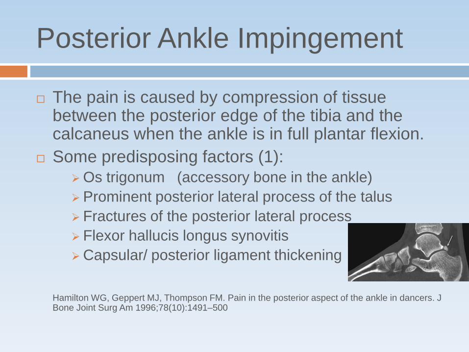
The pain is caused by compression of tissue between the posterior edge of the tibia and the calcaneus when the ankle is in full plantar flexion.
Some predisposing factors (1): Os trigonum (accessory bone in the ankle)
Prominent posterior lateral process of the talus
Fractures of the posterior lateral process
Flexor hallucis longus synovitis
Capsular/ posterior ligament thickening
Hamilton WG, Geppert MJ, Thompson FM. Pain in the posterior aspect of the ankle in dancers. J
Bone Joint Surg Am 1996;78(10):1491–500

Posterior Ankle Impingement
The hyper plantar flexion test often reproduces patient’s symptoms (not yet tested for validation and reliability). This test is performed with the patient in sitting with the knee in 90 degrees (1)
In case there is a concurrent tenosynovitis of the flexor hallucis longus there is a postive Tomassen’s Sign: 1st MTP extension is limited (and painful) with the ankle in dorsi flexion, but not limited with the ankle in plantar flexion
(1) C Niek van Dijk, “Anterior and Posterior Ankle Impingement,” Foot and Ankle Clinics 11, no. 3 (September 2006): 663–683

Posterior Ankle Impingement
Treatment
No randomized clinical trials have been performed on this subject yet. The information below comes from case studies (1),(2), (3) mixed with expert opinion:
Identify the activities that provoke the symptoms (activity modification)
Evaluate and possibly correct the dancing technique (pointe, demi-pointe etc)
Change ballet shoe to a half or ¾ shank that allows the dancer to achieve full pointe positions with less compaction of the posterior structures
Have patients sleep with night splint to prevent ankle plantar flexion
Treat impairments found in involved joints and surrounding joints (1st MTP joint!)
Plantar flexion mobilization
PA mobs and distraction manipulation of the Talo-Crural joint
Subtalar distraction manipulation
Transverse deep friction massage in case of tenosynovitis or FHL
Dry needling FHL muscle
Heel cord stretching
Proprioceptive training and peroneal strengthening
https://connect.regis.edu/p38686942/?launcher=false&fcsContent=true&pbMode=normal
Hedrick MR, McBryde AM. Posterior ankle impingement. Foot and Ankle. 1994;15:2–8.
Terry R. Malone, William T. Hardaker Rehabilitation of Foot and Ankle Injuries in Ballet Dancers J Orthop Sports Phys Ther 1990;11(8):355-361.

Thank you!
Erik De Proost, DPT


![Subclavian vessel injuries: difficult anatomy and difficult ... · evacuation times, and improved survivability [32]. High-velocity-type injuries from explosives and gunshot wounds](https://img.dokumen.tips/doc/110x75/601380c859d6401dbe0bcff5/subclavian-vessel-injuries-dificult-anatomy-and-dificult-evacuation-times.jpg)