Embed Size (px)
Citation preview

Vol. 175, No. 21JO(URNAL OF BACTERIOLOGY, Nov. 1993, P. 7056-70650021-9193/93/217056-1 0$02.00/0Copyright © 1993, American Society for Microbiology
Cloning, Expression in Escherichia coli, and Characterizationof Cellulolytic Enzymes of Azoarcus sp.,
a Root-Invading DiazotrophBARBARA REINHOLD-HUREK,' t* THOMAS HUREK,' t MARC CLAEYSSENS,2
AND MARC VAN MONTAGU'
Laboratorium voor Genetika' and Laboratorium voor Biochemie,2 Universiteit Gent,B-9000 Ghent, Belgium
Received 25 May 1993/Accepted 2 September 1993
We screened members of a new genus of grass-associated diazotrophs (Azoarcus spp.) for the presence ofcellulolytic enzymes. Out of five Azoarcus strains representing different species, only in the endorhizosphereisolate BH72, which is also capable of invading grass roots, was significant endoglucanase activity, in additionto 13-glucosidase and cellobiohydrolase activity, present. Reducing sugars were readily released frommedium-viscosity carboxymethylcellulose (CMC), but neither CMC, cellulose filter strips, Avicel, cellobiose,nor D-glucose served as the sole carbon source for growth of Azoarcus spp. Clones from a plasmid library ofstrain BH72 expressed all three enzymes in Escherichia coli, apparently not from their own promoter. Accordingto restriction endonuclease mapping and subclone analysis, ,-glucosidase and cellobiohydrolase activities werelocalized on a single 2.6-kb fragment not physically linked to a 1.45-kb fragment from which endoglucanase(egl) was expressed. Two isoenzymes of endoglucanase probably resulting from proteolytic cleavage had plvalues of 6.4 and 6.1 and an apparent molecular mass of approximately 36 kDa. Cellobiohydrolase and,I-glucosidase activity were conferred by one enzyme 41 kDa in size with a pl of 5.4, which we classified as an
unspecific exoglycanase (exg) according to substrate utilization and specificity mapping; hydrolysis of variousoligomeric substrates differentiated it from endoglucanase, which degraded substituted soluble cellulosederivatives but not microcrystalline cellulose. Both enzymes were not excreted but were associated with thesurface of Azoarcus cells. Both activities were only slightly influenced by the presence of CMC or D-glucose inthe growth medium but were enhanced by ethanol. egl was located on a large transcript 15 kb in size, whichwas detectable only in cells grown under microaerobic conditions on N2. Surface-bound exo- and endoglu-canases with some unusual regulatory features, detected in this study in a strain which is unable to metabolizecellulose or sugars, might assist Azoarcus sp. strain BH72 in infection of grass roots.
Cellulose is a constituent of lignocellulosic materials of plantorigin and is composed of D-glucopyranosyl units linked by3-1,4 bonds. For its microbial conversion complex, batteries of
several enzymes are required (52). At least three differentclasses of hydrolytic enzymes are thought to participate: endo-1,4-p-glucanase (EC 3.2.1.4), exo- l,4-cellobiohydrolase (EC3.2.1.91), and 1-glucosidase (EC 3.2.1.21).
Cellulose degradation has been reported to occur concom-itantly with nitrogen fixation in aerobic, shipworm-associatedbacteria (57) and in anaerobic freshwater mud and soil isolates(34). Among plant-associated nitrogen-fixing bacteria, cellulo-lytic activity is present in the actinomycete Franckia sp. (49)and in Rhizobium sp. (38, 43). In the legume-Rhizobiumsymbiosis, ultrastructural studies provided evidence that rhizo-bial cellulases may be involved in various steps of the infectionprocess (8, 9). The grass-associated diazotroph Azospirillulmbrasilense, although able to colonize the interior of grass roots(35, 51), is pectinolytic (55), but so far no cellulolytic activityhas been reported to be present. Cellulose only supportedgrowth of Azospirillum cells in coculture with cellulolyticbacteria (22).
Corresponding author.t Present address: Max-Planck-Institut fur Terrestrische Mikrobi-
ologie, Karl-von-Frisch-Str., D-35043 Marburg, Germany.t Present address: Pfarracker 5, D-35043 Bauerbach, Germany.
We were interested in the cellulolytic features of Azoarcusspp., a new genus of gram-negative, nitrogen-fixing bacteriacolonizing grass roots. Taxonomic studies located them in thebeta subclass of the Proteobacteria, in which they cluster in fivegroups differing at the species level (47). Two of them wereproposed as new named species, Azoarcus indigensT andAzoarclis commlunis (47). Members of the genusAzoarcus werefound to be in root-zone-specific association with Kallar grass(Leptochloa fiusca (L.) Kunth), in which they were isolatedfrom the root interior (46). Kallar grass is grown as a pioneerplant on salt-affected, frequently flooded, low-fertility soils inthe Punjab of Pakistan (50). Indirect evidence for colonizationof the root interior obtained by isolation procedures was firstsupported by immunofluorescence with antibodies which areAzoarcus genus specific (44). Light and electron microscopicimmunogold studies of better resolution revealed that thesebacteria colonize the aerenchymatic air spaces but also deeplypenetrate roots into stele and stem bases (27, 28). In gnotobi-otic culture, strain BH72 was capable of systemic infection ofKallar grass and rice seedlings, invading the cortex inter- andintracellularly and even penetrating into xylem vessels. Antic-ipating that plant polymer-degrading enzymes of bacterialorigin were involved, we wanted to screen members of Azoar-cus spp. for occurrence of cellulases. Here, we report cloningand characterization of two classes of cellulolytic enzymes,which share an unusual combination of features indicating theymight be involved in the infection process.
7056
CELLULASES OF AZOARCUS SP. 7057
TABLE 1. Bacterial strains and plasmids"
Strain or plasmid Dcscriptioin Sourcc or reference
StrainsA. indigenis VB32' Wild type 47A. comnhunis SWub3' Wild type 47Azoarcclus sp. strain BH72 Wild type 47Azoarcus sp. strain S5b2 Wild typc 47Azoarcus sp. strain 6a3 Wild type 47E. coli DH5st F' F' ecdA1 hsdRJ7 (rK m-iK+) supE44 thi-i recAI gyrA re/lAl 80/a(cZAM15 A(/acZYA-argF)1,,,X, 23
PlasmidspUC I9 Ap' ColEl replicon 59pUCl8 Apr ColEl replicon 59pBGV5.2 Ap'-, 5.3-kb KpnI fragment of pBGV5 cloned in pUC18 This studypBGVI 1.58 Ap', 1.45-kb SstI-KpsnI fragment of pBGV5 cloned in pUCI8 This studypBGV8.l Apr, 4.8-kb SmaI-XbalI fragment of pBGV8 cloned in pUC18 This study" Plasmitds not shown in this talbIc arc described in Fig. 2.
MATERIALS AND METHODS
Bacterial strains and plasmids. The strains and plasmidsused in this study are listed in Table 1. Azoarcius strainsoriginated from roots of Kallar grass, Leptochloa fusca (L.)Kunth, grown in Pakistan, and represented five groups ofAzoarclus spp. differing at the species level (47). Physical mapsof plasmids are illustrated in Fig. 2.Media and growth conditions. Escherichia coli cells were
grown at 37°C in Luria broth (LB) (2) or on LB agarsupplemented with carbenicillin (150 pg/ml) to maintain plas-mids and were grown, when indicated, with isopropyl-p3-D-thiogalactopyranoside (IPTG; 24 p.g/ml).
Azoarculs spp. were grown on VM medium (47) with rotaryshaking at 28°C as an inoculum for cellulase tests and rotaryshaking at 37°C for cell mass production. To obtain microaero-bic growth, Azoarcus sp. strain BH72 was grown at 33°C in 100ml of liquid SM medium (46), with fourfold-strength phos-phate buffer and 20 mM L-proline added, in a gas-tight sealed1 liter Erlenmeyer vial with reciprocal shaking (70 strokes permin). Cultures were harvested when the initial 02 concentra-tion of 1.7% in the headspace had decreased to 0.8%; atmo-spheric 02 was measured with a GC1 1 gas chromatograph witha thermal conductivity detector (Delsi Instruments, Suresnes,France) by using a Molecular Sieve DS column (2 m, 1/8-mmDS, 80 to 100 mesh; Alltech). To measure the expression ofcellulase by Azoarcus spp., a modification of a medium pro-posed by Kim and Wimpenny (KW medium [29]) was used,which contained 0.4% (NH4)2S04, 0.01% NaCl, 0.01%MgSO4, 0.01% CaCl2, 0.05% yeast extract, 33 mg of Fe(III)-EDTA per liter, 0.05 M potassium phosphate buffer (pH 7.0),and 0.04% medium-viscosity carboxymethylcellulose (CMC;no. C 4888; Sigma, St. Louis, Mo.). Addition of 0.5% filter-sterilized D-glucose or 1.5% agar was optional.When tested for utilization of carbon sources, Azoarcus cells
were grown on KW plates for 4 days, scraped off, and washedtwice in saline. They were used as an inoculum (at 6 [ig ofprotein per ml) for test cultures in liquid KW medium withoutyeast extract (25 ml in 125-ml Erlenmeyer flasks) and wereincubated at 37°C with rotary shaking (150 rpm) with threereplicates each. Complex carbohydrates were autoclaved, andD-glucose and cellobiose were sterilized by filtration and addedat 1% (wt/vol). Nitrogen-fixing growth on the respective car-bon sources was tested with nitrogen-free semisolid malatemedium (46), with potassium malate being replaced by therespective carbohydrate (1% [wt/vol]). Culture tubes wereinoculated with cells washed as described above (at -1 [ig of
protein per culture), and nitrogen fixation was determined bythe acetylene reduction assay (47).
Azoarctus cells for protein extractions were grown in 150 mlof liquid KW medium supplemented with 3 ml of filter-sterilized ethanol per liter in screw-capped 500-ml Erlenmeyerflasks at 28°C with rotary shaking (60 rpm); after 48 h, ethanol(3 ml/liter) was again added and the cells were grown for afurther 12 h.
Screening of bacterial colonies for B-glucosidase, cellobio-hydrolase, and endoglucanase activity. Azoarcus strains grow-ing exponentially on VM medium were streaked on KW plateswith or without D-glucose, incubatcd for 4 days at 37°C, andthen incubated at 37°C with the appropriate overlay to screenfor cellulase activities. Overlays consisted of 8 ml of 0.05 Mpotassium phosphate (pH 7.0)-buffered agarose (0.7%) con-taining 0.5 mg of either 4-methylumbelliferyl-3-D-glucoside(MUG; Sigma) or 4-methylumbelliferyl-3-cellobioside (MUC;Sigma) per ml for 3-glucosidase or cellobiohydrolase detectionor 0.2% CMC for endoglucanase detection. After 4 to 10 h,plates were exposed to 302 nm of UV light on a transillumi-nator, and the active colonies were identified by the appear-ance of blue fluorescence. Endoglucanase activity was detectedafter overnight incubation by staining with Congo red (Merck,Darmstadt, Germany); the appearance of a clear-yellowishhalo on a red background indicated endoglucanase activity(53). When necessary, plates were incubated for 5 min with 10ml of 1 M HCI to enhance contrast (49).To detect 3-glucosidase and cellobiohydrolase activity in E.
coli clones, cells were grown overnight on LB plates with IPTGand then were incubated with a MUG or MUC overlay for I to4 h at 37°C. To visualize expression of endoglucanase, LBplates were overlaid with CMC-agarose supplemented withcarbenicillin and IPTG and clones were inoculated deeply intothe agar. After incubation at 37°C overnight, plates werestained with Congo red as described above.
General cloning and DNA-analysis techniques. Plasmidsfrom E. coli were prepared by the alkaline lysis procedure ofBirnboim and Doly (7), as modified by Ausubel and Frederick(2), for both small-scale and large-scale preparations. Restric-tion endonuclease digestions, ligations, analysis and recoveryof DNA fragments on agarose gels, and autoradiography werecarried out according to standard protocols (2). DNA frag-ments were labeled with 32p with a random primed labeling kit(Boehringer Mannheim). T4 DNA ligase and calf intestinealkaline phosphatase were purchased from Bethesda ResearchLaboratories and Promega, respectively. E. coli competent
V()L. 175, 19)93

7058 REINHOLD-HUREK ET AL.
cells were prepared and transformed according to the methodof Kushner (30).
Construction and screening of a representative genomiclibrary. High-molecular-weight genomic DNA was preparedfrom Azoarcus sp. strain BH72 by the method of Marmur (37),modified as described previously (45), except that furtherpurification by CsCl gradient was omitted. DNA (240 pLg) waspartially digested with Sau3AI and was size fractionated with a
sucrose gradient (2). Fragments 5 to 15 kb in size were used forconstruction of a library in pUC19 which was digested tocompletion with BamHI and dephosphorylated. Vector andinsert DNA in equimolar amounts, as well as vector DNAalone as a control, were ligated and subsequently used fortransformation of E. coli. A total of 2,880 clones positive inblue-white selection with 5-bromo-4-chloro-3-indolyl-,3-D-ga-lactopyranoside (20 ,ug/ml of LB agar) were transferred tomicrotiter plates and replica plated. Bacterial colonies were
screened for expression of 3-glucosidase, cellobiohydrolase,and endoglucanase. Positive clones were restreaked and testedagain to confirm activity.
Preparation of whole-cell extracts and cell fractionation.For enzymatic quantitation, Azoarcus sp. strain BH72 cellswere grown on liquid KW medium supplemented with ethanolunless stated otherwise. Cells from 15 to 25 ml of culture were
processed at 4°C; they were pelleted, washed once in 10 ml of10 mM Tris-HCI (pH 8.0), and suspended in I ml of the same
buffer to give an optical density (at 578 nm) of -60. To obtaintotal cellular extracts, cells were disrupted by ultrasonicationwith a B-10 cell disrupter (Branson Sonic Power Corp.) on iceat a 40-W output for three cycles of 30 s each. Cell debris was
pelleted by centrifugation at 13,000 x g, and supernatantswere used for enzyme assays. For extraction of proteins withTriton X-100, washed cells were shaken for 10 min at 28°C and60 rpm in Tris-HCl buffer containing 0.1% Triton X-100 and10 mM Na2-EDTA. After centrifugation at 9,000 x g for 10min, the supernatant was used.The first step of cell fractionation was performed by centrif-
ugation at 10,000 x g for 10 min. To obtain the extracellularfraction, the culture supernatant was further centrifuged at20,000 x g for 25 min and concentrated 30-fold by ultrafiltra-tion through an Amicon YM10 membrane. Pelleted cells weresubjected to fractionation by the osmotic shock method as
described by Cornelis et al. (14), except that the 25% sucrose
solution was made up in 10 mM Tris-HCl (pH 7.0). Residualcells from which the periplasmic fraction had been preparedwere ultrasonicated as described above to obtain the cytoplas-mic fraction.The total cellular protein of E. coli was prepared by ultra-
sonication as described above or by the freeze-thaw method.Cells suspended in Tris-HCI buffer were rapidly frozen andthawed (dry ice-cooled ethanol, 30°C).
Determination of protein concentrations. The protein con-
tent of cell extracts was determined by the Bio-Rad proteinassay (Bio-Rad Laboratories Ltd.). Cellular protein was esti-mated by the micro-Goa method (4).
IEF, zymograms, and SDS-PAGE. For isoelectric focusing(IEF) LKB Ampholine PAGplates in the pH range 3.5 to 9.5(no. 1804-101, LKB) were used with a Multiphor II electro-phoresis chamber (LKB). Electrophoresis and Coomassie bril-liant blue staining were carried out according to the manufac-turer's instructions, except that experiments were run at lowervoltage (400 V) for 2.5 h. For zymograms, we followed theoutlines given by Coughlan (15). Agarose overlays for IEF gelscontained 0.8% agarose in 0.05 M potassium phosphate buffer(pH 7.0) and the respective substrate at 0.01% (MUG or
MUC) or 0.1% (CMC). After incubation at 37°C, overlays
proceeded as described above, except that CMC overlays werewashed for 10 min in phosphate buffer prior to Congo redstaining.
Proteins from extracts obtained by ultrasonication wereseparated by sodium dodecyl sulfate-polyacrylamide gel elec-trophoresis (SDS-PAGE) on 12% polyacrylamide gels as de-scribed by Laemmli (31).Enzyme assays. ,B-Glucosidase and cellobiohydrolase ac-
tivities were quantified with the chromophoric substrates 2-chloro-4-nitrophenyl-r3-D-glucoside or 2-chloro-4-nitrophenyl-3-cellobioside (11) at 2 mM in 200 mM potassium phosphate
buffer (pH 6.8) with 20 [tg of cell protein extract added (heatinactivated at 95°C as a negative control); the increase in A405was monitored spectrophotometrically upon incubation at37°C. One unit of specific activity is defined as 1 ,umol ofchromophore released min'-'' mg protein', with the extinc-tion coefficient at 405 nm equalling 9,000 M-' cm '. Quali-tative assays with other substrates with the same chromophorewere similarly performed. Enzymic hydrolysis of fluorogenicsubstrates was qualitatively monitored by UV illumination.
Endoglucanase activity was assayed with the chromogenicsubstrate Remazol brilliant blue R-CMC according to themethod of Cleary (13) with modifications. Twenty microgramsof cell protein extract was added to 0.2% Remazol brilliantblue R-CMC dissolved in 50 mM Tris-HCl buffer (pH 6.8), andsamples were incubated at 37°C for 10 h. The specific increasein A5910 was calculated, taking into account a negative control(heat-inactivated extract), as AA599 h -' mg of protein .
Qualitative tests for degradation of Ostazin brilliant red-hydroxyethylcellulose (Sigma) were carried out as described byBiely et al. (6) with the buffer described above. Enzymatichydrolysis of Remazol brilliant blue R-Avicell (0.2%) wastested with 160 mM Tris-HCI (pH 6.8) and calculated from theincrease in A5,0 of the supernatant after centrifugation.
Reducing sugars were quantified by a modification of theSomogyi-Nelson method as described by Marais et al. (36).
Northern analysis. RNA was isolated from bacterial cellsfrom 30 ml of an exponentially growing culture by the hot-phenol method (1). Northern (RNA) blotting and hybridiza-tion were carried out according to standard protocols (2).After separation of nucleic acids on a 0.9% agarose gel, theywere blotted onto a nylon membrane (Hybond N; Amersham),fixed to the membrane by baking at 80°C, and finally werehybridized at 65°C.
RESULTS
Occurrence of cellulolytic activities in Azoarcus strains.Representative members of all five Azoarcus spp. werescreened for the presence of hydrolytic enzymes known to beinvolved in cellulose degradation. Screening of colonies wascarried out with fluorophoric substrates to detect exo-1,4-,B-glucanases (,-glucosidase, EC 3.2.1.21; exo-1,4-cellobiohydro-lase, EC 3.2.1.91), and a CMC-plate-clearing test was used todetect endo-1,4-p-D-glucanase (EC 3.2.1.4). All but one strain,6a3, were positive or weakly positive for f-glucosidase,whereas cellobiohydrolase activity was detected only in A.communis SWub3T. Endoglucanase activity was only detect-able in strain BH72. Whether D-glucose was added to KWmedium or not, no significant difference was observed. How-ever, when ethanol was added to glucose-free KW agar topromote colony growth, detection levels were considerablyenhanced. Strong 3-glucosidase activity could be found instrains BH72 and SWub3T (Fig. IA). Although cellobiohydro-lase activity was generally weaker, it could be detected in allfour strains (Fig. IB). In contrast, considerable endoglucanase
J. BA(CF[-'RIOL.
CELLULASES OF AZOARCUS SP. 7059
FIG. 1. Cellulolytic activities of Azoarcus sp. strains in colony assays. Bacteria werc grown on KW agar plates supplemented with ethnilol (6ml/liter), overlaid with the respective substrate and viewed under UV illumination (A and B) or evaluated by Congo red staining (C). Overlayscontained fluorophoric substrates to detect P-glucosidase (MUG [A]) or cellobiohydrolase (MUC [B]) activity or CMC to detect endoglucanascactivity (C) and were incubated for 4 h, 10 h, or overnight, respectively. Numbers on plates: 1, Azowrcus sp. strain BH72; 2, A. indigens VB32"'; 3,A. communis SWub3?, 4, Azoarcius sp. strain S5b2.
activity was only seen in strain BH72; A. commutnis SWub3Tshowed only very weak activity (Fig. IC). Also, with ethanol asa carbon source, none of these enzymic activities were found instrain 6a3.
All Azoarculs strains were tested for degradation of cellulosefilter strips (9 by 1 cm; Whatman 40) in culture solutionsconsisting of KW medium without CMC, KW medium supple-mented with either D-glucose, potassium malate, or ethanol, orKW medium in the absence of any additional carbon source.Even within 1 month of incubation at 37°C, no structuraldisintegration of the filter strips was visible, indicating thatthere was no significant cellulolytic activity on this substrate.
However, cells of strain BH72 readily released reducingsugars from soluble cellulose CMC. When cells were grown onliquid KW medium with 0.02% CMC and 6 ml of ethanol perliter for 3 days, culture supernatants contained 12 ± 4 pg ofreducing sugar per ml more than controls, corresponding to 29± 10 p.g per mg of protein. Controls consisted of the sum ofthe reducing sugar contents of uninoculated medium andinoculated, CMC-free cultures. All assays were performed intriplicate.
Utilization of cellulose and other carbohydrates. Utilizationby Azoarculs sp. strain BH72 of the following cellulose types,derivatives, and hydrolysis products was tested: medium-vis-cosity CMC, cx-cellulose fiber (C 8002; Sigma), microcrystallineAvicel (Merck), cellobiose, and D-glucose. Growth was moni-tored by measurement of optical density (at 578 nm) or, in thecase of insoluble cellulose, by microscopic examination after 2,7, and 12 days of incubation, and at the end cellular proteinwas determined. There was no evidence for growth on any ofthe carbohydrates. The protein contents found with D-glucose(134 ± 6 pg), cellobiose (132 ± 6 kg), and CMC (129 ± 9pg)coincided with those found in control cultures (131 ± 8 p.g).The same compounds also did not support nitrogen-fixing
growth or nitrogen fixation. Within 2 weeks of incubation,there was no formation of a subsurface pellicle, which is typicalfor nitrogen-fixing growth of Azoarclus spp., and no acetylenereduction could be detected.
Cloning of cellulases and expression in E. coli. E. coli clonesof a total genomic library from Azoarcuts sp. strain BH72constructed in pUC19 were screened by the overlay techniquefor all three cellulase activities found in this strain: P-glucosi-dase, exo-1,4-cellobiohydrolase, and endo-1,4-3-glucanase. Of
2,880 colonies tested, seven clones showed P-glucosidase ac-tivity within 1 to 2 h. The same clones according to theirpositions in the microtiter plates also showed a positive, albeitweaker, reaction for cellobiohydrolase, which was well detect-able within 4 h of incubation. As deduced from differentpositions in the microtiter plates, five different clones werepositive for endoglucanase activity. To allow an initial groupingof the clones, PstI restriction patterns of plasmid DNA fromindividual clones were compared. Clones pBGV7 and pBGV6,as well as pBGV9 and pBGV8, had almost identical patterns,so that only one of each was further analyzed.
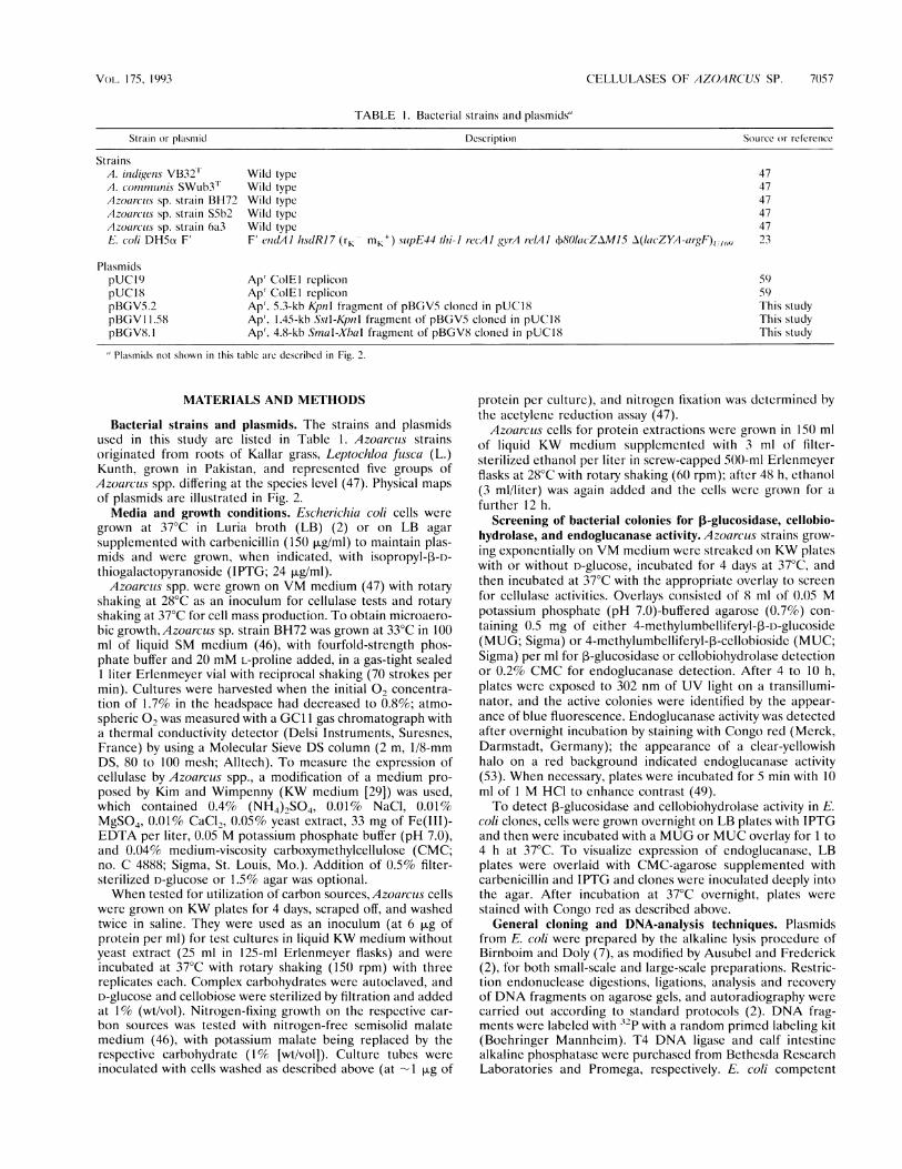
P-Glucosidase and endoglucanase clones were subjected toendonuclease restriction mapping (Fig. 2). Restriction maps ofclones pBGV10, pBGV12, pBGV6, and pBGV8 overlapped ina central genomic region which was likely to carry the 3-glu-cosidase (g,Ic) and cellobiohydrolase (cbh) genes (Fig. 2A). Toconfirm this assumption and to localize the genes more accu-rately, fragments were subcloned into pUCI9 in the sameorientation as in clone pBGV6. A 2.6-kb subclone (pBGV6.6)expressed both activities strongly, whereas smaller subcloneshad no or only weak 3-glucosidase activity (Fig. 2A), indicatingthat parts of the gene necessary for function might be locatedupstream from the HindlIl site. Also the endoglucanaseactivity (egi) seemed to be located as one gene in an overlap-ping region of pBGV2, pBGV3, pBGVl 1, pBGVI, andpBGV5. Subcloning could localize the activity on a 1.45-kbSstI-KpnI fragment (Fig. 2B, pBGV11.5). There was no evi-dence for a second endoglucanase gene located within 5 kbupstream or 12.5 kb downstream of egl in this genomic region(pBGV2.1 and pBGV2.2, respectively).
Restriction maps of genomic regions harboring 3-glucosi-dase or endoglucanase gave no evidence for physical linkage.
Because all clones of one type which we had obtained fromscreening of the library were in the same orientation withrespect to the IacZ promoter of pUC 19, we anticipated thatcellulase genes were not under control of their own promoterwhen expressed in E. coli. When the orientation of egl-activefragments (pBGV1 1.5 or pBGV5) was reversed (pBGV1 1.58or pBGV5.2, respectively), no endoglucanase activity could bedetected. Similarly, reversion of a glc- and cbh-active fragment(pBGV8 to pBGV8. 1) resulted in loss of 3-glucosidasc orcellobiohydrolase activity, indicating that the respective Azo-
VOL. 175, 19933
7060 REINHOLD-HUREK ET AL.
½½~~~~~~~~~-- 1 2 3 2 3 2 3½ 4,
glc/cbh
pBGV10
pBGV12
pBGV6
pBGV8
pBGV6.6
pBGV6.6 K
pBGV6.6H1kb
egl
II
pBGV2
pBGV3
pBGVll
pBGV1
pBGVS
pBGV21
pBGV2.2
pBGV5.1
pBGV11.5lkb
FIG. 2. Physical map of genomic regions carrying putative ,-glu-cosidase and cellobiohydrolase (glc/cbh) genes corresponding to an
exoglycanase (exg) and the endoglucanase gene (egi) of Azoarcus sp.strain BH72 and plasmid inserts in pUCI9 transcribed from the 1acZpromoter (right side of figure). For clones pBGV2 and pBGV3, theposition of the second Sall site from the right is one of two possiblelocations, the second being 0.7 kb to the left. The enzymatic activitiesof P-glucosidase (glc), cellobiohydrolase (cbh), and endoglucanase(egl) determined by colony assays are given to the right: +, positive; w,weak activity; -, no activity detectable.
arcus promoters either were not located on the fragments or
were not active in E. coli.In order to compare cloned and wild-type cellulases, enzyme
activities were located on IEF gels by zymograms. Sonicatedcell extracts of Azoarcus sp. strain BH72 were not separatedinto distinct patterns in IEF-PAG plates but gave smears,probably due to the presence of lipids. We overcame thisproblem by extracting samples once with chloroform, a treat-ment compatible with endoglucanase but detrimental to ,B-glu-cosidase activity. The latter was resistant to Triton X-100 andEDTA, which we used to release cell surface proteins, includ-ing ,-glucosidase (see below). ,B-Glucosidase and cellobiohy-drolase activities of the E. coli clone were focused at the same
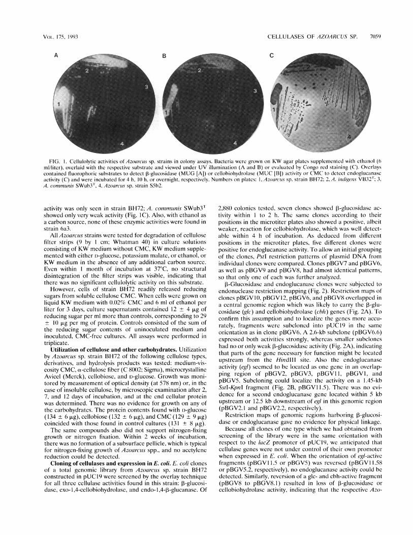
pH (pl 5.4); wild-type enzymes behaved the same way (Fig. 3).In contrast, endoglucanase activity obtained from a clonecarrying a small 1.45-kb insert exhibited two distinct bands withpIs of 6.4 and 6.0, respectively (Fig. 4). These two isoenzymesalso appeared in Azoarcus spp., although with different inten-sity ratios.The apparent molecular masses of the cellulases expressed
by E. coli were estimated by SDS-PAGE (Fig. 5). A new,41-kDa band appeared in E. coli cells expressing 3-glucosidaseand cellobiohydrolase from a 2.7-kb fragment when inducedwith IPTG, which was not seen in uninduced cells or inducedcells carrying insert-free plasmid (Fig. SA). When endoglu-canase was induced from a 1.45-kb fragment, newly formedbands were not clearly visible (not shown). However, with a
different method of protein extraction (SDS-soluble proteins[45]) a new, 36-kDa band was clearly visible (Fig. SB).
Enzymatic properties of the cloned cellulases. Substrate
Phenotypeglc cbh
7.0 -
6.5 -
6.0 -
5.1 -
A B CFIG. 3. IEF and zymograms of cloned (lanes 2) and wild-type
(lanes 3) exoglucanase. Protein extracts were obtained by the freeze-thaw method from E. coli cells carrying plasmid pUC19 as a negativecontrol (lane 1) or pBGV6.6 (lanes 2) and from cell surface proteinsprepared by Triton X- 100 washes from Azoarcus sp. strain BH72 (lanes3). Gels were stained with Coomassie brilliant blue (A) and for13-glucosidase (B) or cellobiohydrolase (C) activity with overlaysincubated for 15 or 45 min, respectively. Twenty and 8 ,utg of protein(lanes Al and A2 and lane A3, respectively) were loaded for Coo-massie staining; 10 and 12 ,ug of protein (lanes B2 and C2 and lanes B3and C3, respectively) were loaded for zymograms. Numbers to the leftcorrespond to pls of protein standards. Enzymatically activc bands atpl 5.1 are found in both cloned (lanes 2) and wild-type (lanes 3)preparations.
utilization was determined for Azoarcus sp. strain BH72 cellsand for individual cloned cellulases expressed in E. coli andtested with cell extracts (Table 2). E. coli cells carryinginsert-free pUC19 were negative for all substrates tested (notshown). The substrate specificity of cloned ,B-glucosidase andcellobiohydrolase (exoglucanase) was clearly different fromthat of endoglucanase. The activity of the cellobiohydrolaseand ,B-glucosidase is incompatible with substituted, solublecellulose derivatives, but a variety of oligomeric sugars aresubstrates, indicating that the presumed glc/cbh gene codes foran exoglucanase (exg). The activity of the endocellulase isclearly in contrast with this behavior. Microcrystalline cellulose(Avicel) was not attacked by any of the enzymes or thewild-type Azoarcus strain. This clearcut difference in substratespecificity allowed us to differentiate activities in cell extractscontaining both enzymes.To elucidate the mode of action and substrate specificity of
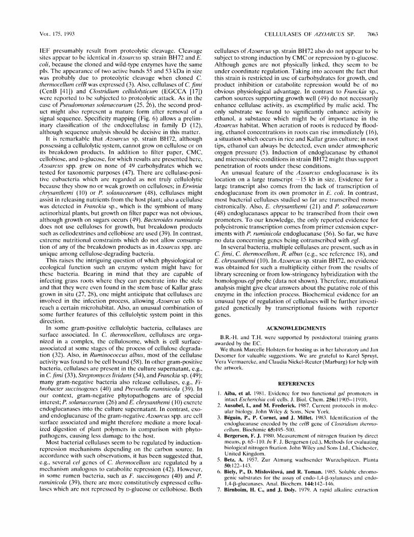
cellulases, cleavage sites of 3-glucosidase and cellobiohydro-lase and endoglucanase were determined with cell extractsfrom E. coli clones. 1,4-,-Cellooligosaccharides carrying afluorophoric group (MUGj) were tested as substrates for therespective enzymes (4 h of incubation), and the products wereseparated by thin-layer chromatography. Inspection of theproducts obtained (Fig. 6) indicated that endoglucanase pref-erably attacked oligosaccharides larger than cellobiose andreleased large oligomers when sufficiently long substrates wereused. 3-Glucosidase attacked dimers as expected (Table 2), butit also attacked larger substrates, preferably cleaving the bondat the fluorophoric group (nonreducing end). Apparently,more-highly oligomeric substrates were also cleaved at internalbonds.Temperature and pH profiles of exoglucanase and endoglu-
canase activities were obtained from cell extracts of Azoarcussp. strain BH72 (not shown). Profiles for exoglucanase mea-sured either as 3-glucosidase or as cellobiohydrolase activitycorrelated satisfactorily. The specific activity was 3- to 10-fold
J. BA(TFI-RIOL..
CELLULASES OF AZOARCUS SP. 7061
1 2 3 2 3
8.1 -
7.0-
6.5-
6.0-
5.1-
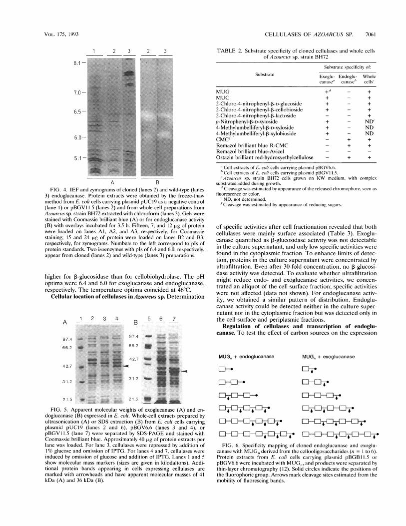
A bFIG. 4. IEF and zymograms of cloned (lanes 2) and wild-type (lanes
3) endoglucanase. Protein extracts were obtained by the freeze-thawmethod from E. coli cells carrying plasmid pUC19 as a negative control(lane 1) or pBGV1 1.5 (lanes 2) and from whole-cell preparations fromAzoarculs sp. strain BH72 extracted with chloroform (lanes 3). Gels werestained with Coomassie brilliant blue (A) or for endoglucanase activity(B) with overlays incubated for 3.5 h. Fifteen, 7, and 12 xg of proteinwere loaded on lanes Al, A2, and A3, respectively, for Coomassiestaining; 15 and 24 ,ug of protein were loaded on lanes B2 and B3,respectively, for zymograms. Numbers to the left correspond to pIs ofprotein standards. Two isoenzymes with pIs of 6.4 and 6.0, respectively,appear from cloned (lanes 2) and wild-type (lanes 3) preparations.
higher for 3-glucosidase than for cellobiohydrolase. The pHoptima were 6.4 and 6.0 for exoglucanase and endoglucanase,respectively. The temperature optima coincided at 46°C.
Cellular location of cellulases in Azoarcus sp. Determination
1 2 3 4A ---
97.4
66.2
42.7
31.2
B 5 6 7
97.4 - "t"
66.2 a _ "f
42.7 _.
31.2 _
21.5 21.5 _
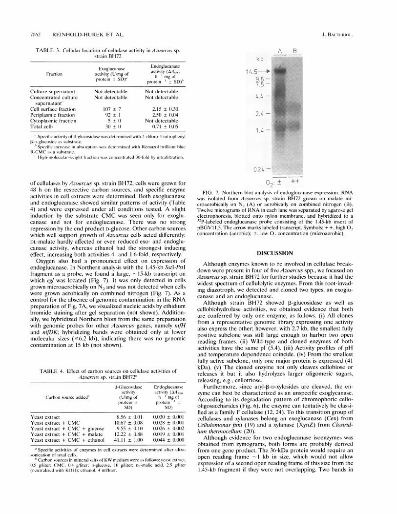
FIG. 5. Apparent molecular weights of exoglucanase (A) and en-doglucanase (B) expressed in E. coli. Whole-cell extracts prepared byultrasonication (A) or SDS extraction (B) from E. coli cells carryingplasmid pUC19 (lanes 2 and 6), pBGV6.6 (lanes 3 and 4), orpBGVI 1.5 (lane 7) were separated by SDS-PAGE and stained withCoomassie brilliant blue. Approximately 40 p.g of protein extracts perlane was loaded. For lane 3, cellulases were repressed by addition of1% glucose and omission of IPTG. For lanes 4 and 7, cellulases wereinduced by omission of glucose and addition of IPTG. Lanes 1 and 5show molecular mass markers (sizes are given in kilodaltons). Addi-tional protein bands appearing in cells expressing cellulases aremarked with arrowheads and have apparent molecular masses of 41kDa (A) and 36 kDa (B).
TABLE 2. Substrate specificity of cloned cellulases and wholc cellsof Azoarcus sp. strain BH72
Substratc specificity of:Substrate Exoglu- Endoglu- Whole
canaIsc" canase" cells"
MUG +" - +MUC + - +2-Chloro-4-nitrophenyl-f3-D-glucoside + - +2-Chloro-4-nitrophenyl-3-cellobioside + - +2-Chloro-4-nitrophenyl-l3-lactoside - - +
p-Nitrophenyl-P-D-xyloside + - ND"4-Methylumbelliferyl-13-D-xyloside + - ND4-Methylumbelliferyl-l3-xylobioside + - NDCMC' - + +Remazol brilliant blue R-CMC - + +Remazol brilliant blue-Avicel -
Ostazin brilliant red-hydroxyethylcellulose - + +
" Cell extracts of E. coli cells carrying plasmid pBGV6.6."'Cell extracts of E. coli cells carrying plasmid pBGVI 1.5.'Azoarcus sp. strain BH72 cells grown on KW medium, with complex
substrates added during growth.d Cleavage was estimated by appearance of the released chromophore, seen as
fluorescence or color.' ND, not determined./'Cleavage was estimated by appearance of reducing sugars.
of specific activities after cell fractionation revealed that bothcellulases were mainly surface associated (Table 3). Exoglu-canase quantified as ,B-glucosidase activity was not detectablein the culture supernatant, and only low specific activities werefound in the cytoplasmic fraction. To enhance limits of detec-tion, proteins in the culture supernatant were concentrated byultrafiltration. Even after 30-fold concentration, no 3-glucosi-dase activity was detected. To evaluate whether ultrafiltrationmight reduce endo- and exoglucanase activities, we concen-trated an aliquot of the cell surface fraction; specific activitieswere not affected (data not shown). For endoglucanase activ-ity, we obtained a similar pattern of distribution. Endoglu-canase activity could be detected neither in the culture super-natant nor in the cytoplasmic fraction but was detected only inthe cell surface and periplasmic fractions.
Regulation of cellulases and transcription of endoglu-canase. To test the effect of carbon sources on the expression
MUG, + endoglucanase
El--
El-Ei---
-
ElE lE4lE4
MUG, + exoglucanase
4El-El-EI-.
4
El4El4 lEl-
FIG. 6. Specificity mapping of cloned endoglucanase and exoglu-canase with MUGn derived from the cellooligosaccharides (n = 1 to 6).Protein extracts from E. coli cells carrying plasmid pBGBI 1.5 orpBGV6.6 were incubated with MUG,,, and products were separated bythin-layer chromatography (12). Solid circles indicate the positions ofthe fluorophoric group. Arrows mark cleavage sites estimated from themobility of fluorescing bands.
VOL. 1 75, 19'93
70)62 REINHOLD-HUREK ET AL.
TABLE 3. Cellular location of cellulase activity in Azoarcus sp.strain BH72
Endoglucanase
Friiction -tity(Exoglucmngasc activity (AA590Fraction acprteivit (U/mg of h mg ofprotcin± SD) protein -- SD)"
Culture supernatant Not detectable Not detectableConcentrated culture Not detectable Not detectable
supernatant"Cell surface fraction 107 ± 7 2.15 ± 0.30Periplasmic fraction 92 ± 1 2.50 ± 0.04Cytoplasmic fraction 5 ± (0 Not detcctableTotal cells 30 ± 0 0.71 ± 0.05
" SpCcific activity of -glUcosidase was determined with 2-chloro-4-nitrophenylP-i)-glucoside as substrate.
/ Specific increase in absorption was determined with Remazol brilliant blueR-CMC as a substrate.
High-molccukar-wcight fraction was concentrated 30-fold by ultratfiltration.
of cellulases by Azoarcuis sp. strain BH72, cells were grown for48 h on the respective carbon sources, and specific enzymeactivities in cell extracts were determined. Both exoglucanaseand endoglucanase showed similar patterns of activity (Table4) and were expressed under all conditions tested. A slightinduction by the substrate CMC was seen only for exoglu-canase and not for endoglucanase. There was no strongrepression by the end product D-glucose. Other carbon sourceswhich well support growth of Azoarcus cells acted differently:DL-malate hardly affected or even reduced exo- and endoglu-canase activity, whereas ethanol had the strongest inducingeffect, increasing both activities 4- and 1.6-fold, respectively.Oxygen also had a pronounced effect on expression of
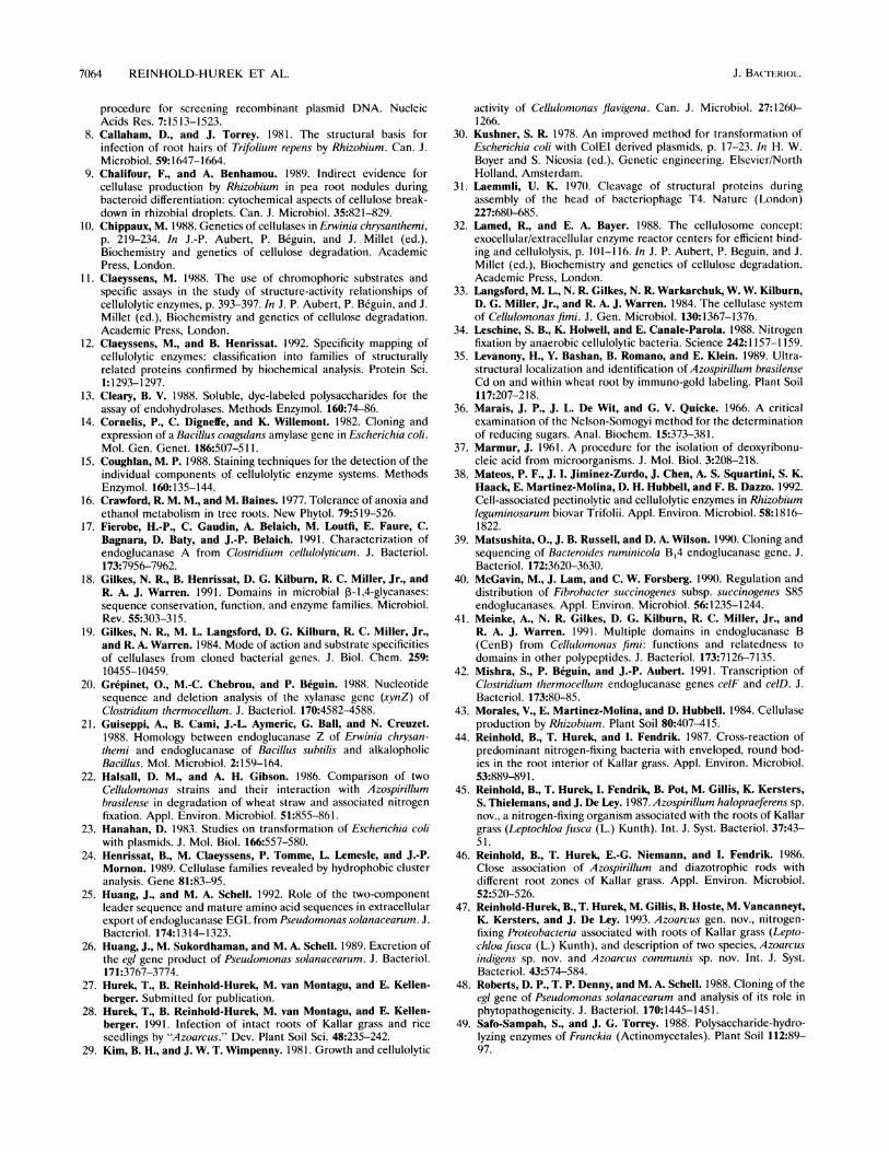
endoglucanase. In Northern analysis with the 1.45-kb SstI-Pstlfragment as a probe, we found a large, -15-kb transcript onwhich egl was located (Fig. 7). It was only detected in cellsgrown microaerobically on N2 and was not detected when cellswere grown aerobically on combined nitrogen (Fig. 7). As acontrol for the absence of genomic contamination in the RNApreparation of Fig. 7A, we visualized nucleic acids by ethidiumbromide staining after gel separation (not shown). Addition-ally, we hybridized Northern blots from the same preparationwith genomic probes for other Azoarcus genes, namely nifHand nifDK; hybridizing bands were obtained only at lowermolecular sizes (-6.2 kb), indicating therc was no genomiccontamination at 15 kb (not shown).
TABLE 4. Effect of carbon sources on cellulase activities ofAzoarcus sp. strain BH72"
P-Glucosidase Endoglucanascactivity activity (AA590
Cairbon source added' (U/mg ot h mg ofprotein + protcin ±
SD) SD)
Yeast extract 8.56 ± 0.01 0.030 ± 0.001Yeast extract + CMC 10.67 ± 0.t)8 0.028 ± 0.001Yeast extract + CMC + glucose 9.55 ± (1.11 0.026 ± 0.002Yeast extract + CMC + malate 12.22 ± 0.88 0.019 ± 0.0(1Yeast extract + CMC + ethanol 41.11 ± 1.0() 0.044 ± 0.000
" Specific aictivitics of cnzymes in cell extracts were determined after ultria-sonication of total cells.
' Carbon sources in mineral salts of KW medium were as follows: yeast extract,11.5 g/liter; CMC, 11.4 g/liter; 1)-glucose, 1) g/liter; il.-malic acid, 2.5 g/liter(neutralized with KOII); ethalnol, 4 ml/litcr.
02 + ++
FIG. 7. Northern blot analysis of endoglucanase expression. RNAwas isolated from Azoar-cus sp. strain BH72 grown on malate mi-croaerobically on N, (A) or aerobically on combined nitrogen (B).Twelve micrograms of RNA in each lane was separated by agarose gel
electrophoresis, blotted onto nylon membrane, and hybridized to a
329P-labeled endoglucanase probe consisting of the 1.45-kb insert ofpBGVI 1.5. The arrow marks labeled transcript. Symbols: + +, high 02concentration (aerobic); +, low 0O concentration (microaerobic).
DISCUSSION
Although cnzymes known to be involved in cellulase break-down were present in four of five Azoarcus spp., we focused on
Azoarcus sp. strain BH72 for further studies because it had thewidest spectrum of cellulolytic enzymes. From this root-invad-ing diazotroph, we detected and cloned two types, an exoglu-canase and an endoglucanase.
Although strain BH72 showed 3-glucosidase as well as
cellobiohydrolase activities, we obtained evidence that bothare conferred by only one enzyme, as follows. (i) All clonesfrom a representative genomic library expressing one activityalso express the other; however, with 2.7 kb, the smallest fullypositive subclone was still large enough to harbor two open
reading frames. (ii) Wild-type and cloned enzymes of bothactivities have the same pl (5.4). (iii) Activity profiles of pHand temperature dependence coincide. (iv) From the smallestfully active subclone, only one major protein is expressed (41kDa). (v) The cloned enzyme not only cleaves cellobiose or
releases it but it also hydrolyzes larger oligomeric sugars,releasing, e.g., cellotriose.
Furthermore, since aryl-3-D-xylosides are cleaved, the en-
zymc can best be characterized as an unspecific exoglycanase.According to its degradation pattern of chromophoric cello-oligosaccharides (Fig. 6), the enzyme can tentatively be classi-fied as a family F cellulase (12, 24). To this transition group ofcellulases and xylanases belong an exoglucanase (Cex) fromCellulomonas fimi (19) and a xylanase (XynZ) from Clostrid-ium thermocellum (20).
Although evidence for two endoglucanase isoenzymes was
obtained from zymograms, both forms are probably derivedfrom one gene product. The 36-kDa protein would require an
open reading frame -1 kb in size, which would not allowexpression of a second open reading frame of this size from the1.45-kb fragment if they were not overlapping. Two bands in
kb
14.5-*9.5 -7.5-
4.4 -
2.4 -
1.4 -
0.24 -
J. BACT11RIOL.
CELLULASES OF AZOARCUS SP. 7063
IEF presumably result from proteolytic cleavage. Cleavagesites appear to be identical in Azoarcuts sp. strain BH72 and E.coli, because the cloned and wild-type enzymes have the samepIs. The appearance of two active bands 55 and 53 kDa in sizewas probably due to proteolytic cleavage when cloned C.thermocellum celB was expressed (3). Also, cellulases of C. fimni(CenB [41]) and Clostridium cell/ulolytictum (EGCCA [17])were reported to be subjected to proteolytic attack. As in thecase of Pseudomonias solanacearuim (25, 26), the second prod-uct might also represent a mature form after removal of asignal sequence. Specificity mapping (Fig. 6) allows a prelim-inary classification of the endocellulase in family D (12),although sequence analysis should be decisive in this matter.
It is remarkable that Azoarcus sp. strain BH72, althoughpossessing a cellulolytic system, cannot grow on cellulose or onits breakdown products. In addition to filter paper, CMC,cellobiose, and D-glucose, for which results are presented here,Azoarcus spp. grew on none of 49 carbohydrates which wetested for taxonomic purposes (47). There are cellulase-posi-tive eubacteria which are regarded as not truly cellulolyticbecause they show no or weak growth on celluloses; in Erwiniachrysanit/lemiii (10) or P. solanaceanlini (48), cellulases mightassist in releasing nutrients from the host plant; also a cellulasewas detected in Franckia sp., which is the symbiont of manyactinorhizal plants, but growth on filter paper was not obvious,although growth on sugars occurs (49). Bacteroides ruminicoladoes not use celluloses for growth, but breakdown productssuch as cellodextrines and cellobiose are used (39). In contrast,extreme nutritional constraints which do not allow consump-tion of any of the breakdown products as in Azoarcuts spp. areunique among cellulose-degrading bacteria.
This raises the intriguing question of which physiological orecological function such an enzyme system might have forthese bacteria. Bearing in mind that they are capable ofinfecting grass roots where they can penetrate into the steleand that they were even found in the stem base of Kallar grassgrown in situ (27, 28), one might anticipate that cellulases areinvolved in the infection process, allowing Azoarcus cells toreach a certain microhabitat. Also, an unusual combination ofsome further features of this cellulolytic system point in thisdirection.
In some gram-positive cellulolytic bacteria, cellulases aresurface associated. In C. thermocellum, cellulases are orga-nized in a complex, the cellulosome, which is cell surface-associated at some stages of the process of cellulose degrada-tion (32). Also, in Ruminococcus albus, most of the cellulaseactivity was found to be cell bound (58). In other gram-positivebacteria, cellulases are present in the culture supernatant, e.g.,in C. fimi (33), Streptomyces lividans (54), and Franckia sp. (49);many gram-negative bacteria also release cellulases, e.g., Fi-brobacter sluccinogenes (40) and Prevotella rluminicola (39). Inour context, gram-negative phytopathogens are of specialinterest; P. solanacearlum (26) and E. chrysanthemi (10) excreteendoglucanases into the culture supernatant. In contrast, exo-and endoglucanase of the gram-negative Azoarcuts spp. are cellsurface associated and might therefore mediate a more local-ized digestion of plant polymers in comparison with phyto-pathogens, causing less damage to the host.Most bacterial cellulases seem to be regulated by induction-
repression mechanisms depending on the carbon source. Inaccordance with such observations, it has been suggested that,e.g., several cel genes of C. therrnocell/tm are regulated by amechanism analogous to catabolite repression (42). However,in some rumen bacteria, such as F. succinogenes (40) and P.ruiminicola (39), there are more constitutively expressed cellu-lases which are not repressed by D-glucose or cellobiose. Both
cellulases ofAzoarcus sp. strain BH72 also do not appear to besubject to strong induction by CMC or repression by D-glucose.Although genes are not physically linked, they seem to beunder coordinate regulation. Taking into account the fact thatthis strain is restricted in use of carbohydrates for growth, endproduct inhibition or catabolite repression would be of noobvious physiological advantage. In contrast to Franckia sp.,carbon sources supporting growth well (49) do not necessarilyenhance cellulase activity, as exemplified by malic acid. Theonly substrate we found to significantly enhance activity isethanol, a substance which might be of importance in theAzoarcuts habitat. When aeration of roots is reduced by flood-ing, ethanol concentrations in roots cain rise immediately (16),a situation which occurs in rice and Kallar grass culture; in roottips, ethanol can always be detected, even under atmosphericoxygen pressure (5). Induction of endoglucanase by ethanoland microaerobic conditions in strain BH72 might thus supportpenetration of roots under these conditions.An unusual feature of the Azoarcus endoglucanase is its
location on a large transcript -15 kb in size. Evidence for alarge transcript also comes from the lack of transcription ofendoglucanase from its own promoter in E. coli. In contrast,most bacterial cellulases studied so far are transcribed mono-cistronically. Also, E. chrysanthemi (21) and P. solanacearlum(48) endoglucanases appear to be transcribed from their ownpromoters. To our knowledge, the only reported evidence forpolycistronic transcription comes from primer extension exper-iments with P. ruminicola endoglucanase (56). So far, we haveno data concerning genes being cotranscribed with eg/.
In several bacteria, multiple cellulases are present, such as inC. fimni, C thermocellum, R. albus (e.g., see reference 18), andE. chrysanthemi (10). InAzoarcius sp. strain BH72, no evidencewas obtained for such a multiplicity either from the results oflibrary screening or from low-stringency hybridization with thehomologous egl probe (data not shown). Therefore, mutationalanalysis might give clear answers about the putative role of thisenzyme in the infection process. Biochemical evidence for anunusual type of regulation of cellulases will be further investi-gated genetically by transcriptional fusions with reportergenes.
ACKNOWLEDGMENTS
B.R.-H. and T.H. were supported by postdoctoral training grantsawarded by the EC.We thank Marcelle Holsters for hosting us in her laboratory and Jan
Desomer for valuable suggestions. We are grateful to Karel Spruyt,Vera Vermaercke, and Claudia Nickel-Reuter (Marburg) for help withthe artwork.
REFERENCES
1. Aiba, et al. 1981. Evidence for two functional gal/ promoters inintact Eschericlhia coli cells. J. Biol. Chem. 256:11905-1 19 t1).
2. Ausubel, I., and M. Frederick. 1987. Current protocols in molec-ular biology. John Wiley & Sons, New York.
3. Beguin, P., P. Cornet, and J. Millet. 1983. Identification of theendoglucanase encoded by the celB gene of Clostuidiiui,i tlilermo-ce/llum. Biochimie 65:495-5(t(.
4. Bergersen, F. J. 198t). Measuremenlt of nitrogen fixation by directmeans, p. 65-110. In F. J. Bergersen (ed.), Methods for evaluatingbiological nitrogen fixation. John Wiley and Sons Ltd., Chichester,United Kingdom.
5. Betz, A. 1957. Zur Atmung wachsender Wurzelspitzen. Planta50:122-143.
6. Biely, P., D. Mislovicova, and R. Toman. 1985. Soluble chromo-genic substrates for the assay of endo-1,4-3-xylanases and endo-1,4-p-glucanases. Anal. Biochem. 144:142-146.
7. Birnboim, H. C., and J. Doly. 1979. A rapid alkaline extraction
Voi.- 1 75? 1 993
7064 REINHOLD-HUREK ET AL.
procedure for screening recombinant plasmid DNA. NucleicAcids Res. 7:1513-1523.
8. Callaham, D., and J. Torrey. 1981. The structural basis forinfection of root hairs of Trifolium repens by Rhizobium. Can. J.Microbiol. 59:1647-1664.
9. Chalifour, F., and A. Benhamou. 1989. Indirect evidence forcellulase production by Rhizobium in pea root nodules duringbacteroid differentiation: cytochemical aspects of cellulose break-down in rhizobial droplets. Can. J. Microbiol. 35:821-829.
10. Chippaux, M. 1988. Genetics of cellulases in Erwinia chrysanthemi,p. 219-234. In J.-P. Aubert, P. Beguin, and J. Millet (ed.),Biochemistry and genetics of cellulose degradation. AcademicPress, London.
11. Claeyssens, M. 1988. The use of chromophoric substrates andspecific assays in the study of structure-activity relationships ofcellulolytic enzymes, p. 393-397. In J. P. Aubert, P. Beguin, and J.Millet (ed.), Biochemistry and genetics of cellulose degradation.Academic Press, London.
12. Claeyssens, M., and B. Henrissat. 1992. Specificity mapping ofcellulolytic enzymes: classification into families of structurallyrelated proteins confirmed by biochemical analysis. Protein Sci.1:1293-1297.
13. Cleary, B. V. 1988. Soluble, dye-labeled polysaccharides for theassay of endohydrolases. Methods Enzymol. 160:74-86.
14. Cornelis, P., C. Digneffe, and K. Willemont. 1982. Cloning andexpression of a Bacillus coagulans amylase gene in Escherichia coli.Mol. Gen. Genet. 186:507-511.
15. Coughlan, M. P. 1988. Staining techniques for the detection of theindividual components of cellulolytic enzyme systems. MethodsEnzymol. 160:135-144.
16. Crawford, R. M. M., and M. Baines. 1977. Tolerance of anoxia andethanol metabolism in tree roots. New Phytol. 79:519-526.
17. Fierobe, H.-P., C. Gaudin, A. Belaich, M. Loutfi, E. Faure, C.Bagnara, D. Baty, and J.-P. Belaich. 1991. Characterization ofendoglucanase A from Clostridium cellulolyticum. J. Bacteriol.173:7956-7962.
18. Gilkes, N. R., B. Henrissat, D. G. Kilburn, R. C. Miller, Jr., andR. A. J. Warren. 1991. Domains in microbial P3-1,4-glycanases:sequence conservation, function, and enzyme families. Microbiol.Rev. 55:303-315.
19. Gilkes, N. R., M. L. Langsford, D. G. Kilburn, R. C. Miller, Jr.,and R. A. Warren. 1984. Mode of action and substrate specificitiesof cellulases from cloned bacterial genes. J. Biol. Chem. 259:10455-10459.
20. Grepinet, O., M.-C. Chebrou, and P. Beguin. 1988. Nucleotidesequence and deletion analysis of the xylanase gene (xynZ) ofClostridium thermocellum. J. Bacteriol. 170:4582-4588.
21. Guiseppi, A., B. Cami, J.-L. Aymeric, G. Ball, and N. Creuzet.1988. Homology between endoglucanase Z of Erwinia chrysan-themi and endoglucanase of Bacillus subtilis and alkalopholicBacillus. Mol. Microbiol. 2:159-164.
22. Halsall, D. M., and A. H. Gibson. 1986. Comparison of twoCellulomonas strains and their interaction with Azospirillumbrasilense in degradation of wheat straw and associated nitrogenfixation. Appl. Environ. Microbiol. 51:855-861.
23. Hanahan, D. 1983. Studies on transformation of Escherichia coliwith plasmids. J. Mol. Biol. 166:557-580.
24. Henrissat, B., M. Claeyssens, P. Tomme, L. Lemesle, and J.-P.Mornon. 1989. Cellulase families revealed by hydrophobic clusteranalysis. Gene 81:83-95.
25. Huang, J., and M. A. Schell. 1992. Role of the two-componentleader sequence and mature amino acid sequences in extracellularexport of endoglucanase EGL from Pseudomonas solanacearum. J.Bacteriol. 174:1314-1323.
26. Huang, J., M. Sukordhaman, and M. A. Schell. 1989. Excretion ofthe egl gene product of Pseudomonas solanacearum. J. Bacteriol.171:3767-3774.
27. Hurek, T., B. Reinhold-Hurek, M. van Montagu, and E. Kellen-berger. Submitted for publication.
28. Hurek, T., B. Reinhold-Hurek, M. van Montagu, and E. Kellen-berger. 1991. Infection of intact roots of Kallar grass and riceseedlings by "Azoarcus." Dev. Plant Soil Sci. 48:235-242.
29. Kim, B. H., and J. W. T. Wimpenny. 1981. Growth and cellulolytic
activity of Cellulomonas flavigena. Can. J. Microbiol. 27:1260-1266.
30. Kushner, S. R. 1978. An improved method for transformation ofEscherichia coli with ColEI derived plasmids, p. 17-23. In H. W.Boyer and S. Nicosia (ed.), Genetic engineering. Elsevier/NorthHolland, Amsterdam.
31. Laemmli, U. K. 1970. Cleavage of structural proteins duringassembly of the head of bacteriophage T4. Nature (London)227:680-685.
32. Lamed, R., and E. A. Bayer. 1988. The cellulosome concept:exocellular/extracellular enzyme reactor centers for efficient bind-ing and cellulolysis, p. 1t)1-1 16. In J. P. Aubert, P. Beguin, and J.Millet (ed.), Biochemistry and genetics of cellulose degradation.Academic Press, London.
33. Langsford, M. L., N. R. Gilkes, N. R. Warkarchuk, W. W. Kilburn,D. G. Miller, Jr., and R. A. J. Warren. 1984. The cellulase systemof Cellulomonas fimi. J. Gen. Microbiol. 130:1367-1376.
34. Leschine, S. B., K. Holwell, and E. Canale-Parola. 1988. Nitrogenfixation by anaerobic cellulolytic bacteria. Science 242:1157-1159.
35. Levanony, H., Y. Bashan, B. Romano, and E. Klein. 1989. Ultra-structural localization and identification of Azospirillum brasilenseCd on and within wheat root by immuno-gold labeling. Plant Soil117:207-218.
36. Marais, J. P., J. L. De Wit, and G. V. Quicke. 1966. A criticalexamination of the Nelson-Somogyi method for the determinationof reducing sugars. Anal. Biochem. 15:373-381.
37. Marmur, J. 1961. A procedure for the isolation of deoxyribonu-cleic acid from microorganisms. J. Mol. Biol. 3:208-218.
38. Mateos, P. F., J. I. Jiminez-Zurdo, J. Chen, A. S. Squartini, S. K.Haack, E. Martinez-Molina, D. H. Hubbell, and F. B. Dazzo. 1992.Cell-associated pectinolytic and cellulolytic enzymes in Rhizobiumleguminosarum biovar Trifolii. Appl. Environ. Microbiol. 58:1816-1822.
39. Matsushita, O., J. B. Russell, and D. A. Wilson. 1990. Cloning andsequencing of Bacteroides ruminicola B,4 endoglucanase gene. J.Bacteriol. 172:3620-3630.
40. McGavin, M., J. Lam, and C. W. Forsberg. 1990. Regulation anddistribution of Fibrobacter succinogenes subsp. succinogenes S85endoglucanases. AppI. Environ. Microbiol. 56:1235-1244.
41. Meinke, A., N. R. Gilkes, D. G. Kilburn, R. C. Miller, Jr., andR. A. J. Warren. 1991. Multiple domains in endoglucanase B(CenB) from Cellulomonas fimi: functions and relatedness todomains in other polypeptides. J. Bacteriol. 173:7126-7135.
42. Mishra, S., P. Beguin, and J.-P. Aubert. 1991. Transcription ofClostridium thermocellum endoglucanase genes celF and celD. J.Bacteriol. 173:80-85.
43. Morales, V., E. Martinez-Molina, and D. Hubbell. 1984. Cellulaseproduction by Rhizobium. Plant Soil 80:407-415.
44. Reinhold, B., T. Hurek, and I. Fendrik. 1987. Cross-reaction ofpredominant nitrogen-fixing bacteria with enveloped, round bod-ies in the root interior of Kallar grass. Appl. Environ. Microbiol.53:889-891.
45. Reinhold, B., T. Hurek, I. Fendrik, B. Pot, M. Gillis, K. Kersters,S. Thielemans, and J. De Ley. 1987. Azospirillum halopraeferens sp.nov., a nitrogen-fixing organism associated with the roots of Kallargrass (Leptochloa fusca (L.) Kunth). Int. J. Syst. Bacteriol. 37:43-51.
46. Reinhold, B., T. Hurek, E.-G. Niemann, and I. Fendrik. 1986.Close association of Azospirillum and diazotrophic rods withdifferent root zones of Kallar grass. Appl. Environ. Microbiol.52:520-526.
47. Reinhold-Hurek, B., T. Hurek, M. Gillis, B. Hoste, M. Vancanneyt,K. Kersters, and J. De Ley. 1993. Azoarcus gen. nov., nitrogen-fixing Proteobacteria associated with roots of Kallar grass (Lepto-chloa fusca (L.) Kunth), and description of two species, Azoarcusindigens sp. nov. and Azoarcus communis sp. nov. Int. J. Syst.Bacteriol. 43:574-584.
48. Roberts, D. P., T. P. Denny, and M. A. Schell. 1988. Cloning of theegl gene of Pseudomonas solanacearum and analysis of its role inphytopathogenicity. J. Bacteriol. 170:1445-145 1.
49. Safo-Sampah, S., and J. G. Torrey. 1988. Polysaccharide-hydro-lyzing enzymes of Franckia (Actinomycetales). Plant Soil 112:89-97.
J. BAC1 ECRIOL.

CELLULASES OF AZOARCUS SP. 7065
50. Sandhu, G. R., and K. A. Malik. 1975. Plant succession-a key tothe utilization of saline soils. Nucleus 12:35-38.
51. Schank, S. C., R. L. Smith, G. C. Weiser, D. A. Zuberer, J. H.Bouton, K. H. Quesenberry, M. E. Tyler, J. R. Milam, and R. C.Littell. 1979. Fluorescent antibody technique to identify Azospiril-lim brasilense associated with roots of grasses. Soil Biol. Biochem.11:287-295.
52. Sharrock, K. R. 1988. Cellulase assay methods: a review. J.Biochem. Biophys. Methods 17:81-106.
53. Teather, R. M., and P. J. Wood. 1982. Use of Congo red-polysaccharide interactions in enumeration and characterizationof cellulolytic bacteria from the bovine rumen. Appl. Environ.Microbiol. 43:777-780.
54. Theberge, M., P. Lacaze, F. Shareck, R. Morosoli, and D. Kluepfel.1992. Purification and characterization of an endoglucanase fromStreptomyces lividans 66 and DNA sequence of the gene. Appl.Environ. Microbiol. 58:815-820).
55. Tien, T. M., H. G. Diem, M. H. Gaskins, and D. H. Hubbell. 1981.Polygalacturonic acid transeliminase production by Azospirilllinspecies. Can. J. Microbiol. 27:426-431.
56. Vercoe, P. E., and K. Gregg. 1992. DNA sequence and transcrip-tion of an endoglucanase gene from Prevotella (Bacwroides)ruminicola AR20. Mol. Gen. Genet. 233:284-292.
57. Waterbury, J. B., C. B. Calloway, and R. D. Turner. 1983. Acellulolytic nitrogen-fixing bacterium cultured from the gland ofdeshayes in shipworms (Bivalvia: Teredinidae). Science 221:1401-1403.
58. Wood, T. M., C. A. Wilson, and C. S. Stewart. 1982. Preparation ofthe cellulase from the cellulolytic anaerobic bacterium Ruinino-coccus albus and its release from the bacterial cell wall. Biochem.J. 205:129-137.
59. Yanisch-Perron, C., J. Vieira, and J. Messing. 1985. ImprovedM13 phage cloning vectors and host strains: nucleotide sequencesof the M13mpl8 and pUC19 vectors. Gene 33:10)3-119.
VOL. 175, 1993