Embed Size (px)
Citation preview

CARDIOLOGY • October 2015 EMJ EUROPEAN MEDICAL JOURNAL CARDIOLOGY • October 2015 EMJ EUROPEAN MEDICAL JOURNAL 94 95
SELF-EXPANDING CORONARY STENTS: RATIONALE, CLINICAL STATUS, FUTURE PROSPECTS
*Rainer Wessely,1 Giovanni Amoroso2
1. Cologne Cardiovascular and Chest Center, Cologne, Germany2. Onze Lieve Vrouwe Gasthuis, Amsterdam, Netherlands
*Correspondence to [email protected]
Disclosure: Prof Dr R. Wessely and Dr G. Amoroso have received minimal consulting speaker fees from STENTYS.Support: The publication of this article was funded by STENTYS. The views and opinions expressed are those of the authors and not necessarily of STENTYS.Received: 01.12.14 Accepted: 15.06.15Citation: EMJ Cardiol. 2015;3[2]:94-106.
ABSTRACT
The mechanical treatment of coronary artery stenoses by placement of balloon-expandable (Bx) coronary stents has become the most widely used invasive treatment for symptomatic coronary artery disease (CAD). However, the mechanical properties of Bx stents may be limited and are frequently not well adapted to the requirements of the biological system. Consequently, there is evidence that the mechanical shortcomings of Bx stents, such as conformability to the vascular wall, stent underexpansion or oversizing, adaptability to vessel tapering, scaffolding of bifurcated lesions, inability to address vessel remodelling, and achieving optimal drug delivery, could translate into adverse clinical events. New, enhanced technology now allows the application of a number of self-expanding (Sx) coronary stents to treat CAD. Various clinical trials have proven coronary applicability and the clinical safety and efficacy of Sx stents. It is expected that this new generation of endovascular prostheses that are specifically tailored to the needs of the coronary arteries can overcome some of the limitations that are associated with Bx stents, while maintaining their valuable, traditional features. Clinical results of Sx stents may be further improved by continuous development of these devices.
Keywords: ST segment elevation myocardial infarction (STEMI), STENTYS, self-apposing stent, self-expanding (Sx) stent, stent underexpansion, stent strut malapposition.
INTRODUCTION
A contemporary coronary stent is an extraordinary piece of engineering and the result of continuous research and application of multidisciplinary knowledge (chemistry, mechanics, physics, hydraulics, etc.). In the ideal device, vessel scaffolding is consistent over time and does not interfere with blood vessel rheology or vascular healing. In contrast, drug delivery should be limited in time but be geographically homogeneous along the treated vessel surface. Despite the tremendous and continuous efforts made to enhance the efficacy and safety of these devices, balloon-expandable (Bx) stents can still encounter failures inherent in their construction and design, both during and after implantation.
Consequently, there is emerging evidence that the mechanical shortcomings of Bx stents can translate into adverse clinical events (ACEs). An overview of the limitations of contemporary coronary stents is provided in Table 1 and further discussed below. However, the metal backbone of Bx stents also exhibits advantageous features that enable the widespread use of this technology. These features comprise a suitable radial force, relatively thin struts that facilitate healing, the possibility of a low profile due to thinner struts and refined crimping technology, and radiopacity.
Self-expanding (Sx) stents were introduced in peripheral transluminal angioplasty during the last decade of the previous millennium and are now widely used in other areas of interventional medicine. Their use is not confined to peripheral

CARDIOLOGY • October 2015 EMJ EUROPEAN MEDICAL JOURNAL CARDIOLOGY • October 2015 EMJ EUROPEAN MEDICAL JOURNAL 94 95
vascular indications and also stretches to gastrointestinal and pulmonary purposes, in particular in the context of obstructive neoplastic disease. Within the endovascular sector, they are now routinely employed not only for classical percutaneous transluminal angioplasty, e.g. of the lower extremities,1 but also in aortic repair2 and neurovascular interventions.3
New, enhanced technology now allows the application of the Sx stent concept in the treatment of coronary artery disease. It is expected that this new generation of endovascular prostheses specifically tailored to the needs of the coronary arteries can overcome the limitations that are associated with Bx stents, while maintaining their valuable, traditional features. This review will focus on the mechanical and biological shortcomings of conventional Bx stents and highlight the intriguing new features that new Sx platforms offer.
LIMITATIONS OF BALLOON-EXPANDABLE STENTS
Conformability: Support of the Vascular Wall
Fundamental mechanical requirements for coronary stents include flexibility to conform to the curved configuration of coronary arteries as well as providing a sustained, uniform support of the vascular wall. Among Bx stents, substantial differences regarding these mechanical properties have been described.4-6 While there is currently no long-term assessment of the clinical impact of stent conformability available, a recent study found no difference within the first year of implantation with regard to stent placement in angulated lesions
versus straight lesions when a second-generation drug-eluting stent (DES) was implanted.7
The potential consequences of suboptimal conformability are: firstly, vessel curvature is altered, which leads to flow alterations that may have an unfavourable impact on the occurrence of restenosis and stent thrombosis;8 secondly, non-conformability greatly increases the likelihood of stent strut malapposition at the vascular wall, which is commonly associated with impaired healing9 and reduced drug delivery to the vascular wall.10 Again, this malapposition may consequently lead to an increase in neointima formation and thus restenosis, as well as providing a conducive environment for stent thrombosis.
Vessel Tapering
Bx stents provide a fixed diameter that can be altered to a certain extent by balloon inflation. However, especially in left main11 as well as long and bifurcated lesions,12 vessel tapering can be substantial,13 which cannot be adequately emulated by conventional Bx stents. It remains almost impossible to appropriately size a conventional stent, at least in longer lesions, and thus inappropriate stent sizing may lead to endothelial dysfunction and increased wall stress.14
Bifurcated Lesions
All of the previously mentioned mechanical properties are of integral and particular importance in bifurcation lesions, in which vessel tapering, flow disturbances, endothelial dysfunction, inappropriate stent expansion, and accelerated, delayed, or inhibited strut coverage may converge and impact on clinical outcome.
Table 1: Current limitations of balloon-expandable and self-expanding stents.
Balloon-expandable stents Self-expanding stents
• Stent strut malapposition• Conformability• No self-adjustment to tapered lesions• No self-adjustment to vessels undergoing positive remodelling• Cell size allowing plaque protrusion/prolapse• Unequivocal strut coverage in bent lesions• Stent overexpansion• Stent underexpansion• Edge dissection• Immediate vascular injury• Side-branch access
• Limited availability of length• Stent deliverability• Price• Radial force• Precise stent deployment • Foreshortening during implantation• Unfamiliar implantation technique
CARDIOLOGY • October 2015 EMJ EUROPEAN MEDICAL JOURNAL CARDIOLOGY • October 2015 EMJ EUROPEAN MEDICAL JOURNAL 96 97
Vessel Remodelling and Stent Underexpansion
Vasospasm and vascular remodelling can make the appropriate choice of stent size quite challenging. This is particularly the case in acute myocardial infarction (AMI), as well as in acutely revascularised chronic total occlusions (CTOs). In this context, a recent study showed that vessel size increases following successful revascularisation in AMI patients,15 which subsequently increases the likelihood of stent thrombosis compared with stable coronary lesions.16,17 This is also frequently the case for CTOs, in which the distal vessel diameter increases over time in the absence of positive remodelling and despite persistent endothelial dysfunction;18 one of the reasons for this is the temporary or ongoing impairment of vasomotor tone after vessel reopening.
Stent Oversizing
Stent underexpansion is considered to be one of the most important predictors of stent thrombosis19 and, therefore, interventionalists try to avoid this situation. However, by doing so, this raises the risk of oversizing the stent, which is associated with
accelerated vascular injury and can precipitate processes that lead to augmented neointima formation. In an accepted animal model of coronary stenting, a DES loses its advantages over a bare metal stent (BMS) with higher balloon-to-vessel ratios in terms of reduction of neointima formation.20
Drug Delivery
Stent over or underexpansion as well as kissing balloon techniques may negatively impact upon the drug delivery from a coated stent.21 Clinical evidence suggests a relationship between biomechanical stent properties and both angiographic and clinical outcome in humans.22
CLINICAL CONSEQUENCES OF BALLOON-EXPANDABLE STENTS
Evidently, there are important mechanical limitations of Bx stents that together may result in ACEs. These may include acute, subacute, late, or very late stent thrombosis, myocardial infarction (MI), restenosis, or neoatherosclerosis. This is
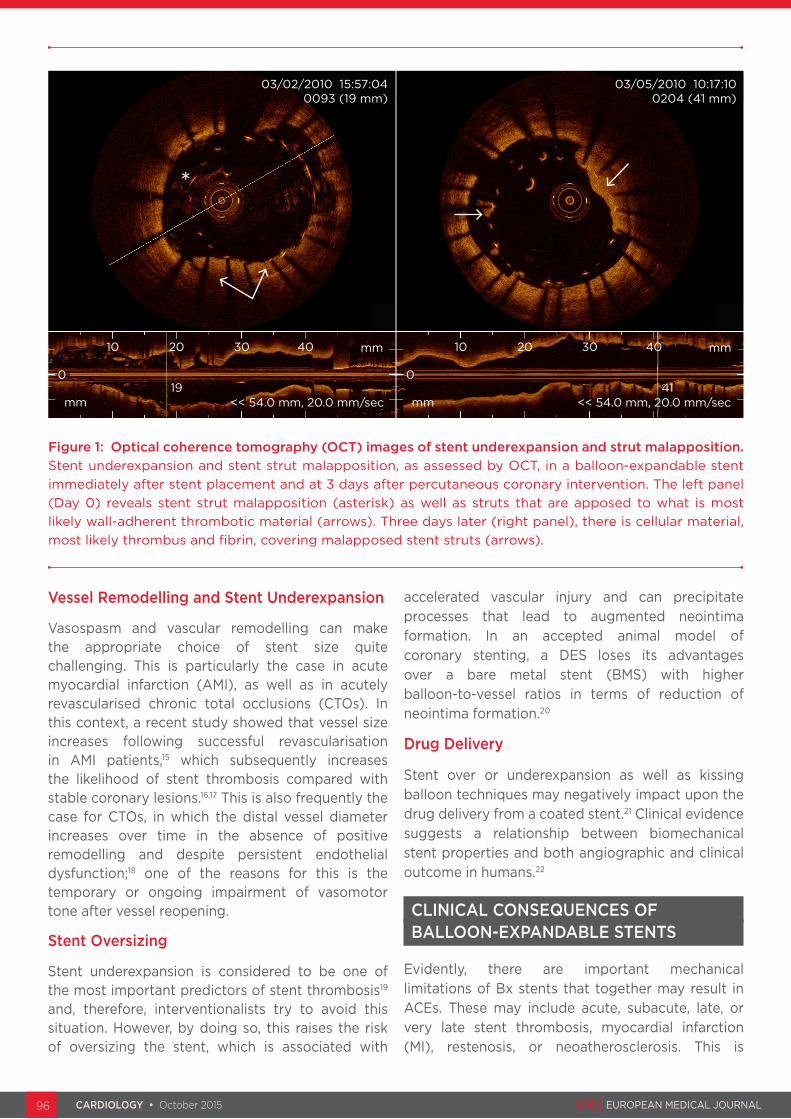
Figure 1: Optical coherence tomography (OCT) images of stent underexpansion and strut malapposition.Stent underexpansion and stent strut malapposition, as assessed by OCT, in a balloon-expandable stent immediately after stent placement and at 3 days after percutaneous coronary intervention. The left panel (Day 0) reveals stent strut malapposition (asterisk) as well as struts that are apposed to what is most likely wall-adherent thrombotic material (arrows). Three days later (right panel), there is cellular material, most likely thrombus and fibrin, covering malapposed stent struts (arrows).
03/05/2010 10:17:100204 (41 mm)
03/02/2010 15:57:040093 (19 mm)
0
mm
10
19<< 54.0 mm, 20.0 mm/sec
0
<< 54.0 mm, 20.0 mm/secmm
mm20 30
41
201040 30 40 mm
*

CARDIOLOGY • October 2015 EMJ EUROPEAN MEDICAL JOURNAL CARDIOLOGY • October 2015 EMJ EUROPEAN MEDICAL JOURNAL 96 97
particularly applicable to the first-generation Bx stents; second-generation stents with improved design show better clinical results,23 although the mechanical limitations remain.
Polymer Damage and Platform Damage/Loss
Most of the current DESs make use of a thin polymeric layer in which the anti-proliferative drug is embedded. The polymer can be durable or biodegradable. Irregularities or defects in the polymer coating have emerged as a potential factor in determining the outcome of DES implantation.24 Since Bx stents are crimped onto the balloon without any protection on the outer side, drug-eluting Bx stents can undergo different degrees of polymer damage due to manual handling, navigation through the catheter, or engagement in complex lesions (tortuosity, calcification, bifurcations). For example, using electron microscope investigation, Wiemer et al.24 reported the occurrence of polymer damage on the abluminal surface of stents for which implantation failed in between 3% (durable polymers) and 20% (bioabsorbable polymers) of cases. Polymer damage leads to non-uniform delivery of the drug, an excessive inflammatory response, and thrombogenicity, with stent failure ranging from restenosis to stent thrombosis. Strikingly, this study also showed polymer damage on the luminal stent surface, i.e. the inner surface, in contact with the balloon before delivery. Polymer-free, drug-eluting Bx stents are under development25 but, so far, have not achieved widespread clinical use.
Although complications such as stent damage/dislocation are quite rare (<1%) thanks to the development of very low-profile and factory-crimped Bx devices, they still cause severe adverse events (AEs) (including death) and/or pose serious technical challenges in acute treatment.26
Stent Strut Malapposition
Malapposed stent struts that have no physical contact to cellular structures of the vascular wall do not allow direct ingrowth of neointimal tissue and are likely to accumulate acellular masses, such as fibrin clots. Fibrin is readily identified in lesions that have been histologically investigated subsequent to an acute coronary event.27 In fact, most studies that have investigated the possible clinical causes of stent thrombosis using contemporary intravascular imaging devices have identified stent underexpansion and stent strut malapposition as a frequent finding.28 These
clinical and pathophysiological observations were confirmed in an ex vivo stent model.29 In simple terms, stent strut malapposition can occur via two distinct mechanisms: stent underexpansion/undersizing and/or vascular remodelling. An example of stent underexpansion, strut malapposition, and their short-term consequences is provided in Figure 1.
Stent underexpansion is observed in 15-20% of stented lesions.30,31 It can, however, be reliably prevented by the use of intravascular imaging techniques such as intravascular ultrasound (IVUS) or optical coherence tomography (OCT), which, unfortunately, are both costly and time-consuming. This can add value particularly in re-opened CTO vessels or bifurcations. Stent strut malapposition can also occur at later time points following stent placement. A serial IVUS study in stented patients found that malapposition can occur months or even years after stent implantation, with the possibility of subsequent healing or, in contrast, persistence of malapposition.32 In addition, this clinical study showed stent strut malapposition in 73.9% of DESs that were involved in very late stent thrombosis.
Otake et al.33 showed that stent strut malapposition is also dependent on symmetrical stent expansion. This group found a linear relationship between the extent of asymmetrical deployment and strut malapposition in cases where stents were deployed asymmetrically.33 Interestingly, the authors also identified increased thrombus formation in asymmetrically deployed Bx stents that showed evidence of strut malapposition.
Stent strut malapposition may appear more frequently in DESs since these particular stents are designed to inhibit neointima formation, which is, to a certain degree, required for strut healing. This was confirmed by a recent study that applied intravascular imaging via OCT. This study was able to confirm that uncovered struts were most likely malapposed to the vascular wall following implantation of a paclitaxel-eluting stent.34
Strut malapposition may be linked to a specific cellular response. This is suggested by a study that compared the cellular responses in patients suffering from late stent thrombosis with or without strut malapposition.35 In this analysis, very late stent thrombosis was associated with histopathological signs of inflammation and evidence of vessel remodelling as assessed by

CARDIOLOGY • October 2015 EMJ EUROPEAN MEDICAL JOURNAL CARDIOLOGY • October 2015 EMJ EUROPEAN MEDICAL JOURNAL 98 99
IVUS. Compared with other causes of MI, eosinophilic infiltrates were more common in thrombi harvested from very late DES thrombosis and positively correlated with the extent of stent strut malapposition.
Strut malapposition is associated with both an increase in neointima formation and thus restenosis, as well as stent thrombosis.36 Besides restenosis and thrombosis, formation of neoatherosclerosis has been associated with delayed healing, in particular in drug-eluting Bx stents.37 A recent imaging study suggests that late coronary events are in part precipitated by the development of neoatherosclerosis in some DESs.38 Another recent report confirms these findings.39
Stent Fracture
Besides stent strut malapposition, stent fracture is not a rare finding in patients who received a Bx stent and who present with stent thrombosis. The Nordic Intravascular Ultrasound Study found that 16% of DESs and 24% of BMSs were fractured in patients that presented with stent thrombosis. Stent fractures are more likely to occur in longer stented lesions, thus reflecting the important mechanical component of the pathogenesis of stent fracture.40
Stent Recoil
Stent recoil refers to the reduction of the internal surface/volume of stents after their placement, due to shrinkage of the stent platform. Different studies have reported stent recoil of about 3-5% for the currently available Bx stents; stent recoil is one of the predictors of restenosis after drug-eluting Bx stent implantation. The degree of stent recoil is due to both the intrinsic properties of the stent material and the specific geometrical design of the stent. A higher-than-expected rate of stent recoil has led, for example, to significant changes in the design of the ABSORB bioabsorbable scaffold.41 Sx stents, because of their intrinsic properties, are not affected by stent recoil.42 Furthermore, in the case of positive remodelling of the coronary vessel, Sx stents follow the growth of the coronary lumen.15
Longitudinal Compression
Longitudinal compression has recently emerged as a serious potential complication of Bx stent placement: this refers to in situ stent shortening, with stent rings coming close to each other or even overlapping, after recrossing the stent with other
devices such as post-dilatation balloons, stent delivery systems, or IVUS catheters. Longitudinal compression can lead to incomplete lesion coverage, stent displacement, intraluminal strut protrusion, or device embolisation.43 Although the clinical consequences of longitudinal compression have not been fully elucidated, we can speculate regarding multiple, potential AEs ranging from restenosis to stent thrombosis to MI. Bench studies have shown that stent design is a major predictor of longitudinal compression, which can reach up to 47% of the nominal length in case of peak-to-peak design and fewer links between stent rings.44 Malapposition of the stent can play a significant role in longitudinal compression by giving an edge to the crossing device upon which to get entrapped. For this reason, Sx stents that provide immediate apposition and active adherence to the vessel wall are apparently devoid of this recently observed complication.
Late mechanical stent deficiencies cannot be readily detected in most patients unless they suffer from an adverse cardiac event. Therefore, interventionalists rely on the quality of the device and the procedure during percutaneous coronary intervention (PCI). Thus, improvements in stent design are warranted to ensure an appropriate long-term outcome for patients not impeded by mechanical stent shortcomings, which may become clinically apparent over time.
THE CONCEPT OF THE SELF-EXPANDING CORONARY STENT
Historical Development
The first Sx stent, the Wallstent, was a stainless steel stent first tested in humans in 1986. This was followed by the development of nitinol stents. A shape-memory alloy was developed at the US Naval laboratories in 1962. It was made of 55% nickel and 45% titanium. The team named their new alloy Nitinol (pronounced night-in-all). The ‘Ni’ and ‘Ti’ are the atomic symbols for nickel and titanium, and the ‘nol’ represents the Naval Ordinance Laboratory where it was discovered. Its unique characteristics are shape-memory and superelasticity. Shape-memory allows the nitinol material to be deformed and then, upon heating above its ‘transition temperature’, it will recover its original ‘undeformed’ shape. The Radius stent, which received FDA approval for coronary use, and the Symbiot covered stent for saphenous

CARDIOLOGY • October 2015 EMJ EUROPEAN MEDICAL JOURNAL CARDIOLOGY • October 2015 EMJ EUROPEAN MEDICAL JOURNAL 98 99
vein graft interventions are examples of early- generation nitinol stents.
Characteristics and Clinical Data of the STENTYS® Self-Expanding Stent
The STENTYS® Coronary Stent (STENTYS S.A., Paris, France) is a self-apposing nitinol stent with a nominal strut width of 68 µm (0.0027”). The stent is compatible with a 6 Fr guide catheter and is delivered using a rapid-exchange delivery system over a conventional 0.014” guide wire. The device is deployed by withdrawal of a retractable sheath, and is available in three lengths (17, 22, and 27 mm) with diameters suitable for vessels ranging from 2.5-3.0 mm (small), 3.0-3.5 mm (medium), and 3.5-4.5 mm (large). The stent is available in a bare- metal version, a paclitaxel-eluting version, and in a sirolimus-eluting version (1.4 µg/mm² of stent), all of which are incorporated in a proprietary coating (ProTeqtor®), a durable polymer matrix of polysulfone and a soluble polyvinylpyrrolidone that acts as an excipient. It has a closed-cell design with a cell area of 0.95 mm², which is much smaller than that of Bx stents.
The expansive property of the STENTYS stent was substantiated in the APPOSITION I study15 (BMS version in ST segment elevation myocardial infarction [STEMI], and as evidenced by a 19% increase in stent area following a 19% increase in lumen area of the distal reference vessel at 3 days post STEMI, as measured by IVUS). In the APPOSITION II trial,45 the self-apposing STENTYS® BMS was compared with Bx stents and proved to be superior with respect to acute stent apposition, as assessed by OCT. At 3 days post implantation, it was shown that the STENTYS mean stent area increased further while the rate of malapposed struts decreased, suggesting that the stent conforms to changes in vessel anatomy during the first days after the index event. On a per-patient basis, none of the STENTYS stents were malapposed (defined as ≥5% malapposed struts) compared with 28% in the Bx stent group at 3 days follow-up (p<0.001). The APPOSITION III registry46 of 1,000 STEMI patients showed highly satisfactory clinical results, in particular in patients that were post-dilated (major adverse cardiac event rate at 12 months: 8.4%). The APPOSITION IV47 study compared the STENTYS® Sirolimus-Eluting Stent (SES) with the Resolute stent. The STENTYS SES was equivalent to a conventional drug-eluting Bx stent with respect to late stent strut apposition and coverage at 9 months. However, stent
strut apposition and coverage at 4 months was significantly better in the STENTYS group. Satisfying results with the STENTYS stent were also demonstrated in the OPEN I and II bifurcation studies.
The recently completed SETUP trial48 evaluated a new delivery system for the STENTYS stent. The self-apposing STENTYS® Xposition S is folded on a delivery balloon that is covered with a distal splittable 0.0032” sheath assembly. The nominal diameter of this delivery balloon is the same as the smallest diameter for which the stent is suitable. When the semi-compliant delivery balloon is inflated within the sheath using low pressures starting at 8 atm, it causes the sheath assembly to split, thus allowing the STENTYS stent to deploy in the coronary artery at the desired location. The results show that this new system facilitates exact positioning and delivery of the stent. The use of this device could be considered in anatomical subsets with a high risk of stent mis-positioning and stent mis-sizing, such as lesions with a very high thrombotic burden, lesions located in ectatic/aneurysmal vessels, or bifurcation lesions with large differences between the proximal and distal diameters, lesions of the left main coronary artery, and ostial located lesions.
Mitigating Balloon-Expandable Stent Shortcomings
Stent strut malapposition, stent under and overexpansion, non-uniform distribution of strut geometry, and cell size are important features that should be resolved in upcoming generations of stent development, in particular for complex lesions such as AMI, bifurcation, and tapered as well as angulated lesions.
Sx stents would likely bridge the gap of acute and acquired stent strut malapposition through self-alignment of the stent struts to the vascular wall, thus facilitating strut healing and optimal drug delivery to the vessel wall. Furthermore, consistent strut apposition in tapered vessels and downstream vessel remodelling, as is consistently the case in AMI lesions and revascularised CTOs for example, would be feasible. Additional features of the STENTYS Sx stent system, such as disconnectable interconnectors, allow further improvements in the scaffolding of bifurcated lesions.
PCI would, in many instances, be easier to perform because the likelihood of acute and subacute strut malapposition is significantly decreased.

CARDIOLOGY • October 2015 EMJ EUROPEAN MEDICAL JOURNAL CARDIOLOGY • October 2015 EMJ EUROPEAN MEDICAL JOURNAL 100 101
Stent conformability and self-adaptation to the natural vessel are likely to improve the properties of blood flow in revascularised lesions (Figure 2). Since divergent patterns of blood flow are negatively associated with vascular healing as well as adverse coronary events,49 this could be of
particular importance for overall long-term stent performance. In addition, it has been suggested that Sx stents may decrease the incidence of no-reflow and side-branch occlusion compared with Bx stents.50 In this context, Sx stents can also be associated with lower edge dissections51 that
Figure 3: Overview of contemporary self-expanding stent platforms.A) Axxess™ stent, B) Cappella stent, C) STENTYS® stent, D) CardioMind Sparrow™ stent, E) vProtect™ stent. Further information about the different stent platforms is provided in the text.
A
D E
B C
Figure 2: Stent conformability.Stent conformability compared between balloon-expandable stents (A and B) and self-expanding stents (C and D). Non-apposed stent struts are visible only in the balloon-expandable stent images (arrow). Optimal stent conformability is provided by the self-expanding stent only. To facilitate stent strut visibility for the reader, stents are followed in B and D by a dotted yellow line. Otherwise, images A and B as well as C and D are identical.
A
Proximal Distal
C
B D
Proximal Distal
Proximal Distal
Proximal Distal

CARDIOLOGY • October 2015 EMJ EUROPEAN MEDICAL JOURNAL CARDIOLOGY • October 2015 EMJ EUROPEAN MEDICAL JOURNAL 100 101
are also known to precipitate adverse cardiac events,52 even when they can be considered minor. The SCORES trial suggested that the lower immediate pressure of Sx stent deployment compared with high-pressure Bx stent placement may induce less biomechanical injury to the vascular wall,53 which is possibly followed by a lower rate of restenosis and presumably improved vascular healing.54
Considering these features, the concept of implanting Sx stents in STEMI lesions is of particular interest. No-reflow is a major complication following stent-based revascularisation and adversely influences short and long-term outcomes.55 No-reflow is multifactorial; however, thrombus fragmentation, mobilisation, and distal embolisation are believed to be the main factors that cause no-reflow in primary PCI for STEMI.56 The STENTYS stent has been used for the specific treatment of STEMI lesions in several studies. This concept builds on the presumption that choice of stent size can be difficult in STEMI lesions because coronary spasm may impede visual vessel size assessment. Secondly, the high pressures that are necessary for Bx stent deployment are generally not necessary for Sx stent implantation. Therefore, the necrotic core may not be penetrated by self-apposing stent platforms, whereas mandatory balloon inflation of a conventional stent has a higher likelihood to penetrate the necrotic core and cause thrombus fragmentation after stenting.57 Besides potential advantages in the acute situation, stent malapposition is frequently detected after primary PCI for STEMI. Indeed, there was significantly lower stent strut malapposition in the APPOSITION II study 3 days post implantation in favour of Sx versus Bx stents (0.58% versus 5.46%, p<0.001).45
Other interventional scenarios that could be potentially targeted by Sx stents are tapered lesions, left main lesions, aneurysms, coronary artery bypass grafts, large vessels, etc. However, limited reliable clinical data regarding these lesion subtypes are available.
The problems inherent with polymer damage during navigation and deployment of Bx stents are not present in the case of Sx stents because the stent is protected by its external sleeve during navigation and the device does not undergo high-pressure balloon deployment. The external sleeve of Sx stents offers the additional advantage of protecting the device from strut damage and
dislocation, and, in the worst case scenario, stent loss during navigation and deployment.
SELF-EXPANDING STENT PLATFORMS
Just as for Bx stents, not all Sx stent systems are the same. They differ in terms of strut length, flexibility and ability to appose, radial and chronic outward forces, strut thickness, ease and accuracy of deployment, and foreshortening. The performance of an Sx stent is dependent on both the material it is made from and the stent design. Sx stents are generally manufactured from nitinol, an alloy of nickel and titanium in roughly equal proportions that incorporates various favourable properties for the purpose of stent design: shape-memory, fatigue resistance, biocompatibility, and superelasticity. Various nitinol Sx stent platforms are currently under clinical investigation or are commercially available in selected countries. An overview of the various Sx stent types is provided in Figure 3. Most contemporary Sx platforms are designed to treat bifurcations (Axxess™, Cappella Sideguard, Galway, Ireland), including the STENTYS Self-Apposing® stent, which has now been clinically evaluated for other additional applications such as AMI. The vProtect™ Luminal Shield (Prescient Medical, Inc., Doylestown, Pennsylvania, USA) was designed to stent intermediate lesions (vulnerable plaques). CardioMind Sparrow™ is a small Sx stent that is mounted on a 0.014” guide wire and specifically designed to treat small vessels. Not all Sx stents are made of nitinol. The Wallstent,58 which is no longer commercially available as a coronary Sx stent, was a stainless steel woven mesh constructed of 16 wire filaments. Due to the difference in material and stent design, the Wallstent is unable to appose completely to the vessel wall compared with, for example, the good apposition of the STENTYS stent.45
Intravascular Imaging Reveals Stent Benefits
OCT is a sensitive method to visualise mechanical as well as biomechanical properties of coronary stents.59 OCT is able to highlight the favourable findings of Sx stents compared with Bx stents, including conformability, strut apposition, and self-alignment to the vascular wall. These properties have been investigated in the APPOSITION II trial,45 which confirmed that strut apposition is significantly improved with Sx stents in patients presenting with AMI compared with conventional Bx stents.

CARDIOLOGY • October 2015 EMJ EUROPEAN MEDICAL JOURNAL CARDIOLOGY • October 2015 EMJ EUROPEAN MEDICAL JOURNAL 102 103
Potential Self-Expanding Stent Limitations
Sx stents in their current form reveal some limitations that are important to acknowledge. The chronic outward force is generally lower than that of balloon pressures used to deploy Bx stents,60 which can be of some limitation specifically in calcified lesions. Deliverability of Sx stents can be cumbersome as the advancement of the delivery system and precise positioning is sometimes challenging and requires some training. Thus, success rates regarding stent delivery are
somewhat lower compared with Bx stents, in particular in tortuous, calcified, and distal vessels. Recently, however, a novel balloon delivery system was developed for the self-apposing STENTYS SES, consisting of the inflation of a balloon at low pressures to split the covering delivery sheath longitudinally, which releases the stent. It then deploys and apposes to the vessel wall where the ‘jailed’ sheath is then retracted. This system aims for easy delivery and a highly precise longitudinal placement of the Sx stent.48
Table 2: Clinical trials involving self-expanding stent systems.
Stent Study name Pts. Background/Primary endpoint
Remarks Reference
Axxess™ AXXESS 43 Safety and efficacy study/6-month in-segment restenosis
Bare-metal version of Axxess™ stent6-month F/U completed
61
AXXESS PLUS
139 Single-arm, safety and efficacy trial/6-month LLL
Axxess™ plus DES (Biolimus A9)6-month MACE: 11.2%; TLR: 7.5%; LLL: 0.09 mmF/U through 3 years completed
62
DIVERGE 302 Single-arm study/MACE De novo bifurcations included only, no control groupMACE rate: 7.7% (0.7 death, 3.3% NQWMI, 1% QWMI, 4,3% TLR)
63
AXXENT 33 Single-arm pilot study in left main/MACE at 6 months
12-month follow-up completedMACE at 6 months: 18.2%
64
Cappella Sideguard™
Doi H et al. 25 Safety and feasibility trial De novo bifurcations only, no control group6-month TLR rate: 12.5%
65
CardioMind Sparrow™
CARE I 21 Safety and feasibility trial Binary restenosis rate 20% at 6 months, single-arm study2 MACE events at 24 months F/U
N/A
CARE II 100 Randomised/LLL Lesions ≤20 mm length between 2.00-2.75 mm36 CardioMind BMS (LLL 0.86 mm), 36 SES (LLL 0.29 mm), 30 BMS (LLL 0.94 mm)
N/A
STENTYS® OPEN I 60 Safety and feasibility trial/6-month MACE
De novo bifurcations only including the STENTYS BMS and DES (paclitaxel), no control group6-month MACE: BMS 27.3%, DES 3.7%LLL BMS 0.86 mm, DES 0.39 mm
66
OPEN II 200 Safety and feasibility trial/6-month MACE
STENTYS DES (paclitaxel), no control group6-month MACE: 10.1%, Death: 0.5%; ST: 1.0%12-month MACE: 13%, Death: 1.4%, ST: 2.0%Kissing balloon has no effect on MACE.
67
APPOSITION I
25 Safety and feasibility trial/6-month MACE
STEMI lesions onlySTENTYS BMS, no control group6 months: binary restenosis 25%, MACE 12% (3 TLR)
15

CARDIOLOGY • October 2015 EMJ EUROPEAN MEDICAL JOURNAL CARDIOLOGY • October 2015 EMJ EUROPEAN MEDICAL JOURNAL 102 103
The variety of stent lengths that are currently available is confined to a limited number; the same is true for the available diameter sizes of some Sx stents. Due to current limitations of the delivery system, very long stents are generally not available at this time. Because precise positioning remains a challenge, overlapping Sx stents can be difficult. In addition, distal positioning of a second Sx stent through an already implanted Sx stent is generally not recommended, at least in smaller vessel sizes, because there can be issues associated with stent injury and shifting of the implanted Sx stent. Post-dilation of the stent is generally recommended to ensure that the stent is well expanded over its entire length.46
STUDIES WITH SELF-EXPANDING STENTS
Most clinical trials that examined the outcome of patients who received Sx stents have been carried out as feasibility and safety studies that were single-arm and included only a limited number of patients. However, as is particularly the case for the STENTYS stent, larger series with appropriate control groups are now available. A detailed overview of contemporary clinical trials is provided in Table 2. Most human trials have been carried out in a selected patient or lesion subgroup, in particular in patients that presented with AMI or bifurcation lesions. Despite all the trials available
Table 2 continued.
Stent Study name Pts. Background/Primary endpoint
Remarks Reference
STENTYS® APPOSITION II
80 Randomised/stent strut malapposition at 3 days
STEMI lesions only treated with BMSSTENTYS BMS versus Abbott Vision/Medtronic Driver Malapposed stent struts by OCT assessment 0.58% for STENTYS vs. 5.46% for Multi-Link Vision (p<0.001). MACE at 6 months 2.3% vs. 0% (p=NS).
45
APPOSITION III
965 Non-randomised,observational study/12-month MACE
STENTYS BMS 74%/DES (paclitaxel) 26%12-month results: MACE 9.3% (after post-dilation: 8.4%), cardiac death 2%24-month results: MACE 11.2%, cardiac death 2.3%
46
APPOSITION IV
150 Randomised, FIM trial for STENTYS DES (sirolimus)/4 or 9-month malapposition and strut coverage
STEMI lesions onlySTENTYS DES (sirolimus) vs. Medtronic Resolute4-month results: malapposed stent struts by OCT 0.07% for STENTYS vs. 1.16% for Resolute (p=0.002). Total stent coverage 33.3% STENTYS vs. 3.8% Resolute (p=0.02)9-month results: No difference in malapposition, strut coverage, or MACE. LLL 0.04 mm STENTYS vs. 0.17 mm Resolute. Greater mean lumen diameter in STENTYS arm (p=0.01).
47
SETUP 25 Feasibility study, FIM trial for balloon-delivery system of STENTYS SES/technical success
1 month: 100% technical success, 0% geographical miss
48
vProtect™ Luminal Shield
SECRITT 21 Safety and feasibility trial TCFA lesions were sealed with vProtect™ Luminal Shield stent only, no control group; no MACE at 6-months F/U
68
DES: drug-eluting stent; MACE: major adverse cardiac event; TLR: target lesion revascularisation; LLL: late lumen loss; ST: stent thrombosis; NQWMI: non-Q wave myocardial infarction; QWMI: Q wave myocardial infarction; NS: not significant; F/U: follow-up; BMS: bare-metal stent; SES: sirolimus-eluting stent; STEMI: ST segment elevation myocardial infarction; OCT: optical coherence tomography; TCFA: thin-cap fibroatheroma; N/A: not yet published.

CARDIOLOGY • October 2015 EMJ EUROPEAN MEDICAL JOURNAL CARDIOLOGY • October 2015 EMJ EUROPEAN MEDICAL JOURNAL 104 105
REFERENCES
1. Mewissen MW. Primary nitinol stenting for femoropopliteal disease. J Endovasc Ther. 2009;16(2 Suppl 2):II63-81.2. Oberhuber A et al. Influence of different self-expanding stent-graft types on remodeling of the aortic neck after endovascular aneurysm repair. J Endovasc Ther. 2010;17(6):677-84.3. Wakhloo AK et al. Advances in interventional neuroradiology. Stroke. 2009;40(5):e305-12.4. Rieu R et al. Assessment of the trackability, flexibility, and conformability of coronary stents: a comparative analysis. Catheter Cardiovasc Interv. 2003;59(4):496-503.5. Foin N et al. Maximal expansion capacity with current DES platforms: a critical factor for stent selection in the treatment of left main bifurcations? EuroIntervention. 2013;8(11):1315-25.6. Mortier P et al. Virtual bench testing of new generation coronary stents. EuroIntervention. 2011;7(3):369-76.7. Gomez-Lara J et al. Risk of target lesion failure in relationship to vessel angiographic geometry and stent conformability using the second
generation of drug-eluting stents. Am Heart J. 2011;162(6):1069-1079.e2.8. Nakazawa G et al. Pathological findings at bifurcation lesions: the impact of flow distribution on atherosclerosis and arterial healing after stent implantation. J Am Coll Cardiol. 2010;55(16):1679-87.9. Gutiérrez-Chico JL et al. Vascular tissue reaction to acute malapposition in human coronary arteries: sequential assessment with optical coherence tomography. Circ Cardiovasc Interv. 2012;5(1):20-9, S1-8.10. Hwang CW et al. Thrombosis modulates arterial drug distribution for drug-eluting stents. Circulation. 2005;111(13):1619-26.11. Zeina AR et al. Dimensions and anatomic variations of left main coronary artery in normal population: multidetector computed tomography assessment. Coron Artery Dis. 2007;18(6):477-82.12. Legrand V et al. Percutaneous coronary intervention of bifurcation lesions: state-of-the-art. Insights from the second meeting of the European Bifurcation Club. EuroIntervention. 2007;3(1):44-9.13. Zubaid M et al. Normal angiographic tapering of the coronary arteries. Can J Cardiol. 2002;18(9):973-80.
14. Chen HY et al. Effects of stent sizing on endothelial and vessel wall stress: potential mechanisms for in-stent restenosis. J Appl Physiol (1985). 2009;106(5):1686-91.15. Amoroso G et al. Assessment of the safety and performance of the STENTYS self-expanding coronary stent in acute myocardial infarction: results from the APPOSITION I study. EuroIntervention. 2011;7(4):428-36.16. Leibundgut G et al. Stent thrombosis up to 3 years after stenting for ST-segment elevation myocardial infarction versus for stable angina--comparison of the effects of drug-eluting versus bare-metal stents. Am Heart J. 2009;158(2):271-6.17. Gonzalo N et al. Incomplete stent apposition and delayed tissue coverage are more frequent in drug-eluting stents implanted during primary percutaneous coronary intervention for ST-segment elevation myocardial infarction than in drug-eluting stents implanted for stable/unstable angina: insights from optical coherence tomography. JACC Cardiovasc Interv. 2009;2(5):445-52.18. Tomasello SD et al. Retrograde approach for revascularization of
to date not being powered to address clinical outcomes, they have mostly provided proof-of-concept. In this context, it is important to acknowledge that contemporary Sx stent platforms are at least as safe and effective as Bx stents and that stent placement is feasible in the scenarios that were covered in these mostly observational trials. Therefore, initial clinical trials comparing the performance of Sx stents with Bx stents in confined clinical scenarios have shown clearly encouraging results and proof-of-concept, despite the fact that clinical outcomes have not been primarily studied.
FUTURE DIRECTIONS AND CONCLUSION
Bx stents and Sx stents came almost simultaneously to the market nearly 30 years ago. Since then, Bx stents have been significantly improved, resulting in favourable clinical results and better ease of use. On the other hand, Sx stents were slowly abandoned after the first experiences and only recently, thanks to the clinical applications of the nitinol alloy, have they experienced a strong reappraisal. For this reason, the currently available devices could take advantage of further refinement in order to close the gap or even to surpass Bx stents.
Device improvement and ‘cultural acceptance’ by interventional cardiologists, who are now more experienced, are critical for the clinical advancement of Sx technology.
Regarding mechanical improvements, there are several challenges that should be considered by manufacturers of Sx stents. These include a smaller profile of the delivery device to facilitate navigation through complex anatomy and enabling 5 Fr compatibility. Visibility should be improved in order to expedite ostial positioning and overlapping stents. This could be accomplished by adding markers or by enhancing radiopacity. Longer devices are needed for diffuse disease. Reduced strut profile and density is needed to enable treatment of small vessels, but needs to be balanced against maintaining a sufficient radial force.
Changing from Bx stents to Sx stents involves a complete change of mindset. One has to get used to them to appreciate the strengths and weaknesses in their application. The combination of the best of both worlds, a balloon delivery system (ease of deployment) with an Sx stent (efficacy), would represent a major advancement in coronary interventions.

CARDIOLOGY • October 2015 EMJ EUROPEAN MEDICAL JOURNAL CARDIOLOGY • October 2015 EMJ EUROPEAN MEDICAL JOURNAL 104 105
coronary chronic total occlusion. Minerva Cardioangiol. 2012;60(5):461-72.19. van Werkum JW et al. Predictors of coronary stent thrombosis: the Dutch Stent Thrombosis Registry. J Am Coll Cardiol. 2009;53(16):1399-409.20. Carter AJ et al. Long-term effects of polymer-based, slow-release, sirolimus-eluting stents in a porcine coronary model. Cardiovasc Res. 2004;63(4): 617-24.21. Wessely R. New drug-eluting stent concepts. Nat Rev Cardiol. 2010;7(4): 194-203.22. König A et al. Influence of stent design and deployment technique on neointima formation and vascular remodeling. Z Kardiol. 2002;91 Suppl 3:98-102.23. von Birgelen C et al. Third-generation zotarolimus-eluting and everolimus-eluting stents in all-comer patients requiring a percutaneous coronary intervention (DUTCH PEERS): a randomised, single-blind, multicentre, non-inferiority trial. Lancet. 2014;383: 413-23.24. Wiemer M et al. Scanning electron microscopic analysis of different drug eluting stents after failed implantation: from nearly undamaged to major damaged polymers. Catheter Cardiovasc Interv. 2010;75(6):905-11.25. Wessely R et al. Inhibition of neointima formation by a novel drug-eluting stent system that allows for dose-adjustable, multiple, and on-site stent coating. Arterioscler Thromb Vasc Biol. 2005;25(4):748-53.26. Kammler J et al. Long-term follow-up in patients with lost coronary stents during interventional procedures. Am J Cardiol. 2006;98(3):367-9.27. Finn AV et al. Pathological correlates of late drug-eluting stent thrombosis: strut coverage as a marker of endothelialization. Circulation. 2007;115(18):2435-41.28. Alfonso F et al. Combined use of optical coherence tomography and intravascular ultrasound imaging in patients undergoing coronary interventions for stent thrombosis. Heart. 2012;98(16):1213-20.29. Kolandaivelu K et al. Stent thrombogenicity early in high-risk interventional settings is driven by stent design and deployment and protected by polymer-drug coatings. Circulation. 2011;123(13):1400-9.30. Kume T et al. Intravascular ultrasound assessment of postprocedural incomplete stent apposition. J Invasive Cardiol. 2012;24(1):13-6.31. van der Hoeven BL et al. Stent malapposition after sirolimus-eluting and bare-metal stent implantation in patients with ST-segment elevation
myocardial infarction: acute and 9-month intravascular ultrasound results of the MISSION! intervention study. JACC Cardiovasc Interv. 2008;1(2):192-201.32. Lee CW et al. Intravascular ultrasound findings in patients with very late stent thrombosis after either drug-eluting or bare-metal stent implantation. J Am Coll Cardiol. 2010;55(18):1936-42.33. Otake H et al. Local determinants of thrombus formation following sirolimus-eluting stent implantation assessed by optical coherence tomography. JACC Cardiovasc Interv. 2009;2(5):459-66.34. Davlouros PA et al. Neointimal coverage and stent strut apposition six months after implantation of a paclitaxel eluting stent in acute coronary syndromes: an optical coherence tomography study. Int J Cardiol. 2011;151(2):155-9.35. Cook S et al. Correlation of intravascular ultrasound findings with histopathological analysis of thrombus aspirates in patients with very late drug-eluting stent thrombosis. Circulation. 2009;120:391-9.36. Liu X et al. A volumetric intravascular ultrasound comparison of early drug-eluting stent thrombosis versus restenosis. JACC Cardiovasc Interv. 2009;2(5): 428-34.37. Park SJ et al. In-stent neoatherosclerosis: a final common pathway of late stent failure. J Am Coll Cardiol. 2012;59(23):2051-7.38. Habara M et al. Morphological differences of tissue characteristics between early, late, and very late restenosis lesions after first generation drug-eluting stent implantation: an optical coherence tomography study. Eur Heart J Cardiovasc Imaging. 2013;14(3):276-84.39. Karanasos A et al. In-stent neoatherosclerosis: a cause of late stent thrombosis in a patient with “full metal jacket” 15 years after implantation: insights from optical coherence tomography. JACC Cardiovasc Interv. 2012;5(7): 799-800.40. Doi H et al. Classification and potential mechanisms of intravascular ultrasound patterns of stent fracture. Am J Cardiol. 2009;103(6):818-23.41. Tanimoto S et al. Comparison of in vivo acute stent recoil between the bioabsorbable everolimus-eluting coronary stent and the everolimus-eluting cobalt chromium coronary stent: insights from the ABSORB and SPIRIT trials. Catheter Cardiovasc Interv. 2007;70(4):515-23.42. Bosiers M et al. Does free cell area influence the outcome in carotid artery stenting? Eur J Vasc Endovasc Surg. 2007;33(2):135-41; discussion 142-3.43. Bartorelli AL et al. Stent longitudinal distortion: strut separation (pseudo-
fracture) and strut compression (“concertina” effect). EuroIntervention. 2012;8(2):290-1.44. Prabhu S et al. Engineering assessment of the longitudinal compression behaviour of contemporary coronary stents. EuroIntervention. 2012;8(2):275-81.45. van Geuns RJ et al. Self-expanding versus balloon-expandable stents in acute myocardial infarction: results from the APPOSITION II study: self-expanding stents in ST-segment elevation myocardial infarction. JACC Cardiovasc Interv. 2012;5(12):1209-19.46. Koch KT et al. One-year clinical outcomes of the STENTYS Self-Apposing® coronary stent in patients presenting with ST-segment elevation myocardial infarction: results from the APPOSITION III registry. EuroIntervention. 2015;10(11);doi:10.4244/EIJY15M02_08. [Epub ahead of print].47. van Geuns RJ et al. STENTYS self-apposing® sirolimus-eluting stent in ST-segment elevation myocardial infarction: results from the randomized APPOSITION IV trial. EuroIntervention. 2015. [Epub ahead of print].48. Lu H et al. First-in-man evaluation of the novel balloon delivery system STENTYS Xposition S for the self-apposing coronary artery stent: impact on longitudinal geographic miss during stenting. EuroIntervention. 2015;11(1);doi:10.4244/EIJY15M05_07. [Epub ahead of print].49. Lüscher TF et al. Drug-eluting stent and coronary thrombosis: biological mechanisms and clinical implications. Circulation. 2007;115(8):1051-8.50. König A et al. Stent design-related coronary artery remodeling and patterns of neointima formation following self-expanding and balloon-expandable stent implantation. Catheter Cardiovasc Interv. 2002;56(4):478-86.51. Hirayama A et al. Angiographic and clinical outcome of a new self-expanding intracoronary stent (RADIUS): results from multicenter experience in Japan. Catheter Cardiovasc Interv. 2000;49(4):401-7.52. Choi SY et al. Intravascular ultrasound findings of early stent thrombosis after primary percutaneous intervention in acute myocardial infarction: a Harmonizing Outcomes with Revascularization and Stents in Acute Myocardial Infarction (HORIZONS-AMI) substudy. Circ Cardiovasc Interv. 2011;4(3):239-47.53. Shin ES et al. Comparison of acute vessel wall injury after self-expanding stent and conventional balloon-expandable stent implantation: a study with optical coherence tomography. J Invasive Cardiol. 2010;22(9):435-9.54. Tanaka S et al. Prospective randomized trial comparing a nitinol

CARDIOLOGY • October 2015 EMJ EUROPEAN MEDICAL JOURNAL 106
self-expanding coronary stent with low-pressure dilatation and a high-pressure balloon expandable bare metal stent. Heart Vessels. 2008;23(1):1-8.
55. Rezkalla SH et al. No-reflow phenomenon following percutaneous coronary intervention for acute myocardial infarction: incidence, outcome, and effect of pharmacologic therapy. J Interv Cardiol. 2010;23(5):429-36.
56. Niccoli G et al. Myocardial no-reflow in humans. J Am Coll Cardiol. 2009;54(4):281-92.
57. Brosh D et al. Effect of no-reflow during primary percutaneous coronary intervention for acute myocardial infarction on six-month mortality. Am J Cardiol. 2007;99(4):442-5.
58. Strauss BH et al. Relative risk analysis of angiographic predictors of restenosis within the coronary Wallstent. Circulation. 1991;84(4):1636-43.
59. Bezerra HG et al. Optical coherence tomography versus intravascular ultrasound to evaluate coronary artery disease and percutaneous coronary intervention. JACC Cardiovasc Interv. 2013;6(3):228-36.
60. Grenacher L et al. In vitro comparison of self-expanding versus balloon-expandable stents in a human ex vivo model. Cardiovasc Intervent Radiol. 2006;29(2):249-54.61. Dubois CL, Wijns W. The AXXESS™ self-expanding biolimus A9™ eluting stent system for coronary bifurcation lesions. EuroIntervention. 2010;6 Suppl J:J130-4.62. Grube E et al. Six-month clinical and angiographic results of a dedicated drug-eluting stent for the treatment of coronary bifurcation narrowings. Am J Cardiol. 2007;99(12):1691-7.63. Verheye S et al. 9-month clinical, angiographic, and intravascular ultrasound results of a prospective evaluation of the Axxess self-expanding biolimus A9-eluting stent in coronary bifurcation lesions: the DIVERGE (Drug-Eluting Stent Intervention for Treating Side Branches Effectively) study. J Am Coll Cardiol. 2009;53(12):1031-9.64. Hasegawa T et al. Analysis of left main coronary artery bifurcation lesions treated with biolimus-eluting DEVAX AXXESS plus nitinol self-expanding stent: intravascular ultrasound results of the AXXENT trial. Catheter Cardiovasc Interv.
2009;73(1):34-41.
65. Doi H et al. Serial intravascular ultrasound analysis of bifurcation lesions treated using the novel self-expanding sideguard side branch stent. Am J Cardiol. 2009;104(9):1216-21.
66. Verheye S et al. Six-month clinical and angiographic results of the STENTYS® Self-Apposing Stent in Bifurcation Lesions. EuroIntervention. 2011;7:580-7.
67. Naber C et al. Final results of a self-apposing paclitaxel-eluting stent fOr the PErcutaNeous treatment of de novo lesions in native bifurcated coronary arteries study. EuroIntervention 2015;11-online publish-ahead-of-print June 2015.
68. Wykrzykowska JJ et al. Plaque sealing and passivation with a mechanical self-expanding low outward force nitinol vShield device for the treatment of IVUS and OCT-derived thin cap fibroatheromas (TCFAs) in native coronary arteries: report of the pilot study vShield Evaluated at Cardiac hospital in Rotterdam for Investigation and Treatment of TCFA (SECRITT). EuroIntervention. 2012;8(8):945-54.
If you would like reprints of any article, contact: 01245 334450.