Embed Size (px)
Citation preview

Clinical Pathology of Parrots
-autotutorial programs
Marie Gunnarsson
Handledare: Harold Tvedten Institutionen för biomedicin och veterinär folkhälsovetenskap avd för
bilddiagnostik och klinisk kemi
__________________________________________________________________________________________________________________________________________________________________
Sveriges lantbruksuniversitet Examensarbete 2006:8 Fakulteten för veterinärmedicin och ISSN 1652-8697 husdjursvetenskap Uppsala 2006 Veterinärprogrammet Swedish University of Agricultural Sciences Degree project 2006:8 Faculty of Veterinary Medicine and ISSN 1652-8697 Animal Sciences Uppsala 2006 Veterinary Medicine Programme

INNEHÅLLSFÖRTECKNING SUMMARY........................................................................................................1
SAMMANFATTNING........................................................................................1
INLEDNING ......................................................................................................1
Collecting Samples for Testing .................................................................2
Avian Hematology .......................................................................................2
Avian Biochemistry.....................................................................................2
Common Avian Diseases ...........................................................................2
Avian Cases .................................................................................................3
LITTERATURFÖRTECKNING.........................................................................4
Internetkällor................................................................................................4

1
SUMMARY At the Veterinary Medicine Programme there is very little time allotted paid to exotic pets, which include psittacine birds, compared to other kinds of animals. However parrots are quite often seen as patients in the animal clinics in Sweden currently. Many veterinarians have unfortunately not enough knowledge to take care of such patients in a proper manner. The aim of this degree project was to put together a CD with information about hematology and clinical chemistry in parrots. The information is supposed to be easily available and designed for interested veterinary students and veterinarians. The CD also includes information about how to collect blood samples and common avian diseases. It consists of five Power Point presentations: Collecting Samples for Testing, Avian Hematology, Avian Biochemistry, Common Avian Diseases and Avian Cases. The language is English to make this project also available for foreign students and veterinarians.
SAMMANFATTNING På veterinärutbildningen vid Sveriges lantbruksuniversitet ägnas knappt någon tid alls åt de udda sällskapsdjuren, dit papegojor räknas, i jämförelse med andra djurslag. Det är dock inte så ovanligt att papegojor kommer in som patienter till djurklinikerna. Många veterinärer har inte de kunskaper som krävs för att kunna ta hand om de här patienterna på ett proffesionellt sätt. Syftet med det här examensarbetet var att skapa en CD-rom där information om framför allt hematologi och klinisk kemi hos papegojor ska finnas lättillgänglig för veterinärstudenter och yrkesverksamma veterinärer. Även information om provtagning och vanliga sjukdomar ingår. På CD-skivan ingår fem Power Point-presentationer: Collecting Samples for Testing, Avian Hematology, Avian Biochemistry, Common Avian Diseases och Avian Cases. Språket som används är engelska för att även utländska studenter och veterinärer ska kunna ta del av utbildningsmaterialet.
INLEDNING Papegojor tillhör den grupp djur som brukar kallas exotiska eller udda sällskapsdjur i veterinärmedicinska sammanhang. På veterinärutbildningen vid Sveriges lantbruksuniversitet är de utan tvekan exotiska, men denna beskrivning stämmer allt sämre när man ser till vilka typer av patienter som dyker upp på djurklinikerna i Sverige. Allt fler djurägare har sina papegojor försäkrade och uppsöker veterinär när fåglarna blir sjuka. Det växande behovet av kunskap hos veterinärer inom detta område möts enligt min mening inte upp av veternärutbildningen. Eftersom undervisningen på veterinärprogrammet inte tar upp udda sällskapsdjur i någon större utsträckning och detta är ett område som speciellt intresserar mig har jag valt att under min sista termin på veterinärprogrammet fördjupa mig inom ämnet. Jag har inriktat mig framför allt på hematologi hos papegojor.

2
För att andra veterinärstudenter och även färdigutbildade veterinärer ska kunna ta del av mina nyförvärvande kunskaper på ett lättillgängligt och inspirerande sätt har jag skapat en CD-rom som innehåller fem olika Power Point-presentationer. Dessa presentationer är utformade så att den intresserade ska kunna följa dem på egen hand i valfri turordning. För bästa utbyte bör presentationerna ses i fullbildsformat. De flesta bilder som ingår är tagna för det här projektet av min handledare Harold Tvedten eller utvalda ur hans arkiv och några är tagna av veterinärstudent Torvald Netterby. Resten av bilderna är bilder från internet som inte är upphovsrättsskyddade. Mer information om dessa bilders ursprung finns på CD-skivan. Språket som används är engelska för att även utländska studenter och veterinärer ska kunna ta del av utbildningsmaterialet. Nedan följer en kort beskrivning av de olika Power Point-presentationerna. Collecting Samples for Testing En förutsättning för att kunna undersöka papegojornas blod är naturligtvis att ett blodprov ska kunnas tas. Detta kan säkert skrämma många som inte är vana att hantera fåglar. Det är dock viktigt att komma ihåg att det inte är svårare att ta ett blodprov från en papegoja än vad det är att ta ett blodprov från en hund eller en katt. Naturligtvis underlättar det om en van person håller i fågeln och lämplig utrustning används. Den här presentationen går igenom förberedelser inför provtagning, provtagningsförfarande och rutiner efter provtagning till exempel hur ett blodutstryk utförs. Avian Hematology Den största skillnaden mellan fågelblod och blod från däggdjur är att alla blodceller i perifert fågelblod är kärnförande. Detta innebär alltså att hos fåglar har både erytrocyter och trombocyter kärnor. En annan skillnad är att fåglar saknar neutrofila granulocyter som finns hos däggdjur. Istället har de en celltyp som kallas heterofila granulocyter. Dessa två olika celltyper motsvarar varandra. Den här presentationen går framför allt igenom den normala morfologin hos de olika celltyperna, men även lite mer speciella fynd såsom toxiska heterofiler och reaktiva lymfocyter tas upp. Anemi och artefakter är två andra ämnen som också ingår i presentationen. Presentationen avslutas med frågor och svar som får läsaren att tänka till och kontrollera sin kunskaper. Avian Biochemistry Vilka biokemiska analyser som är meningsfulla att genomföra skiljer sig mellan djurslag. Presentationen som handlar om biokemi tar upp de analyser som oftast genomförs på papegojor enligt Gunnel Andersson, som arbetar som veterinär på Djurkliniken Roslagstull i Stockholm, där de är specialiserade på papegojor och andra udda sällskapsdjur. Även hur provsvaren sedan ska tolkas ingår i stora drag i presentationen. Common Avian Diseases Papegojor drabbas till viss del av samma sjukdomar som däggdjur, men det finns en del sjukdomar som är specifika för fåglar. De sjukdomar som tas upp i den här presentationen är bland de vanligaste som förekommer hos fågelpatienter på djurklinikerna. Psittacine beak and feather disease är inte vanligt förekommande i Sverige, men ingår ändå eftersom det är en så pass viktig sjukdom. Vissa

3
sjukdomar som drabbar papegojor är zoonoser, t.ex. chlamydios eller papegojsjuka som den även kallas. Dessa sjukdomar är kanske extra viktiga att känna till när man arbetar med fågelpatienter. Avian Cases Denna presentation tar upp två autentiska fall som är hämtade ur patientunderlaget på Regiondjursjukhuset Strömsholm. Bilderna på blodutstryken är tagna av Harold Tvedten från de blodutstryk som tillhör de riktiga patienterna. Syftet med presentationen är att visa typiska fall så att läsaren kan bilda sig en uppfattning om hur det kan vara att ha en papegoja som patient. Det sista fallet visar även att det inte alltid blir riktigt som man tänkt sig.

4
LITTERATURFÖRTECKNING Andersson, Gunnel. Legitimerad veterinär, Djurkliniken Roslagstull, Stockholm. Udda
sällskapsdjur. Föredrag 2005-10-15.
Austic RE & Scott ML. (1991) Nutritional diseases. In: Calnek BW, Barnes HJ, Beard CW, Reid WM & Yoder HW Jr. (Eds.) Diseases of poultry. 9 ed. 45-71. Ames: Iowa state university press.
Campbell TW. (1988) Avian hematology and cytology. Ames: Iowa state university press.
Campbell TW. (1994) Hematology. In: Ritchie BW, Harrison GJ & Harrison LR. (Eds.) Avian medicine: Principles and applications. 176-198. Lake worth: Wingers Publishing, Inc.
Coles BH. (1991) Cage and aviary birds. In: Beynon PH. & Cooper JE. (Eds.) Manual of exotic pets. 150-179. Cheltenham: British Small Animal Veterinary Association
Fudge AM. (2000) Laboratory medicine avian and exotic pets. Philadelphia: W.B. Saunders Company.
Girling SJ. (2003) Veterinary nursing of exotic pets. Oxford: Blackwell Publishing.
Harcourt-Brown NH. (2000) Pcittacine birds. In: Tully TN, Lawton MPC & Dorrestein GM (Eds.) Avian medicine. 112-143. Oxford: Butterworth-Heinemann.
Harris DJ. (2000) Clinical tests. In: Tully TN, Lawton MPC & Dorrestein GM (Eds.) Avian medicine. 43-51. Oxford: Butterworth-Heinemann.
Hawkey CM, Hart MG, Knight JA, Samour JH & Jones DM. (1982) Haematological findings in healthy and sick African grey parrots (Psittacus erithacus). Vet Rec18/25, 580-582.
Kazacos EA & Van Vleet JF. (1989) Sequential ultrastructural changes in the pancreas in zinc toxicosis in ducklings. Am J Pathol 134, 581-595.
Lu J. & Combs GF Jr. (1988) Effects of excess dietary zinc on pancreatic exocrine function in the chick. J Nutr 118, 681-689.
Lucas AM. & Jamroz C. (1961) Atlas of avian hematology. Washington: United States department of Agriculture.
Lumeij JT & Remple JD. (1991) Plasma urea, creatinine and uric acid concentration in relation to feeding peregrine falcons (Falco peregrinus). Avian Pathol 20, 79-83.
Samour JH. (2000) Avian Medicine. London: Mosby.
Zdziarski JM, Mattix M, Bush RM & Montali RJ. (1994) Zinc toxicosis in diving ducks. J Zoo Wildl Med 25, 438-445.
Zinkl JG. (1986) Avian hematology. In: Nemi JC. (Ed.) Schalm’s veterinary hematology. 256-273. Philadelphia: Lea & Febiger.
Internetkällor http://www.californiaavianlaboratory.com/artifact.html. (2005-03-11)
http://www.californiaavianlaboratory.com/bileac.html. (2005-03-11)
http://www.californiaavianlaboratory.com/chempro.html. (2005-03-11)
http://www.californiaavianlaboratory.com/chempro.html. (2005-03-11)
http://www.californiaavianlaboratory.com/leukocount.html. (2005-03-11)

5
http://www.merckvetmanual.com/mvm/htm/bc/170304.htm (2005-12-04)
http://www.merckvetmanual.com/mvm/htm/bc/170310.htm (2005-12-04)
http://www.merckvetmanual.com/mvm/htm/bc/170318.htm (2005-12-04)
http://www.merckvetmanual.com/mvm/htm/bc/170319.htm (2005-12-04)
http://www.merckvetmanual.com/mvm/htm/bc/170322.htm (2005-12-04)
http://www.merckvetmanual.com/mvm/htm/bc/206100.htm (2005-12-04)

1
Avian hematology
Marie GunnarssonInstitute for Clinical Chemistry
Swedish Agricultural University
ErythrocytesBirds have nucleatederythrocytes. The cell is oval with an oval centrally positionednucleus. Avian erythrocytesare much larger thanmost mammalian erythrocytes. The size varies amongspecies.
This 1.5 year old African grey parrot had a problem with feather plucking but had
normal appearing erythrocytes.ErythrocytesThe chromatin
in the nucleus is uniformlyclumped and becomes morecondensed with age. The cytoplasm has a uniform textureand stainsorange-pink.
These are normal erythrocytes from a healthy 11 year old African grey parrot. ErythrocytesIn avian blood
smears variations from the typicalerythrocyte are occasionally seen. The shape can vary(poikilo-cytosis) from irregular to round or elongated. Changes limited to certain areas are often artifactual.

2
The occasional abnormally formederythrocyte was from the same normal African grey parrot seen previously.
Erythrocytes
It is important to seperate artifact from true morphologicchanges. Erythrocytes missingnuclei are callederythroplastids. Theyare often due to preparation artifact.
ErythrocytesOccasional immature polychromatophilcerythrocytes are normally seen. Their cytoplasm is more basophilic and chromatin more dispersed than in mature erythrocytes.
This blood is from a healthy 11 year old African grey parrot.
Erythrocytes
Immatureerythrocytes are alsomore round thanmature erythrocytes.
Question:Which one of theseerythrocytes is the most immature?
Erythrocytes
Answer:This is the mostimmatureerythrocyte.
The blood is from a healthy African grey parrot.
3
Anemia
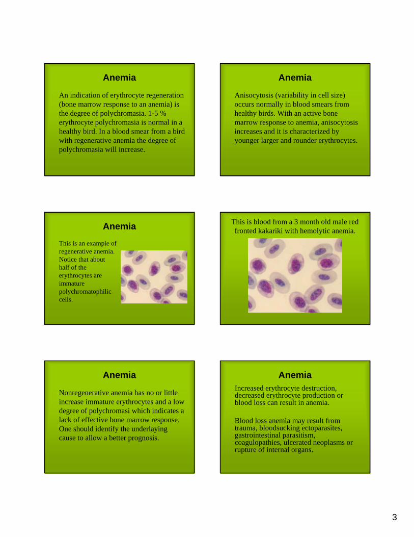
An indication of erythrocyte regeneration (bone marrow response to an anemia) is the degree of polychromasia. 1-5 % erythrocyte polychromasia is normal in a healthy bird. In a blood smear from a birdwith regenerative anemia the degree of polychromasia will increase.
Anemia
Anisocytosis (variability in cell size) occurs normally in blood smears from healthy birds. With an active bonemarrow response to anemia, anisocytosis increases and it is characterized by younger larger and rounder erythrocytes.
Anemia
This is an example of regenerative anemia. Notice that abouthalf of the erythrocytes are immaturepolychromatophilic cells.
This is blood from a 3 month old male red fronted kakariki with hemolytic anemia.
Anemia
Nonregenerative anemia has no or littleincrease immature erythrocytes and a lowdegree of polychromasi which indicates a lack of effective bone marrow response. One should identify the underlayingcause to allow a better prognosis.
AnemiaIncreased erythrocyte destruction, decreased erythrocyte production or blood loss can result in anemia.
Blood loss anemia may result from trauma, bloodsucking ectoparasites, gastrointestinal parasitism, coagulopathies, ulcerated neoplasms or rupture of internal organs.

4
Anemia
Increasederythrocytedestruction may be associated with bacterialsepticemias, acuteaflatoxicosis, toxemias or bloodparasites.
This is blood from the red fronted kakarikiwith hemolytic anemia.
Anemia
Decreased erythrocyte production may be associated with chronic infectious diseasessuch as tuberculosis, chlamydiosis, aspergillosis and chronic hepatic disease.
Other causes are nutritional deficiencies (iron, folic acid), chemicals and toxins (lead, aflatoxin). It may also be associated with neoplasias, such as lymphoid neoplasia.
Anemia
This picturepresent anemia and hypochromicerythrocytescaused by leadpoisoning.
ThrombocytesThrombocytes are nucleated and function like mammalian platelets in hemostasis. Thrombocytes are smaller and morerounded thanmatureerythrocytes.
ThrombocytesCompared to the erythrocyte nuclei, thrombocytenuclei are more rounded and have a higher nuclear/cytoplasmicratio.
Thrombocyte
Lymphocyte

5
ThrombocytesAvian thrombocytesare often mistaken for lymphocytes by beginning hematologists and even automated hematology analyzers. Lymphocyte
ThrombocytesThe cytoplasm is clear but not homogenous. Thrombocytescontain specificgranules in variable number, size and position in the cell. Theytake a pink to reddish color.
ThrombocytesThrombocytes tendto clump, so it is difficult to do a thrombocyte count. A subjectiveestimation can be made. Seeing 1-2 thrombocytes in an average monolayeroil immersion field is normal.
This is blood from a 3 month old femalekakariki with signs of liver disease. The
thrombocytes look normal.
Thrombocytes
Enlargement of the thrombocyte’scytoplasm indicates a reactive change. Thrombocytes have a phagocytic defensefunction and the reactive changes are thought to be associated with this function.
Leukocytes

6
LeukocytesDifferential white cell count:Interspecies variations are great and thesereference valuesare only a veryrough guide.
0-4%Eosinophils0-5%Basophils0-5%Monocytes20-65%Lymphocytes30-75%Heterophils
Leukocytes
Disease or physiologic changes such as ”stress” may cause leukocytosis(increased number of leukocytes in blood). Infection is the most common cause to disease related leukocytosis.
Leukocytes
Stress leukocytosis occur in species like macaws, cockatoos, African greys and ratites. Stress causes endogenous release of cortisone which has many effects on blood and other tissues. Treatment with corticosteroids can also result in stress hemograms. Elevated leukocyte countsare common although the bird may not be diseased.
LeukocytesMild leukocytosis: bacterial, fungal and chlamydial infections.
Moderate leukocytosis: yolk peritonitis, granulomatous disease, some phases of septicemia.
Severe leukocytosis: active chlamydiosis, aspergillosis, tuberculosis, leukemia.
Leukocytes
Leukopenia is reduced leukocytenumbers which are often an artifactrelated to sample handling such as:
• Blood clots before placement in anticoagulant
• Lysis due to excessive shipping and storage time
• Poor quality blood films
Leukocytes
True leukopenia is usually a result of overwhelming bacterial infection, severeviral disease or toxic substances.
Consider the variation of leukocyte countbetween species. Smaller birds tend to have lower leukocyte count than largerbirds.

7
HeterophilsHeterophils, the cells analogouswith mammalian neutrophils, are the most common leukocyte in avianblood. They are round with colorlesscytoplasm and eosinophilic rod-shaped granules.
This is a heterophil from an 11 year old African grey parrot.
HeterophilsThe nucleus is lobed in matureheterophils with clumpedchromatin that stains purple. The cytoplasmicgranules oftenhide the nucleus. Heterophils show a little variabilityin size.
This is another heterophil from the same 11 year old African grey parrot as seen
previously.
Toxic heterophilsHeterophils mayexhibit toxicchanges, includingcytoplasmicbasophilia, nuclearhypersegmentation, vacuolization and basophiliccytoplasmicgranules.
These are two toxic heterophils from a 17 year old Amazon parrot with respiratory
disease.

8
Toxicheterophils
Toxic heterophils are seen with septicemias, viremias and chlamydialinfections. Moresevere toxicchange indicatesmore severe and often infectiousdisease.
Left shiftImmature heterophils when seen indicatessevere inflammation. Both toxicheterophils and immature heterophils have cytoplasmicbasophilia and it is easy to confuse thesetwo.
ArtifactsIt is important to recognize a normal cell even ifthere is a technique artifactinvolved. Stainthat is too old maycause this artifactwhere heterophil granules fail to stain. This is an artifact and not toxic change in these heterophils.
These heterophils were from a cockatoowith no clinical signs of disease.
EosinophilsEosinophils tend to be more irregularthan heterophils. They are typicallyround and haveround granules. Eosinophil cytoplasm is paleblue. Granules maybe red, blue or clear. Cell size varies quitea lot.
This is a eosinophil from an 1.5 year old male African grey parrot.

9
Eosinophils
The nucleus of the eosinophil oftenstains more blue and is more noticeablethan the heterophil nucleus. Eosinophil nuclei are lobed with clumped chromatinthat stains purple.
This is an eosinophil from a healthyAfrican grey parrot.
Basophils
Avian basophils are round with a round nucleus. The nucleus is centrally located and light blue. The cytoplasmicgranules stain deeply basophilic and often hide the nucleus.
LymphocytesIn some avianspecies lymphocytes are the most common leukocyte. They are round but cansometimes look irregular due to molding aroundother adjacentcells. The nucleusis round.
LymphocytesThe amount of cytoplasm may varyfrom a narrow band to abundant cytoplasm in large lymphs. The nuclearto cytoplasmic ratiois high. The cytoplasm is light blue and hyaline.
Reactivelymphocytes
Antigenicstimulation transforms lymphocytes intoreactivelymphocytes. Viral and chlamydialinfections may be responsible butthe nonspecificallyindicate an immune response.

10
This is a reactive lymphocyte from a 15 year old parrot. Reactive lymphocytes
The cytoplasm of reactivelymphocyes is darker blue reflecting protein synthesis. The nucleus often has an immatureappearance.
This is another reactive lymphocyte from a 15 year old parrot. MonocytesAvian monocytes
are large and round or irregular. The nucleus is eccentrically placedin many monocytesand may be round or bilobed. The chromatin is delicate and lacelike, butchromatin clumpscan be present.
This is a monocyte from an 1.5 year old male African grey parrot. Monocytes
The cytoplasm has a finely granularappearance and stains blue-gray. Sometimes it contains vacuoles.

11
This is a monocyte from an 11 year old male African grey parrot. Question
One of theseleukocytes is a heterophil. Whichone is the heterophil and what type of leukocytes is the other one?
Answer
This is the heterophil (arrow) and the otherleukocyte is a lymphocyte.
Question
One of theseleukocytes is a monocyte. Which one is the monocyte and what type of leukocytes is the other one?
Answer
This is the monocyte (arrow) and the otherleukocyte is a heterophil.
Question
How manylymphocytes doyou see in this picture?

12
Answer
There are fourlymphocytes in this picture. It´seasy to get confused by the immatureerythrocyte(arrow).
Question
What caracterize an immatureerythrocyte?
Answer
Immature erythrocytes are polychromatophilic with basophiliccytoplasm. They are also rounder thanmature erythrocytes.
Question
What kind of leukocytes can you see in this picture?
Answer
This is a monocyte(arrow). The other twoleukocytes are lymphocytes.
Question
Identifytheseleukocytes.

13
AnswerThe two leukocytes to the left are heterophils (arrows) and the two leukocytes to the right are lymphocytes.
Question
Name a disease that causes severeleukocytosis.
Answer
Active chlamydiosis, aspergillosis, tuberculosis and leukemia are someexamples of diseases that causes severeleukocytosis.
Question
Identify theseblood cells.
Answer
All the cells are erythrocytes. The one in the upper left corner is the most immature.
Question
This blood is from a bird with anemia. How can you differentiate betweennon-regenerative anemia and regenerative anemia in a blood smear?

14
Answer
In regenerative anemia the degreeof polychromasiand anisocytosis increases.
Question
What kind of leukocyte is this?
Answer
This is an eosinophil (arrow).
Question
Identify theseleukocytes.
Answer
The twoleukocytes are normal heterophils. Theirabnormalappearance is dueto an artifact.
Question
What kind of leukocyte is this?

15
Answer
This is a basophil(arrow).
Question
What kind of cell is this?
Answer
This is a reacticelymphocyte.
Question
What is the cause of reactivelymphocytes?
Answer
Antigenicstimulation causesthe transformation of resting lymphocytes to reactivelymphocytes.
Question
Identify theseblood cells.

16
Answer
The two smallercells in the middleare thrombocytesand the cells surrounding themare erythrocytes.
Question
Identify this leukocyte.
Answer
This is a toxicheterophil. Noticethe dark and round and irregularlyshaped granules.
Good luck!
This was a goodstart but continuereading the litterature for moreinformation aboutavian hematology.

1
Avian Biochemistry
Marie GunnarssonInstitute for Clinical Chemistry
Swedish Agricultural University
Avian biochemistryThe most common analyses are:
• AST – aspartate aminotransferase• CK - creatine kinase• Bile acids• Glucose• Protein• Uric acid• Calcium• Amylase• Cholesterol• Triglycerides• Lead and zinc
Aspartate aminotransferaseAST will get elevated within days followingliver or muscle damage. If both AST and CK are elevated the bird can suffer from liver and/or muscle damage. If only AST is elevatedliver damage is indicated.
Severe hemolysis in the serum sample is another reason for high AST.
Aspartate aminotransferase
AST has high sensitivity but lowerspecificity for it leaks from both liver and muscle whenthose tissues are damaged.
Creatine kinaseElevated CK can be a sign of activemuscle damage. It has both high specificity and high sensitivity for muscledisease. An elevation of CK can also be due to severe samplehemolysis.
Bile acidsFrom cholesterol bile acids are synthesized in the liver. Bile acids are released from the gallbladder after a meal. Even in birds withoutgallbladder there is an increased release after eating.
Elevation in bile acids is a sign of reducedhepatic function. Liver-related enzymes are not as sensitive as bile acids when it comes to detecting reduced hepatic function.

2
Bile acidsChronic hepaticdamage with persistant loss of liver function may be detected by measuringbile acids. AST willoften return to normal after acute insult ifthere is not continuingdamage to hepatocyteswith leakage of AST.
Bile acids
Persistant elevation in bile acids is an indication for a hepatic biopsy. A biopsy can give a more specificmorphologic or etiologic diagnosis.
Glucose
Birds have a much higher normal glucosevalue than mammals.
The main regulatory hormone is glucagon, rather than insulin.
Glucose
Glucocortiocoid hormones may cause hyperglycemia by increased glucoseproduction. Adenergic hormones are glycogenolytic and result in increasedglucose release.
Glucose
Diabetes mellitusis a possiblediagnosis whenglucose is elevatedto very high concentrations(>1000mg/dL).
Glucose
Granivorous birds can not maintainconstant plasma glucose levels as long as carnivorous species.
Very low values are often due to bacterialcontamination or samples stored too long before analysed.

3
Protein
Inflammatorydisorders and hemoconcentrationdue to dehydrationcan result in absolute and relative elevations of protein.
ProteinAcute blood loss or protein losingenteropathies shouldcause hypo-proteinemia. Starvation may cause hypoproteinemia buthomeostasis oftenmaintains plasma protein normal.
Uric acidUric acid is synthesized in the kidney, butto a greater extent in the liver. It is filteredby the glomerules and secreted by the proximal tubules. Elevated blood uric acidindicates either extensive proximal tubulardamage (renal azotemia) or severedehydration (prerenal azotemia) in the noncarnivorous bird. Significantelevations of blood uric acid occur after a high-protein meal in carnivorous birds.
Uric acidUric acid concentration is not a sensitive renal function test, but it is the best way to assess the renal function in birds. Urea and creatinineare not useful.
Calcium
During ovulations large elevations of calcium can be seen. Neurologicalsigns may be caused with hypocalcemia.
CalciumMalnutrition may be cause hypocalcemia butnormal serum values donot rule out disorders in calcium metabolism. Homeostasis can oftenhold calcium and phosphorusconcentrations withinnormal limits.

4
Calcium
Hypocalcemia is often related to specificnutritional deficiencies. Nutritionalimbalance is most commonly due to seeddiets. The classic seed mix contains littleor no vitamin D3, excess fat, small amounts of calcium, and excess phosphorous giving a low Ca/P ratio in the diet.
CalciumGood-qualitysample is neededfor calciumanalysis. Hemolysis, lipemia, bacterialcontamination and unseparatedsamples adverselyaffect calciumassays.
AmylasePlasma amylaseelevations can be associated with pancreatitis. As in mammals some casesof enteritis mayresult in amylaseelevations. To obtaina specific diagnosis pancreatic biopsy is indicated.
Cholesterol and triglyceridsBy measuringcholesterol and triglycids the fat metabolism can be assessed.
Obesity is a common problem for birds that eat to much seeds with excess fat.
Cholesterol and triglycerids
Elevated cholesterol levels are associatedwith hypothyroidism, hepatic lipidosis, high fat diets and starvation, especially in obese birds. But unfortunately there are no clear indicators to determine whetheror not an abnormal cholosterol level is associated with a particular condition.
Lead
Lead is a toxic heavy metal of no nutritional value. It is absorbed through the intestine into the blood and affects a variety of systems.
Common clinical signs of lead poisoning are neurological signs, vomiting and fatigue.

5
LeadWhole blood shouldbe collected for hematology and leadanalysis. Different laboratories requiredifferent samplesizes and somelaboratories prefer a particularanticoagulant.
Zinc
Zinc is an essential element. It is requiredfor many enzymatic systems but cancause toxicity if the intake is too high. Excess zinc exposure interferes with the normal exocrine function of the pancreas, reduce egg production and initiates moultamong other things.
Zinc
When zinc toxicosis is suspectedradiography, hematology and analysis of wholeblood, plasma or serum should be used for diagnosis.
Good luck!Hopefully these comments on avian biochemical analyses helped. Much work still needs to be done to aid in avian diagnosis so keep reading and even think of projects to improve our knowledge.

1
Collecting Samples for Testing
Marie GunnarssonInstitute for Clinical Chemistry
Swedish Agricultural University
Clinical proceduresHandling birds stresses and occasionally kills them. Minimize how often you catch a bird or restrain it.
Have all the equipment you´ll need to take blood ready before clinical examination commences.
Clinical procedures
If the best quality equipment is used the risk for erroneous results decreases. Anticoagulant bottles for example, should have been stored properly.
Since human health may be at risk when taking samples appropriate guidlines should be followed. Be aware of zoonotic infections!
Clinical proceduresFollowing sample collection the bird should be carefully monitored. This provides further information on the condition of the bird and also makes sure of the well-being of the bird.
The following techniques can be carried out on avian blood:
• Hematology• Biochemistry• Parasitology• Toxicology• Microbiology• DNA and chromosomal studies• Blood gas analysis
Collection of blood samples
The total blood volume of a bird is approximately 10% of its body weight and in a healthy bird it is safe to remove 10% of the blood volume. In a severely ill bird this volume should be reduced.

2
Collection of blood samples
A 30 g bird will have approximately 3 ml of blood and therefore 0,3 ml can be safely removed.
Collection of blood samplesThe blood collected is usually of venous origin and can be taken from the right jugular vein which is larger than the left. On the right side of the neck there is an area without feathers where you usually easy find the vein.
Collection of blood sample
Blood can also be drawn from the basilica vein (wing or brachial vein) where it crosses the elbow on the inside of the wing, but this often results in large hematomas. Another alternative is the caudal tibial vein on the medial side of the tibiotarsus above the tarsal joint.
Collection of blood samplesIt is not necessary to collect blood during anestesia but it can reduce the stress for the bird. If the bird has to go through further examination, x-ray for example, it can be a good idea to do all diagnostic investigations at once during anesthesia.
Collection of blood samplesThe blood is taken using a thin needle and a 1-2 cc syringe or an ”insulin syringe”. The blood is drawn into the syring and then transferred into the different tubes needed depending on choice of analysis.
Collection of blood samplesFor biochemistry a
tube with heparin is usually used. A drop of fresh blood should immediately be placed on a slide and spread for differential white cell count. Since all bloodcells have nuclei the count has to performed manually.
3
Preparation of blood smear
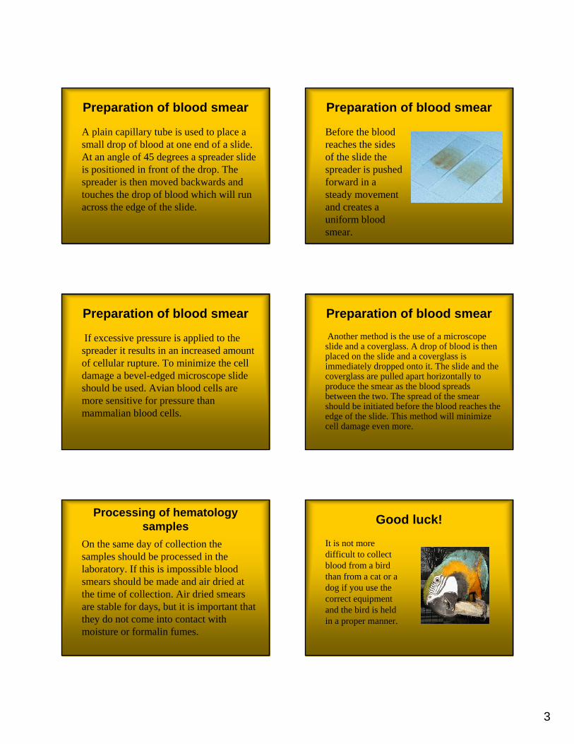
A plain capillary tube is used to place a small drop of blood at one end of a slide. At an angle of 45 degrees a spreader slide is positioned in front of the drop. The spreader is then moved backwards and touches the drop of blood which will run across the edge of the slide.
Preparation of blood smear
Before the blood reaches the sides of the slide the spreader is pushed forward in a steady movement and creates a uniform blood smear.
Preparation of blood smear
If excessive pressure is applied to the spreader it results in an increased amount of cellular rupture. To minimize the cell damage a bevel-edged microscope slide should be used. Avian blood cells are more sensitive for pressure than mammalian blood cells.
Preparation of blood smearAnother method is the use of a microscope slide and a coverglass. A drop of blood is then placed on the slide and a coverglass is immediately dropped onto it. The slide and the coverglass are pulled apart horizontally to produce the smear as the blood spreads between the two. The spread of the smear should be initiated before the blood reaches the edge of the slide. This method will minimize cell damage even more.
Processing of hematology samples
On the same day of collection the samples should be processed in the laboratory. If this is impossible blood smears should be made and air dried at the time of collection. Air dried smears are stable for days, but it is important that they do not come into contact with moisture or formalin fumes.
Good luck!
It is not more difficult to collect blood from a bird than from a cat or a dog if you use the correct equipment and the bird is held in a proper manner.

1
Common avian diseases
Marie GunnarssonInstitute for Clinical Chemistry
Swedish Agricultural University
Common diseasesCommon diseases in caged parrots:
• Trauma• Toxicosis• Neoplastic disease• Compulsive egg laying• Egg-binding• Respiratory disease• Aspergillosis• Chlamydiosis
Common diseases
Common diseases in caged parrots:• Obesity• Ingluvitis• Hepatitis• Proventricular dilatation syndrome• Mites (scaly beak and tassle foot)• Psittacine beak and feather disease• Feather plucking
Trauma
Injuries is mostlyseen among birdsthat are allowed to fly freely inside the house. The mostimportant advice is to prevent the injuries before theyhappen.
Trauma
Common causes to trauma:• The bird sits on top of a door that is being
closed and fractures a leg or toe.• The bird flies into a window and gets a
conclusion or fractures a wing.• The bird lands onto the stove or into a
boiling pot and gets burn injuries.
Trauma
Common causes to trauma:• The bird is being chased/captured by the
family’s cat or dog and gets bitten. Remember that a cat bite is always life threatening to the bird. The bird can die from an infection within 12 h.
• The bird sits on top of another bird’s cage and gets bitten in the toes.
2
Toxicosis
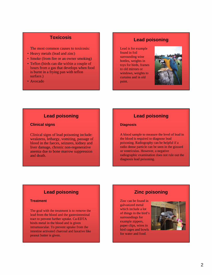
The most common causes to toxicosis:• Heavy metals (lead and zinc)• Smoke (from fire or an owner smoking)• Teflon (birds can die within a couple of
hours from a gas that develops when food is burnt in a frying pan with teflon surface.)
• Avocado
Lead poisoning
Lead is for examplefound in foilsurrounding winebottles, weights in toys for birds, framesto old mirrors or windows, weights to curtains and in old paint.
Lead poisoning
Clinical signs
Clinical signs of lead poisoning include: weakness, lethargy, vomiting, passage of blood in the faeces, seizures, kidney and liver damage, chronic non-regenerative anemia due to bone marrow suppressionand death.
Lead poisoning
Diagnosis
A blood sample to measure the level of lead in the blood is required to diagnose leadpoisoning. Radiography can be helpful if a radio dense particle can be seen in the gizzardor ventriculus. However, a negative radiographic examination does not rule out the diagnosis lead poisoning.
Lead poisoning
Treatment
The goal with the treatment is to remove the lead from the blood and the gastrointestinal tract to prevent further uptake. Ca-EDTAbinds metal in the blood and is given intramuscular. To prevent uptake from the intestine activated charcoal and laxative like peanut butter is given.
Zinc poisoning
Zinc can be found in galvanized metal which include a lot of things in the bird’s surroundings for example zippers, paper clips, wires in bird cages and bowls for water and food.

3
Zinc poisoning
Clinical signs
Clinical signs of zinc poisoning include: hemolytic anemia, kidney and liver damage, weakness, lethargy, vomiting, seizures, convulsions, feather pluckingand death.
Zinc poisoningDiagnostic tests and treatment are the same as with leadpoisoning.
Treatment should be initiated as soon as lead or zincpoisoning are suspected since it will pass severaldays beforediagnosis is confirmed.
Neoplasitic disease
Tumors occur mostly in elderly birds. An exception to this rule is budgerigars who often have tumors already from one yearof age.
Neoplasitic disease
Renal adenocarcinoma in budgerigars is a leading cause of death in males. In females ovarian tumors are morecommon. Males can also have testicularcancer. Clinical signs are in all caseslameness, weight loss and a palpableabdominal mass. Radiographs are helpfulin diagnosis.
Neoplasitic diseaseLipomas are mostcommonly seenamong budgerigars. The lipomaeventually gets troublesome for the bird as it applypressure on surrounding organs and changes the bird’s centre of gravity.
Neoplasitic disease
Caged birds often have fibrosarcomasthat have affinity for wings, legs and face. Some of these can be treated with amputation.
Lymphosarcomas and avian leukosis-likesyndromes are also commonly reported in pet birds.

4
Compulsive egg laying
Compusive egg laying is a rathercommon problem in cockatiels and lovebirds. Instead of laying an ordinarylarge litter of 3-5 eggs she continues to lay egg after egg. To accomplish this a great amount of calcium and nourishmentis required. Eventually the egg layingleads to malnutrition with egg-binding as a possible result.
Compulsive egg laying
It is not easy to break the bird’sunnaturalbehaviour but it can be donewithout medical treatment or surgery.
Compulsive egg laying
To break the behavior, the daylight lengthshould be reduced to 6-8 hours the first threeweeks. The following weeks the light can be turned on 10-12 hours a day. Nesting boxeshave to be removed. The eggs are supposed to be left in with the hen to inhibit further laying. For some birds it is enough to move around the things in the cage or to move the cage to different rooms now and then.
Compulsive egg layingUnfortunately thesemanipulations are seldom enough to inhibit the compulsive egg laying. In those casesmedical treatmentwith hormones or surgical spaying are indicated.
Egg-binding
Egg-binding is most commonly seen among cockatiels, budgerigars and lovebirds. It can be caused by many different factors like age, obesity, cold, dry air, poor diet, calcium deficiency, compulsive egg laying and salpingitis. The hen bird often sits on the floor of its cage and becomes lethargic. Sometimes they breath heavily and get staggering.
Egg-bindingDiagnostics includehistory, clinicalsigns, palpation of the egg and radiography.
Treatment by givingthe bird calcium and provide suitablenesting material and a warm damp environment may be adequate.

5
Egg-bindingIn more difficultcases the egg has to be collapsed with needle and syringeeither per cloaca or via midline throughthe abdominal wall. This procedure is obviously performedduring anesthesia.
Respiratorydisease
Respiratory diseasesare very common among caged birds. Several factorscontribute to developing disease. Examples of suchfactors are vitamin A deficiency, dry indoor air, pollutedair from smoking and food containingmold.
Respiratory disease
Common respiratory diseases:• Chronic rhinitis• Sinusitis• Airsacculitis
• (Aspergillosis)• (Chlamydiosis)
Aspergillosis(Mycotic pneumonia, Pneumomycosis)
Aspergillosis is caused by Aspergillus sppfrequently Aspergillus fumigatus. Inhalation of spores from contaminated litter or feed causesinfection. The fungal spores settle out in the region of the syrinx, the abdominal air sacsand the lung.
Aspergillosis
Clinical signs
Clinical signs of Aspergillosis include: dyspnea, hyperpnea, inappetence, emaciation, increased thirst, somnolenceand other neurological signs. If aflatoxin is produced the bird can contract liver damage, failure and death.
AspergillosisDiagnosis
• Markedly elevated WBC count• Radiographs to show thickening of air sacs• Endoscopy to examine syrinx and internal air
sacs• Tracheal and lung washes

6
AspergillosisBy culture or by microscopicalexamination of freshpreparations the fungus can be demonstrated.
Treatment is difficult. It may haveto go on for the rest of the bird’s life.
Chlamydiosis
(Psittacosis, Ornithosis, Papegojsjuka)
Chlamydiosis is a zoonosis easily spreadto people. It is caused by the bacteriumChlamydophila psittaci. Birds may carry it and develop disease under stress. Shortly after purchase birds should be tested.
ChlamydiosisClinical signs
Clinical signs of chlamydiosis includeweight loss, depression, lime-greenurates, loose feces, emaciated whenexamined and respiratory signs usuallyrelated to an airsacculitis.
Chlamydiosis
Diagnosis
• Radiography – enlarged liver and/or spleen
• Hematology – elevated WBC count, monocytosis
• Biochemistry – elevated AST
Chlamydiosis
Diagnosis
• Serology – can only show if the bird has been in contact with the disease.
• Antigen in feces – risk for false negative• Culture – difficult, often false negative
Chlamydiosis
All birds with a positive test resulthave to be treatedeven if they show no sign of disease. Treatments with doxycycline are veryeffective but require45 days of therapy.

7
Obesity
Obesity is a verycommon diseaseamong budgerigarsand amazons. The problem is related to the fact that the seeddiets commonly fedto the birds haveexcess fat.
Obesity
Obesity increase the risk for:• Hepatic lipidosis• Atherosclerosis• Lipoma• Xanthomatosis
Ingluvitis
Ingluvitis means inflammation of the crop. It can be caused by a variety of pathogens; yeast such as Candidaalbicans, bacteria such as E.coli and parasites such as Trichomonas spp. Regurgitation of seed in budgerigars and cockatiels is often caused by Trichomonas spp.
IngluvitisDiagnosis of the causal agent can be made by cytology or culture of a cropwash.
Clinical signs of ingluvitis mayinclude vomiting, weight loss, depression and a palpable crop filledwith fluid.
Hepatitis
Hepatitis is a very common diseaseamong pet birds. It is often caused by a combination of an increased number of bacteria in the environment and poorfeed. Clinical signs include vomiting, inappetence, loose feaces and yellow-green urates.
Hepatitis
Hepatitis causedby bacteria can be treated with antibiotics for a long period of time in combination with improved feed.

8
Proventricular dilatation syndrome
The disease was initially seen in macawsand therefore also referred to as macawwasting syndrome (arasjuka). Later on it has been seen in other species as well. A viral etiology is suggested, although this remains unproven.
Proventricular dilatation syndrome
Clinical signs
The disease is characterized by lethargy, regurgitation, chronic weight loss, enlarged proventriculus, nervous signs, abnormal droppings and crop impactions.
Proventricular dilatation syndrome
Diagnosis
Diagnosis is based on clinical signs, radiographs showing an enlargedproventriculus and exclusion of differential diagnoses. For a certaindiagnosis a biopsy of the gizzard or cropis required.
Proventricular dilatation syndrome
There is no treatment for this disease. It has always a deadlyoutcome.
MitesCnemoidocoptespilae is common on budgerigars and rare on all otherpsittacines. The disease is thought to depend on a immunodeficiencyand is therefore not thought to be contagious.
Mites
The mites cause conditions known as scaly beak and tassle foot. The signs in the face are crusting of eyelids, corners of the mouth and cere at the base of the beakoften causing beak deformities. Thickening and crusting of the skin are signs of disease on the legs.

9
MitesThe mites canbe recoveredfrom skin scraping and demonstratedby microscopy.
Ivermectin is an effectivetreatment.
Psittacine beak and featherdisease
This disease is caused by psittacinecircovirus. It may infect any psittacinebird. Primarily birds <3 years old appearsto be affected.
The disease is not common in Sweden. When it occurs it is mostly in african greyparrots and lovebirds.
Psittacine beak and featherdisease
Clinical signs
Typical signs are feather loss, abnormalpin feathers, abnormal mature feathers, lack of powder down, beak abnormality, pigment loss in colored feathers and immunosuppression.
Psittacine beak and featherdisease
Diagnosis
• Clinical signs• Detect virus in blood• Biopsies of affected feather follicles
Psittacine beak and featherdisease
There is no treatmentfor this disease. Mostbirds die within fouryears in otherinfections due to immunosuppression. African grey parrotsusually die within a few days due to severe hematologicalchanges.
Feather plucking
It is thought that 25% of the birds that have a problem with feather pluckingsuffer from an actualclinical disease. 75% are due to psychologicalreasons.

10
Feather pluckingInitially disease has to be excluded by performing a completedermatologicalexamination. If no disease is found the bird´s environmenthas to be investigated and hopefully the triggering cause canbe found.
Feather pluckingPossible triggering factors:• Small cage• Boredom• Sexual or social stress or frustration• Attention seeking• Fear, nervousness• Changed diet• Compulsive egg laying• Lack of privacy
Feather pluckingTreatment of feather pluckingbirds with psycologicalproblems impliesgradually changingthe birdsenvironment. Mostoften the birdsunfortunatelybecome constantfeather pluckers.
Good luck!
Please turn to the reference litteraturefor more information about the mentioneddiseases or if you are interested in otheravian diseases.

1
Avian cases
Marie GunnarssonInstitute for Clinical Chemistry
Swedish Agricultural University
Avian cases
The following two cases are authenticcases from Regiondjursjukhuset Strömsholm.
The laboratory reference ranges used are taken from Fudge AM. (2000) Laboratory Medicine Avian and ExoticPets.
Case 1Signalment
20 year old maleamazon.
This is a picture of another amazon
History Case 1Three female amazons had died withinthree days. They sat in different cages a couple of meters from each other. The owner never noticed they were ill. The patient eats ”Nutribird”, apples, oranges, pasta, rice, broccoli and carrots. It drinks community water. The day he came to the clinic he seemed ill and sat with his eyes shut.
Case 1
Blood was collected from the bird withoutanesthesia.Results from blood analyses:
132-618umol/l119Uric acid2.6-5.9ukat/l3.3AST5-17x10.9/l29.8Leukocytes41-53%42Hematocrit
Case 1
0-2%13Monocytes0-0%0Eosinophils0-2%0Basophils31-71%62Heterophils20-67%25Lymphocytes
Polychromasia 12-13%Moderate poikilocytosisThe polychromasia indicates regenerative anemia

2
Case 1This is a picture of a blood smear from the amazon with the unknown illness.
The polychromasia of the erythrocytesindicatesregenerative anemia.
Prominent polychromasia: Regenerative anemia
Case 1
Blood was also sent in for lead and zinc analyses, but the results from those analyses take several days to receive.
Note polychromasia of the erythrocytes.
Case 1
Chlamydialdiagnostics
• Culture – negative• Serology – low titer
This is a picture of blood collectionfrom another bird.
Case 1If this bird had Chlamydia infection, then what kind of anemia should that infection cause?
Case 1Treatment
The amazon wastreated with enrofloxacin, Na-EDTA, vitamin K and fed via a croptube.
This is a picture of yet another amazon.

3
Case 1
The amazon was hospitalized for threedays. When he went home he was feeling a lot better. The owner continued to treatthe bird with Na-EDTA and enrofloxacinat home.
Diagnosis?
Case 1
The results from the lead and zinc analyses:
0-1mmol/l0.10Lead
0-30mmol/l53Zinc
The amazon was suffering from zinc poisoning!
Case 1
Zinc poisoning causes a hemolytic anemia. Hemolytic anemia is a regenerative anemia. Infections cause a nonregenerative anemia, so the hematology results should have steered us away from just an infection like chlamydiosis. A metal foreign body containing zinc was not reported.
Case 2
Signalment
Female moluccancockatoo, ageunknown.
This is a picture of another cockatoo.
Case 2
History
This bird lived in a zoological park and came to the clinic for a health check up. Blood was collected for a number of analyses.
Case 2
Results from blood analyses:
190.3-632.9umol/l446Uric acid2.3-6.2ukat/l1.3AST0-1%1Monocytes0-2%1Eosinophils0-1%1Basophils44-71%78Heterophils19-50%19Lymphocytes8-12x10.9/l14.1Leukocytes

4
Case 2
A few reactivelymphocytes werefound in the bloodsmear from the cockatoo. This is a picture of a reactivelymphocyte and an erythrocyte.
Case 2
This is anotherpicture of the bloodsmear. The twoleukocytes are an eosinophil (left) and a heterophil (right).
Case 2
In this picture you can see oneeosinophil (arrow), two thrombocytes(upper left corner) and one lymphocyte(upper right corner). The rest of the cells are erythrocytes.
Case 2
Chlamydialdiagnostics
• Culture – negative• Serology – high titer
This is a picture of yet another cockatoo.
Case 2The cockatoo wasalso tested for psittacine beak and feather disease. This test was negative.
This is a picture of another reactivelymphocyte from the patient.
Case 2
Since the serology test for chlamydiosisshowed high titer for the disease the birdwas treated with doxycycline.
After a few weeks the bird died. Why? Had something been missed at the healthcheck up?

5
Case 2
Autopsy
The autopsy showed a stenosis in trachea. This turned out to be caused by aspergillosis.
Good luck!The two previouscases illustrate someof the diseases of parrots. Unfortunately the second bird had 2 infections/diseases. We often havetrouble diagnosingthe second unexpected disease.