Embed Size (px)
Citation preview

Research Article Open AccessOpen AccessResearch Article
Clinical & Experimental Ophthalmology
Jirasková et al., J Clin Exp Ophthalmol 2013, S3http://dx.doi.org/10.4172/2155-9570.S3-006
J Clin Exp Ophthalmol Neuro Ophthalmology ISSN:2155-9570 JCEO an open access journal
*Corresponding author: Jan Lešták, MD, PhD, FEBO, JL Clinic, V Hůrkách 1296/10, Prague, Czech Republic, E-mail: [email protected]
Received February 12, 2013; Accepted March 29, 2013; Published April 04, 2013
Citation: Jirasková N, Rozsíval P, Stepanov A, Velika V, Lešták J (2013) Outcomes of Optic Nerve Sheath Decompression for Visual Loss in Idiopathic Intracranial Hypertension. J Clin Exp Ophthalmol S3: 006. doi:10.4172/2155-9570.S3-006
Copyright: © 2013 Jirasková N, et al. This is an open-access article distributed under the terms of the Creative Commons Attribution License, which permits unrestricted use, distribution, and reproduction in any medium, provided the original author and source are credited.
Outcomes of Optic Nerve Sheath Decompression for Visual Loss in Idiopathic Intracranial HypertensionNada Jirasková1,4, Pavel Rozsíval1,4, Alexander Stepanov1,4, Vera Velika1,4 and Jan Lešták2,3,4*1Department of Ophthalmology, University Hospital and Charles Medical Faculty, Hradec Králové, Czech Republic2JL Clinic, V Hurkách 1296/10, Prague, Czech Republic3Faculty of Biomedical Engineering, Czech Technical University in Prague, Czech Republic4Faculty of Medicine in Hradec Kralove, Charles University, Czech Republic
Keywords: Idiopathic intracranial hypertension; Optic nerve sheath decompression; Papilledema; Visual functions
IntroductionIntracranial hypertension (IH) is a multifactorial syndrome
characterized by severe headache, nausea, vomiting, transient visual obscuration and diplopia. Idiopathic intracranial hypertension (IIH) is the terminology used when no underlying etiology is detected. The definition of IIH has evolved with clinical experience and advances in imaging technology. Currently, IIH can be diagnosed only if the following criteria are met: 1) symptoms and signs attributable to increased intracranial pressure (ICP); 2) elevated ICP recorded during lumbar puncture in the lateral decubitus position; 3) normal cerebrospinal fluid (CSF) composition; 4) no imaging evidence of ventriculomegaly or a structural cause for increased ICP, such as a brain parenchymal, ventricular, meningeal, or venous sinus abnormality; and 5) no other cause of intracranial hypertension identified, such as use of certain medications [1].
Idiopathic intracranial hypertension is a surprisingly common disorder. In young overweight women (the typical patients with IIH), the annual incidence is as high as 20 per 100,000 persons. Atypical patients include men, slim women, prepubescent children, and patients older than 44 years [1].
Papilledema is an optic disc edema secondary to increased intracranial pressure. It is the ophthalmologic hallmark of increased intracranial pressure and can lead to permanent visual loss if left untreated. Visual loss is one of the major morbidities, with blindness reported in up to 10% of patients and visual field defects of some type in up to 90% of patients [2].
Primary indications for the treatment of IIH are intractable headache and vision loss [3]. Multiple therapeutic alternatives are available for management of IIH. Medical therapy is directed towards reducing CSF secretion and increasing CSF absorption with carbonic anhydrase inhibitors such as acetazolamide as a first line medication
[1]. Dietary management and weight loss are advocated to reduce intraabdominal pressure and weight loss appeared to have been the catalyst for reducing the severity of papilledema [4,5]. Repeated lumbar punctures are sometimes used in patients with occasional symptom relapses, in pregnant women, or in the setting of rapidly declining vision to temporarily lower the CSF pressure while planning a more aggressive treatment [1]. Surgery is considered under the following circumstances: 1) progressive loss of vision despite maximal medical therapy; 2) severe or rapid visual loss at onset; and 3) severe papilledema causing macular edema exudates [6,7]. Surgical procedures used for the treatment of visual loss include optic nerve sheath decompression (ONSD) and CSF diversion procedures. Whether one procedure is superior to the other remains controversial. Case studies suggest that ONSD is generally effective for managing visual loss, whereas diversion and perhaps stent placement procedures may be more beneficial for managing headache [3].
We report our experience with ONSD for visual loss in IIH.
MethodsIn this retrospective noncomparative study, patients undergoing
ONSD for visual loss due to IIH from January 1995 to December 2012 were included for analysis. Only patients who met the modified Dandy criteria for IIH were included. The decision for performing ONSD was
AbstractPurpose: To report our experience with optic nerve sheath decompression (ONSD) for visual loss in idiopathic
intracranial hypertension (IIH).
Methods: Six patients (7 eyes) with idiopathic intracranial hypertension and visual loss were involved. Five women and 1 man (mean age 42 years, ranged from 12 to 65 years) were operated at the Department of Ophthalmology, University Hospital, Hradec Králové. Surgeries were performed by a standard medial transconjunctival approach and the sheaths were cut by three incisions. We have not seen any serious intra and postoperative complications. Only one patient developed transient double vision after surgery.
Results: Visual functions improved in 5 patients. In one patient permanent visual loss with optic atrophy in both eyes occurred due to long-term duration of papilledema before referral to us.
Conclusions: On the basis of our results, we believe that ONSD is safe and effective treatment of patients with IIH in vision-threatening cases after thoroughly balanced assessment of possible risks and gains.
Citation: Jirasková N, Rozsíval P, Stepanov A, Velika V, Lešták J (2013) Outcomes of Optic Nerve Sheath Decompression for Visual Loss in Idiopathic Intracranial Hypertension. J Clin Exp Ophthalmol S3: 006. doi:10.4172/2155-9570.S3-006
Page 2 of 4
J Clin Exp Ophthalmol Neuro Ophthalmology ISSN:2155-9570 JCEO an open access journal
made on the basis of acuteness, severity of visual loss at presentation or progression despite maximally tolerated medical therapy.
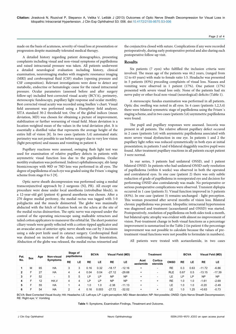
A detailed history regarding patient demographics, presenting complaints including visual and non-visual symptoms of papilledema and raised intracranial pressure was taken. All patients underwent a detailed neurological evaluation including history, clinical examination, neuroimaging studies with magnetic resonance imaging (MRI) and cerebrospinal fluid (CSF) studies (opening pressure and CSF composition). Relevant investigations were done to detect any metabolic, endocrine or hematologic cause for the raised intracranial pressure. Ocular parameters (assessed before and after surgery follow-up) included best-corrected visual acuity (BCVA), perimetry, stereoscopic fundoscopy, pupillary light response and ocular motility. Best corrected visual acuity was recorded using Snellen´s chart. Visual field asessment was performed using a Humphrey field analyzer, SITA standard 30-2 threshold test. One of the global indices (mean deviation, MD) was chosen for obtaining a picture of improvement, stabilization or further worsening of visual field. Mean deviation is a location-weighted mean of the values in the total deviation plot. It is essentially a distilled value that represents the average height of the entire hill of vision [8]. In two cases (patients 3,4) automated static perimetry was not possible to use before surgery due to very low vision (light perception) and nausea and vomiting in patient 4.
Pupillary reactions were assessed, swinging flash light test was used for examination of relative pupillary defects in patients with asymmetric visual function loss due to the papilledema. Ocular motility evaluation was performed. Indirect ophthalmoscopy, slit-lamp biomicroscopy with 90D or 78D lens was performed in all eyes. The degree of papilledema of each eye was graded using the Frisen´s staging scheme from stage 0 to 5 [9].
Optic nerve sheath decompression was performed using a medial transconjunctival approach by 2 surgeons (NJ, PR). All except one procedure were done under local anesthesia (retrobulbar block), in a 12-year-old girl (patient 4) general anesthesia was chosen. After a 270 degree medial peritomy, the medial rectus was tagged with 5-0 polyglactin and the muscle disinserted. The globe was maximally abducted with the hitch of fixation hook on the sclera at the site of the medial rectus disinsertion. The optic nerve was exposed under the control of the operating microscope using malleable retractors and tailed cotton applicators to maneuver the orbital fat. The short posterior ciliary vessels were gently reflected with a cotton-tipped applicator and an avascular area of anterior optic nerve sheath was cut by 3 incisions using a side-port knife used in cataract surgery. Cerebrospinal fluid was drained on incision of the dura, confirming the fenestration. Abduction of the globe was released, the medial rectus reinserted and
the conjunctiva closed with suture. Complications if any were recorded perioperatively, during early postoperative period and also during each postoperative follow-up visit.
ResultsSix patients (7 eyes) who fulfilled the inclusion criteria were
involved. The mean age of the patients was 44.2 years, (ranged from 12 to 65 years) with male to female ratio 1:5. Headache was presented in 5 patients (83%) preceding complaints of visual loss. Nausea and vomiting were observed in 1 patient (17%). One patient (17%) presented with severe visual loss only. None of the patients had six nerve palsy or other focal non-visual (neurological) deficits (Table 1).
A stereoscopic fundus examination was performed in all patients. Optic disc swelling was noted in all eyes. In 4 cases (patients 1,2,3,4) there were bilateral symmetric stage of papilledema using the Frisen´s staging scheme, and in two cases (patients 5,6) asymmetric papilledema were found.
The pupil and pupillary responses were assessed. Isocoria was present in all patients. The relative afferent pupillary defect occured in 2 cases (patients 5,6) with asymmetric papilledema associated with more severe visual dysfunction in one eye. In patients 1 and 2 the pupillary light reflex was reduced symmetrically in both eyes at initial presentation; in patients 3 and 4 bilateral sluggishly reactive pupil were found. After treatment pupillary reactions in all patients except patient 3 were normal.
In our series, 5 patients had unilateral ONSD, and 1 patient bilateral ONSD. In patients who had unilateral ONSD early resolution of papilledema (within 6 weeks) was observed in both the operated and contralateral eyes. In one case (patient 2) there was only subtle reduction of grade of papilledema in nonoperated eye and decision for performing ONSD also contralateraly was made. No perioperative or serious postoperative complications were observed. Transient diplopia occurred in 1 case (patients 5). Visual function improved in 5 patients (83%). In one case (patient 3) remains unchanged - light perception. This woman presented after several months of vision loss. Bilateral chronic papilledema was present. Idiopathic intracranial hypertension was diagnosed and treatment (aceazolamid and ONSD) was started. Postoperatively, resolution of papilledema on both sides took a month, but bilateral optic atrophy was evident with almost no improvement of vision. Post-treatment change in both visual functions as a percentage improvement is summarized in the Table 2 (in patient 4 the percentage improvement was not possible to calculate because the values of pre-treatment visual functions were not possible to formulate in numbers).
All patients were treated with acetazolamide, in two cases
Pat.No. Sex Age Non-visual
Symptoms
Stage ofpapilledema BCVA Visual Field (MD) Treatment BCVA Visual Feld (MD)
RE LE RE LE RE LE Acetazolamide
Corticosteroids ONSD RE LE RE LE
1 M 65 HA 3 3 0.16 0.32 -18.17 -12.16 + - RE 0.3 0.63 -11.72 -6.862 F 27 HA 4 4 0.04 0.04 -27.12 -29.68 + - RLE 0.67 0.5 -13.15 -17.393 F 52 - 5 5 LP LP NP NP + - LE LP LP NP NP4 F 12 HA,V 4 4 LP LP NP NP + + RE 1.0 1.0 -1.61 -2.695 F 55 HA 1 4 1.0 1.0 -2.38 -11.13 + - LE 1.0 1.0 -0.20 -2.496 F 54 HA 2 4 0.16 0.003 -27.72 -32.02 + + LE 1.0 1.25 +0.63 -0.73
BCVA: Best Corrected Visual Acuity; HA: Headache; LE: Left eye; LP: Light perception; MD: Mean deviation; NP: Not possible; ONSD: Optic Nerve Sheath Decompression; RE: Right eye; V: Vomiting
Table 1: Symptoms, Examination Findings, Ttreatment and Outcome.

Citation: Jirasková N, Rozsíval P, Stepanov A, Velika V, Lešták J (2013) Outcomes of Optic Nerve Sheath Decompression for Visual Loss in Idiopathic Intracranial Hypertension. J Clin Exp Ophthalmol S3: 006. doi:10.4172/2155-9570.S3-006
Page 3 of 4
J Clin Exp Ophthalmol Neuro Ophthalmology ISSN:2155-9570 JCEO an open access journal
(patients 4, 6) with rapid progressive loss of vision due to malignant IIH combination of maximal dose of acetazolamide and intravenous methylprednisolone followed by oral steroids was used.
Two cases (patients 5 and 6) presented with asymmetric visual loss. Patient 5 presented with eight months history of headaches and visual obscurations in the left eye followed, 1 month later with progressive declining vision in the left eye. Best-corrected visual acuity was 1.0 in both eyes. Automated static perimetry showed bilateral enlarged blind spot and upper and lower Bjerrum´s scotoma in the left eye (Figure 1). Ophthalmoscopy revealed marked chronic papilledema in the left eye and incipient in the right eye (Figure 2). Results of brain MRI were normal. Lumbar puncture revealed an opening pressure of 280 mm Hg with normal chemistry and no cells. Idiopathic intracranial hypertension was diagnosed and she was treated with 250 mg oral acetazolamid three times daily. One week after starting therapy, she complained of worsening vision. ONSD was performed in the left eye. Papilledema resolved over 6 weeks (Figure 3) and visual field improved within 3 months (Figure 4). Patient 6 presented with one month history of rapid visual loss in the left eye. Ophthalmoscopy revealed marked developed papilledema in the left eye and incipient in the right eye (Figure 5). Also decrease in vision was markedly asymmetric (Table 1and Figure 6). All necessary neurologic and imaging diagnostic tests were performed and “malignant” IIH was diagnosed. Intensive treatment with maximal doses of acetazolamide and intravenous corticosteroids were administered. ONSD was performed in the left eye. After surgery resolution of papilledema and improvement of visual function were observed within four weeks (Table 1 and Figure 7).
In all patients, the clinical course was a single episode, with no relapse.
DiscussionThe syndrome of increased ICP without ventriculomegaly or mass
lesion, and with normal CSF composition, was first described more than a century ago, yet we still know little about pathogenesis [1].
As it was mentioned previously, the typical patient with IIH is an obese woman of childbearing age; atypical patients include men, slim women, prepubescent children, and patients older than age 44 years. In our series 1 patient was prepubescent child, 4 patients were older than age 50 years and the only one woman in childbearing age was slim. This may be in a connection with more severe clinical course of the disease and late diagnosis in some of them.
Additionally, we believe that the “atypicality” of the patients in our series is also in connection with a very long time-frame for recruitment (17 years). According to our experience the “typical” patients with IIH improve after dietary management and/or pharmacological treatment with acetazolamide. During the last 20 years we have diagnosed,
observed and/or treated more than 100 patients with IIH and only those 6 required surgical intervention.
The principal morbidity of IIH is papilledema-associated visual loss. Failure of conservative management to prevent progressive visual loss is an indication for surgery which includes ONSD or CSF shunting procedures. The pathophysiologic mechanism by which ONSD improves visual function is unresolved [10]. Two mechanisms have been proposed in the literature to explain how ONSD works. Cerebrospinal fluid filters through the dural opening into the orbit with a subsequent decrease in subarachnoid CSF pressure around the optic
BCVA: Best Corrected Visual Acuity; LE: Left eye; RE: Right eye
Table 2: Post-treatment change in visual functions as a percentage improvement.
Pat. No.
BCVA Visual Field (MD)RE LE RE LE
1 47 49 36 442 94 92 52 413 0 0 0 04 - - - -5 0 0 92 786 84 100 102 98
Figure 1: Visual field of the patient 5 before treatment.
Figure 2: Fundus of the patient 5 before treatment.
Figure 3: Fundus of the patient 5 after treatment.
Figure 4: Visual field of the patient 5 after treatment.

Citation: Jirasková N, Rozsíval P, Stepanov A, Velika V, Lešták J (2013) Outcomes of Optic Nerve Sheath Decompression for Visual Loss in Idiopathic Intracranial Hypertension. J Clin Exp Ophthalmol S3: 006. doi:10.4172/2155-9570.S3-006
Page 4 of 4
J Clin Exp Ophthalmol Neuro Ophthalmology ISSN:2155-9570 JCEO an open access journal
nerve. Another potential explanation is that the fibrous tissue obliterates the operation site and prevents the direct transmission of elevated CSF pressure to the optic nerve head [11]. After unilateral ONSD, 5 patients in our series (83%) experienced reduction of grade of papilledema in both operated and nonoperated eyes. The reduction in disc edema and visual stabilization was clinically sufficient to obviate the need to proceed with a contralateral ONSD in all but one of our patients. We acknowledge that the mechanism for this contralateral surgical effect is not certain and could be related to decreased intrasheath CSF in both optic nerves after unilateral ONSD, regression toward the mean, spontaneous improvement, or simply better patient compliance with maximum medical therapy after unilateral surgery.
Visual functions after ONSD in our series showed significant improvement in 5 patients (83%), in 1 case (17%) remain stable, and in no case progressive visual loss after surgery was observed. The improvement in visual functions occurred in the early postoperative period (within 4 weeks) and was maintained at all follow-up visits. In all patients, the clinical course was a single episode, with no relapse.
In patient 3 vision after ONSD remains unchanged - light perception. This woman presented after several months of slow-progressing vision
loss. Severe bilateral chronic papilledema was present. Postoperatively, resolution of papilledema on both sides took a month, but bilateral optic atrophy was evident with almost no improvement of vision. We believe that in her case was “too late” for any treatment and even decrease in subarachnoid CSF pressure around the optic nerve after ONSD could not prevent optic nerve atrophy with permanent blindness. This case emphasizes the necessity of early diagnostics and treatment of IIH to prevent severe and permanent visual loss.
There were 2 cases (patients 4, 6) of “malignant” IIH [1] with rapid visual decline and decreased visual acuity in our series. Marked papilledema was observed at presentation. Visual loss occurred in patient 4 within two weeks and in patient 6 unilateraly within four weeks. Intensive treatment with maximal doses of acetazolamide and intravenous corticosteroids were administered. Prompt indication of surgical treatment (ONSD) was necessary and visual outcomes in both cases were excellent.
We recognize the limitations of our study, including the retrospective nature of the design and small sample size. Despite these limitations, we believe our study shows that ONSD is a safe procedure with a short surgical time and fast patients recovery time. Moreover this procedure has essentially less stress for the patients from the procedure itself than CSF diversion procedures.
In conclusion, ONSD is our first-line surgical intervention in patients with visual loss due to IIH in whom medical therapy fails.
Conflict of Interest None of the authors have a financial or proprietary interest in any product
mentioned.
AcknowledgmentsThis study was supported by Charles University in Prague - Programme
P37/07 (PRVOUK).
References1. Friedman DI, Jacobson DM (2004) Idiopathic intracranial hypertension. J
Neuroophthalmol 24: 138-145.
2. Banta JT, Farris BK (2000) Pseudotumor cerebri and optic nerve sheath decompression. Ophthalmology 107: 1907-1912.
3. Feldon SE (2007) Visual outcomes comparing surgical techniques for management of severe idiopathic intracranial hypertension. Neurosurg Focus 23: E6.
4. Johnson LN, Krohel GB, Madsen RW, March GA Jr (1998) The role of weight loss and acetazolamide in the treatment of idiopathic intracranial hypertension (pseudotumor cerebri) Ophthalmology 105: 2313-2317.
5. Kollar C, Parker G, Johnston I (2001) Endovascular treatment of cranial venous sinus obstruction resulting in pseudotumor syndrome. Report of three cases. J Neurosurg 94: 646-651.
6. Corbett JJ, Thompson HS (1989) The rational management of idiopathic intracranial hypertension. Arch Neurol 46: 1049-1051.
7. Carter SR, Seiff SR (1995) Macular changes in pseudotumor cerebri before and after optic nerve sheath fenestration. Ophthalmology 102: 937-941.
8. Walsh TJ (1996) Visual Fields: Examination and Interpretation. (2 edn) Oxford University Press, USA.
9. Frisén L (1982) Swelling of the optic nerve head: a staging scheme. J Neurol Neurosurg Psychiatry 45: 13-18.
10. Nithyanandam S, Manayath GJ, Battu RR (2008) Optic nerve sheath decompression for visual loss in intracranial hypertension: report from a tertiary care center in South India. Indian J Ophthalmol 56: 115-120.
11. Alsuhaibani AH, Carter KD, Nerad JA, Lee AG (2011) Effect of optic nerve sheath fenestration on papilledema of the operated and the contralateral nonoperated eyes in idiopathic intracranial hypertension. Ophthalmology 118: 412-414.
Figure 5: Fundus of the patient 6 before treatment.
Figure 6: Visual field of the patient 6 before treatment.
Figure 7: Visual field of the patient 6 after treatment.