Embed Size (px)
Citation preview

Clinical and population-based studies in multiple myeloma and
monoclonal gammopathy -focus on infections
Cecilie Hveding Blimark
Department of Internal Medicine and Clinical Nutrition Institute of Medicine
Sahlgrenska Academy at University of Gothenburg
Gothenburg 2014

Cover illustration: Bone marrow aspirate with plasma cells in a patient with multiple myeloma. With the courtesy Dr. Stefan Jacobsson, clin. chem. Sahlgrenska University Hospital
Clinical and population-based studies in multiple myeloma and monoclonal gammopathy -focus on infections © Cecilie Hveding Blimark 2014 [email protected] ISBN: 978-91-628-9252-4 http:/hdl.handle.net/2077/36910 Printed in Gothenburg, Sweden 2014 Ale Tryckteam

I Keep six honest serving-men:
(They taught me all I knew)
Their names are What and Where and When
And How and Why and Who.
Rudyard Kipling (1865–1936)
To Christian, Henrik and Johanna


Clinical and population-based studies in multiple myeloma and monoclonal gammopathy -focus on infections
Cecilie Hveding Blimark Department of Internal Medicine and Clinical Nutrition,
Institute of Medicine Sahlgrenska Academy at University of Gothenburg
Göteborg, Sweden

ABSTRACT
Multiple myeloma is a haematological disorder of the bone marrow. It is preceded by the benign precursor monoclonal gammopathy of undetermined significance (MGUS). In multiple myeloma, a progression leading to expansion of malignant plasma cells occurs, causing skeletal lesions, anemia and renal insuffiency. Multiple myeloma is incurable, but the disease can be controlled with chemotherapy and other immunosuppressive drugs. It is known that both conditions have compromised immune responses, which lead to an increased risk of infections. However, there is no population-based data on the occurrence and type of infections in patients with plasma cell disorders compared to the normal population. Considering the cumulative immunodeficiency in patients with multiple myeloma, caused by multiple cytotoxic and immunomodulating therapies, there is a demand for less toxic treatments in the relapse setting, aiming to reduce morbidity and mortality in infections. Recent studies have suggested that immunomodulating treatment is beneficial even in smouldering multiple myeloma. There is a lack of population-based incidence data in smouldering multiple myeloma patients with high risk of progressing to multiple myeloma, and there is a need of identifying patients with smouldering multiple myeloma that could benefit from up-front treatment. In paper I we investigated the treatment with intermediate-dose melphalan (Mel 100) and stem cell support in multiple myeloma patients relapsing after high dose melphalan and autologous transplantation (ASCT) in 66 patients. With an overall response of 62%, limited toxicity and a progression-free survival of 8.5 months, we conclude that Mel 100 is a viable therapeutic option in relapsed patients and the best efficacy was seen in patients with long-lasting response after ASCT. In paper II and III we studied the risk of infections in MGUS and multiple myeloma patients compared to matched controls. Using population-based data from Sweden, in paper II we estimated the risk of infections among 5 326 MGUS patients compared to 20 161 matched controls. We found that patients with MGUS had a 2-fold increased increased risk (hazard ratio (HR) 2.1; 95% confidence interval (CI) 2.0-2.3(p<0.05)) of developing any infection at 5- follow up, and at 10-year follow up the risk was very similar (HR=2.2; 95% CI 2.0-2.3). Patients with M-protein concentration over 2.5 mg/dl had the highest risk of infections. In paper III we compared the risk of infections in 9 253 multiple myeloma patients to 34 931 matched controls. Overall, multiple myeloma patients had a 7-fold (HR =7.1; 95% CI 6.8-7.4) risk of developing any infection compared to matched controls. The increased risk of developing a bacterial infection was 7-fold (7.1; 6.8-7.4), and for viral infections it was 10-fold (10.0; 8.9-11.4). Multiple myeloma patients diagnosed in the more recent calendar periods had significantly higher risk of infections compared to patients diagnosed earlier (p<0.001). We could show, that in patients who died within the first year of diagnosis, 22 % of deaths were infection-related. Our findings provide novel insights into the mechanisms behind infections in patients with plasma cell disorders, and may have clinical implications and could give support to preventive interventions. In paper IV we estimated the risk of progression to symptomatic multiple myeloma in a cohort of smouldering multiple myeloma patients with high-risk features using population-based data from the Swedish Myeloma Registry. The 2-year risk of progressing was 56% and this cohort count for 29% of all smouldering multiple myeloma patients and should be considered for clinical early treatment trials.
Keywords: multiple myeloma, MGUS, infections ISBN: 978-91-628-9252-4 ISBN: 978-91-628-9255-5 http:/hdl.handle.net/2077/36910


POPULÄRVETENSKAPLIG SAMMANFATTNING
Myelom är en typ av blodcancer som uppstår i benmärgen genom en klonal expansion av de antikroppsbildande cellerna (plasmacellerna) i immunsystemet. Den kan inte botas, men cellgiftsbehandling kan förbättra symptomen och förlänga livet på patienten. Både sjukdomen och cellgiftsbehandlingen gör att många myelompatienter får dåligt immunförsvar. Myelom föregås alltid av ett godartat tillstånd kallat MGUS (från engelska: monoclonal gammopathy with undetermined significance) vid vilket en så kallad M-komponent (förhöjd mängd av en sorts obrukbara antikroppar) kan påvisas men där det inte finns några tecken till sjukdomsaktivitet. Vissa MGUS-patienter kan också ha samma immundefekt som myelompatienter (t.ex. brist på effektiva antikroppar, sk. hypogammaglobulinemi) och besväras av upprepade infektioner. Vi är intresserade av risken för infektioner hos MGUS och myelompatienter och hur man kan minska denna.
För patienter upp till 65 år, är standardbehandling för myelom i Sverige en hög dos cellgift med stamcellstöd, så kallas autolog stamcellstransplantation (ASCT), vilket oftast medför en 3 veckors sjukhusvistelse med risk för infektioner. I ett försök att finna mindre giftig behandling har vi på Hematologen Sahlgrenska behandlat 66 myelompatienter, i första återfall, mellan 1996 och 2007 med en så kallad ”Minitransplantation” med halverad cellgiftsdos. Denna behandling gav låg toxicitet och få inläggningar. Patienter höll sig återfallsfria i 8,5 månader i snitt efter behandlingen med betydligt mindre sjuklighet i infektioner jämfört med ASCT.(Delarbete I). Tidigare har mindre studier indikerat att myelompatienternas liv och hälsa kan hotas av speciella typer av infektioner. Inga populationsbaserade data finns publicerade som beskriver hur stor risk MGUS och myelompatienter har för att drabbas av infektioner jämfört med normalbefolkningen. Vi har därför försökt besvara frågor om hur vanligt det är med infektioner hos MGUS och myelompatienter jämfört med normalbefolkningen och vilka typer av infektioner dessa patienter har störst risk att drabbas av. Vi ville ta reda på om nyare och starkare behandlingar på senare tid medfört ökad infektionsrisk och om risken att dö i infektioner för myelompatienter har ökat. Genom analys av bl.a. svenska cancerregistret, olika sjukhusregister och dödsorsaksregistret har vi jämfört svenska myelompatienter och individer med MGUS över en viss tidsperiod med ett representativt matchat urval av den svenska normalbefolkningen. I sjukhusregister har vi hittat 5 326 MGUS-fall och jämfört med 20 161 friska kontrollpersoner matchade avseende kön, ålder och bostadsort och funnit att risken att insjukna i infektion för MGUS-patienter jämfört med normalbefolkningen är 2-falt ökad (Delarbete II). Från Cancerregistret hämtades data för alla 9 253 patienter som fått myelomdiagnosen mellan 1988 och 2004 och vi jämförde deras risk för infektioner med ett köns-, bostadsort- och åldersmatchat urval på 34 931 friska personer av den svenska normalbefolkningen. För myelompatienter jämfört med kontroller var risken 7-falt ökad att drabbas av en infektion. Vi fann också att infektioner är en viktig orsak till tidig död hos myelompatienter (Delarbete III). Sjukdomar som visade sig vara mycket vanligare hos både MGUS och myelompatienter än normalbefolkningen var främst lunginflammation,

hjärnhinneinflammation, blodförgiftning, bältros samt influensa. Vi kunde också visa att risken för infektioner hos myelompatienter har ökat mer på senare år, men att risken att dö i infektioner var konstant under studieperioden. Sammanfattningsvis är detta den största studie som har studerat risken för infektioner hos myelompatienter i ett populationsbaserat material. Genom att rikta uppmärksamheten mot denna potentiellt botbara komplikation hoppas vi att kunna bidra till att förbättra vården och slutligen överlevnaden hos denna patientgrupp.
Det sista delarbetet är baserat på Myelomregistret (en del av Blodcancerregistret). Sverige har ett heltäckande sjukvårdssystem och översikt över alla patienter genom sina olika register kopplade med personnumret. Det svenska nationella Myelomregistret startades i 2008 för att kunna utvärdera förekomsten av myelom, karakteristika vid sjukdomen, behandling, sjuklighet och dödlighet i sjukdomen i Sverige på nationell nivå. Det gör att myelomregistret i Sverige kan ge en mer sann bild av blodsjukdomen myelom än studier från sjukhusregister på stora behandlingscentra där det finns stor risk för patientselektion. Med dagens höga täckningsgrad kan man med myelomregistret leverera högkvalitativa populationsbaserade data på diagnostik, behandling och överlevnad. Patienter med asymtomatiskt myelom kan hålla sig stabila i många år och skall enligt nu gällande riktlinjer inte behandlas, utan man skall avvakta en övergång till symptomatiskt myelom. Det finns dock nu en behandlingsstudie som utmanar detta, där man hävdar att tidig behandling av patienter med högriskfaktorer för progress kan ge en överlevnadsfördel. Den föreslagna behandlingen är dock inte utan biverkningar och studien är kontroversiell. I diskussion och kritik internationellt kom det fram att det råder stor osäkerhet kring förekomsten av högrisk asymtomatiska myelom, då populationsbaserade data är få. Vi ville undersöka asymtomatiska myelom, deras förekomst i ett populationsbaserat material och risken för att övergå i symptomatisk myelom (Delarbete IV). Från Myelomregistret tog vi fram förekomsten av asymtomatiska myelom med högrisk-kriterier i Sverige från 2008 och deras risk att övergå i symptomatiskt myelom. Vi fann att nästan 1/3 av alla asymtomatiska myelom har hög risk för progress. Vi fann också att risken för progress hos asymtomatiska myelompatienter med högrisk-faktorer, att övergå till symptomatisk myelom, var 56 % de första 2 åren. Dessa patienter bör sannolikt komma i fråga för behandlingsstudier.


Cecilie Hveding Blimark
1
LIST OF PUBLICATIONS
This thesis is based on the following studies, referred to in the text by their Roman numerals.
I. Melphalan 100 mg/m2 with stem cell support as first relapse treatment is safe and effective for myeloma patients with long remission after autologous stem cell transplantation.
Cecilie Hveding Blimark, Ljupco Veskovski, Jan Westin, Stig Rödjer, Mats Brune, Martin Hjorth, Erik Holmberg, Per-Ola Andersson, Ulf-Henrik Mellqvist. Eur J Haematol 2011:87(2):117-22.
II. Monoclonal gammopathy of undetermined significance and risk of infections: a population-based study.
Sigurdur Y Kristinsson, Min Tang, Ruth M Pfeiffer, Magnus Björkholm, Lynn R Goldin, Cecilie Hveding Blimark, Ulf-Henrik Mellqvist, Anders Wahlin, Ingemar Turesson, Ola Landgren. Haematologica 2012:97(6):854-8.
III. Multiple Myeloma and Infections: A population-based study on 9,253 multiple myeloma patients.
Cecilie Hveding Blimark, Erik Holmberg, Ulf-Henrik Mellqvist, Ola Landgren, Magnus Björkholm, Malin L Hultcrantz, Christian Kjellander, Ingemar Turesson and Sigurdur Y Kristinsson. Haematologica 2014 Epub 24 Oct.
IV. Treatment for High-Risk Smouldering Myeloma
Sigurdur Y Kristinsson, Erik Holmberg, Cecilie Hveding Blimark. N Engl J Med 2013; 369(18):1762-3

Clinical and population-based studies in multiple myeloma and monoclonal gammopathy -focus on infections
2
TABLE OF CONTENTS
1 Introduction ..................................................................................... 6
1.1 History ...................................................................................... 6 1.2 Multiple myeloma ................................................................. 10
1.2.1 Definition .......................................................................... 10 1.2.2 Epidemiology ................................................................... 11 1.2.3 Clinical features ................................................................ 11 1.2.4 Treatment and prognosis .................................................. 11
1.3 Smouldering multiple myeloma ............................................ 15 1.3.1 Definition and epidemiology ............................................ 15 1.3.2 Treatment and prognosis .................................................. 15
1.4 Monoclonal Gammopathy of undetermined significance (MGUS) .................................................................................. 19
1.4.1 Definition .......................................................................... 19 1.4.2 Epidemiology ................................................................... 19 1.4.3 Etiology and pathogenesis ................................................ 19 1.4.4 Clinical features ................................................................ 21 1.4.5 Prognosis .......................................................................... 21
1.5 Infections in plasma cell disorders ......................................... 23 1.5.1 Inherent immunodeficiency .............................................. 23 1.5.2 Infection as complication to treatment ............................. 24 1.5.3 Prophylaxis and treatment of infections ........................... 25
2 Aims .............................................................................................. 29 3 Mini-transplantation in myeloma (I) ............................................. 30
3.1 Patients and methods .............................................................. 30 3.1.1 Statistical analyses ............................................................ 30
3.2 Results and discussion ............................................................ 31 4 MGUS, Myeloma and infections (II, III) ...................................... 35
4.1 Patients, controls and methods ............................................... 34 4.1.1 MGUS patients and controls ............................................ 34 4.1.2 Multiple myeloma patients and controls .......................... 36
4.2 Results and discussion ............................................................ 37 5 Treatment of high-risk smouldering myeloma (IV) ...................... 44
5.1 Patients and methods .............................................................. 44 5.2 Results and discussion ............................................................ 45

Cecilie Hveding Blimark
3
6 Methodological issues .................................................................. 47 7 Summary and conclusions ............................................................ 50 8 Future perspectives ....................................................................... 51 Acknowledgement .............................................................................. 52 References ........................................................................................... 54

Clinical and population-based studies in multiple myeloma and monoclonal gammopathy -focus on infections
4
ABBREVIATIONS
ASCT CD
Autologous stem cell transplantation Cluster of differentiation
CI Confidence interval CLL Dex ESR FISH FLC
Chronic lymphocytic leukemia Dexamethasone Erythrocyte sedimentation rate Flourescence in situ hybridization Free light-chains
G-CSF HDM HR
Granulocyte colony stimulating factor High-dose melphalan Hazard ratio
HSV Ig
Herpes simplex virus Immunoglobulin
IL-6 IMiDs IVIG ISS IMWG LPL MEL MGUS
Interleukin-6 Immunomodulatory drugs Intravenous immunoglobuline International Staging System International Myelom Working Group Lymphoplasmocytic lymphoma Melphalan Monoclonal gammopathy of undetermined significance
MP Melphalan-prednisone MPT MPV MRI NEJM NIH NMSG NSAID ORR OS PET-CT PETHEMA PFS RCT Rd TRM TTP VAD
Melphalan-prednisone + thalidomide Melphalan-prednisone + Velcade (bortezomib) Magnetic resonance imaging New England Journal of Medicine National Institutes of Health Nordic Myeloma Study Group Non-steroidal anti-inflammatory drug Overall response rate Overall survival Positron emission tomography-computed tomography Programa de Estudio y Tratamiento de las Hemopatías Malignas Progression-free survival Randomized controlled trial Revlimid (lenalidomide) + low dose dexamethasone Treatment-related mortality Time to progression Vincristine-doxorubicine-dexamethasone

Cecilie Hveding Blimark
5

Clinical and population-based studies in multiple myeloma and monoclonal gammopathy -focus on infections
6
1 INTRODUCTION
Multiple myeloma is a malignant haematological disease belonging to the monoclonal gammopathies. The monoclonal gammopathies are a group of disorders characterised by the proliferation of a single clone of plasma cells that produces a homogeneous monoclonal (M) protein.1,2 The size of the M-protein produced is a surrogate marker for tumour cell load. Multiple myeloma progresses from a benign precursor state called monoclonal gammopathy with undetermined significance (MGUS), where the M-protein is measured in the blood, but no evidence of multiple myeloma is present.2,3 In multiple myeloma, the expanding plasma cell clone forms osteolytic lesions; osteoporosis and compression fractures that cause skeletal pain and fractures. Other key symptoms are immunodeficiency with infections, anemia and renal failure.4
1.1 History
Excavations have revealed skeletons with characteristic multiple osteolytic lesions resembling multiple myeloma disseminated all over the world (Egypt, Island Germany).5-7 The oldest are dated to be 3-4000 years, suggesting that multiple myeloma might have existed as a disease in humans for thousands of years.8 The first well-documented case of multiple myeloma was the second patient in a series of cases of “mollities ossium” (i.e., pathological bony softness and fragility) published in 1844 by Samuel Solly (1805–1871), a distinguished London surgeon. The patient’s name was Sarah Newbury, a 39-year-old housewife, who developed fatigue and severe back pain. Two years later, the pain in Mrs. Newbury’s limbs increased, making movement difficult, and she was eventually confined to her room. On one occasion, she developed fractures of her femurs when her husband lifted her and carried her to the bed. This event was followed by fractures of the clavicles, right humerus, and right radius and ulna. (Figure 1)9
Figure 1. (A) Bone destruction in the sternum (B) The patient with fractured femur and right humerus. (C) Bone destruction involving the femur. Solly; 1844: Remarks on the pathology of mollities ossium; with cases.

Cecilie Hveding Blimark
7
On April 15, 1844, Mrs. Newbury was hospitalized at St. Thomas’ Hospital in Southwark, London, where Dr. Solly was a lecturer on anatomy. Treatment consisted of an infusion of orange peel and a rhubarb pill, as well as opiates at night. She also received wine and arrowroot, a mutton chop, and a pint of porter daily. Despite these ministrations, Mrs. Newbury died four years later on April 20, 1844. At autopsy, Dr. Solly found that the cancellous portion of her sternum had been replaced by a peculiar red matter. The bone marrow cells were examined by Dr. Solly and a Mr. Burkett, who described the cells as “very clear, their edge being remarkably distinct and the clear oval outline enclosed one bright central nucleolus, rarely two, never more.” 9
Figure 2. Malignant plasmacells in a bone marrow aspirate
The cells they were describing were probably malignant plasma cells, and by the autopsy, it was clear that these cells were expanding in the normal bone, causing softening of tissue and fractures. (Example of malignant plasma cells shown in figure 2). However, it was first in the year of 1900, Dr Wright described plasma cells as the origin of tumours in the bone in multiple myeloma.10 The term ”multiple myeloma” was introduced in 1873 by von Rustitsky, a Russian pathologist working in the laboratory of Friedrich von Recklinghausen (1833–1910) in Strassburg in 1873.8 At autopsy, a 47-year-old patient examined had eight separate tumours of bone marrow, which Von Rustizky called “multiple myelomas”, and he noted that the nucleus of the tumour cells was located in the periphery of the cell membrane, a morphology highly suggestive of plasma cells.
Clinical and population-based studies in multiple myeloma and monoclonal gammopathy -focus on infections
8
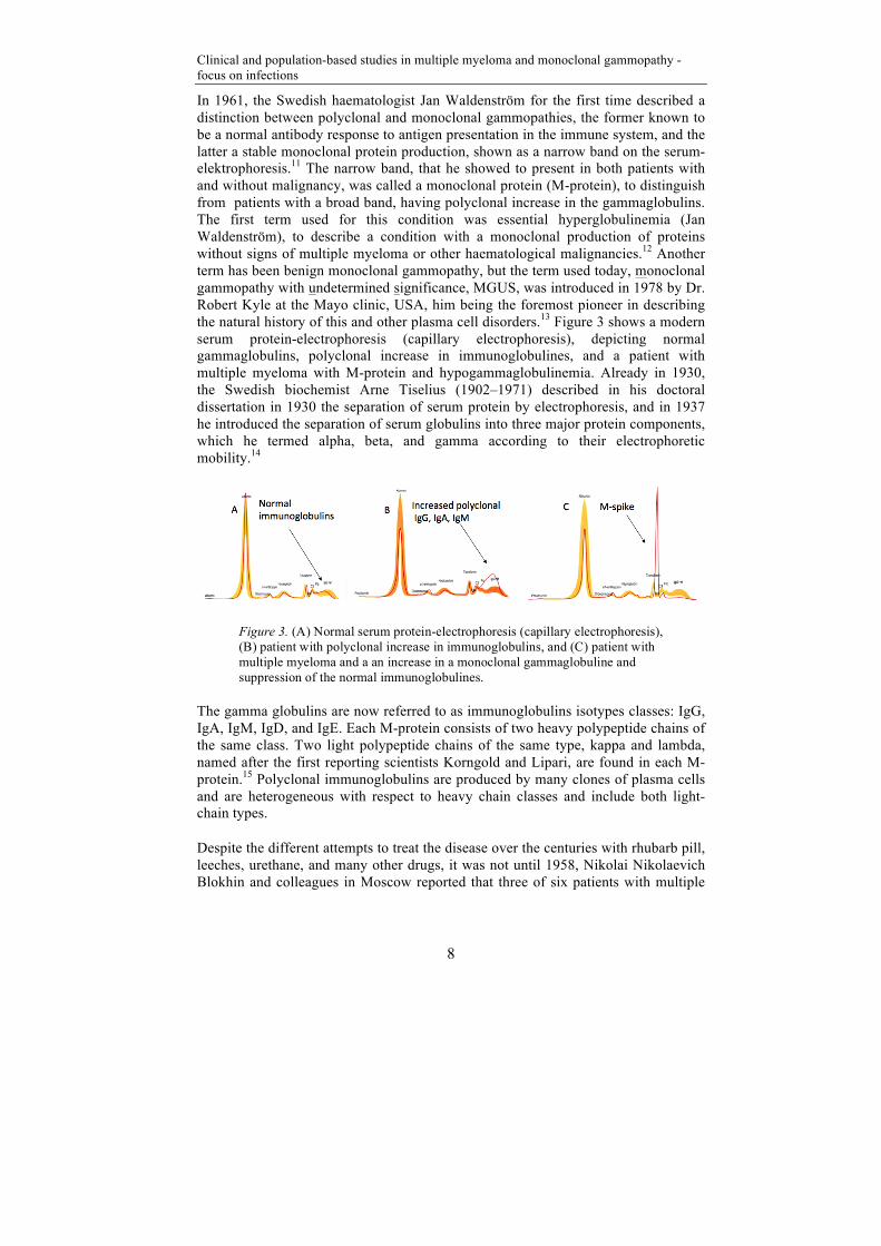
In 1961, the Swedish haematologist Jan Waldenström for the first time described a distinction between polyclonal and monoclonal gammopathies, the former known to be a normal antibody response to antigen presentation in the immune system, and the latter a stable monoclonal protein production, shown as a narrow band on the serum-elektrophoresis.11 The narrow band, that he showed to present in both patients with and without malignancy, was called a monoclonal protein (M-protein), to distinguish from patients with a broad band, having polyclonal increase in the gammaglobulins. The first term used for this condition was essential hyperglobulinemia (Jan Waldenström), to describe a condition with a monoclonal production of proteins without signs of multiple myeloma or other haematological malignancies.12 Another term has been benign monoclonal gammopathy, but the term used today, monoclonal gammopathy with undetermined significance, MGUS, was introduced in 1978 by Dr. Robert Kyle at the Mayo clinic, USA, him being the foremost pioneer in describing the natural history of this and other plasma cell disorders.13 Figure 3 shows a modern serum protein-electrophoresis (capillary electrophoresis), depicting normal gammaglobulins, polyclonal increase in immunoglobulines, and a patient with multiple myeloma with M-protein and hypogammaglobulinemia. Already in 1930, the Swedish biochemist Arne Tiselius (1902–1971) described in his doctoral dissertation in 1930 the separation of serum protein by electrophoresis, and in 1937 he introduced the separation of serum globulins into three major protein components, which he termed alpha, beta, and gamma according to their electrophoretic mobility.14
Figure 3. (A) Normal serum protein-electrophoresis (capillary electrophoresis), (B) patient with polyclonal increase in immunoglobulins, and (C) patient with multiple myeloma and a an increase in a monoclonal gammaglobuline and suppression of the normal immunoglobulines.
The gamma globulins are now referred to as immunoglobulins isotypes classes: IgG, IgA, IgM, IgD, and IgE. Each M-protein consists of two heavy polypeptide chains of the same class. Two light polypeptide chains of the same type, kappa and lambda, named after the first reporting scientists Korngold and Lipari, are found in each M-protein.15 Polyclonal immunoglobulins are produced by many clones of plasma cells and are heterogeneous with respect to heavy chain classes and include both light-chain types.
Despite the different attempts to treat the disease over the centuries with rhubarb pill, leeches, urethane, and many other drugs, it was not until 1958, Nikolai Nikolaevich Blokhin and colleagues in Moscow reported that three of six patients with multiple

Cecilie Hveding Blimark
9
myeloma obtained benefit from sacrolysin (L-phenylalanine mustard, melphalan) (Blokhin et al. 1958).16 Melphalan has together with prednisone been used treating multiple myeloma patients ever since, orally administered melphalan and prednisone (MP) being the most dominating drugs treating to Swedish multiple myeloma patients in the years 1970-2000.17 Alkylating agents are still the backbone of multiple myeloma first line treatment. This is now, in the years after 2000, for the first time challenged by other drugs, like the immunmodulatory drugs proteasome inhibitors and IMiDs.
Summarizing the Swedish treatment strategies in multiple myeloma; up to 1995, most patients were treated with alkylating agents and steroids. After 1995, high-dose melphalan and ASCT was recommended for all patients under 60-65 years of age.17 In studies from the Nordic Myeloma Study Group (NMSG), between 65% and 75% of all eligible patients below 60-66 years were included in studies involving high-dose melphalan (HDM) and ASCT in 1994-2003.18,19 In the Swedish Myeloma registry, recording population-based and clinical data from 2008, 81% of patients 65 years and younger and 4% of patients older than 65 years had undergone HDM-ASCT.20 The novel agents, primarily thalidomide, were used predominantly in Sweden after the year 2000. For elderly patients the most common first line treatment was MP until 2002, when NMSG introduced MP plus thalidomide in a randomized study.17 Bortezomib was approved in Sweden in the year 2004.

Clinical and population-based studies in multiple myeloma and monoclonal gammopathy -focus on infections
10
1.2 Multiple myeloma
1.2.1 Definition
In an early stage, multiple myeloma can be asymptomatic (“smouldering”), and can stay so for years. The majority of patients with multiple myeloma presents with symptoms and are in need of immediate treatment..
Table 1. Definition of multiple myeloma according to the International Myeloma Working Group (IMWG) 2014 21
Smouldering multiple myeloma Multiple myeloma
Both criteria must be met:
Serum monoclonal protein (IgG or IgA) > 30g/L or urinary M-protein >500mg/24h and/or Clonal bone marrow plasma cells 10-60 % Absence of myeloma defining events or amyloidosis
Clonal bone marrow plasma cells ≥ 10% or extramedullary plasmocytoma and
Any one or more myeloma defining events (evidence of end-organ damage attributed to the underlying plasma cell disorder)
or
Any one or more biomarkers of malignancy
Myeloma defining events: • Hypercalcemia: ≥0.25 mmol/L above upper limit or≥ 2.75 mmol/L • Renal insufficiency: creatinine clearencee< 40 mL/min or
s -creatinine > 177 µmol/L • Anemia: haemoglobin 20 g/L or more below normal or < 100g/L • Bone lesions: one or more osteolytic lesionon skeletal x-ray, CT or PET-CT
(Positron emission tomography-computed tomography) Biomarkers of malignancy
• Clonal bone marrow plasma cells ≥60% • Serum FLC-ratio ≥100 • > 1 focal lesion on MRI
The diagnostic criteria of multiple myeloma require the presence of at least 10 % plasma cells on examination of the bone marrow (or biopsy of a tissue with monoclonal plasma cells), and evidence of end-organ damage.21 The myeloma defining events include hypercalcemia, renal, insufficiency, anemia and bone lesions. (Table 1). New in 2014 IMWG criteria`s is the addition biomarkers of malignancy, in studies predicting an imminent onset of symptomatic disease.22-26 The biomarkers of malignancy include clonal bone marrow plasma cells above 60%, Free light-chains (FLC) ratio ≥ 100 and one or more focal lesion on magnetic resonance imaging (MRI) studies. The differential diagnosis includes MGUS, , primary amyloidosis,

Cecilie Hveding Blimark
11
solitary plasmacytoma, low-grade lymphoma, chronic lymphocytic leukemia (CLL), and metastatic carcinoma.
1.2.2 Epidemiology
Multiple myeloma is the second most common haematological malignancy after lymphoma and stands for 1% of all cancer and 13% of all haematological cancer.27 In Sweden, the incidence is 6.8 new cases per 100 000 inhabitants and year.28 The incidence and prevalence of multiple myeloma increase with age; the annual age-adjusted incidence rises from < 1/100,000 for subjects younger than 40 years, to > 40/100,000 for those older than 80 years; the annual prevalence of multiple myeloma in patients aged 65–74 is approximately 31/100,000 and rises to 46/100,000 in patients aged older than 75 years. Both the incidence and prevalence of multiple myeloma in elderly patients are expected to grow in the next future due to the increase in the life expectancy of the general population and the improved survival times achieved with the introduction of novel agents.29 The median age at diagnosis is approximately 70 years, and at diagnosis, 37% are below 65 years, 26% are between 65 and 74 years, and 37% are 75 years and older.30 Multiple myeloma is twice as common in African-Americans compared to caucasians and slightly more common in males than females.4,31
1.2.3 Clinical features
The most common symptoms on presentation are fatigue, bone pain, and recurrent infections.4 Bone pain, due to ostolytic lesions or compression fracture, especially in the spine and chest is present in two third of patients at diagnosis. This is caused by the expansion of malignant plasma cells in the bone marrow, activation of osteoclasts and the inhibition of osteoblast by the myeloma cell.4,32 Hypercalcemia is seen in 25 % of patients at presentation and is a result of bone resorbtion and can lead to acute confusion, dehydration, and coma. The infiltration of the bone marrow induces anemia, neutropenia and thrombocytopenia. Anemia, most often normochrom and normocytic, is common, present in approximately 70% of patients4,20,33 Neutropenia increases the risk of infections and thrombocytopenia the risk of bleeding. The excretion of light chains has a toxic effect on the distal tubulus of the kidneys, and kidney failure is present in approximately 20-25% of patients at diagnosis and can lead to acute need of dialysis.34 Absence of renal function recovery is associated with a worse prognosis. Hypercalcemia, dehydration, infections, non-steroidal anti-inflammatory drugs (NSAIDs), contrast dye for imaging, and bisphosphonates can contribute to the renal failure. In the rare cases of a very high M-component, hyperviscosity syndrome can be seen. Recurrent infections represent a clinical problem in myeloma patients, and 75% of patients are expected to have a serious infection in the course of the disease.35
1.2.4 Treatment and prognosis
In the last 20 years, the spectrum of treatment options in myeloma patients has changed dramatically and many new treatment modalities have been introduced. With new and more effective treatments, both up front and at relapse, patients can

Clinical and population-based studies in multiple myeloma and monoclonal gammopathy -focus on infections
12
now enjoy long periods of remission and the total survival has increased, especially in younger patients.36,37 After the introduction of high dose melphalan with autologous transplantation (ASCT) for patients younger than 65-70 years (ca 1995) and the introduction of new immunomodulatory drugs, this development has become clear. Thalidomide, lenalidomide and pomalidomide belong to the class of IMiDs, immunomodulatory drugs with antiangiogenic properties. Studies on thalidomide and lenalidomide have reported effect in multiple myeloma patients both up front and at relapse.38-43 Pomalidomide has shown effect in relapsed and refractory patients.44 Proteasome inhibitors, like bortezomib, induce cellular apoptosis with malignant, transformed, and proliferating cells being particularly susceptible. Bortezomib alone and in combinations with dexamethasone and conventional chemotherapeutic drugs is proven to be effective in both relapsed and up-front multiple myeloma patients.45-48 In Sweden, thalidomide was registered 2002, bortezomib 2004, lenalidomid 2008, and pomalidomide 2013 for the treatment of multiple myeloma. In years to come, new proteasome inhibitors, especially carfilzomib, monoclonal antibodies, cell cycle-specific drugs, deacetylase inhibitors and many other drugs will play a great role in the continued treatment of multiple myeloma patients.49
Indication for treatment, Myeloma defining events In multiple myeloma, the standard of care has been not to treat until progression to symptomatic disease occurs. In 2003, IMWG introduced the CRAB criterias suggesting which myeloma-related symptoms should indicate treatment.50 These were; Calcium levels increased 0.25 mmol/L (1mg/dL) above the upper limit or > 2.75 mmol/L (<11mg/dL), Renal insuffiency: creatinine > 173 mmo/l, Anaemia: hemoglobin 2 g/dl below the lower limit or Hb < 10 g/dl, Bone lesion: lytic lesion or osteoporosis with compression fractures. Other symptoms indicating treatment were hyperviscosity, amyloidosis, and recurrent infections (> 2 episodes in 12 months). 4 In the revised 2014 IMWG criterias, the indication to treat has been expanded to involve multiple myeloma defining events and biomarkers of malignancy (Table 1).21 This change in definitions and indications will eventually impact multiple myeloma survival estimates and comparisons in survival over time.
Treatment in younger patients (<65-70 years) The standard treatment for patients up to biological age of 65 years is still high dose melphalan with ASCT. This is normally preceded by cycles of tumour reducing induction treatment. As the cells being infused in the patient are the patients own, collected after in vivo purging with induction chemotherapy, this is not really a transplantation. Instead, this is high dose chemotherapy followed by autologous stem cell infusion enabling the patient to overcome the dose of melphalan which otherwise would be lethal due to the bone marrow toxicity.
Autologous transplantation In Sweden, the standard induction treatment preceding ASCT is 3 to 4 cycles of a chemotherapy combination, after 2008 with the addition of any of the new drugs, mainly bortezomib. Cyclophosphamide (2 g/m2) is given after induction and then granulocyte colony stimulation factor (G-CSF) is injected for 5-7 days to stimulate and release stem cells from the bone marrow. CD 34-positive and mononuclear cells

Cecilie Hveding Blimark
13
are collected and stored frozen. At time of transplantation, at least two million CD 34 pos cells/kg body weight are given back as a stem cell rescue two days after high dose melphalan. This procedure is performed at all university hospitals. The high dose chemotherapy causes mucocitis, severe cytopenia and, often, febrile neutropenia. Because of the toxicity, the patients normally need to stay two to three weeks in hospital.51 In the literature, the treatment-related mortality with this procedure is 2-5 %.18,51-54 Relevant to our study, according to the regional guidelines for Western Sweden from 1996, patients were treated with one or two initial ASCT with melphalan dose of 100-200 mg/m2. It was aimed at harvesting CD34-positive cells (stem cells) for at least 2 ASCTs, and some patients performed 2-3 Mel 100 at relapse.
Treatment in elderly patients (>65-70 years) Patients who are not eligble for ASCT are treated with combinations of chemotherapy and any of the new drugs. The combinations with best support in the literature are MPV (MP + Velcade (bortezomib)), MPT (MP+ thalidomide) and Rd (Revlimid (lenalidomide) + dexamethasone). 40,4348 Each treatment cycle last 3-5 weeks, and the treatment is repeated for 6-8 cycles, bringing the total treatment time up to 6 months to one year. There is not convincing evidence to support high dose melphalan and ASCT in patients over the age of 65-70 years due to treatment toxicity.
Salvage at relapse after high dose treatment Despite the advances made in the treatment of this disease, multiple myeloma remains essentially incurable by the current therapy and continues to represent the haematological malignancy with the worst outcome. Multiple myeloma patients have an overall survival (OS) of only 4–5 years17,55 and the vast majority of patients will eventually relapse after initial treatment and require some form of salvage therapy.56 One would argue, that safety, tolerability of the relapse treatment , and quality of life is more important in patients where one cannot offer a cure. HDM + ASCT has increased progression-free survival (PFS) and OS in patients 65 years and younger in several randomized trials compared to conventional treatment up front.18,51,52 However, it is not a curative approach and the patients relapse in median after 2-3 years and are in need of salvage treatment. There is no consensus in the choice and order of the different treatment strategies at relapse; whether patients should receive chemotherapy or novel drugs or repeat the high dose treatment. The current guidelines state, that if the patient responds well to the intial ASCT, it is advised to repeat the initial treatment at relapse.57-59 At Sahlgrenska University Hospital, when possible, we have collected at least 4 million CD 34 pos cells per kg body weight, reserving 2 million for later use.
Salvage ASCT and prognostic factors for survival after high dose treatment Factor predictive of a worse prognosis is high ß-2-microglobulin and low serum albumin, resulting in the Staging system of IMWG. Stage 3 with ß-2-microglobulin of >5.5 µg/mL has a median overall survival of only 29 months.60 Other risk factors associated with a worse prognosis are older age, male sex, and high risk cytogenetic aberrations t(4;14)61, t(14;16), del17p and 1q in FISH-analyses.62 High-risk disease

Clinical and population-based studies in multiple myeloma and monoclonal gammopathy -focus on infections
14
accounts for about 25% of patients with symptomatic multiple myeloma.63 One of the strongest factors for survival after high dose therapy is the time to progression (TTP); that is, the length of the first remission phase. There is data to support that patients progressing 12-18 months after the high dose therapy have a shorter survival, than patients with a longer TTP.64,65 Repeating the initial Mel 200 with ASCT at relapse has been a recommendation if the patient tolerated the initial ASCT and Atanacovic et al. 2012 reviewed all single centre reports of salvage ASCT (mostly Mel 200), and found a median overall response rate (ORR) of 65 %, a median PFS of 12 months and a median OS of 32 months approximately with similar toxicity profile as the first ASCT but a median transplant related mortality (TRM) of approximately 4 %.66 Another report found a considerable nephrotoxicity at the salvage ASCT.67 In most studies there was a cut off TTP where the OS was significantly better after salvage ASCT, in median a TTP of 19 months in Atanacovic´s study, and as mentioned, most did not recommend a second transplant if TTP after the first ASCT was less than 6-12 months. Data on lower doses of Melphalan + ASCT is scarce and this procedure is mainy tested on few patients in advanced relapsed MM patients.68,69

Cecilie Hveding Blimark
15
1.3 Smouldering multiple myeloma
1.3.1 Definition and epidemiology
Smouldering multiple myeloma differs from MGUS in form of a higher degree of bone marrow infiltration, often reflected in a higher M-protein. It accounts for approximately 10-15 % of all newly diagnosed multiple myelomas, and the median time to progression to a symptomatic multiple myeloma ranges from 2 to 3 years.70-73 There should be no symptoms or sign of symptomatic multiple myeloma, including absence of skeletal lesions attributable to multiple myeloma in a whole body skeletal survey (Table 1).21
1.3.2 Treatment and prognosis
The risk of progressing to symptomatic multiple myeloma is approximately 10 % per year, but the risk varies depending on different risk factors for progression. Considering that in the natural course of smouldering multiple myeloma, some patients can stay in the asymptomatic stage for up to 20 years, and many die of other causes, the gold standard up to date has been not to treat asymptomatic patients up front.71 Historically, many attempts have been made in exploring whether smouldering multiple myeloma would profit from up front treatment. Hjorth et al in NMSG treated 50 patients with smouldering multiple myeloma and and 50 patients with multiple myeloma both with MP and they did not find any difference in groups regarding response rate, response duration or survival.74 They have been succeded by numerous colleagues and trials, testing up front treatment with the same result over the years, none of which has resulted in change of the delayed treatment practice.75 Most early treatment studies on smouldering multiple myeloma have been performed on the whole cohort of smouldering patients. However, the risk of smouldering multiple myeloma progressing to multiple myeloma a related disorder is 10% per year for the first 5 years, 3% per year for the next 5 years and 1–2% per year for the next 10 years, finally resulting in a cumulative risk of progression for all patients of 73 % at 15 years.70 Capturing features in patients rapidly progressing from smouldering multiple myeloma to active disease may eventually identify patients profiting from early treatment.
There is no consensus in the definition of high-risk smouldering multiple myeloma, and in the literature a number of factors contributing to progression have been suggested. It has been shown that the risk of progression is increased in cases with monoclonal protein levels of greater than 30 g/L, IgA isotype, Bence-Jones protein excretion (urinary light-chain) greater than 50 mg/24 hours, evolving smouldering multiple myeloma type, greater than 10 % of plasma cells in the bone marrow, and occult bone lesions on magnetic resonance imaging (MRI).22,70,73,76-78

Clinical and population-based studies in multiple myeloma and monoclonal gammopathy -focus on infections
16
At least 2 different risk models for progression have been proposed based on multivariate analysis and Cox proportional hazard models of different factors of progression. In 2007 Kyle70 presented 3 different risk groups based on presence or absence of the following 2 risk factors analyzed in 276 smouldering multiple myeloma patients; bone marrow plasma cells ≥10% and ≥30 g/l monoclonal protein. For the 106 patients with both factors, the 5-year cumulative probability of progression was 69% at 5 years, for patients with one factor and 0 factors the risk was 43% and 15 % respectively. (Figure 3)
Figure 4. MAYO CLINIC MODEL Cumulative probability of progression from smouldering myeloma to symptomatic disease depending on 0, 1 or 2 risk factors for progression, the risk factors being ≥ 30g/l M-protein and ≥ 10% plasma cells70
The Spanish PETHEMA-group (Programa de Estudio y Tratamiento de las Hemopatías Malignas) presented in 2007 a risk score based on 2 independent risk factors for progression from smouldering to multiple myeloma based on as study of 93 smouldering multiple myeloma patients72. The first risk factor was positive multiparametric flowcytometry based on 4 antbodies applied to identify plasma cells among all mononuclear B cells as well as discrimination of phenotypically abnormal plasma cells from their normal counterpart. The antigens most frequently used for the identification of aberrant plasma cell phenotype include CD19, CD45, and CD56 in combination with CD38/CD138. Thus, the overexpression of CD56 together with the absence of reactivity for CD19 and for CD45 and/or decreased amounts of CD38 have been found to be common characteristics of multiple myeloma plasma cells. At a cut of > 95 % of aberrant plasma cell in the bone marrow, this was found to be a independent risk factor for progression. The other risk factor was hypogammaglobulinemia or immunparesis of the uninvolved gammaglobulin.
The score system for smouldering multiple myeloma was built on the basis of the percentage of immunophenotypically aberrant plasma cells within the bone marrow

Cecilie Hveding Blimark
17
compartment (≥ 95 % aberrant plasma cells, score of 0; ≥ 95%, score of 1) and the presence (score of 1) or absence (score of 0) of immunoparesis. In patients with a score of 1, the median time to progression (TTP) was not reached; in patients with a score of 2,the median TTP was 73 months; and in patients with ascore of 3, the median TTP was 23 months (P <.001). PFS at 5 years of 4%, 46%, and 72%, respectively; P < .001(N= 93 smouldering multiple myeloma patients) (Figure 5).
Figure 5. Spanish PETHEMA model. Risk score and TTP in smouldering multiple myeloma with a risk of progression to symptomatic disease at 5 years of 4 %, 46 %, and 72 %, respectively, for patients with none, 1, or 2 risk factors.72
Both the Mayo Clinic and Spanish PETHEMA models are retrospective, single-centre cohort studies. Recently new risk factors for progression have been added describing ultra-high-risk of progression. Rajkumar et al showed that 95 % of patients with more than 60 % plasma cells in bone marrow progressed within 2 years of diagnosis, with a median time to progression of 7 months.24 Larsen et al found that FLC ratio above 100 (kappa) or <0.01 (lambda) with a 72% risk of progressing to active disease within 2 years.23

Clinical and population-based studies in multiple myeloma and monoclonal gammopathy -focus on infections
18
Cherry and coworkers at NIH (National Institute of Health) published 2013 a prospective study designed to compare the 2 risk models for smouldering multiple myeloma of the Mayo clinic and the Spanish PETHEMA group.79 77 patients with smouldering multiple myeloma enrolled in their Smouldering Myeloma Natural History Study (NCT01109407) between 2010 and 2012, and the above risk scores were assigned according to criteria for each model. Only in 22/77 smouldering multiple myeloma patients overall, there was agreement between the two risk models (Table 2).
Table 2. Distribution of 77 patients with smouldering multiple myeloma between two clinical risk models to predict progression from smouldering to multiple myeloma.79
Spanish PETHEMA
low
Spanish PETHEMA intermediate
Spanish PETHEMA
high Mayo Clinic low 11 15 12 Mayo Clinic intermediate 6 7 22 Mayo Clinic high 0 0 4 Overall agreement 22/77 (28.6%) In 2013, Mateos and collegues from the PETHEMA group published a randomized early-treatment trial (RCT) with Len Dex compared to placebo on high-risk smouldering multiple myeloma patients.80 Patients in the treatment-arm received an induction regimen consisting of nine cycles of Lenalidomide +dex followed by a maintenance regimen for up to 2 years. This was the first RCT showing significant improvement in PFS and OS in smouldering multiple myeloma targeting patients with high-risk features. However, in this study they used a combination of the 2 above models to define high- risk smouldering multiple myeloma patients, and 40% of patients had high-risk according to the PETHEMA model above with > 95% aberrant plasma cells plus immunoparesis. 18% of patients were high risk according to the Mayo model and 42 % according to both models. With the proven discordance between risk models this study has caused discussion and has been difficult to interpret. Flow cytometry in multiple myeloma is not widespread as a diagnostic tool and there is still no consensus in what to consider high-risk smouldering multiple myeloma.
Several ongoing trials with interleukin-6 (IL-6) antibody, anti CS1 monoclonal antibody, the proteasome inhibitors bortezomib, ixazomib and carfilzomib, and ImiDs lenalidomide and thalidomide + zolendronate and others will hopefully finally answer this question in the future. The IMWG has defined smouldering myeloma patients with high risk of progression in the first 2 years to be candidates for chemoprevention trials.81 However, off-study, observation is still the standard even in this group.

Cecilie Hveding Blimark
19
1.4 Monoclonal Gammopathy of undetermined significance
(MGUS)
It is now known that virtually all cases of multiple myeloma are preceded by the condition monoclonal gammopathy with undetermined significance and the steps toward progression are not fully understood.3,82
1.4.1 Definition
In the updated 2014 IMWG diagnostic criteria, MGUS is defined as serum M-protein less than 30 g/L, clonal plasma cell population of < 10%, and absence of end-organ damage (CRAB criteria of multiple myeloma)21 This benign precursor condition can be classified in lymphoid (15%) or plasma cell (85%) -MGUS.83 IgG and IgA monoclonal gammopathy of undetermined significance are precursor conditions of multiple myeloma; light-chain monoclonal gammopathy of undetermined significance of light-chain multiple myeloma; and IgM monoclonal gammopathy of undetermined significance of Waldenström’s macroglobulinemia and other lymphoproliferative disorders.84,85 The discussion below will mainly concern non-IgM MGUS.
1.4.2 Epidemiology
MGUS is one of the most common premalignant disorders in western countries and the prevalence increases with age.86 It is present in 3.2 % of white persons >50 years and in 5% > 70 years of age.87 MGUS is more common in men than in women and 2-3 times more common in African-Americans. The prevalence among Japanese and Mexicans is lower than in Caucasians. Studies indicate that farmers exposed to pesticides and toxins have a higher risk of developing MGUS.85,86,88 A familial predisposition for plasma cell disorders is putative as it is observed a 2 to 3 -fold risk of MGUS in first degree relatives.89 Patients with immunosuppression or immunocompromized patients of other reasons have a higher prevalence.
1.4.3 Etiology and pathogenesis MGUS can arise from primary clonal plasma cell disorder or secondary to a immunological derangement, such as a serious infection, immunosuppression (eg. transplant recipients), rheumatologic, neurologic, hepatologic, endocrine or dermatologic diseases. There is evidence to support a role in genetic factors. There is familial aggregation with a 2-fold overrisk in 1st degree relatives of multiple myeloma patients to develop multiple myeloma90 and first degree relatives of multiple myeloma patients have a 2-fold risk of developing MGUS.88 Further, first-degree relatives of MGUS patients have a 2.8 fold risk of developing MGUS, 3-fold for multiple myeloma, a 4-fold risk of lymphoplasmacytic lymphoma (LPL)/Waldenstrom macroglobulinemia (WM) , and 3.4-fold risk of chronic lymphocytic leukemia (CLL).89 In addition, racial disparities in the development of MGUS91 and familial aggregation of solid tumors in patiens with multiple myeloma and MGUS support this hypothesis.31,92 Over the last three decades, there has been consistent evidence from population-based case-control
Clinical and population-based studies in multiple myeloma and monoclonal gammopathy -focus on infections
20
and cohort studies that certain autoimmune diseases, especially rheumatoid arthritis, Sjögren’s syndrome, and systemic lupus erythematosus, are associated with lymphoproliferative diseases.93-95 Possible explanations for these associations include the role of chronic immune stimulation, treatment for autoimmune disease, and shared genetic and/or environmental factors. Recent studies suggest that chronic antigenic stimulation also plays a role in the causation of plasma cell disorders. In a study of 4641 US veterans there was an association to several autoimmune diseases and overall the risk of developing multiple myeloma and MGUS, as a sign of a disrupted immune system causing both conditions or a chronic immune stimulation as trigger in the pathway to MGUS. In a large population-based study from Sweden, the autoimmune diseases polymyalgia rheumatica, and pernicious anemia were associated with increased risk of multiple myeloma.90,96
Multiple myeloma evolves from MGUS via smouldering multiple myeloma to symptomatic multiple myeloma. Many of the clonal abnormalities found in multiple myeloma can be found in MGUS, indicating that there is a genetic susceptibility developing MGUS and later multiple myeloma.83 Plasma cells are characterized by strong bone marrow dependence and extensive somatic hypermutation of Ig genes. The pathogenesis in developing MGUS can briefly be summarized as follows; early, partially over-lapping genetic events common to MGUS and multiple myeloma include at a minimum primary IgH translocations, hyperdiploidy, and del 13 that lead directly or indirectly to dysregulation of a CCND gene.97 Approximately 50 percent of MGUS patients have translocations that involve the immunoglobulin heavy-chain locus, the immunoglobulin switch region on chromosome 14q32 and one of five partner chromosomes, 11q13 (CCND1) (the most common), 4p16.3 (FGFR-3 and MMSET), 6p21 (CCND3), 16q23 (c-maf), and 20q11 (mafB). These and other cytogenetic changes are thought to play an important role in the evolution of MGUS. As the breakpoints usually occur near or within IgH switch regions, it seems likely that the translocations are related to errors in class switch recombination or somatic hypermutation, as normal B cells pass through the germinal centre.
Progression to multiple myeloma The transition from MGUS to MM is associated with increased MYC expression and sometimes KRAS mutations, but can also include del 13 in t(11;14) tumours. Finally, further progression of the multiple myeloma tumour seems to be associated with other events. For example, increased proliferation and genomic instability, and decreased dependence on the bone marrow microenvironment, sometimes including extramedullary spread of disease, can be associated with late MYC rearrangements that often involve an Ig locus, activating mutations of the NF-kappa B pathway, deletion or mutation of TP53, and inactivation of p18INK4c or RB1. Deletion of 17 p and p53 mutation and loss and gain of 1q are regarded later events that predicts for a worse outcome in multiple myeloma.83 Another genetic risk factor for progression to myelomatosis is hypodiploidy, a risk factor for poor outcome.98 Changes also occur in the bone marrow microenvironment, including the induction of angiogenesis, the suppression of cell-mediated immunity, and the development of paracrine signalling loops involving cytokines such as IL-6 and vascular endothelial growth factor, finally leading to bone disease.32

Cecilie Hveding Blimark
21
1.4.4 Clinical features
MGUS patients often present with a high sedimentation rate due to the molecular weight of the protein, giving rise to suspicion of a serious inflammatory or malignant disease. Although MGUS patients are defined as asymptomatic in respect to the plasma cell disorder, they have increased morbidity and mortality compared to the general population.99 There is gathering evidence that MGUS patients have a higher morbidity in osteoporosis, hypercalcemia, hip and vertebra fractures, and thromboses, possibly linked to the genetic aberrations found in MGUS that involve the bone marrow compartment and angiogenesis.100,101 Polyneuropathy is prevalent in 5 % of MGUS cases, MGUS is also associated with rare skin disorders and sometimes the M-proteins have cold-agglutinine qualities, causing cold-agglutinin-syndrome with haemolysis.2 In some MGUS patients, there is a clinically significant hypogammaglobulinemia, and MGUS patients with recurrent serious febrile infections, typically of the respiratory tract, might require infection prophylaxis, such as vaccines and monthly gammaglobulin infusions. 102
1.4.5 Prognosis
The risk in MGUS patients of progressing to multiple myeloma or other lymphoproliferiative diseases (lymphoma, Mb Waldenström, amyloidosis) is 1% per year and 12% in 10 years, 25%, in 20 years and 30% in 25 years.103 The risk of progression is dependent on different risk factors. Cesana et al reported following risk factors for progression; bone marrow plasmocytosis > 5%, detectable Bence-Jones proteinuria, polyclonal serum immunoglobulin reduction and high sedimentation rate (ESR).77 Further, Turesson found following three factors for progression in 728 Swedish MGUS patients; abnormal free light-chain (FLC) ratio (<0.26 or >1.65), M-protein concentration (≥15 g/L), and reduction of 1 or 2 noninvolved immunoglobulin isotype levels (immunoparesis).104

Clinical and population-based studies in multiple myeloma and monoclonal gammopathy -focus on infections
22
Rajkumar et al. developed a risk-stratification model for progression of MGUS.78 Patients with risk factors consisting of a serum M protein <15 g/L, IgA or IgM MGUS and an abnormal serum FLC ratio had a risk of progression at 20 years of 58 %; compared with 37% when two risk factors were present; 21% when one risk factor was present; and only 5% when none of the risk factors were present (Figure 6). Patients with MGUS and smouldering multiple myeloma require indefinite follow-up given their life-long risk of progression to multiple myeloma or related malignancy.
Figure 6. Three risk factors in progression from MGUS to multiple myeloma or related disorders.78
Kristinsson et al showed on a material of over 4000 MGUS patients collected from Swedish hospital registries that MGUS patients have a poorer survival than the general population. MGUS patients had an increased risk of dying from myeloid malignancies, bacterial infections, heart diseases, liver disorders, and renal diseases. More specifically, MGUS patients had an excess risk of dying in lymphoproliferative malignancies (HR 54; CI 31-92), but also in bacterial infections (HR 3.4; CI1.7-6.7) compared to controls.30 The finding that patients diagnosed below the age of 60 have a 35 % risk of dying from a haematological disease, and that MGUS patients diagnosed in older age die mostly from heart-and other diseases have implications on the management of MGUS patients and support a risk-adapted strategy for follow-up and intervention in patients with this disease.

Cecilie Hveding Blimark
23
1.5 Infections in plasma cell disorders MGUS and multiple myeloma patients have an increased risk of infections, and in multiple myeloma patients infection is known to be an important cause of death105,106. Kristinsson et al has earlier showed that MGUS patients have an increased 3.4-fold risk of dying in infections compared to controls.30 Elderly patients without haematological diseases are also known to have an increased risk of infections compared to younger patients due to features more common in the elderly, such as comorbidity, immobility and the in age reduced function of the immune system.107,108 However, there is no population-based data on how common infections are in MGUS and multiple myeloma patients compared to an age-matched normal elderly population.
1.5.1 Inherent immunodeficiency
The multiple myeloma-related immunodeficiencies involve B-cell dysfunction, like hypogammaglobulinemia, as well as T-cell-, dendritic cell-, and NK-cell abnormalities.109 Secondary hypogammaglobulenemia is reported to be present in about 25-40 % of MGUS 110 and multiple myeloma patients 111,112 whereas a reduction of one or more polyclonal immunoglobulins is seen in more than 90% of patients with myeloma.4 Hypogammaglobulinemia is known to increase the risk of life threatenting infections especially caused by encapsulated bacterias. Streptococcus pneumoniae, Haemophilus influenzae and Escherichia coli are the most frequent causes of infection in myeloma patients.113 114
The risk of infections among patients with MGUS has not been studied in great detail. Gregersen et al. analyzed risk of bacteremia in 1,237 MGUS patients in Denmark diagnosed from 1981 to 1993. Based on 40 episodes of bacteremia, there was a 2.2-fold increase in risk compared to the general population.115 In another study based on screening data from Olmsted County in Minnesota, risks of several different diseases, including some infectious disorders, were analyzed among 605 MGUS patients and compared to 16,793 controls.116 An increased risk of upper respiratory bacterial infection, spontaneous bacterial peritonitis, and mycobacterium infection was found.
In 1982, Savage illustrated based on 75 infections in 57 multiple myeloma patients that infections with Streptococcus pneumoniae and Haemophilus influenzae occurred at presentation and gram-negative bacilli and Staphylococcus aureus were responsible for 80% of infections after diagnosis and 92% of deaths from infection.113 Other studies have suggested that infections occur more often in the first 6 months following diagnosis, in active disease, and that the risk decreases with response to treatment (supported by normalization of hypogammaglobulinemia).117-
119
Advanced age, comorbidities and reduced mobility due to skeletal disease contribute to the risk of infections. Other factors are renal failure (cast nephropathy and others), respiratory compromise, caused by collapse of thoracic vertebra, and opiate therapy (which may depress the central nervous system) given to patients with painful

Clinical and population-based studies in multiple myeloma and monoclonal gammopathy -focus on infections
24
fractures, the multisystem involvement by myeloma associated deposition diseases (AL-amyloidosis and light-chain deposit disease).
Interestingly, Augustson et al found a correlation between thoracic pain and the risk of early death in e.g pneumonia, supporting that immobility and restricted respiratory ability in patients with skeletal disease is contributing to the risk of infections.119 It has also been shown that multiple myeloma patients display a low immune response to infections and vaccines, and that it also predicted a higher risk of infection.120,121
1.5.2 Infection as complication to treatment
The recent advances in treatment have prolonged life in remission and in relapse phase in multiple myeloma. However, managing multiple relapses and salvage therapies can lead to a cumulative immunosuppression and a higher risk of infections.
Schütt et al. analyzed 480 blood samples in 77 multiple myeloma patients going through different types of treatment. They could see that untreated myeloma patients exhibited significantly reduced T-cell, B-cell, and natural killer cells, as well as non-myeloma IgA, IgG and IgM. Conventional-dose chemotherapy resulted in significantly reduced CD4+ and even further decline of T-cells and B-cells cells, most notably in relapsed patients. Following ASCT, prolonged immunosuppression and opportunistic infections with Pneumocystis jirovecii, Cytomegalovirus and Clostridium difficile is observed.122 Other risk factors of infection in ASCT patients include the conditioning regimen (inducing mucocitis), duration of neutropenia, renal failure, iron overload, and smoking.106
Much attention has been drawn to the changing spectrum of infections in multiple myeloma, possibly related to the more intensive treatment in recent years and new immunomodulatory drugs.123,124 There has been some concern, as to whether more intense treatment may increase the risk of infections in patients. Chanan-Khan et al found a significant increased incidence of Herpes zoster in bortezomib treated patients compared to Dex-arm (13 vs 5%)124 in the APEX study with 663 patients with routine acyclovir prophylaxis; Offidani et al found that of 202 patients treated with thalidomide, 19% developed severe infections early.125 Augustson et al. could in a study on over 3000 patients in MRC studies show that of the 10 % of patients in their study that died within 6 months of diagnosis, 45 % of the patients died from infections.119 Nucci et al has looked at RCT studies with new drugs, finding that lenalidomide patients suffer from infections twice as often as patients treated with dexamethasone (Dex).106 Even in a study on smouldering multiple myeloma given lenalidomide and Dex, infection was the most important non-haematologic complication.80
Treatment with the new immunomodulating drugs are also increasing the risk of other opportunistic virus and fungal infections.106 Both chemotherapy, radiation and Graft- versus Host-disease after allogeneic transplantation can cause severe alimentary mucosal damage123, hyperglycemia induced by dexamethasone43. Transfusional iron overload can also increase the risk of infections.126 However, most of these hypotheses rely on small studies or studies on selected patients and a

Cecilie Hveding Blimark
25
population-based overview on the risk of different infections compared to the general population is not to be found in the literature.
1.5.3 Prophylaxis and treatment of infections
Prophylaxis In the British guidelines for infection prophylaxis in plasma cell disorders127; multiple myeloma patients with 3 or more febrile infections per year and a coexcisting hypogammaglobulinemia are recommended intravenous gammaglobulins (IVIG) as empirical treatment. However, the role of prophylactic immunoglobulin needs to be established as the rationale for its use is based on one randomized trial in multiple myeloma plateau phase.128 The support for IVIG in MGUS is scarce, and the patients are evaluated for response based on their infections. IVIG therapy is costly, and it has been estimated that six million US dollars would be needed to achieve 1 quality adjusted life year without an increase in life expectancy in patients with CLL129, and therefore IVIG should be limited to patients with immunoglobulin G-levels <500 mg/dL who suffer recurrent infection despite appropriate antimicrobial prophylaxis. According to current guidelines127, selected multiple myeloma patients with recurrent bacterial infections and other comorbidity (e.g. chron bronchitis, lung disease) patients can receive prophylactic antibiotics. In multiple myeloma, some effort has been made in testing prophylactic antibiotic treatment the two first months of treatment. In the most recent study, Vesole et al. performed a RCT130, including 212 multiple myeloma patients, and found no decrease in serious bacterial infections when comparing patients receiving ciprofloxacin, trimethoprim-sulfamethoxazole, or observation only. In their study, they did not include patients treated with novel agents and the study analyzed only infections during the first two months, and thus only included the pre-ASCT period.
In patients receiving chemotherapy or immunosuppressive treatments causing neutropenia, G-CSF can be given in short intervals. It is known to shorten the neutropenic period and reduce time spent in hospital, but does not reduce mortality.131 Patients receiving high-dose cortisone are given trimethoprim-sulfamethoxazole prophylaxis protecting against pneumocystis jirovecii and anti-viral prophylaxis against varicella zoster (VZV). Because of the immunosuppressive effect of high-dose melphalan, it is standard to give multiple myeloma patients after ASCT antiviral- and pneumocystis prophylaxis orally for 6 to 12 months after the procedure. Numerous studies have evaluated the effect of prophylactic vaccines in multiple myeloma patients. Only against Haemophilus influenza the vaccine is proven to provide antibody-titers comparable to the normal population132 However, the current recommendation for multiple myeloma patients receiving treatment is a combination of pneumococcal, haemophilus- and influenza vaccine prophylaxis, and prophylaxis of influenza even in their household members before season. Following ASCT, revaccination is recommended according to local guidelines, repeating most childhood vaccines. Vaccination against VZV is currently tested in several studies.

Clinical and population-based studies in multiple myeloma and monoclonal gammopathy -focus on infections
26
Treatment of infections Recognition and treatment of infections in multiple myeloma represents a challenge. The inherent immunodeficiency of the disease, and cytotoxic treatment for multiple relapses together with immobility and renal insuffiency represent a cumulative risk of infections and 75 % of patients will experience an infection during the course of the disease.35,114 Nearly all treatments for multiple myeloma contain glucocorticosteroids, a drug that can mask rising temperature and increase the blood glucose, another risk factor for infection. Augustson et al reported, that patients, receiving antbiotics orally at home, creating a delay of hospital care, had a higher risk of dying in infections.119 This supports swift assessment and admission to hospital if needed in febrile multiple myeloma patients. Historically, pneumonia, urinary tract infections and bacteremia are reported to be the most common infections, and are typically caused by Streptococcus pneumoniae, Staphylococcus aureus, and Escherichia coli in patients with conventional treatment.113,133,134 Patients in treatment are advised to have antibiotics (floroquinolones or amoxicillin) at home for empirical use by fever with mild symptoms and respiratory tract infections. Broad spectrum antibiotics and admission to hospital is preferred in febrile neutropenia.127 When dexamethasone is used, infection caused by a depression of cell-mediated immunity is more likely to occur, including mucosal candidiasis, herpes zoster virus (HSV) or VZV infection, and others and treatment with antifungal and antiviral medication is necessary. MGUS and multiple myeloma patients have an increased risk of arterial and venous thromboses and this differential diagnosis must be ruled out.135,136 In figure 7, Nucci et al have proposed a management strategy in myeloma patients with infections.106

Cecilie Hveding Blimark
27
Figure 7. Suggested management of suspected infection in multiple myeloma106 (ATB: antibiotics, FQ: flourokinolones, PE: pulmononary embolism)

Clinical and population-based studies in multiple myeloma and monoclonal gammopathy -focus on infections
28

Cecilie Hveding Blimark
29
2 AIMS
The overall aims of this thesis is to evaluate the risk of infections in patients with plasmacell disorders and increase the awareness of this treatable complication and find new less toxic treatments and hopefully contribute to improvement in the morbidity and mortality of multiple myeloma patients. Further to contribute to identifying patients with smouldering multiple myeloma and high risk for progression who are candidates for early treatment trials.
The specific aims of the work underlying this thesis are:
To describe the toxicity, feasibility and efficacy of MEL 100 with stem cell support with special focus of risk of infection in a cohort of relapsed multiple myeloma patients
To assess the risk of infections in multiple myeloma and MGUS patients compared to matched controls in a population-based study
To study the risk of specific infectious diseases in multiple myeloma and MGUS patients
To assess the risk of infection-related death in multiple myeloma patients
To assess whether the changes in treatment strategies in multiple myeloma patients over time has affected the risk of infections and infection-related deaths.
To estimate the incidence of patients with asymptomatic or smouldering multiple myeloma in a population-based cohort
To identify the proportion of smouldering multiple myeloma patients with high-risk features and their risk of progressing to symptomatic multiple myeloma

Clinical and population-based studies in multiple myeloma and monoclonal gammopathy -focus on infections
30
3 MINI-TRANSPLANTATION IN MULTIPLE
MYELOMA (I)
Previously, it has been shown that retreatment with high-dose melphalan (200 mg⁄ m2) and ASCT after first disease progression can be beneficial64,137,138, and current guidelines recommend retreatment with high-dose melphalan if the first remission exceeds 18–24 months.57,139,140 In the first paper, we apply a new treatment modality; intermediate dose (100mg/m2) melphalan with ASCT on multiple myeloma patients relapsing after ASCT, aiming at feasibility, efficacy and less toxicity, measured in surrogate markers for bone marrow suppression, mucositis, infections and outcome.
3.1 Patients and methods
From January 1996 until December 2007, patients in first systemic relapse after initial ASCT at Sahlgrenska University Hospital were offered retreatment with MEL 100 and stem cell support as part of the regional treatment program in Westen Sweden, provided they: (i) had experienced at least a partial response after first ASCT and (ii) had a sufficient number of remaining stored stem cells (n = 64) or (iii) it was possible to harvest stem cells at relapse (n = 2). We did a retrospective cohort study on multiple myeloma patients from all hospitals in the Western region who had received intermediate-dose melphalan (80-100 mg per meter square) with stem cell support in first relapse at the time of the study. Data regarding safety and toxicity of the treatment was collected from medical records including number of days in hospital, days with fever (>38.0 C), days with parental nutrition, days with platelet count ≤20 x 109 ⁄L, and numbers of blood units given. The treatment efficacy was measured as overall response rate (ORR), PFS and OS.
Treatment at diagnosis Initial treatment in all patients was 2–3 courses of VAD-like treatment, stem cell-mobilising treatment with cyclophosphamide (2 g ⁄m2) and G-CSF, followed by apheresis of CD34-positive peripheral blood stem cells. According to the guidelines at the time, no patient received novel drugs as part of induction treatment. If there was a surplus of CD34-positive cells, these were stored for possible later use, aiming for at least 2 transplants. Thereafter, patients received melphalan with stem cell support (>2 million x 106 CD34+ cells ⁄ kg body weight). Conditioning regimens included MEL 200 (n = 37), tandem autografts (MEL 100+ MEL 200; n = 10 or MEL100+ MEL 100; n = 3) and MEL 100 (n = 16). The patients in the MEL 100 group up front were older, with a median age of 64 (44–70) years, and four of them had significant co-morbidities.

Cecilie Hveding Blimark
31
Treatment at relapse Melphalan 100 mg⁄m2 was given as an intravenous injection, and stem cells (>1.8 x 10⁶ cells ⁄ kg) were given 24 h later. G-CSF injections (5 µg⁄kg) were given from day +3 until a white blood count >0.5 x 10¹²⁄L. All patients received antibiotic prophylaxis with oral ciprofloxacin 500 mg twice daily. For various reasons, the intended dose was modified to 80 mg⁄m2 in two cases. Initially, patients were admitted for 2 days and then followed as outpatients with daily control from day 4–14. This procedure was soon abandoned owing to low toxicity, and the last patients were not admitted for treatment. The threshold for prophylactic platelet transfusion was <15 x 109/L, and for blood transfusion, haemoglobin <8.0 g⁄dL. Fever (>38 ºC), uncontrolled vomiting, malnutrition and dehydration were criteria for prompt admission to hospital. Response was evaluated according to modified IMWG response criteria.141
Statistical analyses Time to progression (TTP) was defined as time from initial ASCT (date of stem cell infusion) to date of relapse. PFS after MEL 100 was defined as time from start of relapse therapy to date of second relapse or death from any cause. OS was calculated from the start of relapse treatment. Curves for PFS and OS were plotted according to the method of Kaplan and Meier.142 We calculated the correlation between TTP and PFS after MEL 100 using Cox´s proportional hazards regression, and differences in curves between the two groups were compared by a log rank test. Follow-up time was defined as date from the start of MEL 100 to date of death, and follow-up data were obtained until at least 1 January 2010.
3.2 Results and discussion
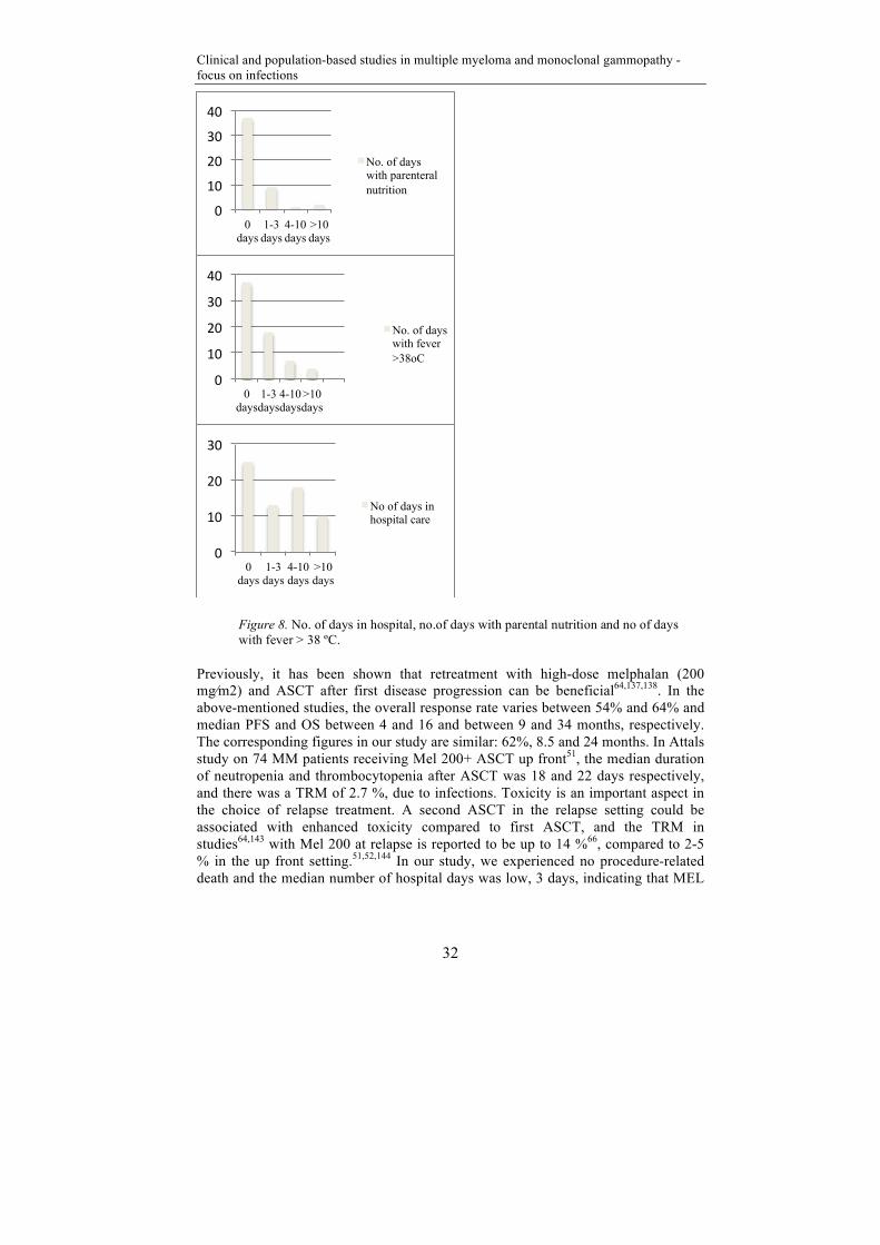
We found that treatment with intermediate-dose melphalan (100 mg⁄m2; MEL 100) and stem cell support was feasible. In total, only 39/66 patients were admitted to the hospital admission, either for melphalan administration, (which was abandoned after some time due to very little acute toxicity), or owing to treatment-related complications. MEL 100 + stem cell support is considered safe in the relapse situation. Less than half of the patients (29 ⁄ 66) experienced a febrile episode and the median number of days with fever was 1. Severe mucositis and prolonged bone marrow depression were infrequent; in median 2 days of a platelet count less than 20 x109 platelets, a median number of erythrocyte and platelet transfusions of 2 and 1, respectively. Mucocitis was limited, illustrated in figure 8, showing days of total parenteral nutrition in of median 0.
Clinical and population-based studies in multiple myeloma and monoclonal gammopathy -focus on infections
32
Figure 8. No. of days in hospital, no.of days with parental nutrition and no of days with fever > 38 ºC.
Previously, it has been shown that retreatment with high-dose melphalan (200 mg⁄m2) and ASCT after first disease progression can be beneficial64,137,138. In the above-mentioned studies, the overall response rate varies between 54% and 64% and median PFS and OS between 4 and 16 and between 9 and 34 months, respectively. The corresponding figures in our study are similar: 62%, 8.5 and 24 months. In Attals study on 74 MM patients receiving Mel 200+ ASCT up front51, the median duration of neutropenia and thrombocytopenia after ASCT was 18 and 22 days respectively, and there was a TRM of 2.7 %, due to infections. Toxicity is an important aspect in the choice of relapse treatment. A second ASCT in the relapse setting could be associated with enhanced toxicity compared to first ASCT, and the TRM in studies64,143 with Mel 200 at relapse is reported to be up to 14 %66, compared to 2-5 % in the up front setting.51,52,144 In our study, we experienced no procedure-related death and the median number of hospital days was low, 3 days, indicating that MEL
0
10
20
30
40
0 days
1-3 days
4-10 days
>10 days
No. of days with parenteral nutrition
0
10
20
30
40
0 days
1-3 days
4-10 days
>10 days
No. of days with fever >38oC
0
10
20
30
0 days
1-3 days
4-10 days
>10 days
No of days in hospital care

Cecilie Hveding Blimark
33
100 could be given with only minor toxicity. The recent development of immunomodulatory drugs (e.g. thalidomide⁄ lenalidomide) and the proteasome inhibitor bortezomib has given us new treatment for patients with myeloma, and these drugs have positively affected response rates, PFS and most probably also OS. However, these drugs may also cause significant side effects and are often given continuously until relapse. Our patients received limited treatment before MEL 100 (50 % did not receive any pretreatment at all), resulting in a short duration of therapy, which possibly could have beneficial effects on quality of life. The overall response rates reported from the randomised trials with bortezomib and lenalidomide (used as a single agent) for relapsed patients vary between 40 % and 60 %, PFS between 6 and 11 months and OS between 29 and 38 months.41,42,145 In a subgroup analysis from the APEX study including only patients in first relapse (n = 132), the PFS was 7 months.145 A subset analysis from the MM-009⁄ 10 studies revealed that patients at first relapse (n = 133) had a better response with a significantly prolonged median PFS (14 vs. 9.5 months) compared with patients treated in later lines of therapy.146 A conservative conclusion is that MEL 100 with stem cell support renders response rates similar to newer drugs in th relpse phase. To assess the possible association between TTP after ASCT and PFS after MEL 100, we grouped the patients by median duration of TTP after ASCT (22.2 months). The group with TTP after ASCT longer than 22.2 months had almost twice as long PFS after MEL 100: 10.4 months (95 % CI; 8.5–12.7) vs. 5.7 months (95 % CI; 4.7–8.6) (P = 0.009) (Figure 9).
Figure 9. Progression-free survival after treatment with MEL 100 depending on time to progression after first autologous stem cell transplantation (TTP<22.2 months: 5.7 months vs TTP>22.2 months 10.4 months; P=0.009).
Patients with TTP after ASCT shorter than the 25th quartile (13 months) had a PFS after MEL 100 of 5.3 months (95 % CI; 3.5–7.9), while patients with TTP after ASCT longer than the 75th quartile (34 months) experienced a PFS after MEL 100 of 12.5 months (95 % CI: 8.8–21.4). This indicate that treatment with the MEL 100

Clinical and population-based studies in multiple myeloma and monoclonal gammopathy -focus on infections
34
regimen could be a suitable option for patients with a first remission after ASCT lasting more than 34 months giving a median PFS of 12.5 months after MEL 100. Previously, it has been concluded that the length of PFS after ASCT is an important predictor of OS with patients having a remission time <12– 18 months appearing not to benefit from a second ASCT.64,65,147 Patients in our study relapsing <22 months after first ASCT had a median time to relapse of <6 months after MEL 100. These patients could instead be considered for an alternative treatment, e.g. combination therapy with newer drugs. To date, there are only a few reports on MEL 100 as salvage therapy. In the report by Olin et al.65, nine patients received Mel 80–180 mg⁄m2 as conditioning therapy, while the others (n = 32) received a more intensive treatment. There were no differences in PFS and OS between the groups: an overall response rate of 55 %, median PFS 8.5 of months and a median OS of 20.7 months, however, with a TRM of 7 %. Krejci et al.68 used MEL 100 as salvage treatment followed by bortezomib⁄ thalidomide consolidation for 31 patients with fulminant progression after first ASCT. They reported an ORR of 58 % but a median time to progression of 5 months and a median OS of only 8 months, indicating a patient population with a very aggressive multiple myeloma. No treatment-related death occurred; however, the median duration of hospital stay was 21 days. Palumbo et al.69 have reported a study on 26 heavily pretreated patients using a conditioning regimen of melphalan 50 mg⁄m2 times 2, bortezomib, thalidomide and corticosteroids (‘‘MVTD’’) followed by stem cell support. The overall response was 66 %, and PFS was 6 months (OS not stated owing to short followup). Cost-effectiveness of different treatments is increasingly important. We have not performed a health economic analysis on the cost benefit of MEL 100 in this study. However, there is a recent report from Cape Town, South Africa, where MEL 100 has been used as upfront induction therapy in an outpatient setting.148 Their estimated cost of MEL 100, including stem cell collection and a 35% risk of hospital admission for 5 d, was 3500 US dollars. They concluded that this cost was still lower than the cost of 3 months of treatment with thalidomide or a single cycle of bortezomib.
In summary, MEL 100 given at first systemic relapse after ASCT is safe and effective, especially for patients who had a durable response after ASCT. In such patients, MEL 100 could be a treatment option with comparable efficacy to newer immunomodulatory drugs, is a therapy with short duration and appears to be associated with low cost.

Cecilie Hveding Blimark
35
4 MGUS, MULTIPLE MYELOMA AND INFECTIONS
(II, III)
Based on data from smaller studies and studies on selected patients we had reason to believe that infections are a serious complication in MGUS and multiple myeloma patients, affecting morbidity and mortality in patients. To our knowledge, no population-based study had previously been performed to evaluate the risk of infections and infection-related mortality in multiple myeloma patients. Therefore, we performed 2 nationwide studies in Sweden, to establish the risk of infections overall and of specific infections in MGUS and multiple myeloma patients, and in multiple myeloma even the risk of infection-related death, compared to matched controls.
4.1 Patients, controls and methods
4.1.1 MGUS patients and controls
Performing population-based studies on MGUS and multiple myeloma patients in Sweden is possible through capturing and following the patients in the different national registries, based on a Swedish patient´s unique social security number. The Swedish Cancer Registry is a nationwide compulsory dual report system developed in 1958, and the Swedish personal identification code system, established in 1947, provides a unique possibility to track all individuals throughout their lifetime. Because MGUS is generally asymptomatic, it is usually an unexpected finding during a medical work-up for another cause. In Sweden, when a clinician detects an MGUS patient, he/she will typically consult with a hematology specialist at a regional hospital, and, if needed, refer the patient for further work-up, especially to rule out an underlying malignancy.
In this study, we established a nationwide MGUS cohort from a national hospital network including MGUS patients recruited from Swedish hospital registries and the Swedish Cancer Registry diagnosed in Sweden between 1965 and 2005. MGUS subtype and concentration of the M-protein at diagnosis was included in the dataset. To minimize the influence of misdiagnosis (eg, smouldering multiple myeloma), MGUS patients with a lymphoproliferative malignancy (including multiple myeloma) diagnosed up to 6 months after MGUS were removed from the MGUS cohort. As an additional quality control measure, we removed any MGUS patient with a recorded preceding lymphoproliferative malignancy. In order evaluate on the occurence of infections in MGUS, for each MGUS patient, 4 population-based controls (matched by sex, year of birth, and county of residence) were chosen randomly from the Swedish Total Population Registry. All controls had to be alive and free of any preceding haematologic malignancy at the time of MGUS diagnosis for the corresponding case. Information on occurrence and date of infections was obtainedfrom the centralized Swedish Patient Registry that captures information on

Clinical and population-based studies in multiple myeloma and monoclonal gammopathy -focus on infections
36
individual patient-based discharge diagnoses and discharge listings from inpatient care. Through linkage with the Cause of Death Register and the Total Population Registry (TPR), we collected information on vital status until December 31, 2006.
4.1.2 Multiple myeloma patients and controls
All multiple myeloma are typically diagnosed and followed clinically by physicians at hospital-based haematology centres. All physicians and pathologists/cytologists in Sweden are since 1958 by law obliged to report each case of incident cancer to the nationwide Swedish Cancer Register. In a recent validation study, the completeness and diagnostic accuracy of the Register was found to be very high (93%) for multiple myeloma patients.149 Record-linkage of the different registries; the Cancer-registry, and In- and Outpatient registries, Cause of Death Registry and the Swedish population database makes it possible to perform population-based studies in a relatively homogenous population with a publicly financed health care, with easy accessible data in public databases.
In the case of the multiple myeloma patients, all patients reported to the nationwide Swedish Cancer Registry from 1988 to 2004 were included in the study. For all included patients, we obtained information on gender, date of birth, date of diagnosis, and region/hospital where the diagnosis was made. For each multiple myeloma patient, four population-based control subjects matched by gender, year of birth, and county of residence were chosen randomly from the Swedish Total Population Register (TPR). The control subjects had to be alive and without preceding haematologic malignancy at the date of diagnosis of the corresponding multiple myeloma patient. From the Swedish Patient Registry, we obtained information on occurrence and date of first infection of every type of infections, with follow-up to 2007.
To assess the role of novel multiple myeloma therapies in relation to the development of infections, patients were divided into three calendar periods; 1988-1993, 1994-1999 and 2000-2004, reflecting time periods with different treatment strategies:
1) Patients diagnosed 1988-1993 (not exposed to high dose melphalan and stem cell support (ASCT)
2) Patients diagnosed 1994-1999 (not exposed to immunomodulatory novel drugs, but some to ASCT)
3) Patients diagnosed 2000-2004 (exposed to modern multiple myeloma treatment)
These cohorts were compared regarding risk of infections compared to controls, over time and risk of infection related death compared to controls and over time.
Statistical analyses
MGUS Cox's proportional hazard models (adjusted for sex, age at diagnosis and year of diagnosis) were used to compare 5- and 10-year risks of infections in MGUS patients compared to controls. Follow up started at age at diagnosis of MGUS (age at registration for controls or January 1, 1987, if MGUS was diagnosed before that

Cecilie Hveding Blimark
37
date). Censoring events were death, emigration, the end of acquisition period or diagnosis of a lymphoproliferative disorder. We excluded all infections occurring in the first six months from MGUS diagnosis (date of selection for controls). To evaluate whether the risk of infection had changed over time, we stratified risk of infections by three calendartime periods of MGUS diagnosis or selection (<1987, 1988-1996, and >1997).
Multiple myeloma To evaluate the overall and one-year risk of infections in multiple myeloma patients compared to controls, Cox proportional hazard models were used. In addition, the effect of gender, age and calendar period of diagnosis was evaluated. Hazard ratios (HR) and confidence intervals (CI) were calculated for the difference in occurrence of infections in patients and controls. We studied patients with multiple myeloma and their controls in the time period of 1988-2004. Follow-up started at date of diagnosis of multiple myeloma (date of multiple myeloma-diagnosis of the corresponding case for controls) and no earlier than January 1, 1988. Censoring events were death, emigration, or the end of acquisition period (December 31, 2007). Event was defined as the diagnosis of a first specific infectious disorder. The median time of follow-up was calculated from the date of multiple myeloma diagnosis (and selection for controls) to the date of censoring. Cumulative incidence at different time periods was calculated as a measure of absolute risk of viral and bacterial infections.
To evaluate the cumulative risk of infections over time (as a measure of absolute risk of infections) and the risk of infection-related death, we also used a competing risk model. The cumulative incidence curve, which explicitly accounted for death as a competing risk, was computed with the method of Gooley et al.150 In these analyses the censoring events were emigration or the end of acquisition period. The competing events were defined as death with diagnosis of an infectious disorder and death without diagnosis of an infectious disorder. To assess the role of novel multiple myeloma therapies in relation to the development of infections, patients were stratified by the mentioned three calendar periods; 1988-1993, 1994-1999 and 2000-2004. Cox proportional hazard models were used to analyze the risk of infections in each cohort. This was also performed separately for age groups younger and older than 65 years at diagnosis. All calculations were performed using Stata version 12 (Stat corp. 2012 Stata Statistical Software: Collage Station, TX, USA).
4.2 Results and discussion
MGUS and infections A total of 5 326 MGUS patients and 20 161 matched population-based controls were included in this study. The median age at diagnosis was 71 years, and 50 % of patients were male. A total of 377 MGUS patients (7.1%) and 550 controls (2.7%) were diagnosed with more than one infection. The average number of infections per MGUS patient was 0.34 and 0.17 per control. At 5-year follow up, compared to controls, MGUS patients had a 2.1-fold increased risk of developing any infection; at

Clinical and population-based studies in multiple myeloma and monoclonal gammopathy -focus on infections
38
10-year follow up, the risk was very similar (Table 3). We further found MGUS patients to have a 2.1-fold and a 2.2-fold increased risk of developing bacterial infections at five and ten years, respectively. When we assessed risks of individual bacterial infections, at 10-year follow up, we found an increased risk of pneumonia, osteomyelitis, septicemia, pyelonephritis, cellulitis, endocarditis, and meningitis (Table 3).
HR: hazard ratio; CI: confidence interval; ctrl: controls; *Cox’s proportional hazard models were used to compare 5- and 10-year risks of infections in MGUS patients compared to controls. The time metric was age. Follow up started at the later of either age at selection or January 1, 1987. Age at selection was age at MGUS diagnosis for a case and for a controlit was age of diagnosis of the matched case. Infections occurring during the first six months were excluded. Follow up ended at the age of diagnosis of a specific infection event or at censoring. Censoring events were death, emigration, the end of acquisition period (December 31, 2006) or diagnosis of a lymphoproliferative disorder. Adjusted (by sex, ageat diagnosis, and year of diagnosis) HRs and 95% CIs were estimated; ** pneumonia, cellulitis, osteomyelitis, endocarditis, meningitis, septicemia, malaria, pyelonephritis, cystitis,sinusitis, tuberculosis, syphilis, gonorrhea, Chlamydia, otitis media, nasopharyngitis/pharyngitis, empyema, HIV, herpes simplex, herpes zoster, hepatitis (A-C), mononucleosis,encephalitis, pericarditis, myocarditis, and influenza; ***pneumonia, cellulitis, osteomyelitis, endocarditis, meningitis, septicemia, malaria, pyelonephritis, cystitis, sinusitis, tuberculosis,syphilis, gonorrhea, Chlamydia, otitis media, nasopharyngitis/pharyngitis, empyema; ****HIV, herpes simplex, herpes zoster, hepatitis (A-C), mononucleosis, encephalitis, pericarditis,myocarditis, and influenza.*****all except those specified above. Our findings that MGUS patients are at a 2-fold increased risk of a broad range of bacterial infections agree with the results of the prior smaller study from Denmark115, and they support the hypothesis that MGUS is associated with an underlying immunodeficiency. It is clear that the major immunological defect in multiple myeloma and Waldenström macroglobulinemia patients is in the humoral system, with a diminished production of polyclonal immunoglobulins which leads to a defective antibody response.35,105,151 In MGUS, prior studies report that hypogammaglobulinemia is present in 25-28% of the cases.110 Interestingly, in contrast to multiple myeloma and Waldenström macroglobulinemia, in the MGUS study from Denmark, presence of hypogammaglobulinemia was not associated with an increased risk of bacteremia.115 Regarding individual viral infections, compared to controls, MGUS patients had a

Cecilie Hveding Blimark
39
2.7-fold increased risk of developing viral infections both at five and ten years. At 10-year follow up, MGUS patients had an increased risk of influenza and herpes zoster (Table 3). To our knowledge, this is the first large population-based study that shows that MGUS patients have an increased risk of viral infections. Interestingly, this risk is similar to that we observed for bacterial infections. Multiple myeloma and Waldenström macroglobulinemia (WM) patients have an increased risk of viral infections. However, this is mainly thought to be related to the therapy given, e.g. herpes zoster infections in patients treated with bortezomib.124 In a case series, MGUS was associated with an increased frequency of Epstein-Barr infections.152 In a study from the Mayo clinic, no increase in several viral infections (chronic hepatitis, cytomegalovirus infection, Epstein-Barr infection, hepatitis C, human immunodeficiency virus) was found among patients with MGUS.116 We found that the risk of infections was similar for the different MGUS isotypes (IgG, IgA and IgM; Table 4) and in an analysis stratified by M-protein concentration, the risk of infection was similar among MGUS cases with an M-protein of 1.0 g/dL and over, and less than 1.0 g/dL, respectively (Table 4). When we assessed the risk of developing multiple myeloma (n=187), WM or related malignancies (n=20) among MGUS patients with (vs. without) an infectious event, we found no statistical difference (HR=0.72; 95% CI 0.40-1.30). In a sensitivity analysis, we also excluded MGUS patients who developed myeloma and the risk estimates were similar (data not shown).
Prior studies have found a history of infectious disease to increase the risk of developing MGUS and multiple myeloma, suggesting that infections may trigger MGUS or multiple myeloma in susceptible patients.96,153 19,20. Furthermore, low levels of polyclonal immunoglobulins in MGUS patients have been found to be a risk factor for progression to multiple myeloma or a related lymphoproliferative malignancy. Taken together, the predisposing role of infections in MGUS and multiple myeloma remains for the most part unclear. Lastly, when we stratified risk of infections by three calendar time periods of MGUS diagnosis or selection (<1987, 1988-1996, and >1997 ), MGUS patients had somewhat different, but consistently

Clinical and population-based studies in multiple myeloma and monoclonal gammopathy -focus on infections
40
increased risk of infections for all calendar periods with HR=2.8 (95% CI 2.1-3.6), HR=1.9 (95% CI 1.7-2.3), and 2.1 (95% CI 1.9-2.4) for before 1987, 1988-1996, and after 1997, respectively (P heterogeneity <0.001). In summary, we found MGUS patients to have a significantly increased risk of several types of both bacterial and viral infections. High M-protein concentration at diagnosis was associated with the highest risks of infections. However, the occurrence of infection was not associated with MM or lymphoproliferative disease progression. Our study provides novel insights into the underlying mechanisms behind infections in patients with MGUS, and may have clinical implications for management and surveillance of MGUS patients Multiple myeloma and infections In study III, a total of 9 253 multiple myeloma patients, diagnosed between 1988 and 2004, and 34 931 population-based controls were included. The median age at multiple myeloma diagnosis was 72 years. The median time of follow-up was 2.6 years for multiple myeloma patients and 7.4 years for controls. The relative 3-year survival of multiple myeloma patients for the calendar periods 1988-1993, 1994-1999, and 2000-2004 was 42.3%, 45.4%, and 47.3%, respectively (no statistically significant difference).The majority of infections were bacterial, 87% in multiple myeloma patients and 85% in controls. Overall, multiple myeloma patients had a significantly 7-fold increased risk of developing any infection compared to matched controls. The risk of developing a bacterial infection in multiple myeloma patients was 7-fold and during the first year following diagnosis the risk was 11-fold to controls. The overall risk for viral infections was 10-fold elevated and during the first year 18-fold higher compared to controls (Table 5).

Cecilie Hveding Blimark
41
This is in accordance with earlier reports, confirming the susceptibility to infections in multiple myeloma. The risk of all included infections was highest during the first year following multiple myeloma diagnosis. Our results are coherent with previous smaller studies that have suggested that infections occur more often in the first 6 months following diagnosis (Table 5)109,113,117,119 Specifically, multiple myeloma patients had an increased risk (p<0.05) of the following bacterial infections compared to matched controls: meningitis septicemia pneumonia, endocarditis, osteomyelitis, cellulitis, and pyelonephritis. Multiple myeloma patients had a significantly increased risk of the viral infections: herpes zoster and influenza compared to matched controls. The results are quite similar to what we found in MGUS, confirming a shared susceptibility to infections. Increasing age was significantly associated with a higher risk of infections (HR=1.02 (per 1 year increment); 95% CI 1.01-1.02, p<0.001) In multiple myeloma patients, we could observe a 20% lower risk of infections in women compared to men the first year after diagnosis. This was true also for controls. This is in good agreement with coherent to population-based data on elderly patients with community-acquired respiratory tract infections in the UK.154
*Risk of infections in all patients and controls ** Risk of infections in all patients, internal comparison by calendar periods ***Risk of infections in patients/controls >65 years, internal comparison by calendar periods
The elevated risk of infections in multiple myeloma patients compared to controls increased significantly with calendar period (p<0.001) and was 6-fold in the period between 1988 and 1993, 7-fold in the period from 1994 to 1999, and 9-fold in 2000-2004 (Table 6). Compared to patients diagnosed during 1988-1993, multiple myeloma patients diagnosed during 1994-1999 and 2000-2004 had a significantly higher risk of infections (Table 6). This is a particularly interesting finding, and raises the question whether modern multiple myeloma therapy increases the risk of infections. The increase in risk of infections was observed in both young and elderly multiple myeloma patients and can thus not solely be explained by HDM-ASCT. The increase in infections was however more pronounced in younger patients and based on data from the Swedish Myeloma Registry20; younger patients are to a larger extent

Clinical and population-based studies in multiple myeloma and monoclonal gammopathy -focus on infections
42
exposed to newer drugs and HDM-ASCT. We would therefore argue that the introduction of HDM-ASCT and novel agents both contribute to the increase in infections. It has been suggested earlier, that the novel agents, probably through their effect on the immune system, make multiple myeloma patients more susceptible to infections. Afessa et al. found a new pattern of bacterial and fungal infections in autologous and allogeneic stem cell recipients.123 Offidani et al. described that 42% of thalidomide-treated patients developed infections, of which 19% were severe.125 Chanan-Khan et al. described in the APEX-study an increasing incidence of herpes zoster in bortezomib-treated patients.124 Our results are important as they suggest that more intensive treatment given to multiple myeloma patients, which has undoubtedly contributed to major improvements in survival in multiple myeloma, probably contribute to an increased susceptibility to infections that needs to be studied in more detail. When we analyzed viral and bacterial infections in different time cohorts, the most significant increase in viral infections was observed between the first and the two latter time periods. In controls, we could not see a corresponding development. There was a continuous increase in the risk of bacterial infections over time.
The increase in risk of infections in multiple myeloma patients compared to controls was statistically significant during the first and five years after diagnosis. This was observed in stratified analyses based on patients diagnosed both under and above the age of 65 years. The absolute risk of an infection expressed in cumulative incidence at five years for all multiple myeloma patients was 19.4% in the years 1988-1993, 40.6% in 1994-1999, and 49.5% in 2000-2004. The 5-year cumulative risk of a bacterial infection in the same time periods was 14.4%, 35.8%, and 46.0%, respectively for multiple myeloma patients and 4.5%, 9.4%, and 10.4% for controls. For viral infections, the 5-year cumulative incidence in the same time periods was 4.8%, 6.3%, and 6.2% for multiple myeloma patients and 0.8%, 0.9%, and 0.7% for controls. In a competing risk model, the 5-year cumulative risk of infections overall and specific infections was essentially the same as in the Cox regression analyses (data not shown). We found that the risk of dying due to infection was 22%, both at two months and one year following diagnosis. This is in contrast to the study from The Medical Research Council (MRC), which showed that nearly 50% of deaths within 2 months were infection-related.119 Despite these differences, both studies stress the importance of this complication in the management of multiple myeloma patients. One important observation in our study is that as the risk of infections increased with calendar period, the risk of infection-related death remained the same during the whole study period. This may be explained by the better supportive care currently available. In summary, in this large population-based study from Sweden we found that bacterial and viral infections represent a major threat to multiple myeloma patients. We found risk of specific infections like pneumonia, and septicemia to be over ten-fold higher than for controls in the first year after multiple myeloma diagnosis, and the risk of infections is increasing in recent years. The risk of dying from an infection is significantly elevated for a multiple myeloma patient compared to age-matched

Cecilie Hveding Blimark
43
controls. With the introduction of the novel therapies, survival in multiple myeloma patients has improved. However, the effect of these drugs on the risk of infection remains to be established and new trials on prophylactic measures are needed.

Clinical and population-based studies in multiple myeloma and monoclonal gammopathy -focus on infections
44
5 TREATMENT OF HIGH-RISK SMOULDERING
MYELOMA (IV)
5.1 Patients and methods
The Swedish Myeloma Registry is a prospective observational registry designed to document real-world treatment and outcomes in all patients with newly diagnosed multiple myeloma in Sweden. It comprises web-reported clinical and laboratory data on all patients diagnosed with smouldering and symptomatic multiple myeloma, plasmocytoma, and plasma cell leukemia from 2008 in Sweden, at time of diagnosis and after one year of follow-up. Coverage is analyzed through the compulsory Swedish Cancer Registry. Survival is achieved from the Swedish Tax Agency. Missing data are actively requested. Multiple myeloma patients diagnosed by autopsy are included in then Swedish Cancer Registry, but not in the Swedish Myeloma Registry. Multiple myeloma patients are reported by treating haematologists and their staff as a part of the everyday practice and quality assurance of coherence to current treatment guidelines. It was founded in 2008 by the Swedish Society of Hematology, is supported by the Swedish National Board of Health and Welfare, and run in collaboration with the Regional Tumour Registries in each of the 6 Swedish health care regions, each covering populations ranging from 0.9 to 1.9 million people, with a total of 9 million.
Smouldering multiple myeloma is with the current guidelines not to be treated outside clinical trials, but to await the debut of symptomatic multiple myeloma. This report was created as a comment to the article of Dr Mateos of the PETHEMA group in New England Journal of Medicine (NEJM) 201380, presenting significant response, and survival improvement in high-risk smouldering myeloma with lenalidomide and dexamethasone treated till progression compared with placebo. In the discussion following this potentially game-changing RCT in smouldering multiple myeloma, one of the major objections was that the 2 current models for risk stratification in high risk smouldering multiple myeloma are based on 2 single centre cohort studies and no population-based incidence was available. In the current study from Mateos, 40% of patients were high-risk according to the PETHEMA model, 18% to the Mayo clinic model70 and 42% according to both models, making the study difficult to interpret. We then decided to analyze the incidence and outcome of smouldering multiple myeloma in a population-based material using the Swedish Myeloma Registry to find:
I. The incidence of high-risk smouldering multiplemyeloma in Sweden
II. The risk of progression for high-risk smouldering multiple myeloma

Cecilie Hveding Blimark
45
As flow-cytometry (used in the PETHEMA risk model) is not widely spread in diagnostics of multiple myeloma, we chose the Mayo clinic model as a simple tool for stratifying high-risk smouldering multiple myeloma patients. Bone-marrow percentage and level of M-protein are available in > 95 % of patients with MM in Sweden.28 Survival and freedom of progression was calculated with the Kaplan - Meier method.142
5.2 Results and discussion
From January 1, 2008, through December 31, 2011, a total of 2494 patients (median age, 72 years) received a diagnosis of multiple myeloma. In the Swedish Myeloma Registry, first a validation study was done of the 437 (18 %) smouldering multiple myeloma patients reported After excluding all patient reported as smouldering multiple myeloma and having osteolytic lesions, n=360 (14.4%) of patients had smouldering multiple myeloma. Of the patients with smouldering multiple myeloma, n=104 (28.8%) had high-risk disease (defined as an M-protein level of ≥30 g per litre and plasma cell infiltration of ≥10%); these patients accounted for 4.2% of all patients with multiple myeloma. On the basis of the world population as reference, the age-standardized incidence of smouldering multiple myeloma is 0.44 cases per 100,000 persons, and the incidence of high-risk disease is 0.14 cases per 100,000 persons. After 2 years, 56.6% of the patients with high-risk smouldering multiple myeloma had progression to symptomatic disease, and after a median follow-up time of 29.8 months, 70.4% had progression. (Figure 10).
Figure 10. Progression of patients with high-risk smouldering multiple myeloma at 2 years of 56.6 %.
Given the high rate of progression observed among patients with high-risk smouldering multiple myeloma in our study, we conclude that approximately 29 % of all patients with newly diagnosed smouldering multiple myeloma will be considered candidates for early treatment, according to the study by Mateos et al. IMWG has proposed that all smouldering multiple myeloma patients with a high risk of progression to multiple myeloma in 2 years should be candidates for early treatment trials with new drugs.81 Identifying high-risk patients with simple diagnostic

Clinical and population-based studies in multiple myeloma and monoclonal gammopathy -focus on infections
46
measures like M-protein and bone marrow samples already available seems appealing. We have in our study identified a cohort of patients with a 57% risk of progression in the first 2 years. There is an unmet need for a consensus definition of high-risk smouldering multiple myeloma based on prospective models that will allow for more refined incorporation of experimental treatment to clinical management of this disease.
Cecilie Hveding Blimark
47
6 METHODOLOGICAL ISSUES
Mel 100 The limitations in this study include the retrospective nature, a possible selection bias of patients owing to the availability of frozen stem cells, and the lack of independent prognostic markers (e.g. chromosomal aberrations or serum ß-2-microglobulin). Nevertheless, Mel 100 was part of the regional treatment guidelines during this time period. Also, the patients in our study were older than those in the above-mentioned relapse studies with a median age of 61 years and about 20% of patients older than 65 years. There were more men than women that received Mel 100 (75%). We therefore investigated whether this was reflected in a less ambitious collecting of stem cells in women. We found that men and women had saved stem cells in equal manners. Regarding that the median age at relapse was 61 years of age in this study, we would however expect more men as there are more younger men (<65 years) than women with multiple myeloma.28 Another fact supporting that our cohort was a representative group of patients in first relapse after ASCT was the survival after relapse, similar to the NMSG studies. In the NMSG-study of Lenhoff and coworkers, the median survival in patients who actually were transplanted and who relapsed thereafter, was 23 months for pts < 60 years, and 16 months for patients 60-64 years (P=0.18)18 and in our study the median survival after MEL 100 was 24 months.
Monoclonal gammopathies and infections . The MGUS cohort in our study is hospital-based and the observed excess risks of infections among MGUS patients may partly reflect various underlying medical illnesses that led to the medical workup and the detection of the M-protein. It is possible that we have a selection of MGUS patients with a higher degree of comorbidity, contibuting to the risk of infections, as they have surfaced in the clinic. However, for clinicians in hospital, this observation is irrelevant, as we encounter and have to manage these patients there. To lessen the impact of this problem in the study, we excluded MGUS patients with a diagnosis of a lymphoproliferative malignancy and infections diagnosed within six months following MGUS diagnosis from our analyses. After this time, during the time of follow-up, 5-10% of MGUS-patients probably have developed multiple myeloma, thus this might theoretically have contributed to an increased risk of infections. Since our controls were population-based and not screened for M-protein, one has to be cautious and consider possible bias. For example, given the fact that MGUS patients are followed clinically, it may have contributed to the reporting of more infections (i.e. surveillance bias).
One future strategy to assess the potential influence of detection and surveillance bias may be the launching of a large record-linkage study based on a screened MGUS population. Although the risk determined in a screened MGUS population is likely to be more conservative and would also reflect the biological underpinning involved in infectious complications following MGUS, this study is based on the general clinical setting. Another limitation is that some of the controls are expected to have

Clinical and population-based studies in multiple myeloma and monoclonal gammopathy -focus on infections
48
undiagnosed MGUS. Furthermore, there is also the potential for inaccuracy and the lack of independent validation of infectious diagnosis obtained from the centralized Patient Registry as the infections may not be microbiologically verified. However, this problem should affect MGUS cases and matched controls equally and thus any bias should be towards a null association. For multiple myeloma patients, the surveillance of infections is probably more vigilant than in the general population that might lead to more reported infections of all sorts in the multiple myeloma cohort. However, most of the infections recorded, and later showing increased risks, were severe infections that would be captured in the general population as well, as they generally require hospitalization. In the MGUS and infections-study we have patients diagnosed from 1965 but follow up on infections only from the hospital registries from 1987. This might lead to a total underestimate of infections in earlier years, but should be equally distributed between patients and controls. The same is for the multiple myeloma cohort, where we lack data from the out-patient registry from the time before 2000, but this should also affect patients and controls alike and should only underestimate the total number of infected patients and controls together. Lack of clinical data on bacterial and viral (fungal) agens form actual cultures, infections on the other hand clinically relevant when mentioned on discharge list, which in not always the case, finding a pathogenic agens in a blood or urine culture. We chose to record only the first infection of each type and not counting infection in the same organ twice in the same individual, and as a result we do not include all infections in all patients, as some patients are diagnosed with the same infection more than once. Thus we performed this to get a more accurate measure of the excess risk of each infection. Otherwise few patients with many infection of the same type could drive the risk increase. We considered this to be a superior choice over the option of eventually overestimating the risk for all multiple myeloma patients due to a few subjects with repeated infections. In large hospital registries, there is a risk of registration bias. There may also be a certain degree of underreporting where in multiple myeloma patients only the multiple myeloma diagnosis is registered on the discharge list and not the infections. The same might occur, obtaining the cause of death for a multiple myeloma patient in the Cause of death registry, causing fewer infections to be reported in the registries. We chose to record only the first infection of each type in every patient and not counting infection in the same organ twice in the same individual. This means that we record only first pneumonia in each patient/control, however that individual can be diagnosed with all other infections, for example viral, meningitis, osteomyelitis etc). As individuals with a specific infection, for example pneumonia, are at an excess risk of getting another pneumonia, it is usual in cohort studies to censor after first event, so that the risk is not overestimated due to this.
The fact that 3-year relative survival has increased by approximately 2% between the last two calendar periods can unlikely explain the increase in infections observed, and thus we believe that improved survival is not a bias in our study.

Cecilie Hveding Blimark
49
High-risk smouldering multiple myeloma As mentioned, in a recent validation study, we have reported that ascertainment and diagnostic accuracy for lymphoproliferative disorders (including multiple myeloma) is very high (>90-95%) in Sweden.149 We discovered a confusion amongst reporting doctors about the terms asymptomatic/smouldering multiple myeloma vs multiple myeloma not requiring immediate treatment, but with obvious signs of symptomatic multiple myeloma. In a validation study, we found that as many as 18% of patients were reported as smouldering multiple myeloma, but at the same time reported to have osteolytic lesions. Correcting for this, we found an incidence of smouldering multiple myeloma of 14.4 %, more consistent with other studies in the literature.71-73

Clinical and population-based studies in multiple myeloma and monoclonal gammopathy -focus on infections
50
7 SUMMARY AND CONCLUSIONS
Minitransplantation at relapse (I) In the retrospective study of 66 patients with myeloma in first systemic relapse after ASCT, we found that treatment with intermediate-dose melphalan (100 mg⁄m2; MEL 100) and stem cell support was feasible, effective, safe with low number of infections. Thus, Mel 100 could be considered as a viable therapeutic option in patients who are not considered for Mel 200 salvage and have a first time to progression of > 22 months.
Monoclonal gammopathies and infections (II, III) We found MGUS patients (II) to have a significantly increased risk of several types of both bacterial and viral infections. High M-protein concentration at diagnosis was associated with the highest risks of infections. Our study provides novel insights into the underlying mechanisms behind infections in patients with MGUS, and may have clinical implications for infection-treatment strategies, prophylactic measures and vaccinations, as well as surveillance of MGUS patients.
We found bacterial and viral infections to represent a major threat to multiple myeloma patients (III). We found the risk of specific infections like pneumonia, and septicemia to be over ten-fold higher than for controls in the first year after multiple myeloma diagnosis, and the risk of infections is increasing in recent years. The risk of dying from an infection is significantly elevated for a multiple myeloma patient compared to age-matched controls.
High-risk smouldering myeloma (IV) We found that 14.4% of patients reported to the Swedish Myeloma Registry had smoldering multiple myeloma. Of the patients with smouldering multiple myeloma, 28.8% had high-risk disease, defined as an M-protein level of ≥30 g per litre and plasma-cell infiltration of ≥10%. We have in our study identified a cohort of patients with a 57 % risk of progression in the first 2 years. These patients could be candidates for treatment trials as recommended by the International Myeloma Working Group.

Cecilie Hveding Blimark
51
8 FUTURE PERSPECTIVES
Our future plans in the field of research on infections in plasma cell disorders will be to analyze the bacterial, viral and fungal cultures on all patients diagnosed with multiple myeloma in a period of ten years in two major university hospitals in Sweden. With a reasonable large cohort of approximately 2000 patients it will be possible to characterize, in a population-based setting, bacterial, viral and fungal pathogens most commonly found in multiple myeloma patients in Sweden. It will be possible to detect a eventual shift in different pathogens over time. Our goal is to verify more profoundly which pathogens causes the infections morbidity and mortality in Swedish multiple myeloma patients, and our work can contribute to facilitating proper prophylactic measures
The fact that we found an increased risk of infections even in elderly multiple myeloma patients, it would be tempting to intensify the antibiotic prophylaxis in a prospective randomized trial, as this group (patients over the age of 65 years) are, in the current national guidelines, recommended antibacterial prophylaxis only if treated with high-dose glucocorticoids.

Clinical and population-based studies in multiple myeloma and monoclonal gammopathy -focus on infections
52
ACKNOWLEDGEMENT
I want to express my gratitude to all the people helping me in my research and making this work possible. I especially wish to thank:
My principal supervisor, Ulf-Henrik Mellqvist, for sharing your expertise in multiple myeloma and research work. Your patience and support has been highly significant to this thesis. A record holder in fast mail and text answers, your generosity and kindness is inspiring, always listening and being supportive.
Sigurdur Kristinsson, my co-supervisor and Icelandic friend, co-leader of the Swedish Myeloma Registry, who has generously included me in his research group, sharing his vast knowledge in research and writing papers. I have learned so much from you, and thank you for being thorough and having so high ambitions.
Mats Brune, for sharing your data and great knowledge on stem cell transplantation, for late dinners and animated discussions.
Per-Ola Andersson, co-supervisor and former boss. Thank you for creating an excellent research environment in the Department of Haematology and Coagulation, and being there for me when I was writing my first paper, you are greatly missed.
Ingemar Turesson, grand old man in myeloma in Sweden. It is an honor to be coauthoring with you, your are always thorough and wise commenting on papers and participating in the Swedish Myeloma Registry Group.
Ola Landgren, co-author, for your enormous generosity, letting me take part in your research, and introducing me to the NIH.
All physicians and pathologists for providing information on their patients, in particular all the haematologist in the Western Region who helped me filling in all case reports on transplanted patients.
All my colleagues and friends at the Department of Haematology and Coagulation, for input, cheering and support, lunch hour discussions, seeing my patients and taking my clinic-hours.

53
My co-authors Anders Wahlin, Malin Hultcrantz, Magnus Björkholm and Christian Kjellander for valuable input.
Erik Holmberg, at the RCC West, for your invaluable statistical input and wisdom, for ever-lasting patience, always helpful.
Stig Rödjer, my clinical mentor and co-author, the first person introducing me to research. Teaching me a lot about multiple myeloma, and about life in general.
My experienced co-authors Jan Westin and Martin Hjorth for your excellent input and fatherly advice along the way.
Stefan Jacobsson, Göran Oleröd and Inger Ögärd, clin.chem SU/S for providing haematological images for this thesis.
My lovely fellow students and ballet teachers at Dansforum, for wonderful times and for trying to teach me discipline and triple turns.
My many friends and family in Sweden, Norway and Germany, especially I want to mention Yvonne, Birgit, Karin, Tony and Helen, Jan and Maria, Anders and Mia, Malin and Claes, Annika, sisters and brother, and my parents, - for late dinners, dry martinis, friendship, pleasant lunches, late night phone calls, coaching in life, and great partys, for sharing your time and helping me, especially this last year, I will never forget it.
And finally to Christian, Henrik and Johanna, my gems and great love. This thesis is dedicated to you.
This thesis was supported by grants from the Göteborg Medical Society, Sahlgrenska University Hospital Foundations, Research and Development Foundations of Västra Götaland, Roche through the Swedish Society of Haematology, the Swedish Cancer Society and Karolinska Institutet foundation.
Clinical and population-based studies in multiple myeloma and monoclonal gammopathy -focus on infections
54
REFERENCES
1. Swerdlow SH, Campo E, Harris NL, et al. WHO Classification of Tumours of Haematopoietic and Lymphoid Tissues. Lyon, France: IARC Press; 2008. 2. Kyle RA, Rajkumar SV. Monoclonal gammopathy of undetermined significance. Br J Haematol. 2006;134(6):573-589. 3. Landgren O, Kyle RA, Pfeiffer RM, et al. Monoclonal gammopathy of undetermined significance (MGUS) consistently precedes multiple myeloma: a prospective study. Vol. 113; 2009. 4. Kyle RA, Gertz MA, Witzig TE, et al. Review of 1027 patients with newly diagnosed multiple myeloma. Mayo Clin Proc. 2003;78(1):21-33. 5. Zink A, Rohrbach H, Szeimies U, Hagedorn HG, Haas CJ, Weyss C, Bachmeier B, Nerlich AG Malignant tumors in an ancient Egyptian population. Anticancer Res 19:4273–4277. Anticancer Res;19:4273–4277. 6. Gestsdottir H, Eyjolfsson GI. Myeloma in an archaeological skeleton from Hofstadir in Myvatnssveit. Laeknabladid 2005(91):505–509. 7. Nerlich AG, Rohrbach H, Bachmeier B, Zink A Malignant tumors in two ancient populations: an approach to historical tumor epidemiology. Oncol Rep. 2006;16:197–202. 8. Kyle RA, Steensma DP. History of multiple myeloma. Recent Results Cancer Res. 2011;183:3-23. 9. Solly S. Remarks on the pathology of mollities ossium; with cases. Med Chir Trans. 1844;27:435-498 438. 10. Wright JH. A CASE OF MULTIPLE MYELOMA. J Boston Soc Med Sci. 1900;4(8):195-204 195. 11. Heremans JF, Laurell AH, Martensson L, et al. Studies on "abnormal" serum globulins (M-components) in myeloma, macroglobulinemia and related diseases. Acta Med Scand Suppl. 1961;367:1-126. 12. Waldenstrom J. Abnormal proteins in myeloma. Adv Intern Med. 1952;5:398-440. 13. Kyle RA. Monoclonal gammopathy of undetermined significance. Natural history in 241 cases. Am J Med. 1978;64(5):814-826. 14. A T. Electrophoresis of serum globulin.Electrophoretic analysis of normal and immune sera. Biochem J. 1937;31(9):1464-1477.
15. Korngold L, Lipari R. Multiple-myeloma proteins. III. The antigenic relationship of Bence Jones proteins to normal gammaglobulin and multiple-myeloma serum proteins. Cancer. 1956;9(2):262-272.

55
16. Blokhin N, Larionov L, Perevodchikova N, Chebotareva L, Merkulova N Clinical experiences with sarcolysin in neoplastic diseases. Ann NY Acad Sci. 1958;68:1128–1132. 17. Turesson I, Velez R, Kristinsson SY, Landgren O. Patterns of improved survival in patients with multiple myeloma in the twenty-first century: a population-based study. J Clin Oncol. 2010;28(5):830-834. 18. Lenhoff S, Hjorth M, Holmberg E, et al. Impact on survival of high-dose therapy with autologous stem cell support in patients younger than 60 years with newly diagnosed multiple myeloma: a population-based study. Nordic Myeloma Study Group. Blood. 2000;95(1):7-11. 19. Hippe E, Westin J, Wislöff F. Nordic Myeloma Study Group, the first 15 years: scientific collaboration and improvement of patient care. European Journal of Haematology. 2005;74(3):185-193. 20. Blimark C Holmberg E, Juliusson G, Nahi H, Forsberg K, Turesson I, Ahlberg L, Svensson R, Linder O, Kristinsson SY. Real World data in Myeloma: Experiences from the Swedish Population-based Registry on 2494 Myeloma Patients Diagnosed 2008-2011. Blood 2013;122:1972. 2013 (Abstract). 21. Rajkumar SV, Dimopoulos MA, Palumbo A, et al. International Myeloma Working Group updated criteria for the diagnosis of multiple myeloma. The Lancet Oncology. 2014;15(12):e538-e548. 22. Hillengass J, Fechtner K, Weber MA, et al. Prognostic significance of focal lesions in whole-body magnetic resonance imaging in patients with asymptomatic multiple myeloma. J Clin Oncol. 2010;28(9):1606-1610. 23. Larsen JT, Kumar SK, Dispenzieri A, Kyle RA, Katzmann JA, Rajkumar SV. Serum free light chain ratio as a biomarker for high-risk smoldering multiple myeloma. Leukemia. 2013;27(4):941-946. 24. Rajkumar SV, Larson D, Kyle RA. Diagnosis of smoldering multiple myeloma. N Engl J Med. 2011;365(5):474-475. 25. Landgren O, Rajkumar SV. Development of early treatment strategies for high-risk myeloma precursor disease in the future. Semin Hematol. 2011;48(1):66-72. 26. Dispenzieri A, Kyle RA, Katzmann JA, et al. Immunoglobulin free light chain ratio is an independent risk factor for progression of smoldering (asymptomatic) multiple myeloma. Vol. 111; 2008. 27. Altekruse SF, Kosary C, Krapcho M, et al. SEER cancer statistics review,1975-2007. Bethesda, MD: National Cancer Institute. 28. Myelomregistret Svenska. Myelomrapport nr 3. 2013. www.sfhem.se/upl/files/91646.pdf. 29. Palumbo A, Mina R. Management of older adults with multiple myeloma. Blood Rev. 2013 27(3):133-42:133-142.
30. Kristinsson SY, Landgren O, Dickman PW, Derolf AR, Bjorkholm M. Patterns of survival in multiple myeloma: a population-based

Clinical and population-based studies in multiple myeloma and monoclonal gammopathy -focus on infections
56
study of patients diagnosed in Sweden from 1973 to 2003. J Clin Oncol. 2007;25(15):1993-1999. 31. Landgren O, Gridley G, Turesson I, et al. Risk of monoclonal gammopathy of undetermined significance (MGUS) and subsequent multiple myeloma among African American and white veterans in the United States. Blood. 2006;107(3):904-906. 32. Roodman GD. Pathogenesis of myeloma bone disease. Leukemia. 2009;23(3):435-441. 33. Birgegard G, Gascon P, Ludwig H. Evaluation of anaemia in patients with multiple myeloma and lymphoma: findings of the European CANCER ANAEMIA SURVEY. Eur J Haematol. 2006;77(5):378-386. 34. Blade J, Fernandez-Llama P, Bosch F, et al. Renal failure in multiple myeloma: presenting features and predictors of outcome in 94 patients from a single institution. Arch Intern Med. 1998;158(17):1889-1893. 35. Broder S, Humphrey R, Durm M, et al. Impaired synthesis of polyclonal (non-paraprotein) immunoglobulins by circulating lymphocytes from patients with multiple myeloma Role of suppressor cells. N Engl J Med. 1975;293(18):887-892. 36. Kumar SK, Rajkumar SV, Dispenzieri A, et al. Improved survival in multiple myeloma and the impact of novel therapies. Blood. 2008;111(5):2516-2520. 37. Kristinsson SY, Anderson WF, Landgren O. Improved long-term survival in multiple myeloma up to the age of 80 years. Leukemia. 2014;28(6):1346-1348. 38. Barlogie B, Desikan R, Eddlemon P, et al. Extended survival in advanced and refractory multiple myeloma after single-agent thalidomide: identification of prognostic factors in a phase 2 study of 169 patients. Blood. 2001;98(2):492-494. 39. Singhal S, Mehta J, Desikan R, et al. Antitumor activity of thalidomide in refractory multiple myeloma. N Engl J Med. 1999;341(21):1565-1571. 40. Fayers PM, Palumbo A, Hulin C, et al. Thalidomide for previously untreated elderly patients with multiple myeloma: meta-analysis of 1685 individual patient data from 6 randomized clinical trials. Blood. 2011;118(5):1239-1247. 41. Weber DM, Chen C, Niesvizky R, et al. Lenalidomide plus dexamethasone for relapsed multiple myeloma in North America. N Engl J Med. 2007;357(21):2133-2142. 42. Dimopoulos M, Spencer A, Attal M, et al. Lenalidomide plus dexamethasone for relapsed or refractory multiple myeloma. N Engl J Med. 2007;357(21):2123-2132. 43. Rajkumar SV, Jacobus S, Callander NS, et al. Lenalidomide plus high-dose dexamethasone versus lenalidomide plus low-dose dexamethasone as initial therapy for newly diagnosed multiple myeloma: an open-label randomised controlled trial. Lancet Oncol. 2010;11(1):29-37.

57
44. Richardson PG, Siegel DS, Vij R, et al. Pomalidomide alone or in combination with low-dose dexamethasone in relapsed and refractory multiple myeloma: a randomized phase 2 study. Blood. 2014;123(12):1826-1832. 45. Jagannath S, Durie BG, Wolf J, et al. Bortezomib therapy alone and in combination with dexamethasone for previously untreated symptomatic multiple myeloma. Br J Haematol. 2005;129(6):776-783. 46. Richardson PG, Barlogie B, Berenson J, et al. A Phase 2 Study of Bortezomib in Relapsed, Refractory Myeloma. New England Journal of Medicine. 2003;348(26):2609-2617. 47. Harousseau JL, Attal M, Leleu X, et al. Bortezomib plus dexamethasone as induction treatment prior to autologous stem cell transplantation in patients with newly diagnosed multiple myeloma: results of an IFM phase II study. Haematologica. 2006;91(11):1498-1505. 48. San Miguel JF, Schlag R, Khuageva NK, et al. Bortezomib plus melphalan and prednisone for initial treatment of multiple myeloma. N Engl J Med. 2008;359(9):906-917. 49. Ocio EM, Richardson PG, Rajkumar SV, et al. New drugs and novel mechanisms of action in multiple myeloma in 2013: a report from the International Myeloma Working Group (IMWG). Leukemia. 2014;28(3):525-542. 50. Group IMW. Criteria for the classification of monoclonal gammopathies, multiple myeloma and related disorders: a report of the International Myeloma Working Group. Br J Haematol. 2003;121(5):749-757. 51. Attal M, Harousseau JL, Stoppa AM, et al. A prospective, randomized trial of autologous bone marrow transplantation and chemotherapy in multiple myeloma. Intergroupe Francais du Myelome. N Engl J Med. 1996;335(2):91-97. 52. Child JA, Morgan GJ, Davies FE, et al. High-dose chemotherapy with hematopoietic stem-cell rescue for multiple myeloma. N Engl J Med. 2003;348(19):1875-1883. 53. Barlogie B, Kyle RA, Anderson KC, et al. Standard chemotherapy compared with high-dose chemoradiotherapy for multiple myeloma: final results of phase III US Intergroup Trial S9321. J Clin Oncol. 2006;24(6):929-936. 54. Fermand JP, Katsahian S, Divine M, et al. High-dose therapy and autologous blood stem-cell transplantation compared with conventional treatment in myeloma patients aged 55 to 65 years: long-term results of a randomized control trial from the Group Myelome-Autogreffe. J Clin Oncol. 2005;23(36):9227-9233. 55. Ludwig H, Beksac M, Blade J, et al. Current multiple myeloma treatment strategies with novel agents: a European perspective. Oncologist. 2010;15(1):6-25.

Clinical and population-based studies in multiple myeloma and monoclonal gammopathy -focus on infections
58
56. Moreau P, Giralt SA. Optimizing therapy for transplant-eligible patients with newly diagnosed multiple myeloma. Leuk Res. 2012;36 Suppl 1:S13-18. 57. Lonial S. Relapsed multiple myeloma. Hematology Am Soc Hematol Educ Program. 2010;2010:303-309. 58. Mehta J, Tricot G, Jagannath S, et al. Salvage autologous or allogeneic transplantation for multiple myeloma refractory to or relapsing after a first-line autograft? Bone Marrow Transplant. 1998;21(9):887-892. 59. Tricot G, Jagannath S, Vesole DH, Crowley J, Barlogie B. Relapse of multiple myeloma after autologous transplantation: survival after salvage therapy. Bone Marrow Transplant. 1995;16(1):7-11. 60. Greipp PR, San Miguel J, Durie BG, et al. International staging system for multiple myeloma. J Clin Oncol. 2005;23(15):3412-3420. 61. Avet-Loiseau H, Durie BGM, Cavo M, et al. Combining fluorescent in situ hybridization data with ISS staging improves risk assessment in myeloma: an International Myeloma Working Group collaborative project. Leukemia. 2013;27(3):711-717. 62. Landgren O, Morgan GJ. Biologic frontiers in multiple myeloma: from biomarker identification to clinical practice. Clin Cancer Res. 2014;20(4):804-813. 63. Kyle RA, Rajkumar SV. Treatment of multiple myeloma: a comprehensive review. Clin Lymphoma Myeloma. 2009;9(4):278-288. 64. Alvares CL, Davies FE, Horton C, Patel G, Powles R, Morgan GJ. The role of second autografts in the management of myeloma at first relapse. Haematologica. 2006;91(1):141-142. 65. Olin RL, Vogl DT, Porter DL, et al. Second auto-SCT is safe and effective salvage therapy for relapsed multiple myeloma. Bone Marrow Transplant. 2009;43(5):417-422. 66. Atanackovic D, Schilling G. Second autologous transplant as salvage therapy in multiple myeloma. Br J Haematol. 2013;163(5):565-572. 67. Burzynski JA, Toro JJ, Patel RC, et al. Toxicity of a second autologous peripheral blood stem cell transplant in patients with relapsed or recurrent multiple myeloma. Leuk Lymphoma. 2009;50(9):1442-1447. 68. Krejci M, Adam Z, Buchler T, et al. Salvage treatment with upfront melphalan 100 mg/m(2) and consolidation with novel drugs for fulminant progression of multiple myeloma. Ann Hematol. 2010;89(5):483-487. 69. Palumbo A, Avonto I, Bruno B, et al. Intermediate-dose melphalan (100 mg/m2)/bortezomib/thalidomide/dexamethasone and stem cell support in patients with refractory or relapsed myeloma. Clin Lymphoma Myeloma. 2006;6(6):475-477. 70. Kyle RA, Remstein ED, Therneau TM, et al. Clinical course and prognosis of smoldering (asymptomatic) multiple myeloma. N Engl J Med. 2007;356(25):2582-2590.
59
71. Kyle RA, Rajkumar SV. Monoclonal gammopathy of undetermined significance and smoldering multiple myeloma. Curr Hematol Malig Rep. 2010;5(2):62-69. 72. Perez-Persona E, Vidriales MB, Mateo G, et al. New criteria to identify risk of progression in monoclonal gammopathy of uncertain significance and smoldering multiple myeloma based on multiparameter flow cytometry analysis of bone marrow plasma cells. Blood. 2007;110(7):2586-2592. 73. Rosinol L, Blade J, Esteve J, et al. Smoldering multiple myeloma: natural history and recognition of an evolving type. Br J Haematol. 2003;123(4):631-636. 74. Hjorth M, Hellquist L, Holmberg E, Magnusson B, Rodjer S, Westin J. Initial versus deferred melphalan-prednisone therapy for asymptomatic multiple myeloma stage I--a randomized study. Myeloma Group of Western Sweden. Eur J Haematol. 1993;50(2):95-102. 75. Waxman AJ, Kuehl M, Balakumaran A, Weiss B, Landgren O. Smoldering (asymptomatic) multiple myeloma: revisiting the clinical dilemma and looking into the future. Clin Lymphoma Myeloma Leuk. 2010;10(4):248-257. 76. Weber DM, Dimopoulos MA, Moulopoulos LA, Delasalle KB, Smith T, Alexanian R. Prognostic features of asymptomatic multiple myeloma. Br J Haematol. 1997;97(4):810-814. 77. Cesana C, Klersy C, Barbarano L, et al. Prognostic factors for malignant transformation in monoclonal gammopathy of undetermined significance and smoldering multiple myeloma. J Clin Oncol. 2002;20(6):1625-1634. 78. Rajkumar SV, Kyle RA, Therneau TM, et al. Serum free light chain ratio is an independent risk factor for progression in monoclonal gammopathy of undetermined significance. Vol. 106; 2005. 79. Cherry BM, Korde N, Kwok M, et al. Modeling progression risk for smoldering multiple myeloma: results from a prospective clinical study. Leuk Lymphoma. 2013;54(10):2215-2218. 80. Mateos MV, San Miguel JF. Treatment for high-risk smoldering myeloma. N Engl J Med. 2013;369(18):1764-1765. 81. Kyle RA, Durie BG, Rajkumar SV, et al. Monoclonal gammopathy of undetermined significance (MGUS) and smoldering (asymptomatic) multiple myeloma: IMWG consensus perspectives risk factors for progression and guidelines for monitoring and management. Leukemia. 2010;24(6):1121-1127. 82. Weiss BM, Abadie J, Verma P, Howard RS, Kuehl WM. A monoclonal gammopathy precedes multiple myeloma in most patients. Blood. 2009;113(22):5418-5422. 83. Landgren O. Monoclonal gammopathy of undetermined significance and smoldering multiple myeloma: biological insights and early

Clinical and population-based studies in multiple myeloma and monoclonal gammopathy -focus on infections
60
treatment strategies. Hematology Am Soc Hematol Educ Program. 2013;2013:478-487. 84. Dispenzieri A, Katzmann JA, Kyle RA, et al. Prevalence and risk of progression of light-chain monoclonal gammopathy of undetermined significance: a retrospective population-based cohort study. Lancet. 2010;375(9727):1721-1728. 85. Rajkumar SV, Dispenzieri A, Kyle RA. Monoclonal Gammopathy of Undetermined Significance, Waldenström Macroglobulinemia, AL Amyloidosis, and Related Plasma Cell Disorders: Diagnosis and Treatment. Mayo Clinic Proceedings. 2006;81(5):693-703. 86. Wadhera RK, Rajkumar SV. Prevalence of monoclonal gammopathy of undetermined significance: a systematic review. Mayo Clin Proc. 2010;85(10):933-942. 87. Kyle RA, Vincent Rajkumar S. Monoclonal gammopathy of undetermined significance. British Journal of Haematology. 2006;134(6):573-589. 88. Vachon CM, Kyle RA, Therneau TM, et al. Increased risk of monoclonal gammopathy in first-degree relatives of patients with multiple myeloma or monoclonal gammopathy of undetermined significance. Blood. 2009;114(4):785-790. 89. Landgren O, Kristinsson SY, Goldin LR, et al. Risk of plasma cell and lymphoproliferative disorders among 14621 first-degree relatives of 4458 patients with monoclonal gammopathy of undetermined significance in Sweden. Blood. 2009;114(4):791-795. 90. Landgren O, Linet MS, McMaster ML, Gridley G, Hemminki K, Goldin LR. Familial characteristics of autoimmune and hematologic disorders in 8,406 multiple myeloma patients: a population-based case-control study. Int J Cancer. 2006;118(12):3095-3098. 91. Landgren O, Graubard BI, Katzmann JA, et al. Racial disparities in the prevalence of monoclonal gammopathies: a population-based study of 12,482 persons from the National Health and Nutritional Examination Survey. Leukemia. 2014;28(7):1537-1542. 92. Lynch HT, Snyder CL, Lynch JF, Thome S. Familial plasma cell disorders and associated cancers. Clin Lymphoma Myeloma Leuk. 2011;11(1):80-81. 93. Kristinsson SY, Koshiol J, Bjorkholm M, et al. Immune-related and inflammatory conditions and risk of lymphoplasmacytic lymphoma or Waldenstrom macroglobulinemia. J Natl Cancer Inst. 2010;102(8):557-567. 94. Koshiol J, Gridley G, Engels EA, McMaster ML, Landgren O. Chronic immune stimulation and subsequent Waldenstrom macroglobulinemia. Arch Intern Med. 2008;168(17):1903-1909. 95. Smedby KE, Hjalgrim H, Askling J, et al. Autoimmune and chronic inflammatory disorders and risk of non-Hodgkin lymphoma by subtype. J Natl Cancer Inst. 2006;98(1):51-60.

61
96. Brown LM, Gridley G, Check D, Landgren O. Risk of multiple myeloma and monoclonal gammopathy of undetermined significance among white and black male United States veterans with prior autoimmune, infectious, inflammatory, and allergic disorders. Blood. 2008;111(7):3388-3394. 97. Kuehl WM, Bergsagel PL. Molecular pathogenesis of multiple myeloma and its premalignant precursor. J Clin Invest. 2012;122(10):3456-3463. 98. Smadja NV, Bastard C, Brigaudeau C, Leroux D, Fruchart C. Hypodiploidy is a major prognostic factor in multiple myeloma. Blood. 2001;98(7):2229-2238. 99. Kristinsson SY, Bjorkholm M, Andersson TM, et al. Patterns of survival and causes of death following a diagnosis of monoclonal gammopathy of undetermined significance: a population-based study. Haematologica. 2009;94(12):1714-1720. 100. Kristinsson SY, Tang M, Pfeiffer RM, et al. Monoclonal gammopathy of undetermined significance and risk of skeletal fractures: a population-based study. Blood. 2010;116(15):2651-2655. 101. Kristinsson SY, Fears TR, Gridley G, et al. Deep vein thrombosis after monoclonal gammopathy of undetermined significance and multiple myeloma. Blood. 2008;(112):3582-3586. 102. Bird J, Behrens J, Westin J, et al. UK Myeloma Forum (UKMF) and Nordic Myeloma Study Group (NMSG): guidelines for the investigation of newly detected M-proteins and the management of monoclonal gammopathy of undetermined significance (MGUS). Br J Haematol. 2009;147(1):22-42. 103. Kyle RA, Therneau TM, Rajkumar SV, et al. A long-term study of prognosis in monoclonal gammopathy of undetermined significance. N Engl J Med. 2002;346(8):564-569. 104. Turesson I, Kovalchik SA, Pfeiffer RM, et al. Monoclonal gammopathy of undetermined significance and risk of lymphoid and myeloid malignancies: 728 cases followed up to 30 years in Sweden. Blood. 2014;123(3):338-345. 105. Kyle RA, Rajkumar SV. Multiple myeloma. N Engl J Med. 2004;351(18):1860-1873. 106. Nucci M. AE. Infections in patients with multiple myeloma in the era of high-dose therapy and novel agents. . Clin Infect Dis. 2009.(49):1211–1225. 107. Simonetti AF, Viasus D, Garcia-Vidal C, Carratala J. Management of community-acquired pneumonia in older adults. Ther Adv Infect Dis. 2014;2(1):3-16. 108. Meyer KC. The role of immunity in susceptibility to respiratory infection in the aging lung. Respir Physiol. 2001;128(1):23-31. 109. Pratt G, Goodyear O, Moss P. Immunodeficiency and immunotherapy in multiple myeloma. Br J Haematol. 2007;138(5):563-579.

Clinical and population-based studies in multiple myeloma and monoclonal gammopathy -focus on infections
62
110. Kyle RA, Therneau TM, Rajkumar SV, et al. Prevalence of monoclonal gammopathy of undetermined significance. N Engl J Med. 2006;354(13):1362-1369. 111. Blade J, Lopez-Guillermo A, Rozman C, et al. Malignant transformation and life expectancy in monoclonal gammopathy of undetermined significance. Br J Haematol. 1992;81(3):391-394. 112. Baldini L, Guffanti A, Cesana BM, et al. Role of different hematologic variables in defining the risk of malignant transformation in monoclonal gammopathy. Blood. 1996;87(3):912-918. 113. Savage DG LJ, Garret TJ. Biphasic pattern of bacterial infection in multiple myeloma. . Am Int Med. 1982 (96(1)):47-50 114. Jacobson DR, Zolla-Pazner S. Immunosuppression and infection in multiple myeloma. Semin Oncol. 1986;13(3):282-290. 115. Gregersen H MK, Sorensen HT, Schonheyder HC, Ibsen JS, Dahlerup JF. The risk of bacteremia in patients with monoclonal gammopathy of undetermined significance. . Eur J Haematol. 1998(61):140-144. 116. Bida JP, Kyle RA, Therneau TM, et al. Disease associations with monoclonal gammopathy of undetermined significance: a population-based study of 17,398 patients. Mayo Clin Proc. 2009;84(8):685-693. 117. Twomey J. Infections complicating multiple myeloma and chronic lymphocytic leukemia.Arch Int med 1973(132(4)): ; 562-565.
118. Perri RT, Hebbel R, Oken MM. Influence of treatment and response status on infection in multiple myeloma Am J Med. 1981(71(6)): 935-940.
119. Augustson BM, Begum G, Dunn JA, et al. Early mortality after diagnosis of multiple myeloma: analysis of patients entered onto the United kingdom Medical Research Council trials between 1980 and 2002--Medical Research Council Adult Leukaemia Working Party. J Clin Oncol. 2005;23(36):9219-9226. 120. Karlsson J, Andreasson B, Kondori N, et al. Comparative study of immune status to infectious agents in elderly patients with multiple myeloma, Waldenstrom's macroglobulinemia, and monoclonal gammopathy of undetermined significance. Clin Vaccine Immunol. 2011;18(6):969-977. 121. Hargreaves R. M. ea. Immunological factors and risk of infection in plateau phase myeloma. J Clin Pathol. 1995(48):260–266.
122. Schutt P, Brandhorst D, Stellberg W, et al. Immune parameters in multiple myeloma patients: influence of treatment and correlation with opportunistic infections. Leuk Lymphoma. 2006;47(8):1570-1582. 123. Afessa B, Peters SG. Major complications following hematopoietic stem cell transplantation. Semin Respir Crit Care Med. 2006;27(3):297-309.
63
124. Chanan-Khan A SP, Schuster MW, et al. Analysis of herpes zoster events among bortezomib-treated patients in the phase III APEX study. . J Clin Oncol. 2008. 125. Offidani M CL, Polloni C, Gentili S, Brioni A, Visani G, Galieni P, Brunori M, Alesiani F, Catarini M, Centurioni R, Samori A, Blasi N, Ferranti M, Fraticelli P, Mele A, Rizzi R, Larocca F, Leoni P. Infectious complications in patients with multiple myeloma treated with new drug combinations containing thalidomide. Leuk Lymphoma. 2011 (52(5)):776-785.
126. Miceli MH, Dong L, Grazziutti ML, et al. Iron overload is a major risk factor for severe infection after autologous stem cell transplantation: a study of 367 myeloma patients. Bone Marrow Transplant. 2006;37(9):857-864. 127. Smith A, Wisloff F, Samson D, the Uk Myeloma Forum NMSG, British Committee for Standards in H. Guidelines on the diagnosis and management of multiple myeloma 2005. British Journal of Haematology. 2006;132(4):410-451. 128. Chapel HM, Lee M. Randomised trial of intravenous imunoglobulin as prophylaxis against infection in plateau phase multiple myeloma. The UK Group for Immunoglobulin Replacement Therapy in Multiple myeloma. Lancet 1994(343(8905)):1059-1063.
129. Weeks JC, Tierney MR, Weinstein MC. Cost effectiveness of prophylactic intravenous immune globulin in chronic lymphocytic leukemia. N Engl J Med. 1991;325(2):81-86. 130. Vesole DH, Oken MM, Heckler C, Greipp PR, Katz MS, Jacobus S, Morrow GR. Oral antibiotic prophylaxis of early infection in multiple myeloma: a URCC/ECOG randomized phase III study Leukemia. 2012 131. Clark OA, Lyman GH, Castro AA, Clark LG, Djulbegovic B. Colony-stimulating factors for chemotherapy-induced febrile neutropenia: a meta-analysis of randomized controlled trials. J Clin Oncol. 2005;23(18):4198-4214. 132. Robertson JD, et al. Immunogenicity of vaccination against influenza, Streptococcus pneumoniae and Haemophilus influenzae type B in patients with multiple myeloma. Br J Cancer 2000( 82):1261–1265.
133. Rayner HC, Haynes AP, Thompson JR, Russell N, Fletcher J. Perspectives in multiple myeloma: survival, prognostic factors and disease complications in a single centre between 1975 and 1988. Q J Med. 1991;79(290):517-525. 134. Shaikh BS, Lombard RM, Appelbaum PC, Bentz MS. Changing patterns of infections in patients with multiple myeloma. Oncology. 1982;39(2):78-82.

Clinical and population-based studies in multiple myeloma and monoclonal gammopathy -focus on infections
64
135. Kristinsson SY, Pfeiffer RM, Bjorkholm M, et al. Arterial and venous thrombosis in monoclonal gammopathy of undetermined significance and multiple myeloma: a population-based study. Blood. 2010;115(24):4991-4998. 136. Kristinsson SY, Pfeiffer RM, Bjorkholm M, Schulman S, Landgren O. Thrombosis is associated with inferior survival in multiple myeloma. Haematologica. 2012;97(10):1603-1607. 137. Qazilbash MH, Saliba R, De Lima M, et al. Second autologous or allogeneic transplantation after the failure of first autograft in patients with multiple myeloma. Cancer. 2006;106(5):1084-1089. 138. Lee CK, Barlogie B, Zangari M, et al. Transplantation as salvage therapy for high-risk patients with myeloma in relapse. Bone Marrow Transplant. 2002;30(12):873-878. 139. Ludwig H, Sonneveld P, Davies F, et al. European perspective on multiple myeloma treatment strategies in 2014. Oncologist. 2014;19(8):829-844. 140. Rajkumar SV. Multiple myeloma: 2013 update on diagnosis, risk-stratification, and management. Am J Hematol. 2013;88(3):226-235. 141. Durie BG, Harousseau JL, Miguel JS, et al. International uniform response criteria for multiple myeloma. Leukemia. 2006;20(9):1467-1473. 142. Kaplan EL, Meier P. Non parametric estimation from incomplete observations. J Am Stat Assos. 19958;53:457-481. 143. Elice F, Raimondi R, Tosetto A, et al. Prolonged overall survival with second on-demand autologous transplant in multiple myeloma. Am J Hematol. 2006;81(6):426-431. 144. Fermand JP, Ravaud P, Chevret S, et al. High-dose therapy and autologous peripheral blood stem cell transplantation in multiple myeloma: up-front or rescue treatment? Results of a multicenter sequential randomized clinical trial. Blood. 1998;92(9):3131-3136. 145. Richardson PG, Sonneveld P, Schuster MW, et al. Bortezomib or high-dose dexamethasone for relapsed multiple myeloma. N Engl J Med. 2005;352(24):2487-2498. 146. Stadtmauer EA, Weber DM, Niesvizky R, et al. Lenalidomide in combination with dexamethasone at first relapse in comparison with its use as later salvage therapy in relapsed or refractory multiple myeloma. Eur J Haematol. 2009;82(6):426-432. 147. Sirohi B, Powles R, Rudin C, et al. Re-use of the original infusional induction chemotherapy as salvage therapy in myeloma patients relapsing after one autograft. Hematology. 2005;10(5):361-364. 148. Novitzky N, Thomson J, Thomas V, du Toit C, Mohamed Z, McDonald A. Combined submyeloablative and myeloablative dose intense melphalan results in satisfactory responses with acceptable toxicity in patients with multiple myeloma. Biol Blood Marrow Transplant. 2010;16(10):1402-1410.

65
149. Turesson I, Linet MS, Bjorkholm M, et al. Ascertainment and diagnostic accuracy for hematopoietic lymphoproliferative malignancies in Sweden 1964-2003. Int J Cancer. 2007;121(10):2260-2266. 150. Gooley TA, Leisenring W, Crowley J, Storer BE. Estimation of failure probabilities in the presence of competing risks: new representations of old estimators. Stat Med. 1999;18(6):695-706. 151. Hunter ZR, Manning RJ, Hanzis C, et al. IgA and IgG hypogammaglobulinemia in Waldenström’s macroglobulinemia. Vol. 95; 2010. 152. Babel N, Schwarzmann F, Pruss A, Volk HD, Reinke P. Monoclonal gammopathy of undetermined significance (MGUS) is associated with an increased frequency of Epstein-Barr Virus (EBV) latently infected B lymphocytes in long-term renal transplant patients. Transplant Proc. 2004;36(9):2679-2682. 153. Landgren O RJ, Mellemkjaer L, Gridley G, Goldin LR, Engels EA. . Respiratory tract infections in the pathway to multiple myeloma: a population-based study in Scandinavia. Haematologica. 2006(91):1697-1700. 154. Millett ER, Quint JK, Smeeth L, Daniel RM, Thomas SL. Incidence of community-acquired lower respiratory tract infections and pneumonia among older adults in the United Kingdom: a population-based study. PLoS One. 2013;8(9):e75131.