Embed Size (px)
Citation preview
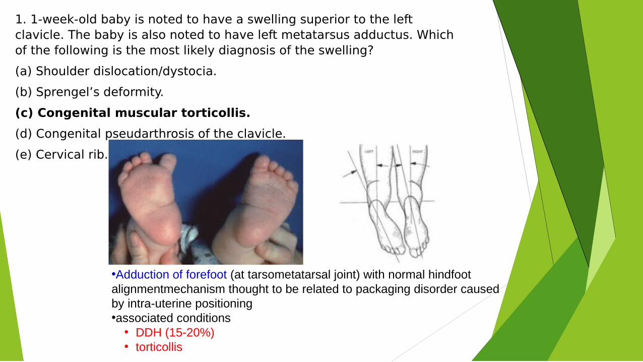
1. 1-week-old baby is noted to have a swelling superior to the left clavicle. The baby is also noted to have left metatarsus adductus. Which of the following is the most likely diagnosis of the swelling?
(a) Shoulder dislocation/dystocia.
(b) Sprengel’s deformity.
(c) Congenital muscular torticollis.
(d) Congenital pseudarthrosis of the clavicle.
(e) Cervical rib.
•Adduction of forefoot (at tarsometatarsal joint) with normal hindfoot alignmentmechanism thought to be related to packaging disorder caused by intra-uterine positioning•associated conditions
• DDH (15-20%)• torticollis
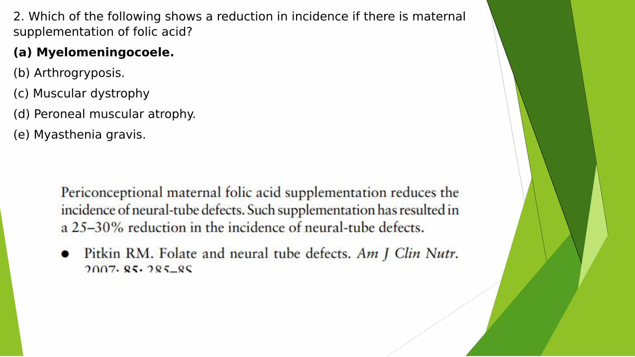
2. Which of the following shows a reduction in incidence if there is maternal supplementation of folic acid?
(a) Myelomeningocoele.
(b) Arthrogryposis.
(c) Muscular dystrophy
(d) Peroneal muscular atrophy.
(e) Myasthenia gravis.
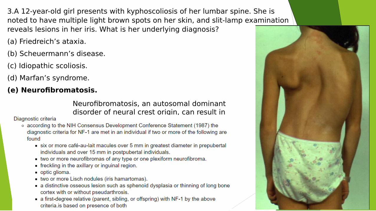
3.A 12-year-old girl presents with kyphoscoliosis of her lumbar spine. She is noted to have multiple light brown spots on her skin, and slit-lamp examination reveals lesions in her iris. What is her underlying diagnosis?
(a) Friedreich’s ataxia.
(b) Scheuermann’s disease.
(c) Idiopathic scoliosis.
(d) Marfan’s syndrome.
(e) Neurofibromatosis.
Neurofibromatosis, an autosomal dominant disorder of neural crest origin, can result in neuromuscular scoliosis.
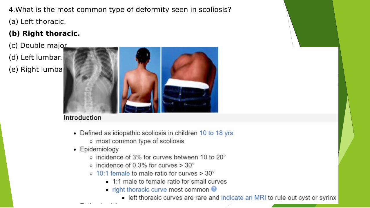
4.What is the most common type of deformity seen in scoliosis?
(a) Left thoracic.
(b) Right thoracic.
(c) Double major.
(d) Left lumbar.
(e) Right lumbar.
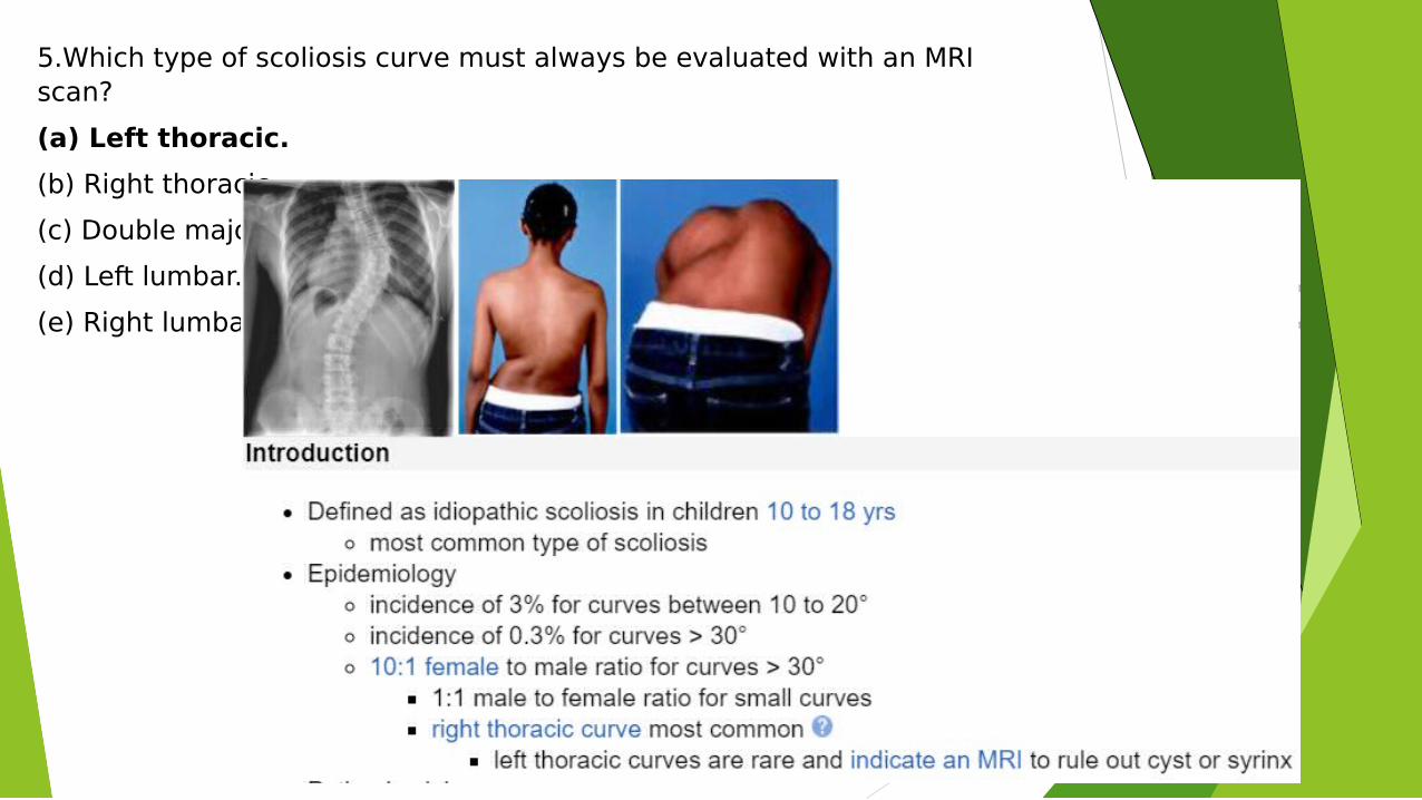
5.Which type of scoliosis curve must always be evaluated with an MRI scan?
(a) Left thoracic.
(b) Right thoracic.
(c) Double major.
(d) Left lumbar.
(e) Right lumbar.
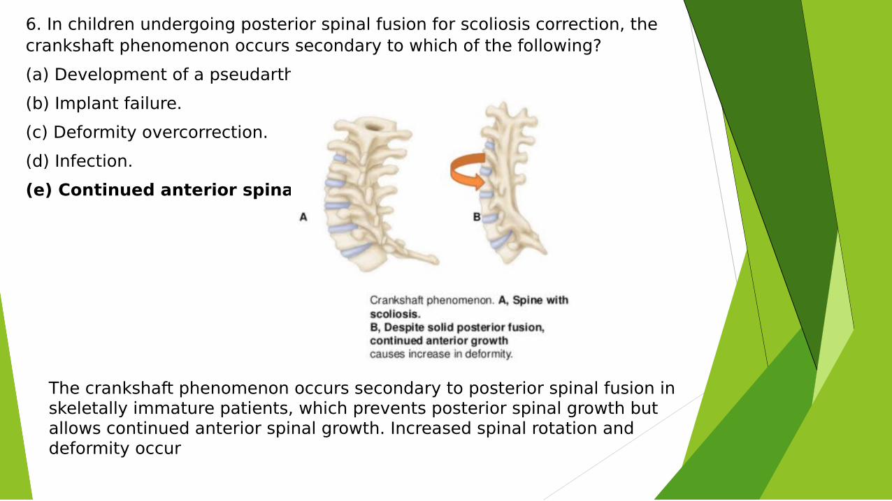
6. In children undergoing posterior spinal fusion for scoliosis correction, the crankshaft phenomenon occurs secondary to which of the following?
(a) Development of a pseudarthrosis.
(b) Implant failure.
(c) Deformity overcorrection.
(d) Infection.
(e) Continued anterior spinal growth
The crankshaft phenomenon occurs secondary to posterior spinal fusion in skeletally immature patients, which prevents posterior spinal growth but allows continued anterior spinal growth. Increased spinal rotation and deformity occur
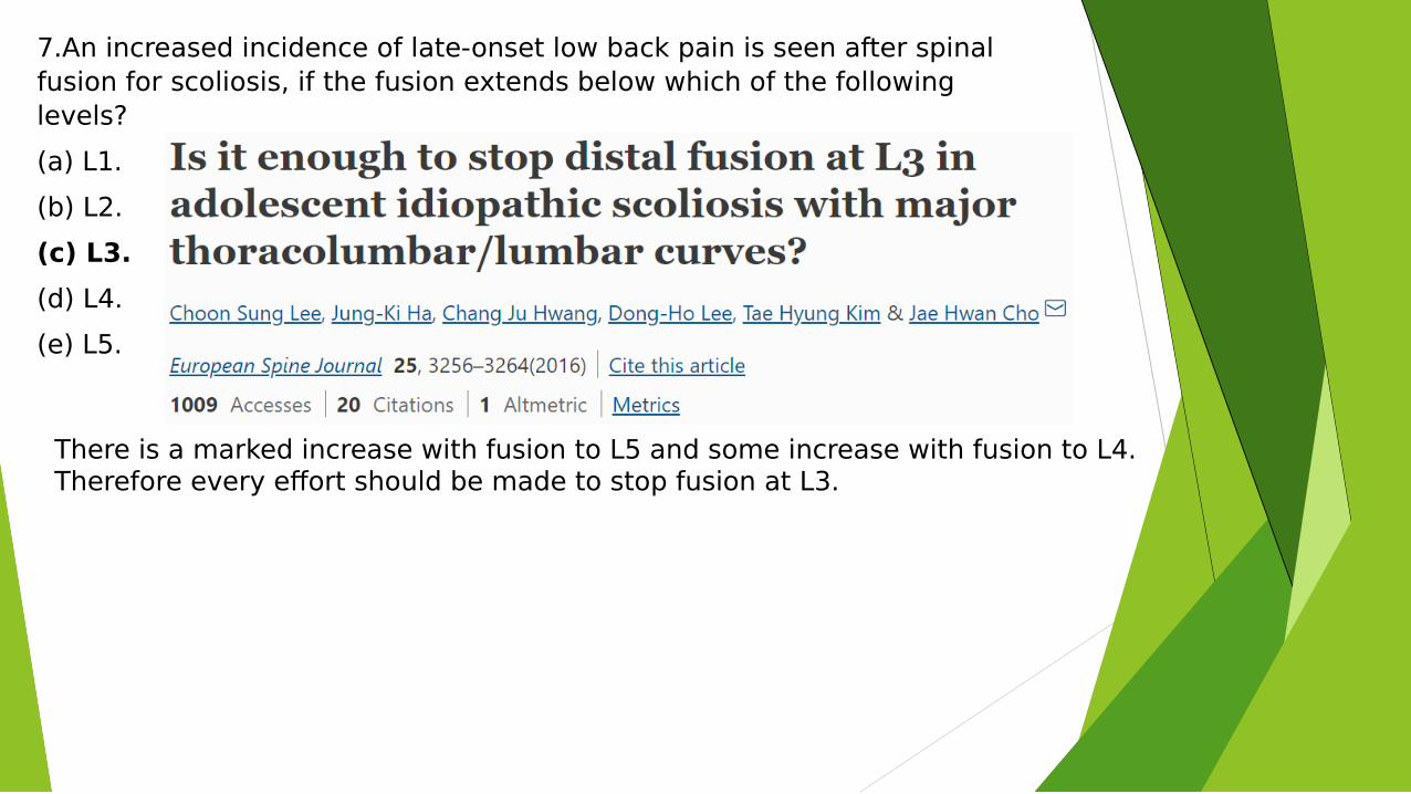
7.An increased incidence of late-onset low back pain is seen after spinal fusion for scoliosis, if the fusion extends below which of the following levels?
(a) L1.
(b) L2.
(c) L3.
(d) L4.
(e) L5.
There is a marked increase with fusion to L5 and some increase with fusion to L4. Therefore every effort should be made to stop fusion at L3.
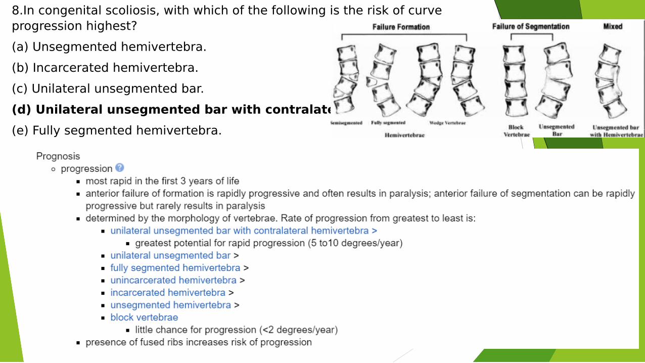
8.In congenital scoliosis, with which of the following is the risk of curve progression highest?
(a) Unsegmented hemivertebra.
(b) Incarcerated hemivertebra.
(c) Unilateral unsegmented bar.
(d) Unilateral unsegmented bar with contralateral hemivertebra.
(e) Fully segmented hemivertebra.
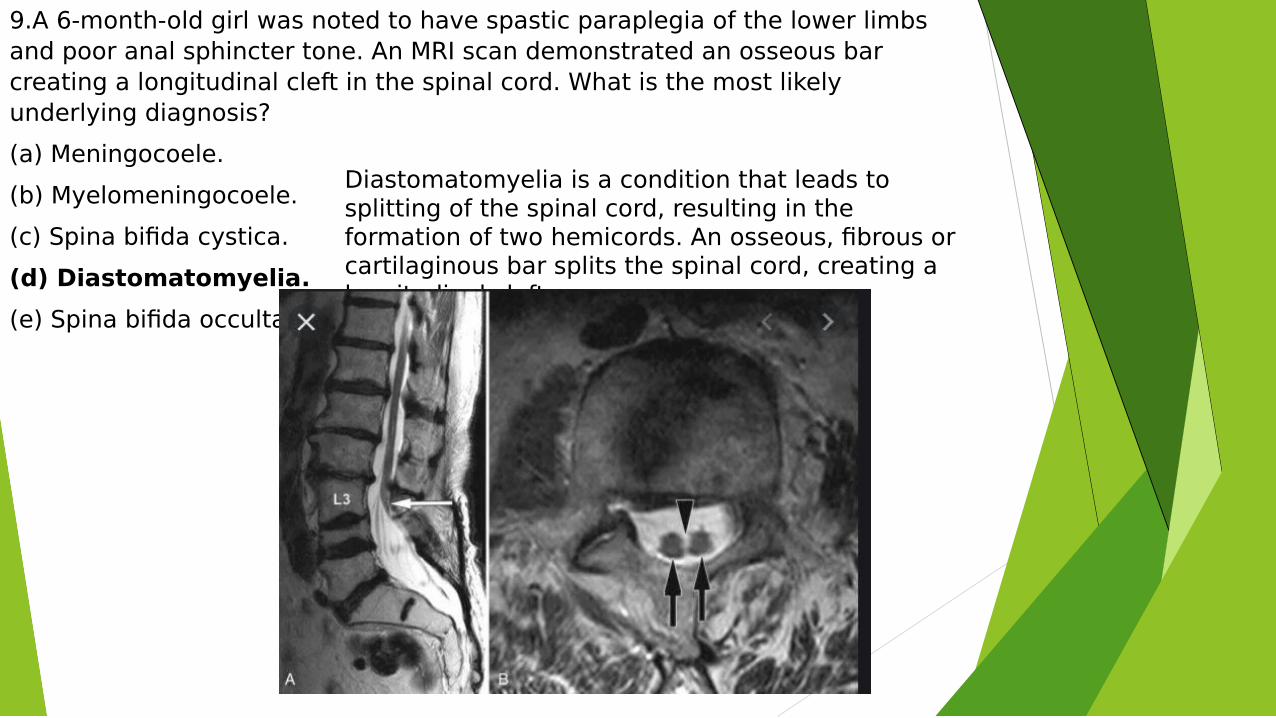
9.A 6-month-old girl was noted to have spastic paraplegia of the lower limbs and poor anal sphincter tone. An MRI scan demonstrated an osseous bar creating a longitudinal cleft in the spinal cord. What is the most likely underlying diagnosis?
(a) Meningocoele.
(b) Myelomeningocoele.
(c) Spina bifida cystica.
(d) Diastomatomyelia.
(e) Spina bifida occulta.
Diastomatomyelia is a condition that leads to splitting of the spinal cord, resulting in the formation of two hemicords. An osseous, fibrous or cartilaginous bar splits the spinal cord, creating a longitudinal cleft.
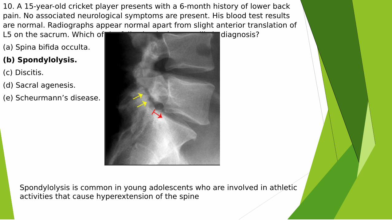
10. A 15-year-old cricket player presents with a 6-month history of lower back pain. No associated neurological symptoms are present. His blood test results are normal. Radiographs appear normal apart from slight anterior translation of L5 on the sacrum. Which of the following is the most likely diagnosis?
(a) Spina bifida occulta.
(b) Spondylolysis.
(c) Discitis.
(d) Sacral agenesis.
(e) Scheurmann’s disease.
Spondylolysis is common in young adolescents who are involved in athletic activities that cause hyperextension of the spine
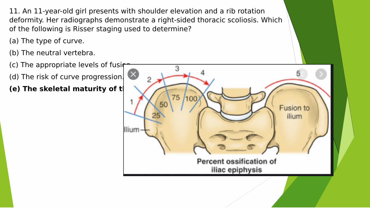
11. An 11-year-old girl presents with shoulder elevation and a rib rotation deformity. Her radiographs demonstrate a right-sided thoracic scoliosis. Which of the following is Risser staging used to determine?
(a) The type of curve.
(b) The neutral vertebra.
(c) The appropriate levels of fusion.
(d) The risk of curve progression.
(e) The skeletal maturity of the patient.
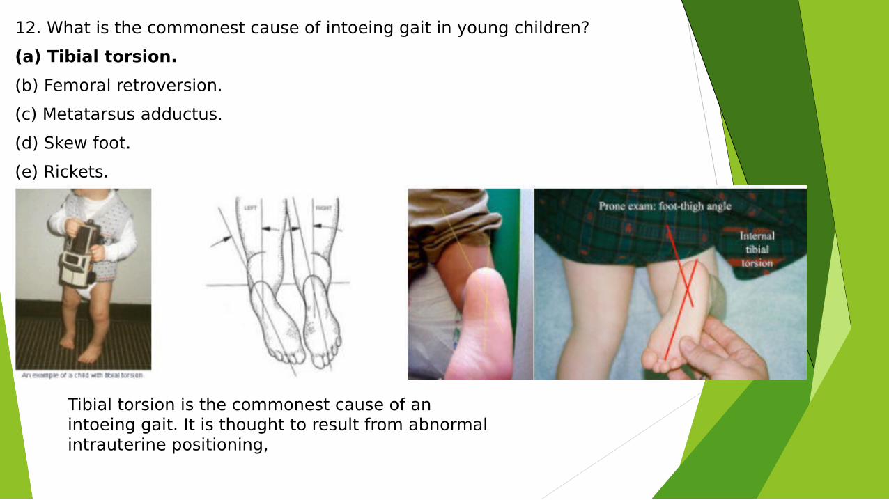
12. What is the commonest cause of intoeing gait in young children?
(a) Tibial torsion.
(b) Femoral retroversion.
(c) Metatarsus adductus.
(d) Skew foot.
(e) Rickets.
Tibial torsion is the commonest cause of an intoeing gait. It is thought to result from abnormal intrauterine positioning,
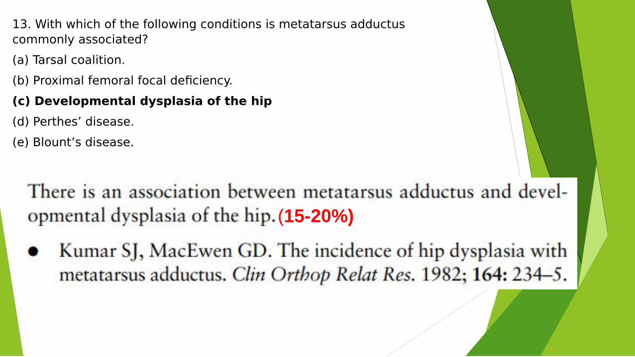
13. With which of the following conditions is metatarsus adductus commonly associated?
(a) Tarsal coalition.
(b) Proximal femoral focal deficiency.
(c) Developmental dysplasia of the hip
(d) Perthes’ disease.
(e) Blount’s disease.
(15-20%)
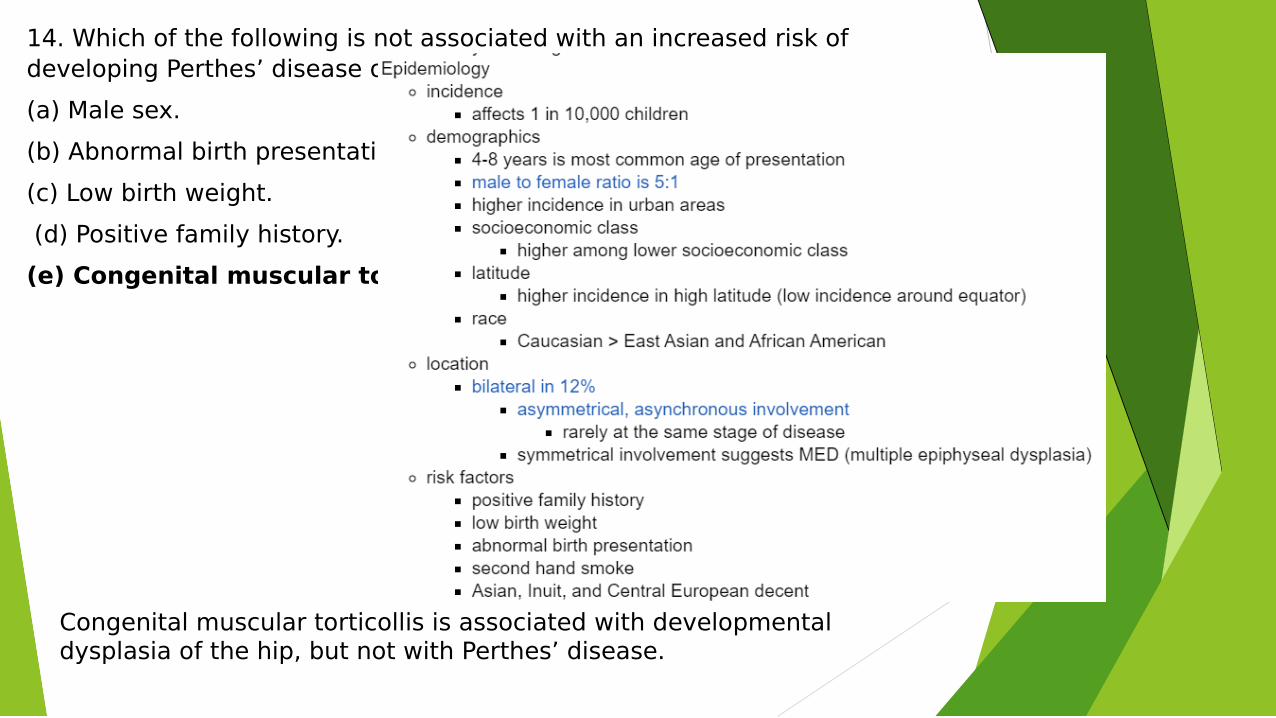
14. Which of the following is not associated with an increased risk of developing Perthes’ disease of the hip?
(a) Male sex.
(b) Abnormal birth presentation.
(c) Low birth weight.
(d) Positive family history.
(e) Congenital muscular torticollis.
Congenital muscular torticollis is associated with developmental dysplasia of the hip, but not with Perthes’ disease.
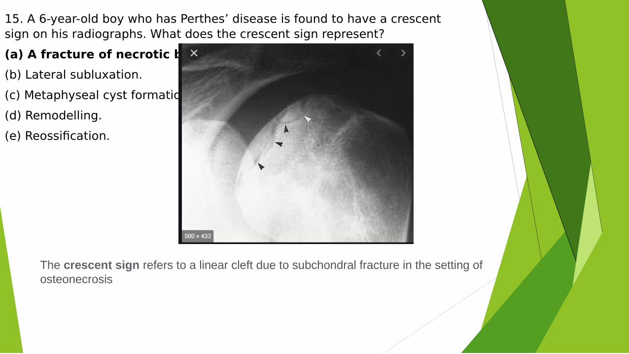
15. A 6-year-old boy who has Perthes’ disease is found to have a crescent sign on his radiographs. What does the crescent sign represent?
(a) A fracture of necrotic bone.
(b) Lateral subluxation.
(c) Metaphyseal cyst formation.
(d) Remodelling.
(e) Reossification.
The crescent sign refers to a linear cleft due to subchondral fracture in the setting of osteonecrosis
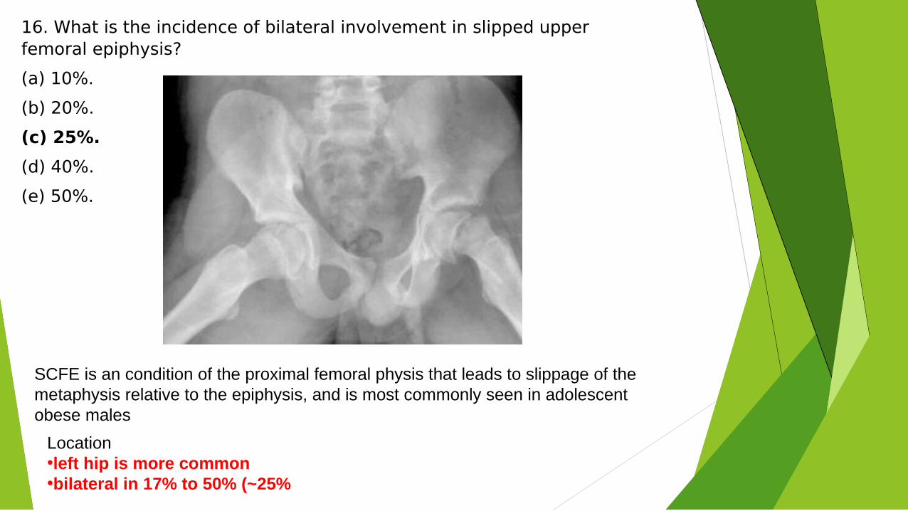
16. What is the incidence of bilateral involvement in slipped upper femoral epiphysis?
(a) 10%.
(b) 20%.
(c) 25%.
(d) 40%.
(e) 50%.
SCFE is an condition of the proximal femoral physis that leads to slippage of the metaphysis relative to the epiphysis, and is most commonly seen in adolescent obese males
Location•left hip is more common•bilateral in 17% to 50% (~25%
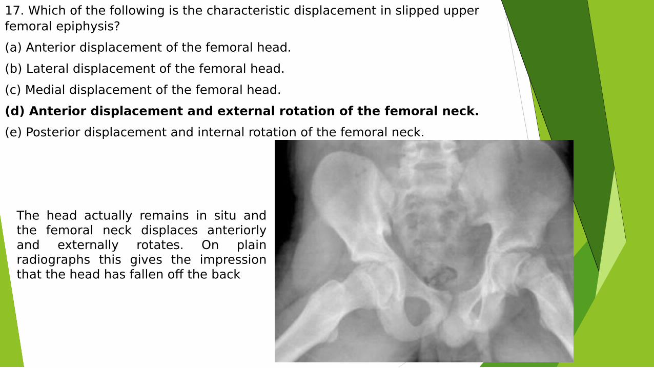
17. Which of the following is the characteristic displacement in slipped upper femoral epiphysis?
(a) Anterior displacement of the femoral head.
(b) Lateral displacement of the femoral head.
(c) Medial displacement of the femoral head.
(d) Anterior displacement and external rotation of the femoral neck.
(e) Posterior displacement and internal rotation of the femoral neck.
The head actually remains in situ and the femoral neck displaces anteriorly and externally rotates. On plain radiographs this gives the impression that the head has fallen off the back
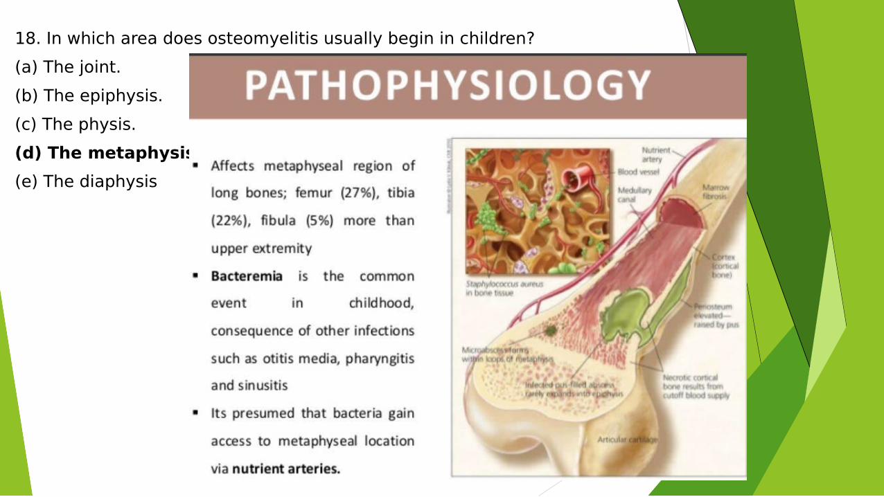
18. In which area does osteomyelitis usually begin in children?
(a) The joint.
(b) The epiphysis.
(c) The physis.
(d) The metaphysis.
(e) The diaphysis
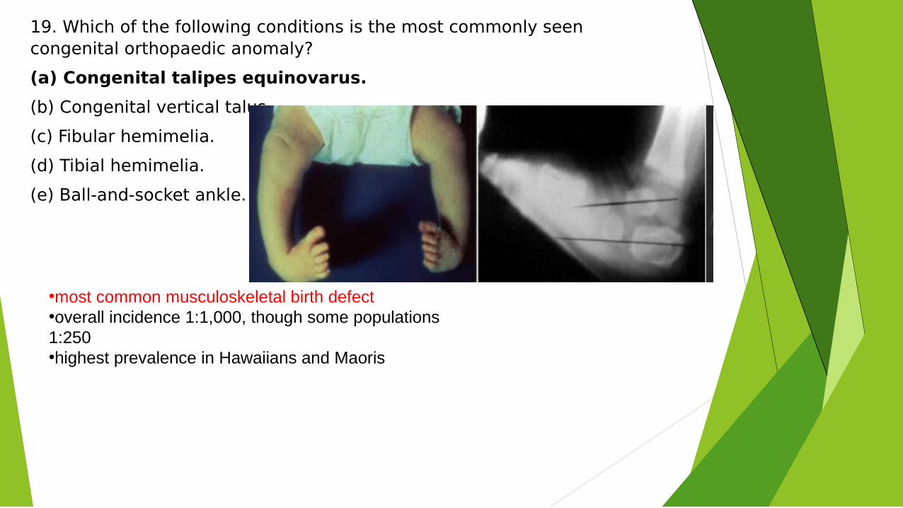
19. Which of the following conditions is the most commonly seen congenital orthopaedic anomaly?
(a) Congenital talipes equinovarus.
(b) Congenital vertical talus.
(c) Fibular hemimelia.
(d) Tibial hemimelia.
(e) Ball-and-socket ankle.
•most common musculoskeletal birth defect•overall incidence 1:1,000, though some populations 1:250 •highest prevalence in Hawaiians and Maoris
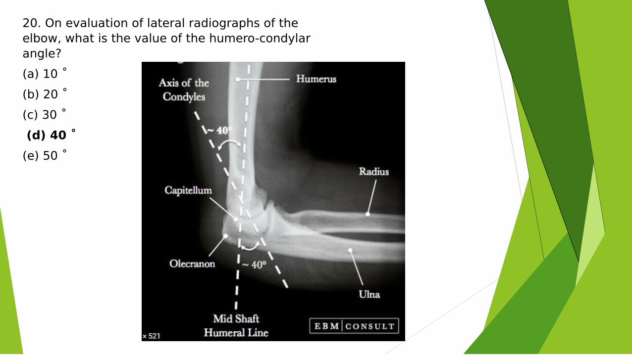
20. On evaluation of lateral radiographs of the elbow, what is the value of the humero-condylar angle?
(a) 10 ˚
(b) 20 ˚
(c) 30 ˚
(d) 40 ˚
(e) 50 ˚
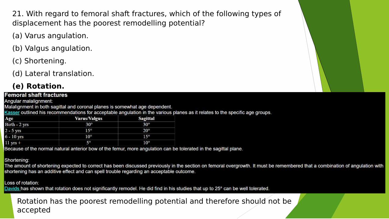
21. With regard to femoral shaft fractures, which of the following types of displacement has the poorest remodelling potential?
(a) Varus angulation.
(b) Valgus angulation.
(c) Shortening.
(d) Lateral translation.
(e) Rotation.
Rotation has the poorest remodelling potential and therefore should not be accepted
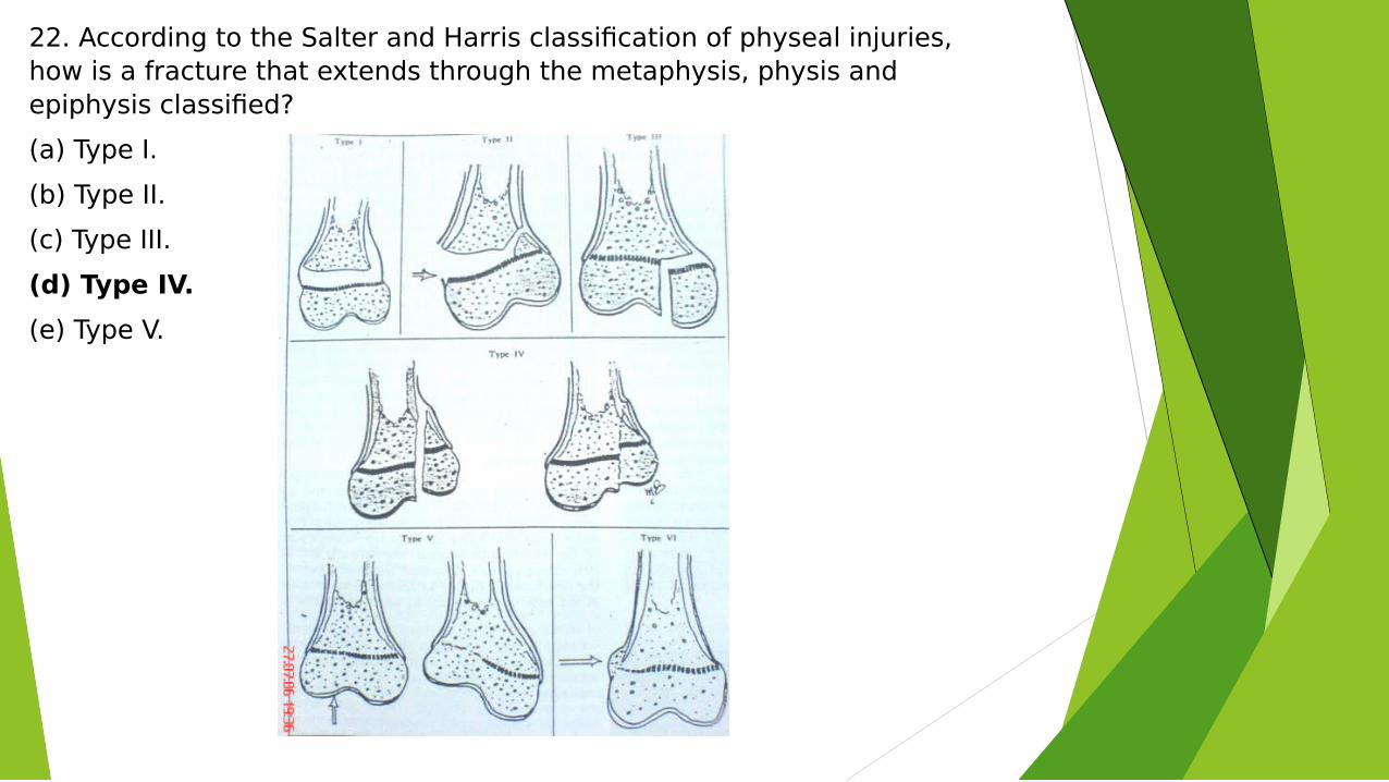
22. According to the Salter and Harris classification of physeal injuries, how is a fracture that extends through the metaphysis, physis and epiphysis classified?
(a) Type I.
(b) Type II.
(c) Type III.
(d) Type IV.
(e) Type V.
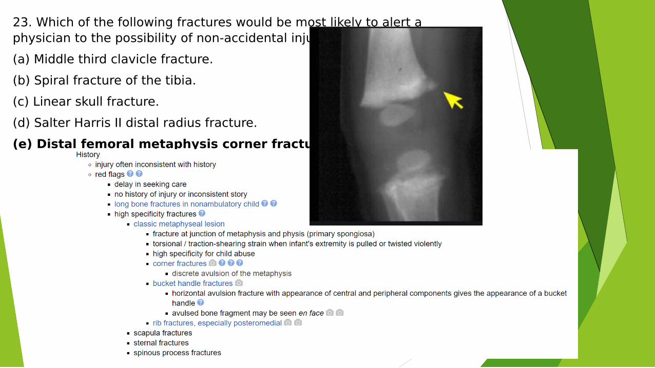
23. Which of the following fractures would be most likely to alert a physician to the possibility of non-accidental injury?
(a) Middle third clavicle fracture.
(b) Spiral fracture of the tibia.
(c) Linear skull fracture.
(d) Salter Harris II distal radius fracture.
(e) Distal femoral metaphysis corner fracture.
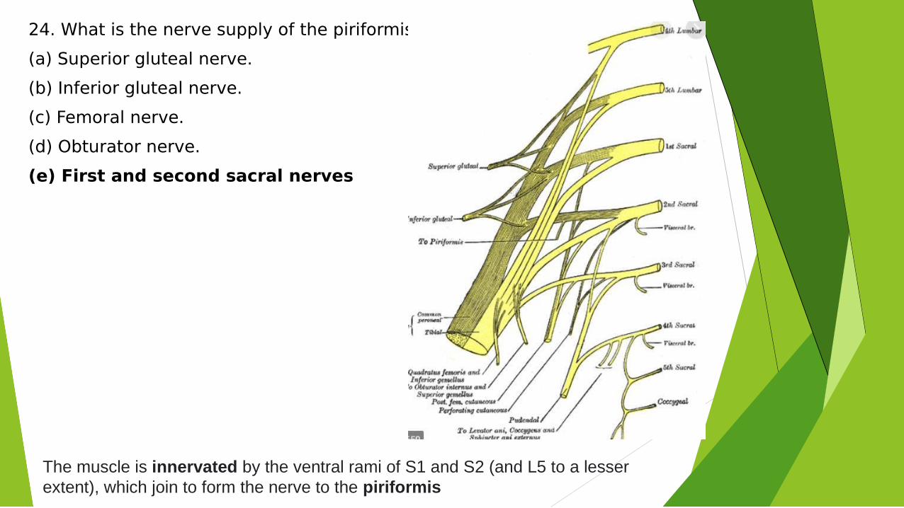
24. What is the nerve supply of the piriformis muscle?
(a) Superior gluteal nerve.
(b) Inferior gluteal nerve.
(c) Femoral nerve.
(d) Obturator nerve.
(e) First and second sacral nerves
The muscle is innervated by the ventral rami of S1 and S2 (and L5 to a lesser extent), which join to form the nerve to the piriformis
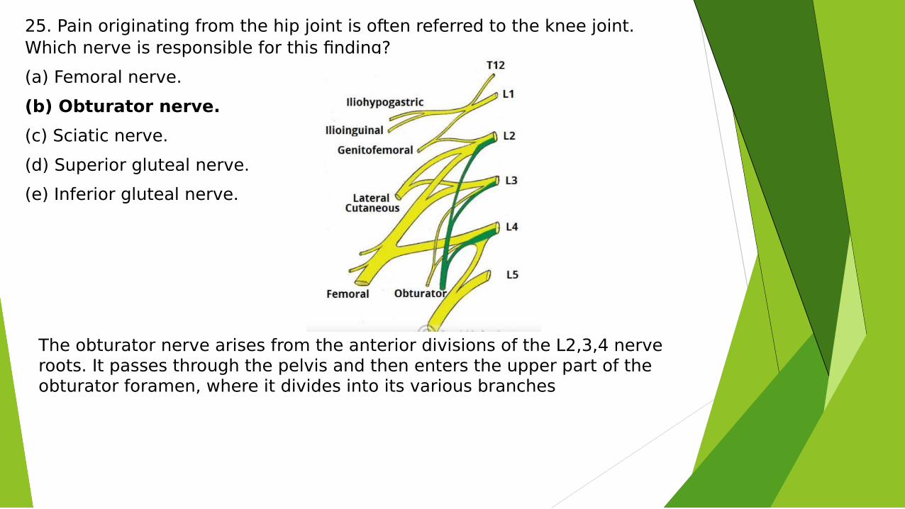
25. Pain originating from the hip joint is often referred to the knee joint. Which nerve is responsible for this finding?
(a) Femoral nerve.
(b) Obturator nerve.
(c) Sciatic nerve.
(d) Superior gluteal nerve.
(e) Inferior gluteal nerve.
The obturator nerve arises from the anterior divisions of the L2,3,4 nerve roots. It passes through the pelvis and then enters the upper part of the obturator foramen, where it divides into its various branches