Embed Size (px)
Citation preview

This journal is c The Royal Society of Chemistry 2011 Chem. Soc. Rev., 2011, 40, 2279–2292 2279
Cite this: Chem. Soc. Rev., 2011, 40, 2279–2292
Evolution in the understanding of [Fe]-hydrogenasew
Michael J. Corr and John A. Murphy*
Received 19th October 2010
DOI: 10.1039/c0cs00150c
Hydrogenases catalyse redox reactions with molecular hydrogen, either as substrate or product.
The enzymes harness hydrogen as a reductant using metals that are abundant and economical,
namely, nickel and iron, and should provide new pointers for the economic use of hydrogen in
manmade devices. The most recently discovered and perhaps the most enigmatic of the
hydrogenases is the [Fe]-hydrogenase, used by certain microorganisms in the pathway that
reduces carbon dioxide to methane. Since its discovery some twenty years ago, [Fe]-hydrogenase
has consistently provided structural and mechanistic surprises, often requiring complete
re-evaluation of its mechanism of action. This tutorial review combines recent advances in X-ray
crystallography and other analytical techniques, as well as in computational studies and in
chemical synthesis to provide a platform for understanding this remarkable enzyme type.
Introduction
Production of ‘biogas’ fuel, rich in methane, is a topic of
growing economic and ecological importance. The methanogen
microorganisms that perform this task are archaea, and the
enzymes within the archaea that take part in catalytic cycles
where carbon dioxide is reduced to methane use various
feedstocks as the reducing source.1 Among these, our fascination
is with the use of dihydrogen as the reducing agent. Through
syntrophy,2 different microorganisms conspire to produce and
to utilise dihydrogen in the reduction of carbon dioxide. For
example, the so-called Methanobacillus omelianskii culture3 is
a co-culture of two partner organisms, strain S and strain
M.o.H.;4 the two strains cooperate in the conversion of
ethanol to acetate and carbon dioxide to methane by intimate
inter-species dihydrogen transfer (Scheme 1).
The two most prevalent types of hydrogenases are known
as [FeFe]-hydrogenase (containing iron–sulfur clusters) and
[NiFe]-hydrogenase (containing nickel and iron–sulfur
clusters).5–10 The [NiFe] hydrogenase family includes
many different types including selenium-based [NiFeSe]
hydrogenases.1,5–10 A third type of hydrogenase was discovered
in 1990 that did not contain nickel or iron–sulfur clusters.11
This is known as the [Fe]-hydrogenase [previously known as
iron–sulfur-cluster free hydrogenase or H2-forming methyl-
enetetrahydromethanopterin dehydrogenase (Hmd)] and is
obtained from methanogenic archaea grown in nickel-limiting
conditions. It catalyses the reversible reduction of N5,N10-
methenyltetrahydromethanopterin (CHRH4MPT+) 1
with H2 to N5,N10-methylenetetrahydromethanopterin
(CH2QH4MPT) 2 and a proton (Scheme 2) as a key step in
the production of methane from CO2 in methanogenic archaea
(DG10 = �5.5 kJ mol�1).12
The [Fe]-hydrogenase catalyses reversible stereospecific hydride
transfer from H2 to C14a of CHRH4MPT+1, providing
the pro-R methylene hydrogen in CH2QH4MPT 2.13,14
Scheme 1 Cooperative use of H2 by syntrophic organisms.
WestCHEM, Department of Pure and Applied Chemistry, Universityof Strathclyde, 295 Cathedral Street, Glasgow G1 1XL, UK.E-mail: [email protected]; Tel: +44 (0)141 548 2389w Note added in proof: after submission of this review, two reviews onthis type of enzyme, with different emphases, appeared.61,62
Michael Corr achieved his M.Sci. degree at the University ofStrathclyde in Glasgow. He gained his PhD from the sameuniversity in 2010 following his studies with Prof. John Murphyon the synthesis and reactivity of superelectrophiles. He iscurrently working as a Postdoctoral Research Associate at theUniversity of St Andrews.
John Murphy was born in Dublin and educated at TrinityCollege, Dublin (BA 1976) and the University of Cambridge(PhD 1980). He held Fellowships at the University of Albertaand the University of Oxford, before being appointed to theUniversity of Nottingham to start his independent research. Hemoved to his current position as Merck-Pauson Professor ofChemistry at the University of Strathclyde in 1995. He obtainedhis DSc University of Strathclyde in 2002. His research interestsare in reactivity in chemistry and biology and he is Director ofthe Glasgow Centre for Physical Organic Chemistry.
Chem Soc Rev Dynamic Article Links
www.rsc.org/csr TUTORIAL REVIEW
Dow
nloa
ded
by L
udw
ig M
axim
ilian
s U
nive
rsita
et M
uenc
hen
on 2
8 Ju
ne 2
011
Publ
ishe
d on
01
Mar
ch 2
011
on h
ttp://
pubs
.rsc
.org
| do
i:10.
1039
/C0C
S001
50C
View Online

2280 Chem. Soc. Rev., 2011, 40, 2279–2292 This journal is c The Royal Society of Chemistry 2011
Initially, it was believed that H2-forming methyl-
enetetrahydromethanopterin dehydrogenase did not contain
functional metal (although 1 mol iron per mol enzyme was
detected at this stage),11 making it a unique, purely organic
hydrogenase.15 This discovery led to much excitement. The
proposal by Berkessel and Thauer16 for the mechanism of
action of this enzyme proved particularly interesting, as this
served to extend the concept of superelectrophilic activation
into the realm of enzymatic reactions, a concept that had
previously been postulated only under superacid conditions.
Whereas this activation may still occur in the enzymatic
conversion, the enzyme was subsequently discovered to
contain an iron cofactor essential for activity17 and the enzyme
crystal structure has been reported,18,19 leading to a new range
of mechanistic investigations and interest in this unusual
hydrogenase. This review examines the investigations carried
out on the [Fe]-hydrogenase, from its initial discovery and
analysis to an assessment of the current position and prospects
for the utilisation of the knowledge of its mode of action for
the future.
Initial studies on H2-forming
methylenetetrahydromethanopterin dehydrogenase
5,6,7,8-Tetrahydromethanopterin (H4MPT) 3 (Fig. 1) serves
as a carrier of C1 fragments in the metabolism of methanogenic
archaea.1 During the course of methanogenesis, a C1 fragment
is transferred to H4MPT 3 at the oxidation level of formic
acid, thereby forming 1, which is subsequently reduced to the
methyl oxidation level in a stepwise manner. During this
process CHRH4MPT+ 1 is converted to CH2QH4MPT 2
by the action of a hydrogenase, then named H2-forming
methylenetetrahydromethanopterin dehydrogenase. In 1990,
Thauer et al.11 reported the hydrogenase activity found
in Methanobacterium thermoautotrophicum, a thermophilic
methanogen that reduces CO2 to CH4. The hydrogenase
activity was rapidly lost under aerobic conditions or in the
presence of dithiothreitol (DTT) or mercaptoethanol.3 Hmd is
a homodimer composed of only one type of subunit that has
an apparent molecular mass from SDS polyacrylamide gel
electrophoresis of 43 kDa with a specific activity about
1000 U mg�1.3 Determination of the primary structure gave
37 788 Da as the actual mass of the polypeptide subunit.20
Both [NiFe]-hydrogenase and [FeFe]-hydrogenase mediate
the reduction of viologen dyes with hydrogen.21 In the absence
of an electron–acceptor, these hydrogenases can catalyse the
exchange of hydrogens between water and H2. However, Hmd
does not catalyse the reduction of viologen dyes and catalyses
the exchange between H2O and H2 only in the presence of
CHRH4MPT+ 1 or CH2QH4MPT 2.12 Hmd was also
unusual in that it appeared to contain no functional metal.11,12
UV-visible spectroscopy showed no absorbance above
340 nm, indicating that Hmd was not an iron–sulfur protein
or flavoprotein. In addition, analysis of non-heme iron showed
o0.4 mol iron per mol enzyme (in contrast with that found
earlier)11 and ICP-MS and AAS analysis showed no nickel or
transition metals (levels o0.1 mol per mol enzyme).12 The
only metal found in significant quantities was zinc, found to
vary between 0.5–4 mol zinc per mol enzyme depending upon
the enzyme culture. However, the zinc content did not
correlate with the specific activity of the enzyme.12 The effect
of CO, CN�, acetylene, nitrite and azide as inhibitors was also
investigated.12 It was found that CO concentrations of 5% and
50% and as well as a CN� concentration of 5% did not cause
any significant loss in activity of the enzyme. Only a CN�
concentration of 50% caused a loss in activity below 50%.
Nitrite, azide and acetylene were not inhibitors under the
conditions of the assays.
Next, tritium exchange in the enzyme was examined.12 When
pure Hmd alone was used, no exchange between 3H2 and1H2O
was observed at various pH values and temperatures. In the
presence of CHRH4MPT+ 1 or CH2QH4MPT 2, exchange
between 3H2 and1H2O was observed. Using CHRH4MPT+ 1,
isolation of the products by HPLC showed that per mol of
CH2QH4MPT 2 formed, 0.5 mol of 3H was incorporated into
the methylene group of CH2QH4MPT 2, consistent with transfer
of a single 3H to the substrate from the 3H2 and with the release
of a corresponding 0.5 mol of 3H into the aqueous medium.
Similarly using 3H-labelled CH2QH4MPT 2, with the label
distributed in the CH2 group, the CHRH4MPT+ formed had
only half the specific radioactivity of the labelled CH2QH4MPT
2, again consistent with transfer of just one of the CH2
hydrogens.
Various isotope studies have been conducted in order to
probe the reaction between CHRH4MPT+ 1 and dihydrogen
in the presence of Hmd. Schworer et al.22 examined the
production of dihydrogen from CH2QH4MPT 2 using hydrogen
isotopes. The purpose of these reactions was to determine if
the reaction of CHRH4MPT+1 with hydrogen occurred via
hydride transfer. If this was the case, the dehydrogenation of
CD2QH4MPT 4 in H2O or the dehydrogenation of
CH2QH4MPT 2 in D2O would ideally produce only HD as
a product, as only one of the hydrogen atoms (or deuterium
atoms) could come from either substrate 2 or 4.
The results showed that for CD2QH4MPT 4 in H2O, HD
and H2 were produced, whereas for CH2QH4MPT 2 in D2O,
only HD and D2 were produced. The production of H2 from
Scheme 2 Reaction catalysed by H2-forming methylenetetrahydro-
methanopterin dehydrogenase, i.e. [Fe]-hydrogenase.
Fig. 1 5,6,7,8-Tetrahydromethanopterin 3.
Dow
nloa
ded
by L
udw
ig M
axim
ilian
s U
nive
rsita
et M
uenc
hen
on 2
8 Ju
ne 2
011
Publ
ishe
d on
01
Mar
ch 2
011
on h
ttp://
pubs
.rsc
.org
| do
i:10.
1039
/C0C
S001
50C
View Online

This journal is c The Royal Society of Chemistry 2011 Chem. Soc. Rev., 2011, 40, 2279–2292 2281
CD2QH4MPT 4 in H2O (or D2 from CH2QH4MPT 2 in D2O)
was explained by the exchange between HD and the solvent,
which was caused by CHRH4MPT+-dependent Hmd catalysis.
A summary of the reactions is shown in Scheme 3.
In the absence of CH2QH4MPT 2 (or CD2QH4MPT 4), no
exchange between H2 and D2O (or D2 and H2O) was observed,
even when high concentrations of the enzyme were used. No
generation of D2 was observed when using CD2QH4MPT 4 in
H2O and no generation of H2 was observed when using
CH2QH4MPT 2 in D2O. This indicated that only one of the
hydrogens from CH2QH4MPT 2 underwent reaction to form
dihydrogen.
The stereochemical course of the reaction was determined
by Schleucher et al.13 using two-dimensional NMR spectro-
scopy. Reaction of CHRH4MPT+ 1 with D2 in D2O led to
formation of CHDQH4MPT 6 and CD2QH4MPT 7
(Scheme 4). Formation of 6 was due to deuteride abstraction
from D2 at the C14a carbon to give labelled-6.
It was found that for a solution of Hmd and
CHRH4MPT+ 1 in D2O, the signal for the methenyl hydrogen
gradually disappeared, indicating exchange of the proton with
D+ from D2O. This side-reaction was much slower than the
enzyme-catalysed reaction; it was not catalysed by the enzyme
and its rate increased with increasing pH. Although no
discussion of its mechanism has appeared in the literature,
this is most likely due to deprotonation to form 9, which
would be more usually represented as the N-heterocyclic
carbene 10, (Scheme 5) a reaction type more familiar in the
biological chemistry of thiamine salts as well as in synthetic
chemistry.23,24 Due to this, formation of 7 in the reaction
could be explained by H/D exchange of the methenyl H
of 1 with D from D2O, then reaction with D2 to give
CD2QH4MPT 7 (Scheme 5). Correspondingly, reaction of
CDRH4MPT+ 5 with H2 in H2O led to formation
of CDHQH4MPT 8 and CH2QH4MPT 2 (Scheme 4).
Determination of the absolute stereochemistry showed that
hydride was transferred from hydrogen to take up the pro-R
position of C14a in CH2QH4MPT 2.
Thauer et al.25 then showed the presence of a low molecular
weight cofactor tightly bound to the enzyme and apparently
containing no redox-active transition metal. Hmd was
heterologously expressed from Methanococcus jannaschii
and, surprisingly, showed no activity in the reduction of 1.
Hmd from Methanothermobacter marburgensis was denatured in
urea and the ultrafiltrate (componentso 10 kDa molecular mass)
Scheme 3 Hydrogen isotope studies on the dehydrogenation of
CH2QH4MPT 2.
Scheme 4 NMR-labelling studies showing stereochemistry of the
reaction.
Scheme 5 H/D exchange observed in CHRH4MPT+ 1.
Fig. 2 Representation of Hmd ultrafiltrate reaction.
Dow
nloa
ded
by L
udw
ig M
axim
ilian
s U
nive
rsita
et M
uenc
hen
on 2
8 Ju
ne 2
011
Publ
ishe
d on
01
Mar
ch 2
011
on h
ttp://
pubs
.rsc
.org
| do
i:10.
1039
/C0C
S001
50C
View Online

2282 Chem. Soc. Rev., 2011, 40, 2279–2292 This journal is c The Royal Society of Chemistry 2011
was collected. When the heterologously produced Hmd in
M. jannaschii was added to the ultrafiltrate fromM. marburgensis,
the combination showed activity in the reaction of 1 with
hydrogen (Fig. 2 represents the reaction performed). Similarly,
Hmd heterologously expressed from M. marburgensis
showed activity when added to the ultrafiltrate collected
fromM. jannaschii. Analysis of the ultrafiltrate showed compo-
nents with a molecular mass below 1000 Da, shown, at the
time, to contain no nickel and only traces of iron and zinc
by TXRF analysis (Total Reflection X-ray Fluorescence
spectrometry).
Superelectrophilic activation in Hmd
Although many activity studies and spectroscopic labelling
studies had conclusively shown that Hmd catalysed the transfer
of hydride from molecular hydrogen to CHRH4MPT+1 to
form CH2QH4MPT 2, the mechanistic details of the reaction
still remained a mystery.
In 1995, Berkessel and Thauer16 presented an intriguing
mechanistic proposal for the reaction of Hmd based on super-
electrophilic activation of alkanes.26 The proposed mechanism
is shown in Schemes 6 and 7. They proposed that protonation
of CHRH4MPT+ 1 in the enzyme active site would lead to
formation of superelectrophilic dicationic species 11 (or 12).
This, in turn, would increase the carbocationic character of
C14a in CHRH4MPT+1, allowing it to abstract hydride from
molecular hydrogen to give CH2QH4MPT 2 (Scheme 6). This
proposal was based on the work of Olah et al.26–29 on the
activation of alkanes for hydride exchange in the presence of
superacids.
According to their proposal,16 when CHRH4MPT+ 1 is in
the enzyme active site, it can undergo protonation on either N5
or N10 to give dication species 11/12 (Scheme 6). Berkessel and
Thauer proposed that this protonation would be accompanied
by a conformational change in the imidazolinium ring to give a
distorted 5-membered ring such as 14, and that the transition
state for reaction with H2 would involve a pentacoordinated
carbonium cation (Scheme 7). The antiperiplanar arrangement
of the nitrogen lone pair of electrons to the C14a–H bond in 14
would make it sufficiently reactive to abstract hydride from
molecular hydrogen to give conformationally locked species
15. Removal from the enzyme active site frees the nitrogen
lone pair of electrons to give reduced product 2.
Thauer et al.30 later examined the rate of dihydrogen
production from the reaction of CD2QH4MPT 4 in H2O,
based on the proposal above that the reaction occurs through
a pentacoordinated carbonium cation such as 14. Two
mechanisms were proposed for the formation of H2 in the
reaction (Scheme 8).
Scheme 6 Superelectrophilic activation of CHRH4MPT+ 1.
Scheme 7 Schematic representation of superelectrophilic activation
in Hmd.
Scheme 8 Mechanisms of formation of H2 by CD2QH4MPT 4.
Dow
nloa
ded
by L
udw
ig M
axim
ilian
s U
nive
rsita
et M
uenc
hen
on 2
8 Ju
ne 2
011
Publ
ishe
d on
01
Mar
ch 2
011
on h
ttp://
pubs
.rsc
.org
| do
i:10.
1039
/C0C
S001
50C
View Online

This journal is c The Royal Society of Chemistry 2011 Chem. Soc. Rev., 2011, 40, 2279–2292 2283
In mechanism 1, protonation of CD2QH4MPT 4 gives
cation [CD2HQH4MPT]+ 16, which then loses HD to give
CDRH4MPT+ 5 (mechanism 1(a), Scheme 8). The HD
produced can then undergo exchange with acid to give H2
and D+ (mechanism 1, eqn (b), Scheme 8). The second
proposed mechanism again involves protonation of
CD2QH4MPT 4 and gives cation [CD2HQH4MPT]+ 16.
Instead of loss of HD, exchange of D+ and H+ occurs to
give onium cation [CH2DQH4MPT]+ 17, with subsequent
loss of H2 to give CDRH4MPT+ 5. The exchange could be
envisaged as involving the transition state 160 involving
specific activation of the pro-R deuteron. In mechanism 1,
HD is formed as an intermediate in the reaction, whereas in
mechanism 2, free HD is not an intermediate in the reaction.
Kinetic investigation of the mechanism could then discriminate
between mechanisms 1 and 2.
They studied the kinetics of formation of H2 and HD from
CD2QH4MPT 4 in H2O and the formation of HD and D2
from CH2QH4MPT 2 in D2O. They found that the rate of
production of H2 or D2 was independent of the concentration
of HD produced in either reaction. Based on this, the authors
concluded that free H–D was unlikely to be produced in the
reaction.31 Of the two mechanisms considered, mechanism 2
was therefore preferred, in which production of H2 or D2
would likely occur through a transition state 160 which under-
goes stereospecific exchanges with the protons of water.
The superelectrophilic mechanism proposed by Berkessel
and Thauer16,35 precipitated a number of computational
studies32–34 on the feasibility of the reaction of amidinium
ions with molecular hydrogen. However, interest in the
hydrogenase was about to intensify further, thanks to an
important discovery that was to be reported.36
From metal-free to [Fe]-hydrogenase
When Lyon et al.36 reexamined the spectral properties of the
Hmd hydrogenase, they made a startling discovery. They
showed that Hmd was inactivated by UV-A/blue light
irradiation and, from this, determined that the ‘‘metal-free’’
hydrogenase did in fact contain an ‘‘active’’ iron cofactor.
Hmd was prepared under anaerobic conditions using an
anaerobic chamber filled with 95%N2/5%H2. These preparations
showed 1 � 0.2 mol iron per mol Hmd monomer (determined
by trapping the iron in a complex with absorption maximum
at 563 nm, and measuring against a FeCl3 calibration curve).
The Hmd stored in the dark showed no loss in activity, in
contrast to samples exposed to white light, where activity
decreased to zero over 30 minutes. Tests using monochromatic
light showed Hmd to be inactivated by UV-A (320–400 nm)
and blue light (400–500 nm). In addition, experiments showed
that iron-leaching increased linearly with decrease in Hmd
activity when EDTA was present and that the inactive Hmd
contained no iron. When EDTA was not present, light
irradiation caused bleaching and inactivation of Hmd, but
almost no iron was released from the enzyme.
Shima et al. also re-examined the effect of CO inhibition on
Hmd.37 Previous studies12 had shown that Hmd was not
inactivated by CO concentrations of 5% or 50%. Now,
however, at 100% CO concentration, Hmd activity was
inhibited by over 50%. The CO concentration was B100-fold
higher than that required to inhibit most [FeFe]-hydrogenases
andB10-fold higher than for [NiFe]-hydrogenases, which was
why the high level had not been previously examined. The CO
inhibition was shown to be reversible, and removal of CO
resulted in a return in Hmd activity.
Studies on the cofactor alone showed a light sensitivity, with
the cofactor being bleached with resulting loss of iron, upon
exposure to light. From these studies, Lyon et al.37 proposed
that Hmd contained an iron-containing cofactor (later called
the FeGP cofactor, where GP is an abbreviated form of
guanylylpyridinol or guanylylpyridone) that is not redox-active
and is necessary for the enzyme activity. With this important
discovery, the H2-forming methylenetetrahydromethanopterin
dehydrogenase could no longer be considered as a purely
organic hydrogenase and a great deal of research was conducted
into determining the structure of this new iron active site and
its importance in the hydrogenase activity.
Light- or heat-inactivation of the cofactor released iron and
CO. Chemical analysis revealed 2.4 � 0.2 mol of CO per mol
Fe in the cofactor.38 In addition, a relatively stable product of
mass 542 Da could be isolated by HPLC and analysed.38
Structure elucidation on the stable product suggested a
pyridone-type ligand consistent with 18 (Fig. 3). The pyridone
structure was supported by fluorescence spectroscopy on the
inactivated cofactor.
Next, Lyon et al.37 examined the IR spectrum of Hmd in
order to determine the ligands that were attached to the iron
in the cofactor. IR analysis of the enzyme showed two
absorption bands at 2011 and 1944 cm�1, derived from the
two intrinsic CO molecules bound to the iron atom in the
cofactor. No change in the IR spectrum was observed when
the enzyme’s spectrum was measured under nitrogen, oxygen
or hydrogen. Analysis of the intensity of the IR bands
suggested the presence of an Fe(CO)2 unit where the CO
groups were present at an angle of 901.
They then examined the IR spectra of CO-inhibited Hmd,
CN�-inhibited Hmd and the spectrum of Hmd in the presence of
1 or 2. Incubation of Hmd under CO led to three CO bands
(2074, 2020 and 1981 cm�1) of similar intensity. Incubation
under 13CO resulted in three bands (2050, 1999 and 1980 cm�1),
one of which was determined as coming from 13CO. The IR
spectrum reverted back to the spectrum recorded for Hmd
(two CO bands at 2011 and 1944 cm�1) when the gas phase
was replaced with argon, H2, N2 or air. The authors suggested
that the CO-inhibited enzyme contains three CO units and
that the extrinsic CO units will not replace the two intrinsic
CO molecules under any incubation times or temperatures. In
addition, the extrinsic CO is readily removed upon removal of
Fig. 3 Pyridone ligand isolated from Hmd cofactor.
Dow
nloa
ded
by L
udw
ig M
axim
ilian
s U
nive
rsita
et M
uenc
hen
on 2
8 Ju
ne 2
011
Publ
ishe
d on
01
Mar
ch 2
011
on h
ttp://
pubs
.rsc
.org
| do
i:10.
1039
/C0C
S001
50C
View Online

2284 Chem. Soc. Rev., 2011, 40, 2279–2292 This journal is c The Royal Society of Chemistry 2011
CO from the gas phase, but the intrinsic CO ligands are not
removed.
The CN�-inhibited Hmd contained three bands at 2091, 2020
and 1956 cm�1. When 13CN� was used as the inhibiting reagent,
three bands were observed at 2047, 2017 and 1956 cm�1. Based
on the shifts measured, the cyanide bands were assigned to 2091
and 2047 cm�1 for CN� and 13CN� respectively. Calculations
based on the spectrum intensities indicated that the two CO units
were at 901 to one another. Competition for inhibition binding
between CO and CN� showed the CN�-inhibited enzyme as the
major product. This indicated that Hmd had a higher affinity for
CN� than for CO as a ligand.
Addition of 1 or 2 under the presence of argon or H2 led to
small changes in the IR spectrum of Hmd. The IR bands of the
CO signals shifted slightly (from 1–7 cm�1) and sharpening of the
absorption bands occurred. The results suggested that 1 and 2
bind close to or at the iron site in the cofactor, resulting in a slight
shift to higher frequency in the CO bands. The 20–30% decrease
in the width of the absorption bands indicated less vibration of
the CO positions in space, consistent with a decrease in flexibility
of the active-site pocket caused by binding of 1 to the iron centre
or in close proximity to it. With the addition of H2, the effects of
1 on the IR spectrum of Hmd were amplified. This could indicate
that hydrogen may directly interact with the iron centre in the
presence of 1, although the authors could find no evidence of
Fe–H or Fe–D stretches in the IR spectrum.
When the iron-containing cofactor was isolated and analysed,
the CO bands were found to shift from 2011 and 1944 cm�1 to
2031 and 1972 cm�1, indicative of decreased electron density in
the iron centre or a change in the total number of ligands
attached.
Based on the IR analysis of the CO ligands, Lyon et al.37
concluded that the iron in the Hmd cofactor was coordinated to
two CO molecules. From this they concluded that the iron
present in Hmd is most likely to be Fe(II); coordination
to three CO molecules in the CO-inhibited Hmd makes Fe(III)
unlikely, as iron(III) tricarbonyl complexes have not been
reported. Fe(0) as the species is unlikely as iron(0) cyanide
complexes are rare (but not unknown, [Fe(CO)4CN]� has been
reported).39 Hmd was shown to be EPR-silent,40 which ruled out
Fe(I) as the iron species. This makes Fe(II) the most probable
species to be present in Hmd, but the results did not prove the
oxidation state.
In order to gain insight into the electronic structure of iron
in Hmd, Shima et al.40 measured the Mossbauer spectra of57Fe-labelled Hmd prepared under dark conditions. The re-
sults showed the presence of a low-spin iron in a low oxidation
state. Inhibition of the enzyme by CO or CN� showed changes
in the recorded Mossbauer spectrum, an indication that
the inhibitors were bound to the iron site. However, the
addition of hydrogen and/or CHRH4MPT+1 did not lead
to any significant change in the Mossbauer spectrum. This
would seem to indicate that H2 or 1 bind near the iron in the
enzyme, but are not direct ligands to the iron site, that
when Hmd interacts with the substrates (H2 and/or 1), the
electronic state of the iron doesn’t change significantly.40
This is surprising, as even if 1 does not bind to the Fe centre,
it would be expected that hydrogen would need to bind
to or interact with the Fe centre (and thereby to change its
spectroscopic properties) in order to become activated for
heterolytic cleavage.
Quantification of the Mossbauer signal intensities gave a
calculated 1.14 � 0.25 mol iron per mol protein monomer.
Analysis of the magnetic properties of the three Hmd preparations
(Hmd, CO-inhibited Hmd and CN�-inhibited Hmd) showed
them to be diamagnetic with spin S = 0, consistent with Fe(II)
or Fe(0) low-spin and not paramagnetic Fe(I) or Fe(III) low-
spin. Based on the IR41 and Mossbauer40 studies, it appears
likely that the iron present in Hmd is Fe(II). The case for an
Fe(II) complex received further strong support in an infrared
study of carbonyl stretch frequencies, performed by Pickett,
Liu et al.41 Here the enzyme and the isolated cofactor
showed bands that, on comparison with known mono-iron cis-
dicarbonyl complexes were clearly consistent only with Fe(II).
More recently, Meyer-Klaucke et al.42 examined the
iron-complex structure of [Fe]-hydrogenase and model
systems, using X-ray absorption near-edge spectroscopy, and
concluded that an Fe(II) complex was consistent with the
[Fe]-hydrogenase in M. jannaschii.
The crystal structure revealed
In 2006, Pilak et al.43 reported the crystal structure of the
apoenzyme of Hmd (the enzyme without the iron-containing
cofactor present) fromM. jannaschii andM. kandleri at 1.75 A
and 2.4 A resolution respectively.
Hmd from M. jannaschii was found to consist of a homo-
dimer with approximate dimensions of 90 A � 50 A � 40 A,
subdivided into a central globular unit attached to two
peripheral units in a linear manner. Each peripheral unit
corresponded to the N-terminal domain of one subunit and
was composed of an a/b structure that belongs to the Rossmann
fold protein family (Fig. 4). The central globular unit was
composed of the C-terminal segment of both subunits, each
containing four a-helices that form an intertwined intersubunit
helix bundle. The Hmd from M. jannaschii was present in a
closed conformational state (Fig. 5(a)), whereas the Hmd from
M. kandleri was found to be in an open conformational state
(Fig. 5(b)). Within the crystal structure, the authors located a
U-shaped electron density 13 A long, located close to the
bottom of the cleft; this is shown in green in Fig. 5 and 6. The
electron density was found to be consistent with polyethyl-
eneglycol, present in the crystallising solution. This was
thought to indicate the presence of a channel for substrate
delivery within the enzyme.
Activity measurements on Hmd reconstituted from
M. jannaschii showed that for the three conserved cysteine
residues (Cys10, Cys176 and Cys250), only that at Cys176 was
essential for enzyme activity.43 Based on this, Cys176 was
assigned as the iron-ligating sulfur ligand.
The authors modelled the iron active site and binding of
CH2QH4MPT 2 in the enzyme based on the structure of
the formaldehyde-activating enzyme of M. extorquens in a
complex with CH2QH4MPT 2.43 They found that 2 sits in the
iron active site at the base of the U-shaped electron density
(Fig. 5 and 6). In this position, the C14a of 2 is B6 A from the
assumed Cys176-ligated iron atom, with sufficient space for
binding of molecular hydrogen. In this model, the phenyl ring
Dow
nloa
ded
by L
udw
ig M
axim
ilian
s U
nive
rsita
et M
uenc
hen
on 2
8 Ju
ne 2
011
Publ
ishe
d on
01
Mar
ch 2
011
on h
ttp://
pubs
.rsc
.org
| do
i:10.
1039
/C0C
S001
50C
View Online

This journal is c The Royal Society of Chemistry 2011 Chem. Soc. Rev., 2011, 40, 2279–2292 2285
adjacent to the imidazolidine ring of 2 is B4.5 A from the
pyridone ring of the cofactor ligand 18, which the authors
proposed could mean that the phenyl ring was present for
binding or conformational purposes.
A later crystal structure showed that the FeGP cofactor18
was anchored to the enzyme by a guanosine monophosphate.
Within the active site, the iron atom exists in a distorted
octahedron ligation shell. One of the ligands is from the
nitrogen of the pyridinol ligand 19 (Fig. 7)—from X-ray
analysis it is not possible to see whether the 2-substituent on
the pyridine is in its protonated (hydroxy) state, or if the
hydroxyl group is deprotonated. The carboxyl group in ligand
19 was disordered in the crystal structure, which led to a
tentative determination of its orientation in the crystal structure.
The other ligands on the iron were composed of two CO
molecules at 901 to each other, consistent with previous
studies,37,40 a sulfur from the cysteine in Cys176, an unknown
ligand (‘U’ in Fig. 7) the structure of which could not be solved
due to high levels of disorder and a solvent water molecule
(‘O’ in Fig. 7). The structure of the unknown ligand could not
be determined from the electron density in the crystal structure.
However, soaking the crystals in 3 mM cyanide led to a 1.6-fold
increase in the electron density at this site, suggesting that it
could be the site of reversible cyanide inhibition.
The ligand site anti- to the nitrogen of 20 was shown to
contain a water molecule. However, the distance between the
oxygen atom of water and the iron atom was 2.7 A, considered
too far away to be a ‘‘proper’’ ligand to the iron. The authors
suggested that this could be the site of binding for H2 or
extrinsic CO in the CO-inhibited enzyme. The solvent
molecule interacts with another solvent molecule, which in
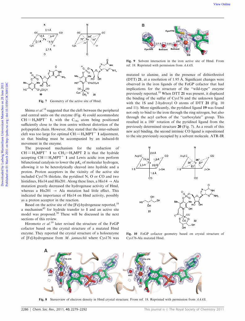
turn is linked to the carbonyl group of Cys250 (Fig. 8 and 9).
Fig. 5 (a) Closed conformational state of Hmd in M. jannaschii.
(b) Open conformational state of Hmd in M. kandleri. The U-shaped
electron density (see text) is shown in green. Reprinted from the
ref. 43, copyright 2006 with permission from Elsevier.
Fig. 4 Crystal structure of Hmd apoenzyme of M. jannaschii. Reprinted from the ref. 43, copyright 2006 with permission from Elsevier.
Fig. 6 Model of the iron cofactor and substrate 2 in the presumed
active site of Hmd in M. jannaschii. The model of the iron-containing
cofactor is shown in yellow, the conformation of CH2QH4MPT 2
taken from the crystal structure of M. extorquens44 is in grey and
is unchanged. Reprinted from the ref. 43, copyright 2006 with
permission from Elsevier.
Dow
nloa
ded
by L
udw
ig M
axim
ilian
s U
nive
rsita
et M
uenc
hen
on 2
8 Ju
ne 2
011
Publ
ishe
d on
01
Mar
ch 2
011
on h
ttp://
pubs
.rsc
.org
| do
i:10.
1039
/C0C
S001
50C
View Online
2286 Chem. Soc. Rev., 2011, 40, 2279–2292 This journal is c The Royal Society of Chemistry 2011
Shima et al.18 suggested that the cleft between the peripheral
and central units on the enzyme (Fig. 4) could accommodate
CHRH4MPT+ 1, with the C14a atom being positioned
sufficiently close to the iron centre without distortion of the
polypeptide chain. However, they stated that the inter-subunit
cleft was too large for optimal CHRH4MPT+1 adjustment,
so that binding must be accompanied by an induced-fit
movement in the enzyme.
The proposed mechanism for the reduction of
CHRH4MPT+ 1 to CH2QH4MPT 2 is that the hydride
accepting CHRH4MPT+ 1 and Lewis acidic iron perform
bifunctional catalysis to lower the pKa of molecular hydrogen,
allowing it to be heterolytically cleaved into hydride and a
proton. Proton acceptors in the vicinity of the active site
included Cys176 thiolate, the pyridinol N, O or CO and two
histidines, His14 and His201. Along these lines, a His14-Ala
mutation greatly decreased the hydrogenase activity of Hmd,
whereas a His201 - Ala mutation had little effect. This
indicated the importance of His14 on Hmd activity, possibly
as a proton acceptor in the reaction.
Based on the active site of the [Fe]-hydrogenase reported,18
a mechanism45 for hydride transfer to 1 and an active site
model was proposed.50 These will be discussed in the next
sections of this review.
Hiromoto et al.19 later revised the structure of the FeGP
cofactor based on the crystal structure of a mutated Hmd
enzyme. They reported the crystal structure of a holoenzyme
of [Fe]-hydrogenase from M. jannaschii where Cys176 was
mutated to alanine, and in the presence of dithiothreitol
(DTT) 21, at a resolution of 1.95 A. Significant changes were
observed in the iron ligands of the FeGP cofactor that had
implications for the structure of the ‘‘wild-type’’ enzyme
previously reported.18 When DTT 21 was present, it displaced
the binding of the sulfur of Cys176 and the unknown ligand
with the 1S and 2-hydroxyl O atoms of DTT 21 (Fig. 10
and 11). More significantly, the pyridinol ligand 19 was found
not only to bind to the iron through the ring nitrogen, but also
through the acyl carbon of the ‘‘carboxylate’’ group. This
resulted in a 1801 rotation of the pyridinol ligand from the
previously determined structure 20 (Fig. 7). As a result of this
new acyl binding, the second intrinsic CO ligand is repositioned
to the site previously occupied by a solvent molecule. ATR-IR
Fig. 7 Geometry of the active site of Hmd.
Fig. 8 Stereoview of electron density in Hmd crystal structure. From ref. 18. Reprinted with permission from AAAS.
Fig. 9 Solvent interaction in the iron active site of Hmd. From
ref. 18. Reprinted with permission from AAAS.
Fig. 10 FeGP cofactor geometry based on crystal structure of
Cys176-Ala mutated Hmd.
Dow
nloa
ded
by L
udw
ig M
axim
ilian
s U
nive
rsita
et M
uenc
hen
on 2
8 Ju
ne 2
011
Publ
ishe
d on
01
Mar
ch 2
011
on h
ttp://
pubs
.rsc
.org
| do
i:10.
1039
/C0C
S001
50C
View Online

This journal is c The Royal Society of Chemistry 2011 Chem. Soc. Rev., 2011, 40, 2279–2292 2287
analysis of the mutated enzyme before and after the crystal
structure determination still showed the presence of two CO
ligands with a calculated angle of 901 between them.46,47
In the mutated enzyme, the hydroxyl group on the pyridinol
ligand now interacted via a hydrogen bond to the imidazole
group of His14. In addition, the oxygen of the acyl group of
the pyridinol ligand was linked via a hydrogen bond to the
amide group of Ala176.
Based on this new geometry, Hiromoto et al.19 reexamined
the previously reported18 EXAFS data on the ‘‘wild-type’’
Hmd enzyme. Modifying the iron ligation to include the newly
proposed bidentate ligation by the pyridinol ligand led
to a better data fit upon re-refining. The authors stated
that the ‘‘previous interpretation of the electron density was
biased by the lack of imagination concerning the possibility of
an acyl group as iron ligand and on the subsequent conclusion
that the orientation of a negatively charged carboxylate
group towards the rather unpolar protein interior is unlikely’’.19
As a result of this, a new structure for the FeGP cofactor was
proposed (Fig. 12 and 13). In this new structure, the authors
state that the most probable site of the second intrinsic CO is
that previously occupied by a solvent water molecule (Fig. 7).
This was based on the crystal structure obtained for the
Cys176-Ala mutated Hmd enzyme, but its position could not
be identified on a structural basis from analysis of the crystal
structure of the ‘‘wild-type’’ Hmd enzyme.
Based on all of the above studies it appears likely that the
iron in the active site of the [Fe]-hydrogenase is ligated by the
ring nitrogen and acyl carbon of the pyridinol ligand, two
intrinsic CO molecules, the sulfur of Cys176 and an unknown
ligand U, affording an overall complex represented in different
styles as 23 or 24. It is thought that both CHRH4MPT+1
and H2 can enter the active site via a channel in the protein and
that the action of the cationic substrate 1 and Lewis acidic Fe
heterolytically cleave molecular hydrogen, though the exact
mechanism is not known. CHRH4MPT+ 1 can be orientated
close to the iron centre and even interact with the ligands of
the iron, but it is not thought to bond directly to the iron
centre during the reaction, although it has not been fully
discounted. Mossbauer studies40 on the iron centre showed
no significant change on addition of 1 to the enzyme, however,
no significant changes were observed on the addition of H2.
With the structure of [Fe]-hydrogenase determined and the
geometry of the active site elucidated, the next stage in
development will be obtaining a plausible mechanism for the
reaction of CHRH4MPT+ 1 with molecular hydrogen.
Computational modelling involving the iron complex
Since reporting of the crystal structure of Hmd, it was entirely
as expected that much work would be conducted in order to
try and understand the mechanism of reaction of this unusual
enzyme. As a result of this, a variety of mechanistic studies
have been reported in an attempt to explain its unusual
reactivity. This section examines these literature reports.
First reported crystal structure
Yang and Hall45 reported a trigger mechanism for the reaction
of Hmd using density functional theory calculations. They
used complex 25 (Scheme 9) as a model for the then-determined
active-site 20. Several differences in model 25 to the active site
are apparent. The hydroxyl group in the model had been
moved from the 2-position of the pyridine in the real group to
the 3-position in this model in order to prevent it from strongly
interacting with the sulfur or CO groups bound to the iron.
Furthermore the replacement of the phosphate ester at the
4-position of the pyridine ring by a pyridinol hydroxyl group is
a significant deviation from the structure of the true cofactor
(unless the true cofactor were to undergo phosphate ester
hydrolysis as part of the catalytic cycle). Another significant
proposal was inclusion of an oxygen bound to the iron in
place of the unknown ligand, the oxygen coming from the
Fig. 11 Stereoview of electron density in mutated Hmd (Cys176-Ala)
crystal structure. Reprinted from ref. 19, copyright 2009 with permission
from Elsevier.
Fig. 12 Revised crystal structure for ‘‘wild-type’’ Hmd.
Fig. 13 Reinterpretation (stereoview) of iron-ligation structure in
[Fe]-hydrogenase. Reprinted from ref. 19, copyright 2009 with permission
from Elsevier.
Dow
nloa
ded
by L
udw
ig M
axim
ilian
s U
nive
rsita
et M
uenc
hen
on 2
8 Ju
ne 2
011
Publ
ishe
d on
01
Mar
ch 2
011
on h
ttp://
pubs
.rsc
.org
| do
i:10.
1039
/C0C
S001
50C
View Online

2288 Chem. Soc. Rev., 2011, 40, 2279–2292 This journal is c The Royal Society of Chemistry 2011
carboxylate chain on the pyridone ring. The calculated IR
values of the CO ligands in model 25 were 1957 and 2014 cm�1
(cf. 1944 and 2011 cm�1 in Hmd).45
The proposed mechanism is shown in Scheme 9. The
conversion of complex 25 where an anionic carboxylate is
bound to iron into complex 27 where MPT+ 26 has bound to
iron, involves the intramolecular transfer of two electrons
from the pyridine ring to the iron, thereby inverting polarity
at the iron and allowing it to attack the MPT+ cation. The
conversion of 28 to 29 involves formal proton transfer to the
MPT carbon and hydride transfer to the iron atom. The
conversion of 30 to 31 reverses the previous intramolecular
two-electron transfer, leaving the iron polarized to accept the
carboxyl group as a ligand, prior to proton transfer. In the
absence of MPT+ 26, the splitting of H2 was calculated with a
barrier of 292 kJ mol�1. In the presence of MPT+ 26, the
activation barrier decreased to 92.0 kJ mol�1. The authors
stated that the exchange of H2/H+ in the mechanism is
strongly dependent on MPT+26 being present. Without it,
H2 splitting and the exchange of the proton with the protons
of water will not occur. They stated that MPT+ 26 or H2 could
arrive in any order at the active site, but that both must be
present for the reaction to occur.
Most recent crystal structure models
With the reporting of a recalculated crystal structure19 of
the active-site of the [Fe]-hydrogenase, further proposed
mechanisms and active-site models have been reported in the
literature. Yang and Hall48 once again reported a trigger
mechanism for the newly re-determined active-site based on
model 32 (Scheme 10).
The authors proposed that complex 35 represents the
cofactor within the resting state of the enzyme. Evidence for
this was from the calculated vibrational frequencies of the cis
CO units at 2007 and 1949 cm�1 closely resembling those of
the wild-type Hmd. Also, upon addition of H2, the Mossbauer
spectrum of the iron in Hmd does not change significantly.40
The authors proposed that complex 35 could be formed
without significant alteration to the IR spectrum of Hmd.
Complex 35 reacts with Ph–MPT+41 undergoing hydride
abstraction through transition state 36 (65.3 kJ mol�1) to
intermediate 37 (47.3 kJ mol�1) (Scheme 11). Ph–HMPT then
detaches from the complex to be replaced by H2, resulting in
formation of more stable complex 38. Proton loss from 38
could then occur from Cys176 (to give 40, 8.8 kJ mol�1) or
from the pyridinol ligand (to give 39, 0.4 kJ mol�1). This is
where the observed H2O/H+ exchange catalysed by Hmd and
1/2 occurs. Subsequent H2 cleavage occurs in 39 or 40 to give
complex 35 and the catalytic cycle continues.
The authors stated that the model showed that the sulfur of
Cys176 and the pyridine hydroxyl group were essential for the
reaction to proceed, aiding in the splitting of H2. It was also
stated that in the absence of Ph–MPT+ 41, no H2O/H+
exchange (or similarly D2O/H+ exchange) would occur
as the calculated deprotonation energies were too high
(42–84 kJ mol�1). The arrival of Ph–MPT+ 41 triggers a
breaking of the strong Fe–H–H–O bond in 35, allowing the
transfer of hydride to occur with an energy barrier of
65.3 kJ mol�l (excluding the energy of H2 splitting as shown
in Scheme 10).
The calculated deprotonation energies are shown in Table 1.
As can be seen, the exothermic deprotonations occur in
complexes 37 and 38, which occur after the reaction with
Ph–MPT+ 41. These calculations are consistent with the
observed reactivity of Hmd, where exchange was observed only
in the presence of Hmd and CHRH4MPT+ 1/CH2QH4MPT
2. However, it is unusual that the deprotonation of the sulfur of
MeSH in complex 35 is calculated as being so endothermic
(105 kJ mol�1). If complexed to the iron centre, a species such
Scheme 9 Proposed mechanism, based on computation, of H2
splitting by model 25.45 Energies are in kJ mol�1. The representation
of localised charges in 25 and 31 represents our (MJC, JAM)
interpretation of the published scheme to allow easy visualization of
reactivity. We thank Prof. Hall for helpful discussions.
Scheme 10 Splitting of H2 using computational model 32.48 Energies
in kJ mol�1.
Dow
nloa
ded
by L
udw
ig M
axim
ilian
s U
nive
rsita
et M
uenc
hen
on 2
8 Ju
ne 2
011
Publ
ishe
d on
01
Mar
ch 2
011
on h
ttp://
pubs
.rsc
.org
| do
i:10.
1039
/C0C
S001
50C
View Online

This journal is c The Royal Society of Chemistry 2011 Chem. Soc. Rev., 2011, 40, 2279–2292 2289
as R2SH+ would be expected to be easily deprotonated. If this
was to occur, it would allow H+/H2O exchange in the absence of
CHRH4MPT+ 1/CH2QH4MPT 2, a reactivity that is not
observed in Hmd.
A further development was reported by Shima et al.49 They
reported the crystal structure of a binary complex of
Cys176–Ala-mutated [Fe]-hydrogenase with CH2QH4MPT 2
at 2.15 A resolution. The results showed that CH2QH4MPT 2
fitted into the active-site cleft of [Fe]-hydrogenase (Fig. 14).
In the crystal structure obtained, the N5 and N10 of the
imidazolidine ring were found to be planar, rather than in
the non-planar active form of the substrate. The authors stated
that the non-planar active form could be induced by rotation
of the phenyl ring adjacent to the imidazolidine ring that could
occur in the closed form of the enzyme. The authors stated
that in the open-form of the enzyme crystallised, no carboxy
group was present to protonate one of the nitrogens of
CHRH4MPT+ 1. This protonation would be necessary for
the superelectrophilic activation mechanism proposed previously
(Schemes 6 and 7). In the open form of the enzyme crystallised,
it was found that the C14a of CH2QH4MPT 2 and iron of the
active-site were separated by a distance of 9.3 A, too far for
hydride transfer.
Shima et al.49 modelled the closed form of the enzyme with
CH2QH4MPT 2 present (Fig. 15). In this model, the distance
between C14a of CH2QH4MPT 2 and iron of the active-site
decreased to only 3 A. The model showed that the C14a of
CH2QH4MPT 2 lay trans to the acyl carbon of the pyridinol
ligand 31, suggesting that the site of H2 activation was in the
position trans to the acyl carbon inhabited by the unknown
ligand (U in 24, Fig. 12).
Based on the crystal structure, they proposed a catalytic
mechanism based on an open/closed conformational transition
(Scheme 12). The catalytic cycle is initiated by binding of
CHRH4MPT+1 to the open form of the enzyme (42,
Scheme 12), causing the closure of the cleft to give the closed
conformation (43, Scheme 12). This closure could induce
conformational changes in CHRH4MPT+ 1, enhancing its
carbocationic character. H2 is then captured at the site of the
unknown ligand, the proposed site of H2-activation, binding
side-on to the iron (44, Scheme 12). The H2 molecule becomes
polarised and can be heterolytically cleaved by the adjacent
carbocation (C14a of CHRH4MPT+ 1). The hydride is
accepted by CHRH4MPT+ 1 and the resulting proton is
proposed to be taken by a base (either the Cys176 thiol or
pyridinol oxygen). The results in formation of CH2QH4MPT
2 and a proton, which can undergo the exchange reactions
detailed above.
It was stated that no carboxy group was present to
protonate one of the nitrogens of CHRH4MPT+ 1, however,
in the modelled active-site 43, it was also stated that the
hydroxyl group of the pyridinol ring could interact with the
N10 of CHRH4MPT+ 1, which the authors proposed could
modify the properties of the pyridinol. The possibility of such
Scheme 11 Catalytic mechanism of reaction of Ph–MPT+ 41 with
H2.48 Energies in kJ mol�1.
Table 1 Calculated deprotonation energies of various complexes(Schemes 10 and 11)
Deprotonation site DG/kJ mol�1
Pyridinol O in 33 84Pyridinol O in 35 163Cys176 S in 35 105Cys176 S in 37 �38Pyridinol O in 38 �21Thiol S in 38 �13Thiol S in 39 96
Fig. 14 Active-site crystal structure of Cys176-Ala-mutated [Fe]-
hydrogenase. Ref. 49. Copyright Wiley-VCH Verlag GmbH & Co.
KGaA. Reproduced with permission.
Dow
nloa
ded
by L
udw
ig M
axim
ilian
s U
nive
rsita
et M
uenc
hen
on 2
8 Ju
ne 2
011
Publ
ishe
d on
01
Mar
ch 2
011
on h
ttp://
pubs
.rsc
.org
| do
i:10.
1039
/C0C
S001
50C
View Online

2290 Chem. Soc. Rev., 2011, 40, 2279–2292 This journal is c The Royal Society of Chemistry 2011
an interaction enabling the superelectrophilic activation has
previously been discussed.
Chemical models of the cofactor and substrate
Modelling of the FeGP cofactor is being undertaken by many
groups. Royer et al.50 reported the synthesis of iridium
complex 46 as a model for the (then reported) active-site of
Hmd. This model showed binding to the metal centre by the
ring nitrogen in the pyridinol form (as shown by X-ray
crystallography). In terms of hydrogenase-related activity,
model 46 was able to dehydrogenate 1-phenylethanol 47
catalytically to acetophenone 48 (Scheme 13). In relation to
the more recent proposals that the FeGP cofactor contains an
iron acyl (structure 22 in Fig. 10) linkage, and noting also
the presence of the cysteine thiolate ligand, Royer, Rauchfuss
and Gray51,52 used oxidative addition of iron to thioester
49 to generate an unstable compound that they assigned as
their target, 50; in the presence of cyanide, the unstable
compound was transformed into stable cyanide complex 51
(Scheme 14).
Hu et al. prepared53 model 52, incorporating a 2-pyridone
ligand, while Popescu, Darensbourg et al., published54 the
analogous complex 53 with a pyridine-2-thione in place of the
2-pyridone shortly afterwards. More recently, Hu’s group
prepared55 complexes 54. With a pyridine, a thiolate, a cis-
dicarbonyl and an acyl all complexed to iron(II), this complex
has a number of similarities to the FeGP cofactor.
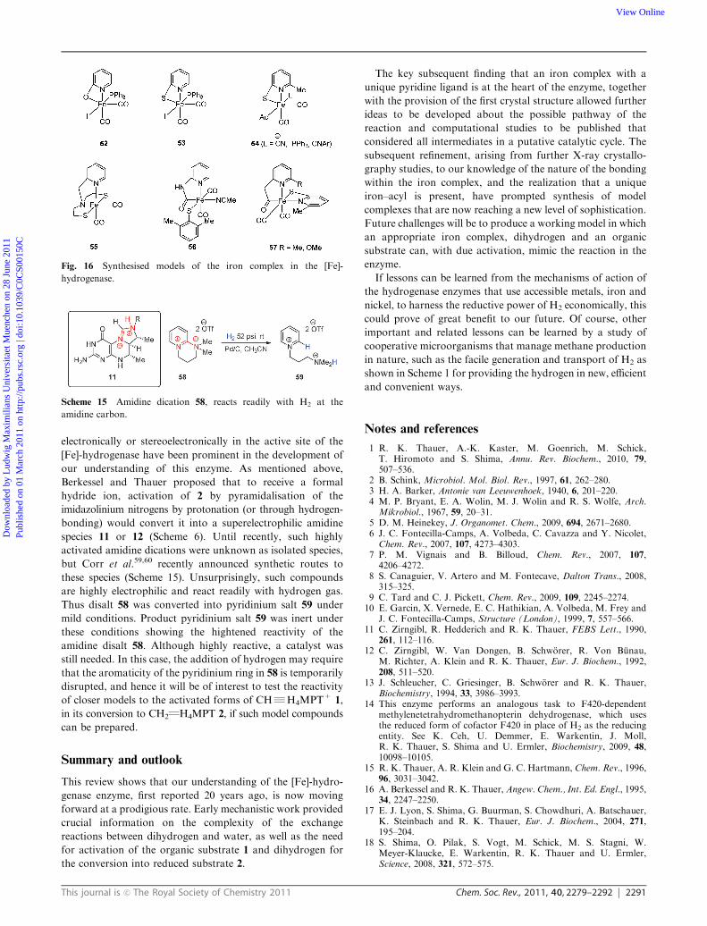
The group of Pickett has also been active in preparation of
model iron complexes. Their earlier work41 had prepared the
cis-dicarbonyliron complex 55, which had been useful in their
study assigning iron(II) as the metal oxidation state in FeGP.
In 2010, Pickett et al. prepared the advanced analogue 56,56
giving CO stretches in the IR spectrum at 2026 and 1958 cm�1
(compared to 2031 and 1972 cm�1 in the FeGP complex). Also
in 2010, the Hu group reported the preparation and properties
of sophisticated iron acyl complexes 57 (Fig. 16).57,58
The organic substrates for the transformations of this
enzyme CHRH4MPT+ 1 and CH2QH4MPT 2 are known,
but proposals on how these substrates may be activated
Fig. 15 (A) Crystal structure representation of Cys176-Ala-mutated
[Fe]-hydrogenase and CH2QH4MPT 2. B) Modelled closed-form of
enzyme with CH2QH4MPT 2 present. Ref. 49. Copyright Wiley-VCH
Verlag GmbH & Co. KGaA. Reproduced with permission.
Scheme 12 Proposed catalytic mechanism for open/closed conforma-
tional transition.49
Scheme 13 Hmd active site model 46.
Scheme 14 Oxidative addition with a thioester.
Dow
nloa
ded
by L
udw
ig M
axim
ilian
s U
nive
rsita
et M
uenc
hen
on 2
8 Ju
ne 2
011
Publ
ishe
d on
01
Mar
ch 2
011
on h
ttp://
pubs
.rsc
.org
| do
i:10.
1039
/C0C
S001
50C
View Online
This journal is c The Royal Society of Chemistry 2011 Chem. Soc. Rev., 2011, 40, 2279–2292 2291
electronically or stereoelectronically in the active site of the
[Fe]-hydrogenase have been prominent in the development of
our understanding of this enzyme. As mentioned above,
Berkessel and Thauer proposed that to receive a formal
hydride ion, activation of 2 by pyramidalisation of the
imidazolinium nitrogens by protonation (or through hydrogen-
bonding) would convert it into a superelectrophilic amidine
species 11 or 12 (Scheme 6). Until recently, such highly
activated amidine dications were unknown as isolated species,
but Corr et al.59,60 recently announced synthetic routes to
these species (Scheme 15). Unsurprisingly, such compounds
are highly electrophilic and react readily with hydrogen gas.
Thus disalt 58 was converted into pyridinium salt 59 under
mild conditions. Product pyridinium salt 59 was inert under
these conditions showing the hightened reactivity of the
amidine disalt 58. Although highly reactive, a catalyst was
still needed. In this case, the addition of hydrogen may require
that the aromaticity of the pyridinium ring in 58 is temporarily
disrupted, and hence it will be of interest to test the reactivity
of closer models to the activated forms of CHRH4MPT+ 1,
in its conversion to CH2QH4MPT 2, if such model compounds
can be prepared.
Summary and outlook
This review shows that our understanding of the [Fe]-hydro-
genase enzyme, first reported 20 years ago, is now moving
forward at a prodigious rate. Early mechanistic work provided
crucial information on the complexity of the exchange
reactions between dihydrogen and water, as well as the need
for activation of the organic substrate 1 and dihydrogen for
the conversion into reduced substrate 2.
The key subsequent finding that an iron complex with a
unique pyridine ligand is at the heart of the enzyme, together
with the provision of the first crystal structure allowed further
ideas to be developed about the possible pathway of the
reaction and computational studies to be published that
considered all intermediates in a putative catalytic cycle. The
subsequent refinement, arising from further X-ray crystallo-
graphy studies, to our knowledge of the nature of the bonding
within the iron complex, and the realization that a unique
iron–acyl is present, have prompted synthesis of model
complexes that are now reaching a new level of sophistication.
Future challenges will be to produce a working model in which
an appropriate iron complex, dihydrogen and an organic
substrate can, with due activation, mimic the reaction in the
enzyme.
If lessons can be learned from the mechanisms of action of
the hydrogenase enzymes that use accessible metals, iron and
nickel, to harness the reductive power of H2 economically, this
could prove of great benefit to our future. Of course, other
important and related lessons can be learned by a study of
cooperative microorganisms that manage methane production
in nature, such as the facile generation and transport of H2 as
shown in Scheme 1 for providing the hydrogen in new, efficient
and convenient ways.
Notes and references
1 R. K. Thauer, A.-K. Kaster, M. Goenrich, M. Schick,T. Hiromoto and S. Shima, Annu. Rev. Biochem., 2010, 79,507–536.
2 B. Schink, Microbiol. Mol. Biol. Rev., 1997, 61, 262–280.3 H. A. Barker, Antonie van Leeuwenhoek, 1940, 6, 201–220.4 M. P. Bryant, E. A. Wolin, M. J. Wolin and R. S. Wolfe, Arch.Mikrobiol., 1967, 59, 20–31.
5 D. M. Heinekey, J. Organomet. Chem., 2009, 694, 2671–2680.6 J. C. Fontecilla-Camps, A. Volbeda, C. Cavazza and Y. Nicolet,Chem. Rev., 2007, 107, 4273–4303.
7 P. M. Vignais and B. Billoud, Chem. Rev., 2007, 107,4206–4272.
8 S. Canaguier, V. Artero and M. Fontecave, Dalton Trans., 2008,315–325.
9 C. Tard and C. J. Pickett, Chem. Rev., 2009, 109, 2245–2274.10 E. Garcin, X. Vernede, E. C. Hathikian, A. Volbeda, M. Frey and
J. C. Fontecilla-Camps, Structure (London), 1999, 7, 557–566.11 C. Zirngibl, R. Hedderich and R. K. Thauer, FEBS Lett., 1990,
261, 112–116.12 C. Zirngibl, W. Van Dongen, B. Schworer, R. Von Bunau,
M. Richter, A. Klein and R. K. Thauer, Eur. J. Biochem., 1992,208, 511–520.
13 J. Schleucher, C. Griesinger, B. Schworer and R. K. Thauer,Biochemistry, 1994, 33, 3986–3993.
14 This enzyme performs an analogous task to F420-dependentmethylenetetrahydromethanopterin dehydrogenase, which usesthe reduced form of cofactor F420 in place of H2 as the reducingentity. See K. Ceh, U. Demmer, E. Warkentin, J. Moll,R. K. Thauer, S. Shima and U. Ermler, Biochemistry, 2009, 48,10098–10105.
15 R. K. Thauer, A. R. Klein and G. C. Hartmann, Chem. Rev., 1996,96, 3031–3042.
16 A. Berkessel and R. K. Thauer, Angew. Chem., Int. Ed. Engl., 1995,34, 2247–2250.
17 E. J. Lyon, S. Shima, G. Buurman, S. Chowdhuri, A. Batschauer,K. Steinbach and R. K. Thauer, Eur. J. Biochem., 2004, 271,195–204.
18 S. Shima, O. Pilak, S. Vogt, M. Schick, M. S. Stagni, W.Meyer-Klaucke, E. Warkentin, R. K. Thauer and U. Ermler,Science, 2008, 321, 572–575.
Fig. 16 Synthesised models of the iron complex in the [Fe]-
hydrogenase.
Scheme 15 Amidine dication 58, reacts readily with H2 at the
amidine carbon.
Dow
nloa
ded
by L
udw
ig M
axim
ilian
s U
nive
rsita
et M
uenc
hen
on 2
8 Ju
ne 2
011
Publ
ishe
d on
01
Mar
ch 2
011
on h
ttp://
pubs
.rsc
.org
| do
i:10.
1039
/C0C
S001
50C
View Online

2292 Chem. Soc. Rev., 2011, 40, 2279–2292 This journal is c The Royal Society of Chemistry 2011
19 T. Hiromoto, K. Ataka, O. Pilak, S. Vogt, M. S. Stagni, W.Meyer-Klaucke, E. Warkentin, R. K. Thauer, S. Shima andU. Ermler, FEBS Lett., 2009, 583, 585–590.
20 R. von Bunau, C. Zirngibl, R. K. Thauer and A. Klein, Eur. J.Biochem., 1991, 202, 1205–1208.
21 S. P. J. Albracht, in The Molecular Basis of Bacterial Metabolism,ed. G. Hauska, R. K Thauer, Springer, Berlin, 1990, pp. 40–51.
22 B. Schworer, V. M. Fernandez, C. Zirngibl and R. K. Thauer, Eur.J. Biochem., 1993, 212, 255–261.
23 R. Breslow, J. Am. Chem. Soc., 1957, 79, 1762–1763.24 M. Scholl, S. Ding, C. W. Lee and R. H. Grubbs, Org. Lett., 1999,
1, 953–956.25 G. Buurman, S. Shima and R. K. Thauer, FEBS Lett., 2000, 485,
200–204.26 G. A. Olah, A. Germain, H. C. Lin and D. A. Forsyth, J. Am.
Chem. Soc., 1975, 97, 2928–2929.27 G. A. Olah, G. K. Surya Parakash and J. Sommer, Superacids,
Wiley, New York, 1985.28 G. A. Olah, in The Chemistry of Alkanes and Cycloalkanes,
ed. S. Patai, Wiley, New York, 1992, pp. 609–652.29 G. A. Olah and D. A. Klumpp, Superelectrophiles and Their
Chemistry, Wiley-Interscience, 2007.30 A. R. Klein, G. C. Hartmann and R. K. Thauer, Eur. J. Biochem.,
1995, 233, 372–376.31 A. R. Klein, V. M. Fernandez and R. K. Thauer, FEBS Lett.,
1995, 368, 203–206.32 J. Cioslowski and G. Bhe, Angew. Chem., Int. Ed. Engl., 1997, 36,
107–109.33 J. H. Teles, S. Brode and A. Berkessel, J. Am. Chem. Soc., 1998,
120, 1345–1346.34 A. P. Scott, B. T. Golding and L. Radom, New J. Chem., 1998, 22,
1171–1173.35 A. Berkessel, Curr. Opin. Chem. Biol., 2001, 5, 486–490.36 E. J. Lyon, S. Shima, G. Buurman, S. Chowdhuri, A. Batschauer,
K. Steinbach and R. K. Thauer, Eur. J. Biochem., 2003, 271,195–204.
37 E. J. Lyon, S. Shima, R. Boecher, R. K. Thauer, F.-W. Grevels,E. Bill, W. Roseboom and S. P. J. Albracht, J. Am. Chem. Soc.,2004, 126, 14239–14248.
38 S. Shima, E. J. Lyon, M. Sordel-Klippert, M. Kaub, J. Kahnt,R. K. Thauer, K. Steinbach, X. Xie, L. Verdier and C. Griesinger,Angew. Chem., Int. Ed., 2004, 43, 2547–2551.
39 S. A. Goldfield and K. N. Raymond, Inorg. Chem., 1974, 13,770–775.
40 S. Shima, E. J. Lyon, R. K. Thauer, B. Mienert and E. Bill, J. Am.Chem. Soc., 2005, 127, 10430–10435.
41 X. Wang, Z. Li, X. Zeng, Q. Luo, D. J. Evans, C. J. Pickett andX. Liu, Chem. Commun., 2008, 3555–3557.
42 M. Salomone-Stagni, F. Stellato, C. M. Whaley, S. Vogt,S. Morante, S. Shima, T. R. Rauchfuss and W. Meyer-Klaucke,Dalton Trans., 2010, 39, 3057–3064.
43 O. Pilak, B. Mamat, S. Vogt, C. H. Hagemeier, R. K. Thauer,S. Shima, C. Vonrhein, E. Warkentin and U. Ermler, J. Mol. Biol.,2006, 358, 798–809.
44 P. Acharya, M. Goenrich, C. H. Hagemeier, U. Semmer,J. A. Vorholt, R. K. Thauer and U. Ermler, J. Biol.Chem., 2005,280, 13712–13719.
45 X. Yang and M. B. Hall, J. Am. Chem. Soc., 2008, 130,14036–14037.
46 The Fe–GP cofactor in the active enzyme shows CO stretchingfrequencies similar to Z5-(cyclopentadienyl)dicarbonylacylironcomplexes in organic solvents or in the dried state. S. T. Belt,D. W. Ryba and P. C. Ford, J. Am. Chem. Soc., 1991, 113,9524–9528.
47 E. Baranowska, W. Danikiewicz, Z. Pakulski and A. Zamojski,J. Mass Spectrom., 1995, 30, 158–162.
48 X. Yang and M. B. Hall, J. Am. Chem. Soc., 2009, 131,10901–10908.
49 T. Hiromoto, E. Warkentin, J. Moll, U. Ermler and S. Shima,Angew. Chem., Int. Ed., 2009, 48, 6457–6460.
50 A. M. Royer, T. B. Rauchfuss and S. R. Wilson, Inorg. Chem.,2008, 47, 395–397.
51 A. M. Royer, T. B. Rauchfuss and D. L. Gray, Organometallics,2009, 28, 3618–3620.
52 A. M. Royer, M. Salomone-Stagni, T. B. Rauchfuss andW. Meyer-Klaucke, J. Am. Chem. Soc., 2010, 132, 16997–17003.
53 B. V. Obrist, D. Chen, A. Ahrens, V. Schunemann, R. Scopellitiand X. Hu, Inorg. Chem., 2009, 48, 3514–3516.
54 B. Li, T. Liu, C. V. Popescu, A. Bilko and M. Y. Darensbourg,Inorg. Chem., 2009, 48, 11283–11289.
55 D. Chen, R. Scopelliti and X. Hu, J. Am. Chem. Soc., 2010, 132,928–929.
56 P. J. Turrell, J. A. Wright, J. M. T. Peck, V. S. Oganesyan andC. J. Pickett, Angew. Chem., Int. Ed., 2010, 49, 7508–7511.
57 D. Chen, R. Scopelliti and X. Hu, Angew. Chem., Int. Ed., 2010,49, 7512–7515.
58 For a recent simpler 2-methoxypyridine complex see S. Tanino,Y. Ohki and K. Tatsumi, Chem.–Asian J., 2010, 5, 1962–1964.
59 M. J. Corr, K. F. Gibson, A. R. Kennedy and J. A. Murphy,J. Am. Chem. Soc., 2009, 131, 9174–9175.
60 M. J. Corr, M. Roydhouse, K. F. Gibson, S. Zhou, A. R. Kennedyand J. A. Murphy, J. Am. Chem. Soc., 2009, 131, 17980–17985.
61 J. A. Wright, P. J. Turrell and C. J. Pickett, Organometallics, 2010,29, 6146–6156.
62 S. Shima and U. Ermler, Eur. J. Inorg. Chem., DOI: 10.1002/ejic.201000955 published on line 2010.
Dow
nloa
ded
by L
udw
ig M
axim
ilian
s U
nive
rsita
et M
uenc
hen
on 2
8 Ju
ne 2
011
Publ
ishe
d on
01
Mar
ch 2
011
on h
ttp://
pubs
.rsc
.org
| do
i:10.
1039
/C0C
S001
50C
View Online