Embed Size (px)
Citation preview

This journal is c The Royal Society of Chemistry 2011 Chem. Commun., 2011, 47, 3793–3795 3793
Cite this: Chem. Commun., 2011, 47, 3793–3795
Single-walled carbon nanotubes chemiresistor aptasensors for small
molecules: picomolar level detection of adenosine triphosphatew
Basanta K. Das,za Chaker Tlili,za Sushmee Badhulika,bLakshmi N. Cella,
cWilfred Chen
a
and Ashok Mulchandani*a
Received 1st November 2010, Accepted 14th January 2011
DOI: 10.1039/c0cc04733c
Here we report single walled-carbon nanotubes (SWNTs)-based
chemiresistor aptasensors for highly sensitive and selective
detection of weakly or uncharged molecules using the displacement
format. As a proof-of-concept we demonstrate the detection of
ATP, a small weakly charged molecule, by displacement of the
ssDNA anti-ATP aptamer hybridized to a small capture oligo-
nucleotide covalently attached on SWNTs, with picomolar
sensitivity and selectivity over GTP.
Aptamers are short synthetic nucleic acids, either single
stranded DNA (ssDNA) or RNA, that can bind a variety
of targets. Since the first reports in 1990 that short RNA,
and subsequently ssDNA, with high binding affinity can be
selected from a large randomized library through in vitro
selection, ‘‘aptamers’’ has captivated the interest of researchers
in the fields of therapeutics and analytical methodologies.1,2
The growing interest in the latter field stems from the many
benefits such as ease of producing mass quantity in vitro by
PCR without the need of an animal host or elaborate and
expensive cell culturing facilities, simple isolation and purifica-
tion, reproducibility of synthesis and better stability, over the
traditional bio-recognition/capture agent, antibodies.3 Using
the Systematic Evolution of Ligands by EXponential enrich-
ment (SELEX) technique, aptamers for a variety of targets
ranging from metal ions, small organic molecules (adenosine
triphosphate (ATP), caffeine and cocaine) and biomolecules to
entire organisms have been isolated and applied as probes in
analytical methodologies.4,5
Nano-sensors based on one-dimensional (1-D) nanostructure
(nanowire, nanotube and nanobelt) chemiresistor/field-effect
transistor (FET) transducers are becoming very promising candi-
dates for the development of label-free affinity/immuno-sensors.
Besides label-free detection, 1-D chemiresistor/FET biosensor
has advantages of extremely high sensitivity, ease of miniaturiza-
tion, low power requirement and development of high density
arrays. Single-walled carbon nanotubes (SWNTs) have been
extensively studied as the transducer element of biosensors as
they meet the important requirements of an efficient biosensor:
excellent electrical, chemical and mechanical properties and a
large surface area to volume ratio results in surface phenomena
predominating over the chemistry and physics that happen in
the bulk.6,7 The resistance/conductance of these devices is
extremely sensitive to any surface adsorption/perturbation
and is a function of the analyte charge. SWNTs-based
chemiresistor/FET transducers have been modified with anti-
bodies, enzymes, and aptamers, for highly sensitive and selective
detection of macromolecules (IgE, thrombin, PA toxin, bacteria,
and virus) with distinct charge properties.8–12 The objective
of this communication was to investigate the sensing of a
target that is small in size displaying minimal charge using a
SWNTs-based aptasensor. Adenosine triphosphate (ATP),
which is important for controlling the cellular mechanism
and many biochemical pathways in a living organism, was
used as a model system. An anti-ATP ssDNA aptamer (50-ACC
TGGGGG AGT ATT GCG GAGGAAGGT GTC ACA-30)
reported by Li and Ho13 was employed as the bioreceptor.
We initially investigated ATP sensing using the traditional
1-D nanostructure chemiresistor affinity-sensor architecture in
which the modulation of the resistance of the bioreceptor
(ssDNA anti-ATP aptamer) modified gate was monitored.
(Please see ESIw for fabrication details.)
Upon the addition of different concentrations (1 pM to 10 nM)
of ATP to the biosensor its response (normalized resistance
change) for each of the concentrations over the investigated
range averaged to a meager 1.72 � 0.7% and had no relation-
ship to the concentration (data not shown). The very weak
response, not much different from that for blank, is not
completely unexpected considering that the response of 1-D
nanostructure-based chemiresistor/FET biosensors is a function
of the analyte charge; which for ATP is small in comparison
to the targets that have been successfully detected by 1-D
nanostructure-based chemiresistor/FET aptasensors and affinity/
immuno-sensors.14–16 In order to sense/detect ATP using
1-D nanostructures-based chemiresistor/FET transducers, we
report a modified aptasensor architecture, depicted in Fig. 1.
a Chemical and Environmental Engineering Department,University of California, Riverside, CA 92521, USA.E-mail: [email protected]; Fax: +1 951-827-5696;Tel: +1 951-827-6419
b Electrical Engineering Department, University of California,Riverside, CA 92521, USA
cCell, Molecular and Developmental Biology Graduate Program,University of California, Riverside, CA 92521, USAw Electronic supplementary information (ESI) available: Protocols foraptasensor fabrication and sensing measurements, and I–V curves. SeeDOI: 10.1039/c0cc04733cz B. K. Das and C. Tlili are equal contributors.
ChemComm Dynamic Article Links
www.rsc.org/chemcomm COMMUNICATION
Dow
nloa
ded
by U
nive
rsity
of
Cal
ifor
nia
- R
iver
side
on
29 M
arch
201
1Pu
blis
hed
on 0
2 Fe
brua
ry 2
011
on h
ttp://
pubs
.rsc
.org
| do
i:10.
1039
/C0C
C04
733C
View Online
3794 Chem. Commun., 2011, 47, 3793–3795 This journal is c The Royal Society of Chemistry 2011
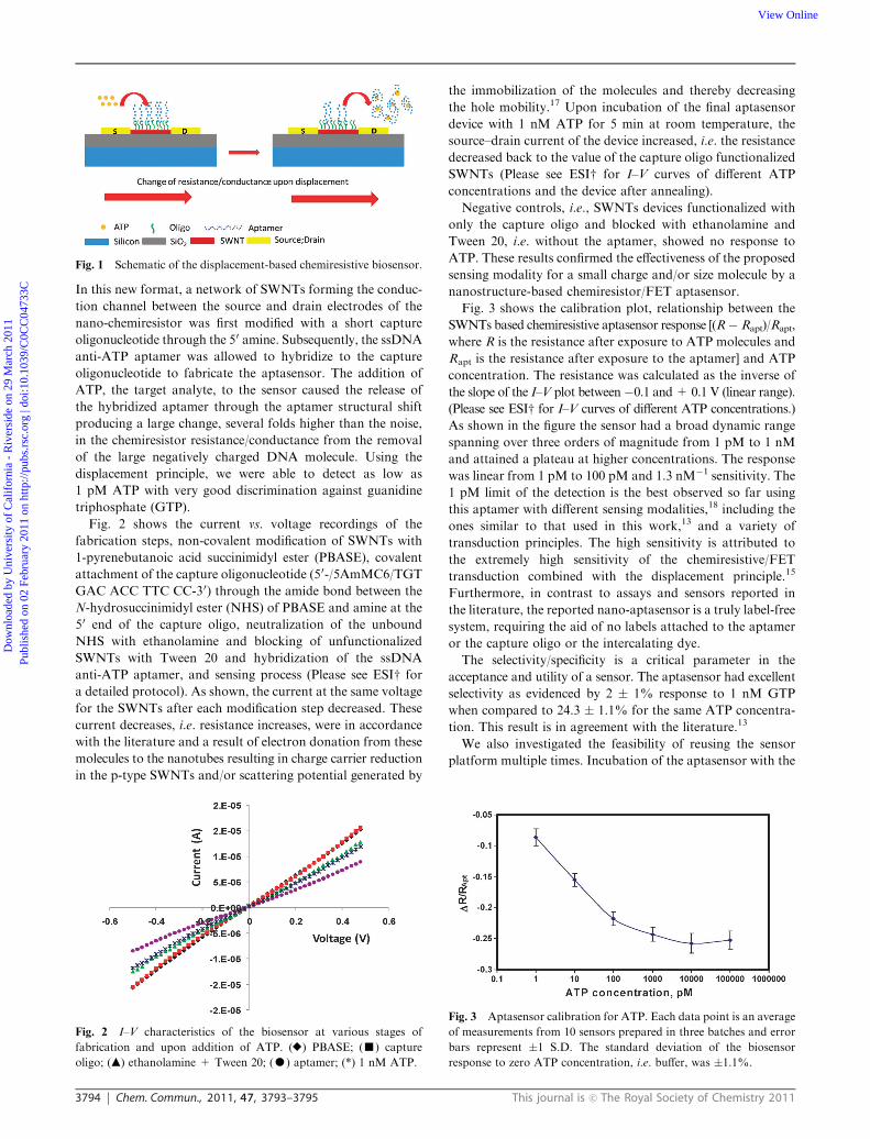
In this new format, a network of SWNTs forming the conduc-
tion channel between the source and drain electrodes of the
nano-chemiresistor was first modified with a short capture
oligonucleotide through the 50 amine. Subsequently, the ssDNA
anti-ATP aptamer was allowed to hybridize to the capture
oligonucleotide to fabricate the aptasensor. The addition of
ATP, the target analyte, to the sensor caused the release of
the hybridized aptamer through the aptamer structural shift
producing a large change, several folds higher than the noise,
in the chemiresistor resistance/conductance from the removal
of the large negatively charged DNA molecule. Using the
displacement principle, we were able to detect as low as
1 pM ATP with very good discrimination against guanidine
triphosphate (GTP).
Fig. 2 shows the current vs. voltage recordings of the
fabrication steps, non-covalent modification of SWNTs with
1-pyrenebutanoic acid succinimidyl ester (PBASE), covalent
attachment of the capture oligonucleotide (50-/5AmMC6/TGT
GAC ACC TTC CC-30) through the amide bond between the
N-hydrosuccinimidyl ester (NHS) of PBASE and amine at the
50 end of the capture oligo, neutralization of the unbound
NHS with ethanolamine and blocking of unfunctionalized
SWNTs with Tween 20 and hybridization of the ssDNA
anti-ATP aptamer, and sensing process (Please see ESIw for
a detailed protocol). As shown, the current at the same voltage
for the SWNTs after each modification step decreased. These
current decreases, i.e. resistance increases, were in accordance
with the literature and a result of electron donation from these
molecules to the nanotubes resulting in charge carrier reduction
in the p-type SWNTs and/or scattering potential generated by
the immobilization of the molecules and thereby decreasing
the hole mobility.17 Upon incubation of the final aptasensor
device with 1 nM ATP for 5 min at room temperature, the
source–drain current of the device increased, i.e. the resistance
decreased back to the value of the capture oligo functionalized
SWNTs (Please see ESIw for I–V curves of different ATP
concentrations and the device after annealing).
Negative controls, i.e., SWNTs devices functionalized with
only the capture oligo and blocked with ethanolamine and
Tween 20, i.e. without the aptamer, showed no response to
ATP. These results confirmed the effectiveness of the proposed
sensing modality for a small charge and/or size molecule by a
nanostructure-based chemiresistor/FET aptasensor.
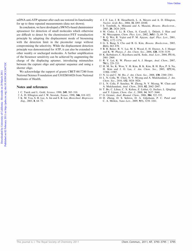
Fig. 3 shows the calibration plot, relationship between the
SWNTs based chemiresistive aptasensor response [(R� Rapt)/Rapt,
where R is the resistance after exposure to ATP molecules and
Rapt is the resistance after exposure to the aptamer] and ATP
concentration. The resistance was calculated as the inverse of
the slope of the I–V plot between�0.1 and+ 0.1 V (linear range).
(Please see ESIw for I–V curves of different ATP concentrations.)
As shown in the figure the sensor had a broad dynamic range
spanning over three orders of magnitude from 1 pM to 1 nM
and attained a plateau at higher concentrations. The response
was linear from 1 pM to 100 pM and 1.3 nM�1 sensitivity. The
1 pM limit of the detection is the best observed so far using
this aptamer with different sensing modalities,18 including the
ones similar to that used in this work,13 and a variety of
transduction principles. The high sensitivity is attributed to
the extremely high sensitivity of the chemiresistive/FET
transduction combined with the displacement principle.15
Furthermore, in contrast to assays and sensors reported in
the literature, the reported nano-aptasensor is a truly label-free
system, requiring the aid of no labels attached to the aptamer
or the capture oligo or the intercalating dye.
The selectivity/specificity is a critical parameter in the
acceptance and utility of a sensor. The aptasensor had excellent
selectivity as evidenced by 2 � 1% response to 1 nM GTP
when compared to 24.3 � 1.1% for the same ATP concentra-
tion. This result is in agreement with the literature.13
We also investigated the feasibility of reusing the sensor
platform multiple times. Incubation of the aptasensor with the
Fig. 1 Schematic of the displacement-based chemiresistive biosensor.
Fig. 2 I–V characteristics of the biosensor at various stages of
fabrication and upon addition of ATP. (E) PBASE; (’) capture
oligo; (m) ethanolamine + Tween 20; (K) aptamer; (*) 1 nM ATP.
Fig. 3 Aptasensor calibration for ATP. Each data point is an average
of measurements from 10 sensors prepared in three batches and error
bars represent �1 S.D. The standard deviation of the biosensor
response to zero ATP concentration, i.e. buffer, was �1.1%.
Dow
nloa
ded
by U
nive
rsity
of
Cal
ifor
nia
- R
iver
side
on
29 M
arch
201
1Pu
blis
hed
on 0
2 Fe
brua
ry 2
011
on h
ttp://
pubs
.rsc
.org
| do
i:10.
1039
/C0C
C04
733C
View Online
This journal is c The Royal Society of Chemistry 2011 Chem. Commun., 2011, 47, 3793–3795 3795
ssDNA anti-ATP aptamer after each use restored its functionality
for up to three repeated measurements (data not shown).
In conclusion, we have developed a SWNTs-based chemiresistor
aptasensor for detection of small molecules which otherwise
are difficult to detect by the chemiresistor/FET transduction
principle by adapting the displacement mode of biosensing
with the detection limit in the picomolar range without
compromising the selectivity. While the displacement detection
principle was demonstrated for ATP, it can also be extended to
other weakly or uncharged molecules. A further amplification
of the biosensor sensitivity can be achieved by augmenting the
charge of the displacing aptamer, introducing mismatches
between the capture oligo and aptamer sequence and using a
shorter oligo.
We acknowledge the support of grants CBET-0617240 from
National Science Foundation and U01ES016026 from National
Institutes of Health.
Notes and references
1 C. Tuerk and L. Gold, Science, 1990, 249, 505–510.2 A. D. Ellington and J. W. Szostak, Nature, 1990, 346, 818–822.3 K. M. You, S. H. Lee, A. Im and S. B. Lee, Biotechnol. BioprocessEng., 2003, 8, 64–75.
4 J. F. Lee, J. R. Hesselberth, L. A. Meyers and A. D. Ellington,Nucleic Acids Res., 2004, 32, D95–D100.
5 S. Tombelli, A. Minunni and A. Mascini, Biosens. Bioelectron.,2005, 20, 2424–2434.
6 M. Cinke, J. Li, B. Chen, A. Cassell, L. Delzeit, J. Han andM. Meyyappan, Chem. Phys. Lett., 2002, 365(1–2), 69–74.
7 B. Q. Wei, R. Vajtai and P. M. Ajayan, Appl. Phys. Lett., 2001,79(8), 1172–1174.
8 G. S. Bang, S. Cho and B. G. Kim, Biosens. Bioelectron., 2005,21(6), 863–870.
9 B. R. Baker, R. Y. Lai, M. S. Wood, E. H. Doctor, A. J. Heegerand K. W. Plaxco, J. Am. Chem. Soc., 2006, 128, 3138–3139.
10 K. Ikebukuro, C. Kiyohara and K. Sode, Anal. Lett., 2004, 37(14),2901–2909.
11 R. Y. Lai, K. W. Plaxco and A. J. Heeger, Anal. Chem., 2007,79(1), 229–233.
12 H. M. So, K. Won, Y. H. Kim, B. K. Kim, B. H. Ryu, P. S. Na,H. Kim and J. O. Lee, J. Am. Chem. Soc., 2005, 127(34),11906–11907.
13 N. Li and C. M. Ho, J. Am. Chem. Soc., 2008, 130, 2380–2381.14 L. N. Cella, W. Chen, N. V. Myung and A. Mulchandani, J. Am.
Chem. Soc., 2010, 132, 5024–5026.15 L. N. Cella, P. Sanchez, W. Zhong, N. V. Myung, W. Chen and
A. Mulchandani, Anal. Chem., 2010, 82, 2042–2047.16 T. Bo, C. Lihua, C. X. Kehua, Z. Linhai, G. Jiechao, L. Qingling
and Y. Lijuan, Chem.–Eur. J., 2008, 14, 3637–3644.17 G. Gruner, Anal. Bioanal. Chem., 2006, 384, 322–335.18 D. Zheng, D. S. Seferos, D. A. Giljohann, P. C. Patel and
C. A. Mirkin, Nano Lett., 2009, 9(9), 3258–3261.
Dow
nloa
ded
by U
nive
rsity
of
Cal
ifor
nia
- R
iver
side
on
29 M
arch
201
1Pu
blis
hed
on 0
2 Fe
brua
ry 2
011
on h
ttp://
pubs
.rsc
.org
| do
i:10.
1039
/C0C
C04
733C
View Online