Embed Size (px)
Citation preview
RESEARCH ARTICLE
Circulating levels of sclerostin but not DKK1
associate with laboratory parameters of CKD-
MBD
Geert J. Behets1, Liesbeth Viaene2, Bjorn Meijers2, Frank Blocki3, Vincent
M. Brandenburg4, Anja Verhulst1, Patrick C. D’Haese1, Pieter Evenepoel2*
1 University of Antwerp, Dept. Biomedical Sciences, Laboratory of Pathophysiology, Wilrijk, Belgium, 2 KUL
Leuven, Department of Immunology and Microbiology, Laboratory of Nephrology, Leuven, Belgium,
3 DiaSorin, Inc., Stillwater, Minnesota, United States of America, 4 University Hospital RWTH Aachen,
Department of Cardiology, Aachen, Germany
Abstract
Introduction
Mounting evidence indicates that a disturbed Wnt–β-catenin signaling may be involved in
the pathogenesis of chronic kidney disease-mineral and bone and mineral disorder (CKD-
MBD). Data on the impact of CKD on circulating levels of the Wnt antagonists sclerostin and
Dickkopf related protein 1 (DKK1) and the relationship with laboratory parameters of CKD-
MBD are incomplete.
Methods
We analyzed serum sclerostin and DKK1 in 308 patients across the stages of chronic kidney
disease (kDOQI stage 1–2 n = 41; CKD stage 3 n = 54; CKD stage 4–5 n = 54; hemodialysis
n = 100; peritoneal dialysis n = 59) as well as in 49 healthy controls. We investigated associ-
ations with demographics, renal function, parameters of mineral metabolism including 25
(OH) vitamin D, 1,25(OH)2 vitamin D, biointact fibroblast growth factor 23 (FGF23), and
parathyroid hormone (PTH), and bone turnover markers.
Results
Serum sclerostin, but not DKK1, increases in more advanced stages of CKD and associates
with PTH, phosphate, and 1,25(OH)2 vitamin D concentrations. Bone turnover markers
are highest in hemodialysis patients presenting the combination of high PTH with low scler-
ostin level. Serum DKK1 levels are lower in CKD patients than in controls and are not asso-
ciated with laboratory parameters of mineral metabolism. Interestingly, a direct association
between DKK1 and platelet count was observed.
Conclusion
In CKD, serum levels of the Wnt inhibitors DKK1 and sclerostin are unrelated, indicating dif-
ferent sites of origin and/ or different regulatory mechanisms. Sclerostin, as opposed to
PLOS ONE | https://doi.org/10.1371/journal.pone.0176411 May 11, 2017 1 / 12
a1111111111
a1111111111
a1111111111
a1111111111
a1111111111
OPENACCESS
Citation: Behets GJ, Viaene L, Meijers B, Blocki F,
Brandenburg VM, Verhulst A, et al. (2017)
Circulating levels of sclerostin but not DKK1
associate with laboratory parameters of CKD-MBD.
PLoS ONE 12(5): e0176411. https://doi.org/
10.1371/journal.pone.0176411
Editor: Yin Tintut, University of California, Los
Angeles, UNITED STATES
Received: January 3, 2017
Accepted: April 9, 2017
Published: May 11, 2017
Copyright: © 2017 Behets et al. This is an open
access article distributed under the terms of the
Creative Commons Attribution License, which
permits unrestricted use, distribution, and
reproduction in any medium, provided the original
author and source are credited.
Data Availability Statement: All relevant data are
within the paper and its Supporting Information
files.
Funding: FB is an employee of Diasorin Inc. The
funder provided support in the form of salaries for
author FB and also supplied research materials for
the current study. PCD has received previous
research grants from Diasorin SAP.
Competing interests: The authors of this
manuscript have read the journal’s policy and have
the following competing interests: FB is an

DKK1, may qualify as a biomarker of CKD-MBD, particularly in dialysis patients. DKK1
serum levels, remarkably, correlate almost uniquely with blood platelet counts.
Introduction
The (canonical) Wnt–β-catenin pathway is increasingly recognized to play an important role
in bone [1] and vascular biology [2]. This pathway is tightly regulated by several antagonists, of
which the soluble Wnt inhibitors Dickkopf related protein 1 (DKK1, 26kD) and especially
sclerostin (28kD) have been studied most intensively. While sclerostin expression is largely
limited to bone [3] and calcifying vascular tissue [4], DKK1 is expressed in a number of other
tissues and cells including platelets, the prostate and the kidneys [5]. Since sclerostin and
DKK1 not only exert local (paracrine) effects, but are also released in the systemic circulation,
inhibition of Wnt signaling in distant tissues and organs can also occur. In SOST-/- mice, for
instance, it has been shown that kidney repair after unilateral urether obstruction is delayed
[6] whilst in animal models of early CKD, incomplete recovery from acute kidney injury led to
increased expression of Wnt inhibitors including DKK1 and sclerostin in the injured kidney
and to increased levels in the systemic circulation [7]. Thus, DKK1 and sclerostin may also be
involved in the many regulatory feedback loops that govern and fine-tune bone and mineral
metabolism [8].
Circulating sclerostin levels increase with severity of chronic kidney disease (CKD) and are
reported to reach levels that are 2 to 4-fold higher in patients with end stage renal disease as
compared to individuals with normal renal function [9–15]. Data on circulating levels of
DKK1 in CKD, conversely, are scarce and inconsistent with some investigators demonstrating
increments already occurring in early stage CKD [16], while others showing levels in the nor-
mal range even in patients with advanced CKD [15, 17]. It is an ongoing debate to what extent
sclerostin and DKK1 may serve as biomarkers of CKD-mineral and bone disorder (MBD)
[18–20]. The purpose of this study was to evaluate circulating DKK1 and sclerostin levels in
CKD and to describe for the first time the relationship between DKK1, sclerostin and proto-
typic laboratory parameters of mineral metabolism across stages of disease.
Materials and methods
Study population
The study population consisted of 308 prevalent CKD stage 1-5D patients and 49 controls. All
patients were recruited from an ongoing observational study at the University Hospitals Leu-
ven, Belgium, investigating uremic toxicity and bone and mineral metabolism in CKD patients
(NCT 00441623). All patients were enrolled between February 2006 and July 2008. CKD stage
5D patients were treated either with thrice weekly conventional hemodialysis (n = 100) or peri-
toneal dialysis (PD, n = 59; continuous ambulatory PD: n = 30; Automated PD: n = 29). Dialy-
sis adequacy was targeted in all patients according to the NKF K-DOQI guidelines. Controls,
defined as individuals with no history of CKD and CKD-EPI estimated GFR > 60 ml/min 1.73
m2, were recruited from the dermatology outpatient clinic at the University Hospital Antwerp.
All participants were 18 years of age or older and provided written informed consent. All stud-
ies were performed according to the Declaration of Helsinki, and approved by the Ethics Com-
mittees of the University Hospital Leuven and the University Hospital of Antwerp.
Wnt signaling and bone markers in CKD
PLOS ONE | https://doi.org/10.1371/journal.pone.0176411 May 11, 2017 2 / 12
employee of Diasorin Inc. The funder provided
support in the form of salaries for author FB and
supplied research materials for the current study.
The funder did not have any additional role in the
study design, data collection and analysis, decision
to publish, or preparation of the manuscript. The
commercial affiliation does not alter the authors’
adherence to all PLOS ONE policies on sharing data
and material.

Biochemical measurements
In all participants but HD patients, blood samples were collected in the morning (random,
non-fasted). In HD patients, blood samples were collected before the mid-week dialysis ses-
sion. After standard centrifugation, serum was aliquoted and stored at -80˚C pending further
analysis. Creatinine, hemoglobin, calcium, phosphate, C-reactive protein (CRP), total alkaline
phosphatase (tAP), and cholesterol were all measured using standard laboratory techniques.
Serum C-terminal cross-linked telopeptide (CTX-I) was measured using an electrochemilumi-
nescence immunoassay (Roche Diagnostics, Switzerland). Albumin was measured using the
bromocresol green method. Bone specific alkaline phosphatase (Bone ALP), calcidiol (25(OH)
D), calcitriol (1,25(OH)2D) and PTH (N-TACT II) (i.e. a 2nd generation PTH assay) were mea-
sured using a LIAISON XLautomated analyzer with the appropriate analyzer kits (DiaSorin,
USA). Serum sclerostin (Biomedica, Austria), DKK1 (Biomedica, Austria), and biointact fibro-
blast growth factor 23 (FGF23, Kainos, Japan) were measured using ELISA kits according to
the manufacturer’s instructions. As a complementary 3rd generation PTH assay, whole (1–84)
PTH (CAP PTH) was also measured using the Scantibodies CAP assay (USA). Detection limits
of the various assays were: Bone ALP (0.1 μg/l); sclerostin (8.9 pmol/l); DKK1 (0.38 pmol/l); 25
(OH)D (4.0 ng/ml); 1,25(OH)2D (< 2.0 pg/ml); CAP PTH (1.0 pg/ml); N-TACT PTH (1.7
pg/ml); FGF23 (3 pg/ml). Available reference values for healthy subjects are: sclerostin (11.9–
47.9 pmol/l); DKK1 (47.7±20 pmol/l); calcitriol (25.1–66.1 pg/ml); CAP PTH (5–39 pg/ml);
N-TACT PTH (14.5–87.1 pg/ml); FGF23 (8.2–54.3 pg/ml). All assays used report intra- and
inter-assay variations below 15%. The eGFR was calculated using the CKD-EPI equation. Sin-
gle pool Kt/V (spKt/V), a measure of dialysis efficacy was calculated using the second-genera-
tion logarithmic formula of Daugirdas [21]. Anuria was defined as a urine output <100 ml.
Statistical analysis
Data are expressed as mean (standard deviation) for normally distributed variables or median
(IQR) for non-normally distributed variables. Differences between groups were tested using
parametric ANOVA, Kruskal-Wallis or chi-squared test as appropriate. Correlations between
circulating levels of DKK1 and sclerostin and other variables were calculated by Spearman’s
rank correlation coefficients. Multivariate linear regression analysis was performed including all
univariately associated variables (p<0.2) to identify independent determinants of serum DKK1
and sclerostin. After excluding collinearity, the best subset of variables was selected by backward
elimination on p<0.2. This subset was then subjected to a final elimination procedure on p
<0.05. Inspection of residual plots assured that the a priori assumptions for linear regression
were justified. For all statistical analysis, p-values less than 0.05 were considered significant. All
statistical analyses were performed using SAS (version 9.3, the SAS institute, Cary, NC, USA).
Results
Demographics
Relevant clinical and biochemical characteristics of the healthy controls and CKD patients, cat-
egorized according to stage of disease, are summarized in Table 1. Serum sclerostin levels were
higher and serum DKK1 levels were lower in CKD patients as compared to controls. Fig 1
shows serum sclerostin and DKK1 levels across CKD stages. In patients with chronic kidney
disease treated with dialysis, sclerostin was approximately 2.5-fold higher than in non-CKD
controls. Serum DKK1 levels, conversely were approximately 2-fold lower in dialysis patients
as compared to non-CKD controls. S1 Fig shows temporal aspects of disordered mineral
metabolism and sclerostin in CKD stage 1-5D.
Wnt signaling and bone markers in CKD
PLOS ONE | https://doi.org/10.1371/journal.pone.0176411 May 11, 2017 3 / 12
Serum sclerostin in CKD
In CKD patients not yet on dialysis (S1 Table), male gender, history of CVD, higher age, phos-
phate, FGF23, PTH (both assays), and lower eGFR, bicarbonate, calcitriol, blood platelets all
were significantly associated with higher serum sclerostin levels. In multivariable analysis, only
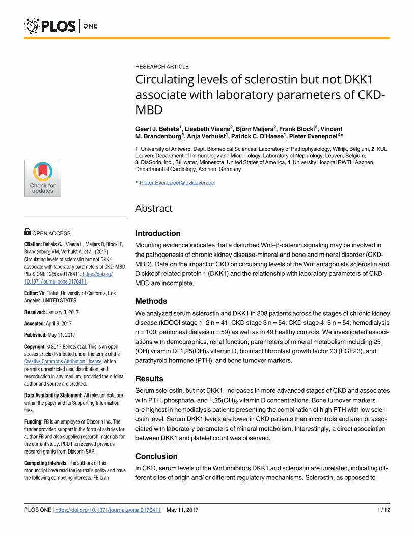
Table 1. Demographics, biochemistry and therapy in healthy volunteers and CKD patients across stages.
HV CKD 1–2 CKD3 CKD 4–5 CKD5D P (CKD)
N 49 41 54 54 159
Age 50.88 ± 16.19 45.37 ± 14.69 64.07 ± 14.04 66.95 ± 11.47 63.47 ± 15.25 <0.0001
BMI 23.99 ± 33.64 25.84 ± 4.92 27.04 ± 5.60 27.59 ± 5.59 23.56 ± 4.14 <0.0001
Renal dx Diabetes (%) - 0 1.9 1.9 17.6 <0.0001
Glomerular (%) - 63.4 29.6 13.0 28.9
Interstitial (%) - 0 5.6 3.7 4.4
Vascular (%) - 2.4 7.4 20.4 13.8
Cystic Heriditary (%) - 9.8 11.1 14.8 4.4
Miscellaneous, unknown (%) - 24.4 44.4 46.3 30.8
Male gender, % 41 32 56 70 60 0.002
CVD, % - 14.6 31.5 44.4 40.9 0.21
DM, % - 10 13 20 26 <0.05
Smoking, % (never/previous/current) NA 62/22/16 66/20/14 45/37/18 46/37/18 0.14
Anti-platelet agents, % 0 10 35 52 51 <0.0001
Non-calcium PB, % 0 0 0 0 24.4
Phosphate binder, % 0 12.2 14.8 35.2 86.5 <0.0001
Nutritional VitD, % 0 4.9 18.5 31.5 48.4 <0.0001
Active VitD, % 0 2.4 7.4 14.8 53.2 <0.0001
Calcimimetics, % 0 0 0 0 9 0.003
Bisphosphonates, % 0 5 15 6 4 <0.05
Hb, g/dL - 14.1 ± 1.56 13.6 ± 1.5 12.5 ± 1.4 11.8 ± 1.3 <0.0001
Platelets, ×103/mm3 - 280 ± 73 227 ± 68 208 ± 67 245 ± 83 <0.0001
Tchol, mg/dL - 187 ± 32 181± 38 178 ± 35 163± 37 0.0009
CRP, mg/L - 3.66 ± 6.15 3.55 ± 4.85 10.89 ± 26.93 8.30 ± 12.73 <0.0001
Albumin, g/L - 44.79 ± 3.33 45.59 ± 2.11 44.48 ± 3.04 39.42 ± 3.78 <0.0001
Urea Nitrogen, mg/dL - 33.5 ± 10,6 62.7± 21.2 110.3 ± 42.5 116.2± 32.4 <0.0001
Creatinine, mg/dL 0.94 ± 0.13 0.86 ± 0.14 1.49± 0.22 3.04 ± 1.43 7.25± 2.75 <0.0001
eGFR, mL/min 1.73m2 81.25± 14.45 79.75± 18.90 38.71± 7.67 19.07± 6.53 - <0.0001
Ca, mg/dL 10.18± 1.16 9.18± 0.45 9.24± 0.35 9.09± 0.54 9.35± 0.73 <0.0001
Phos, mg/dL 4.02± 0.87 3.05± 0.58 3.13± 0.62 3.71± 0.83 4.51± 1.31 <0.0001
Bicarbonate, mmol/L - 25.6 ±2.0 25.0 ±2.5 23.6 ± 2.6 25.0 ± 2.8 0.002
tAP, U/L - 167.34 ± 45.77 182.23 ± 60.64 220.26 ± 111.55 254.25 ± 150.09 <0.0001
Bone ALP, μg/L - 10.9 ± 4.7 12.1 ± 6.0 16.3 ± 14.5 18.7 ±15.0* 0.0003
CTX-I, ng/L - - - - 2539 ± 2392 -
25(OH)D, ng/mL 22.8 (15.7–27.5) 18.2 (12.1–26.2) 17.3 (13.3–25.0) 14.6 (10.9–19.3) 14.8 (10.3–20.6)* 0.08
1,25(OH)2D, pg/mL - 82.0 (47.2–111.1) 54.0 (29.1–92.0) 30.5 (23.0–43.6) - <0.0001
N-TACT PTH, pg/mL 19.7 (14.0–24.7) 18.4 (11.4–27.6) 29.9 (22.6–44.3) 62.95 (46.0–137.0) 80.95 (45.2–154.0) * <0.0001
CAP PTH, pg/mL 45.7 (39.2–63.3) 20.4 (12.4–38.0) 32.1 (25.4–47.6) 77.6 (48.7–123.4) 166.5 (67.7–341.4) <0.0001
Sclerostin, pmol/L 40.5 (34.8–46.2) 27.0 (18.1–35.45) 66.7 (39.1–79.6) 85.0 (63.8–138.2) 102.4 (72.0–148.9) <0.0001
DKK1, pmol/L 64.3 (51.0–82.0) 41.0 (31.0–49.1) 39.1 (31.9–47.9) 32.9 (24.9–38.4) 35.30 (25.6–45.0) 0.02
FGF23, ng/L 41.54 (35.1–49.1) 35.9 (30.3–45.9) 65.2 (49.1–91.4) 155.7 (93.0–279.2) 3725.0 (824.4–9963.1) <0.0001
https://doi.org/10.1371/journal.pone.0176411.t001
Wnt signaling and bone markers in CKD
PLOS ONE | https://doi.org/10.1371/journal.pone.0176411 May 11, 2017 4 / 12

gender, age, eGFR and calcitriol independently associated with serum sclerostin levels,
explaining 54% of its variability (p<0.0001).
In CKD stage 5D patients (S2 Table), male gender, hemodialysis as modality, history of car-
diovascular disease (CVD), higher age and phosphate and lower bicarbonate, PTH (both assays),
residual renal function, and blood platelets all were significantly associated with higher serum
sclerostin levels. In multivariate analysis, only gender, phosphate and N TACT PTH indepen-
dently associated with serum sclerostin levels, explaining 16% of its variability (p<0.0001).
Serum DKK1 in CKD
In CKD patients not yet on dialysis (S1 Table), higher bicarbonate, eGFR, blood platelets, and
lower FGF23 all are significantly associated with higher serum DKK1 levels. In multivariable
Fig 1. Serum sclerostin (A) and DKK1 (B) levels according to CKD stages and in healthy volunteers
(HV).
https://doi.org/10.1371/journal.pone.0176411.g001
Wnt signaling and bone markers in CKD
PLOS ONE | https://doi.org/10.1371/journal.pone.0176411 May 11, 2017 5 / 12
analysis, only bicarbonate and blood platelets independently associated with serum DKK1 lev-
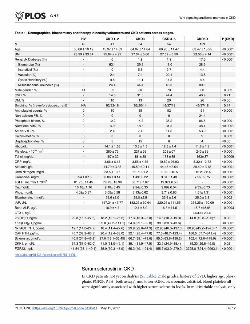
els, explaining 25% of its variability (p<0.0001). Fig 2 shows the correlation between blood
platelet count and serum DKK1 concentration in CKD patients not yet on dialysis.
In CKD stage 5D patients (S2 Table), higher calcium, CRP, and blood platelets and lower
PTH all were significantly associated with higher serum DKK1 levels. In multivariable analysis,
only calcium and blood platelets independently associated with serum DKK1 levels, explaining
14% of its variability (p<0.0001). Of note, no association was observed between serum DKK1
levels and use of antiplatelet agents. Serum sclerostin and DKK1 levels did not correlate, nei-
ther in the overall cohort nor in subgroups.
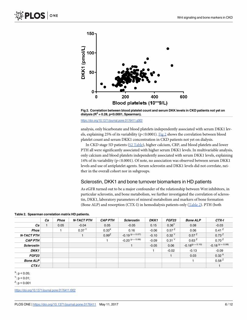
Sclerostin, DKK1 and bone turnover biomarkers in HD patients
As eGFR turned out to be a major confounder of the relationship between Wnt inhibitors, in
particular sclerostin, and bone metabolism, we further investigated the correlation of scleros-
tin, DKK1, laboratory parameters of mineral metabolism and markers of bone formation
(Bone ALP) and resorption (CTX-I) in hemodialysis patients only (Table 2). PTH (both
Fig 2. Correlation between blood platelet count and serum DKK levels in CKD patients not yet on
dialysis (R2 = 0.28, p<0.0001, Spearman).
https://doi.org/10.1371/journal.pone.0176411.g002
Table 2. Spearman correlation matrix HD patients.
Ca Phos N-TACT PTH CAP PTH Sclerostin DKK1 FGF23 Bone ALP CTX-I
Ca 1 0.05 -0.04 0.05 -0.05 0.15 0.36Y 0.08 -0.03
Phos 1 0.37 Y 0.33X 0.16 -0.06 0.57 Z 0.06 0.41 Z
N-TACT PTH 1 0.99Z -0.19 (p = 0.07) -0.10 0.32 Y 0.57 Z 0.73 Z
CAP PTH 1 -0.20 (p = 0.06) -0.09 0.31 Y 0.63 Z 0.70 Z
Sclerostin 1 -0.05 0.06 -0.18(p = 0.10) -0.18 (p = 0.08)
DKK1 1 -0.02 -0.13 -0.09
FGF23 1 0.03 0.32 X
Bone ALP 1 0.58 Z
CTX-I 1
X: p < 0.05;Y: p < 0.01;Z: p < 0.001
https://doi.org/10.1371/journal.pone.0176411.t002
Wnt signaling and bone markers in CKD
PLOS ONE | https://doi.org/10.1371/journal.pone.0176411 May 11, 2017 6 / 12
assays) strongly and directly correlated with Bone ALP and CTX-I. Sclerostin, as opposed to
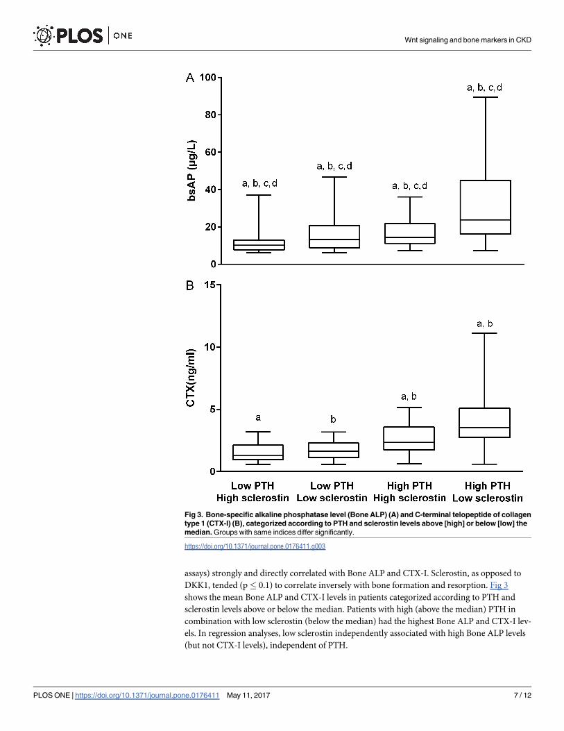
DKK1, tended (p� 0.1) to correlate inversely with bone formation and resorption. Fig 3
shows the mean Bone ALP and CTX-I levels in patients categorized according to PTH and
sclerostin levels above or below the median. Patients with high (above the median) PTH in
combination with low sclerostin (below the median) had the highest Bone ALP and CTX-I lev-
els. In regression analyses, low sclerostin independently associated with high Bone ALP levels
(but not CTX-I levels), independent of PTH.
Fig 3. Bone-specific alkaline phosphatase level (Bone ALP) (A) and C-terminal telopeptide of collagen
type 1 (CTX-I) (B), categorized according to PTH and sclerostin levels above [high] or below [low] the
median. Groups with same indices differ significantly.
https://doi.org/10.1371/journal.pone.0176411.g003
Wnt signaling and bone markers in CKD
PLOS ONE | https://doi.org/10.1371/journal.pone.0176411 May 11, 2017 7 / 12
Discussion
A first finding of the present study is that circulating levels of sclerostin, as opposed to DKK1,
increase with severity of CKD, reaching levels that are 2–3 fold higher than in non-CKD
controls.
This increase is likely the result of an increased production of sclerostin, since a recent clini-
cal study in 120 patients with CKD stage 1–5 showed an increased rather than a decreased
absolute and fractional urinary excretion of sclerostin with declining kidney function [10].
Furthermore, in jck mouse, a genetic model of polycystic kidney disease that exhibits progres-
sive renal disease, a transient increase in bone sclerostin was observed already in early stage
disease [22]. Till today, it is not clear which mechanism underlies signaling to the skeleton to
increase the production of sclerostin in the setting of CKD.
Besides an association with kidney function, we observed significant associations between
circulating sclerostin levels and various laboratory parameters of CKD-MBD. These associa-
tions were most pronounced in CKD stage 5D patients. Most probably, the overwhelming
impact of eGFR on circulating sclerostin levels obscured associations with laboratory parame-
ters of mineral metabolism in CKD patients not yet on dialysis.
Serum 1,25(OH)2D levels negatively associated with circulating sclerostin levels in CKD
patients not yet on dialysis, independent of eGFR, 25(OH)D, PTH and FGF23. This observation
is in line with recent experimental evidence by Ryan et al. [8]. These investigators showed
increased 25-hydroxyvitamin D 1α-hydroxylase cytochrome P450 (cyp27B1) mRNA in kidneys
of SOST KO mice as compared to their wild types. Moreover, treatment of cultured proximal
tubule cells with mouse recombinant sclerostin decreased cyp27B1 mRNA transcripts. Whether
vitamin D, reciprocally, affects SOST expression and circulating sclerostin levels remains to be
investigated. Of note, circulating sclerostin in the present study did not differ between patients
on and off therapy with active and/or nutritional vitamin D (data not shown).
In agreement with previous studies, we observed a positive and independent association
between serum phosphate and sclerostin levels [9, 13, 14]. Additional studies are required to
unravel the underlying regulatory mechanisms. In this context it is worth to be mentioned that
cross-sectional studies investigating the association between sclerostin and FGF23, yielded
conflicting results with some studies (including present study) reporting no association [11]
and other studies observing a positive association [23].
In agreement with previous studies [11, 24], we observed an inverse relationship between
sclerostin and PTH concentrations in dialysis patients. These data confirm and extend clinical
observations in patients with non-renal parathyroid disorders [25–27] and are consistent with
experimental data demonstrating downregulation of SOST by PTH [28]. Of note, high scleros-
tin levels coexist with high PTH levels in patients with advanced CKD. This observation sug-
gests skeletal resistance to the action of PTH [29–31], similar to FGF23 resistance explaining
the coexistence of high FGF23 and PTH levels in advanced stage CKD [32]. Of note, gender,
serum phosphate and PTH levels determined only 16% of the variability of sclerostin levels in
CKD stage 5D patients, implying that many other systemic and local determinants remain to
be identified.
Consistent with the biological effects of sclerostin on bone, we observed an inverse relation-
ship between serum sclerostin and Bone ALP, a bone formation marker, and between serum
sclerostin and CTX-I, a bone resorption marker. As such, our data in HD patients confirm
and extend previous clinical data [11, 12, 33]. Of interest, levels of the bone turnover biomark-
ers were highest in patients with high PTH levels in combination with low sclerostin levels.
Contrary to sclerostin, circulating DKK1 levels were not or only marginally associated with
kidney function and parameters of mineral metabolism. Previous studies investigating the
Wnt signaling and bone markers in CKD
PLOS ONE | https://doi.org/10.1371/journal.pone.0176411 May 11, 2017 8 / 12
association between DKK1 and kidney function yielded conflicting results with some investi-
gators observing unaltered [15, 17], while others reported increased [16] DKK1 levels in CKD.
Of note, serum levels of DKK1 and sclerostin were unrelated in the present study, pointing to
different origin and different regulatory mechanisms.
Importantly, the expression of sclerostin and DKK1 is not restricted to bone. Substantial
evidence indicates that platelets may be a major source of circulating levels of DKK1. More-
over, it has been demonstrated that during clotting ex vivo, DKK1 is released from platelets to
a significant but variable extent [34]. In a cohort of healthy volunteers, levels of DKK1 levels
were 2.7-fold higher in serum samples as compared to plasma samples [34]. Whether the
degree of this ex vivo release is a random phenomenon or relates to the in vivo activation state
of the platelets, as suggested by some investigators, is a matter of ongoing discussion [35]. Of
interest, in the present study, only platelet count and calcium, playing a crucial role in platelet
activation [36], were found to be independently associated with serum DKK1 levels in dialysis
patients and CKD patients not yet on dialysis. As opposed to others [37], we failed to demon-
strate lower DKK1 levels in CKD patients receiving antiplatelet drugs compared with those
not on antiplatelet therapy.
Significant but often complex associations have been reported between circulating levels of
sclerostin and DKK1 and indices of vascular health [13, 15, 20, 38, 39]. Both sclerostin and
DKK1 may be considered mediators and markers of cardiovascular disease (CVD). In the
present study, and opposite to recent studies in non-CKD patients [35, 40], we failed to find
higher circulating DKK1 and sclerostin levels in CKD patients with CVD as compared to
counterparts free of CVD.
In conclusion, serum levels of the Wnt inhibitors DKK1 and sclerostin are unrelated in
CKD, reflecting a different origin and different regulatory mechanisms. Sclerostin, as opposed
to DKK1, may qualify as a biomarker of CKD-MBD, particularly in dialysis patients. DKK1
serum levels mainly relate to platelet count and/or activity. Additional experimental and clini-
cal studies are required to elucidate the (path)physiological role of circulating sclerostin and
DKK1 in bone disease and beyond. This information is mandatory as anti-sclerostin and anti-
DKK1 monoclonal antibodies emerge as very promising pharmaceuticals in the treatment of
osteoporosis.
Supporting information
S1 Fig. Sclerostin and mineral metabolism markers according to CKD stage.
(TIF)
S1 Table. Linear regression analysis with Ln sclerostin and ln DKK1 as dependent variable
in CKD patients, not yet in dialysis.
(DOCX)
S2 Table. Linear regression analysis with Ln sclerostin and ln DKK1 as dependent variable
in CKD patients on maintenance hemodialysis.
(DOCX)
Author Contributions
Conceptualization: VMB PCD PE.
Formal analysis: GJB LV BM PE.
Funding acquisition: FB PCD PE.
Wnt signaling and bone markers in CKD
PLOS ONE | https://doi.org/10.1371/journal.pone.0176411 May 11, 2017 9 / 12

Investigation: GJB LV BM AV.
Methodology: GJB LV FB PCD PE.
Project administration: GJB PCD PE.
Resources: FB VMB PCD PE.
Supervision: PCD PE.
Validation: GJB BM AV.
Visualization: GJB PE.
Writing – original draft: GJB PCD PE.
Writing – review & editing: GJB VMB AV PCD PE.
References1. Baron R, Kneissel M. WNT signaling in bone homeostasis and disease: from human mutations to treat-
ments. Nat Med. 2013; 19(2):179–92. https://doi.org/10.1038/nm.3074 PMID: 23389618
2. Thompson B, Towler DA. Arterial calcification and bone physiology: role of the bone-vascular axis. Nat
Rev Endocrinol. 2012; 8(9):529–43. https://doi.org/10.1038/nrendo.2012.36 PMID: 22473330
3. van Bezooijen RL, Roelen BA, Visser A, van der Wee-Pals L, de Wilt E, Karperien M, et al. Sclerostin is
an osteocyte-expressed negative regulator of bone formation, but not a classical BMP antagonist. J
Exp Med. 2004; 199(6):805–14. https://doi.org/10.1084/jem.20031454 PMID: 15024046
4. Brandenburg VM, Kramann R, Koos R, Kruger T, Schurgers L, Muhlenbruch G, et al. Relationship
between sclerostin and cardiovascular calcification in hemodialysis patients: a cross-sectional study.
BMC Nephrol. 2013; 14:219. https://doi.org/10.1186/1471-2369-14-219 PMID: 24112318
5. Ke HZ, Richards WG, Li X, Ominsky MS. Sclerostin and Dickkopf-1 as therapeutic targets in bone dis-
eases. Endocr Rev. 2012; 33(5):747–83. https://doi.org/10.1210/er.2011-1060 PMID: 22723594
6. Wang LF, Wu H, Xu Y, Deng M, Han XL, Bai D. Effect of SOST gene deletion on the progression of
renal interstitial fibrosis in obstructive kidney injury. Ren Fail. 2015; 37(9):1514–7. https://doi.org/10.
3109/0886022X.2015.1077323 PMID: 26337453
7. Hruska KA, Sugatani T, Agapova O, Fang Y. The chronic kidney disease—Mineral bone disorder
(CKD-MBD): Advances in pathophysiology. Bone. 2017.
8. Ryan ZC, Ketha H, McNulty MS, McGee-Lawrence M, Craig TA, Grande JP, et al. Sclerostin alters
serum vitamin D metabolite and fibroblast growth factor 23 concentrations and the urinary excretion of
calcium. Proc Natl Acad Sci U S A. 2013; 110(15):6199–204. https://doi.org/10.1073/pnas.1221255110
PMID: 23530237
9. Cejka D, Herberth J, Branscum AJ, Fardo DW, Monier-Faugere MC, Diarra D, et al. Sclerostin and Dick-
kopf-1 in renal osteodystrophy. Clin J Am Soc Nephrol. 2011; 6(4):877–82. https://doi.org/10.2215/
CJN.06550810 PMID: 21164019
10. Cejka D, Marculescu R, Kozakowski N, Plischke M, Reiter T, Gessl A, et al. Renal elimination of scler-
ostin increases with declining kidney function. J Clin Endocrinol Metab. 2014; 99(1):248–55. https://doi.
org/10.1210/jc.2013-2786 PMID: 24187403
11. Delanaye P, Krzesinski JM, Warling X, Moonen M, Smelten N, Medart L, et al. Clinical and biological
determinants of sclerostin plasma concentration in hemodialysis patients. Nephron Clin Pract. 2014;
128(1–2):127–34. https://doi.org/10.1159/000366449 PMID: 25377055
12. Ishimura E, Okuno S, Ichii M, Norimine K, Yamakawa T, Shoji S, et al. Relationship between serum
sclerostin, bone metabolism markers, and bone mineral density in maintenance hemodialysis patients.
J Clin Endocrinol Metab. 2014; 99(11):4315–20. https://doi.org/10.1210/jc.2014-2372 PMID: 25093620
13. Kanbay M, Siriopol D, Saglam M, Kurt YG, Gok M, Cetinkaya H, et al. Serum sclerostin and adverse
outcomes in nondialyzed chronic kidney disease patients. J Clin Endocrinol Metab. 2014; 99(10):
E1854–61. https://doi.org/10.1210/jc.2014-2042 PMID: 25057883
14. Pelletier S, Dubourg L, Carlier MC, Hadj-Aissa A, Fouque D. The relation between renal function and
serum sclerostin in adult patients with CKD. Clin J Am Soc Nephrol. 2013; 8(5):819–23. https://doi.org/
10.2215/CJN.07670712 PMID: 23430206
Wnt signaling and bone markers in CKD
PLOS ONE | https://doi.org/10.1371/journal.pone.0176411 May 11, 2017 10 / 12
15. Thambiah S, Roplekar R, Manghat P, Fogelman I, Fraser WD, Goldsmith D, et al. Circulating sclerostin
and Dickkopf-1 (DKK1) in predialysis chronic kidney disease (CKD): relationship with bone density and
arterial stiffness. Calcif Tissue Int. 2012; 90(6):473–80. https://doi.org/10.1007/s00223-012-9595-4
PMID: 22527202
16. Fang Y, Ginsberg C, Seifert M, Agapova O, Sugatani T, Register TC, et al. CKD-induced wingless/inte-
gration1 inhibitors and phosphorus cause the CKD-mineral and bone disorder. J Am Soc Nephrol.
2014; 25(8):1760–73. https://doi.org/10.1681/ASN.2013080818 PMID: 24578135
17. Malluche HH, Davenport DL, Cantor T, Monier-Faugere MC. Bone mineral density and serum biochem-
ical predictors of bone loss in patients with CKD on dialysis. Clin J Am Soc Nephrol. 2014; 9(7):1254–
62. https://doi.org/10.2215/CJN.09470913 PMID: 24948144
18. Brandenburg VM, D’Haese P, Deck A, Mekahli D, Meijers B, Neven E, et al. From skeletal to cardiovas-
cular disease in 12 steps-the evolution of sclerostin as a major player in CKD-MBD. Pediatr Nephrol.
2016; 31(2):195–206. https://doi.org/10.1007/s00467-015-3069-7 PMID: 25735207
19. Evenepoel P, D’Haese P, Brandenburg V. Sclerostin and DKK1: new players in renal bone and vascular
disease. Kidney Int. 2015; 88(2):235–40. https://doi.org/10.1038/ki.2015.156 PMID: 26083653
20. Pelletier S, Confavreux CB, Haesebaert J, Guebre-Egziabher F, Bacchetta J, Carlier MC, et al. Serum
sclerostin: the missing link in the bone-vessel cross-talk in hemodialysis patients? Osteoporos Int.
2015; 26(8):2165–74. https://doi.org/10.1007/s00198-015-3127-9 PMID: 25910747
21. Daugirdas JT. Second generation logarithmic estimates of single-pool variable volume Kt/V: an analysis
of error. J Am Soc Nephrol. 1993; 4(5):1205–13. PMID: 8305648
22. Sabbagh Y, Graciolli FG, O’Brien S, Tang W, dos Reis LM, Ryan S, et al. Repression of osteocyte Wnt/
beta-catenin signaling is an early event in the progression of renal osteodystrophy. J Bone Miner Res.
2012; 27(8):1757–72. https://doi.org/10.1002/jbmr.1630 PMID: 22492547
23. Moyses RM, Jamal SA, Graciolli FG, dos Reis LM, Elias RM. Can we compare serum sclerostin results
obtained with different assays in hemodialysis patients? Int Urol Nephrol. 2015; 47(5):847–50. https://
doi.org/10.1007/s11255-015-0971-7 PMID: 25862239
24. Drechsler C, Evenepoel P, Vervloet MG, Wanner C, Ketteler M, Marx N, et al. High levels of circulating
sclerostin are associated with better cardiovascular survival in incident dialysis patients: results from
the NECOSAD study. Nephrol Dial Transplant. 2015; 30(2):288–93. https://doi.org/10.1093/ndt/gfu301
PMID: 25248363
25. Ardawi MS, Al-Sibiany AM, Bakhsh TM, Rouzi AA, Qari MH. Decreased serum sclerostin levels in
patients with primary hyperparathyroidism: a cross-sectional and a longitudinal study. Osteoporos Int.
2012; 23(6):1789–97. https://doi.org/10.1007/s00198-011-1806-8 PMID: 22041864
26. Costa AG, Cremers S, Rubin MR, McMahon DJ, Sliney J Jr., Lazaretti-Castro M, et al. Circulating scler-
ostin in disorders of parathyroid gland function. J Clin Endocrinol Metab. 2011; 96(12):3804–10. https://
doi.org/10.1210/jc.2011-0566 PMID: 21937621
27. van Lierop AH, Witteveen JE, Hamdy NA, Papapoulos SE. Patients with primary hyperparathyroidism
have lower circulating sclerostin levels than euparathyroid controls. Eur J Endocrinol. 2010; 163
(5):833–7. https://doi.org/10.1530/EJE-10-0699 PMID: 20817762
28. Kramer I, Loots GG, Studer A, Keller H, Kneissel M. Parathyroid hormone (PTH)-induced bone gain is
blunted in SOST overexpressing and deficient mice. J Bone Miner Res. 2010; 25(2):178–89. https://doi.
org/10.1359/jbmr.090730 PMID: 19594304
29. Berdud I, Martin-Malo A, Almaden Y, Tallon S, Concepcion MT, Torres A, et al. Abnormal calcaemic
response to PTH in the uraemic rat without secondary hyperparathyroidism. Nephrol Dial Transplant.
1996; 11(7):1292–8. PMID: 8672025
30. Massry SG, Coburn JW, Lee DB, Jowsey J, Kleeman CR. Skeletal resistance to parathyroid hor-
mone in renal failure. Studies in 105 human subjects. Ann Intern Med. 1973; 78(3):357–64. PMID:
4571863
31. Wesseling-Perry K, Harkins GC, Wang HJ, Elashoff R, Gales B, Horwitz MJ, et al. The calcemic
response to continuous parathyroid hormone (PTH)(1–34) infusion in end-stage kidney disease varies
according to bone turnover: a potential role for PTH(7–84). J Clin Endocrinol Metab. 2010; 95(6):2772–
80. https://doi.org/10.1210/jc.2009-1909 PMID: 20382692
32. Komaba H, Goto S, Fujii H, Hamada Y, Kobayashi A, Shibuya K, et al. Depressed expression of Klotho
and FGF receptor 1 in hyperplastic parathyroid glands from uremic patients. Kidney Int. 2010; 77
(3):232–8. https://doi.org/10.1038/ki.2009.414 PMID: 19890272
33. Cejka D, Jager-Lansky A, Kieweg H, Weber M, Bieglmayer C, Haider DG, et al. Sclerostin serum levels
correlate positively with bone mineral density and microarchitecture in haemodialysis patients. Nephrol
Dial Transplant. 2012; 27(1):226–30. https://doi.org/10.1093/ndt/gfr270 PMID: 21613383
Wnt signaling and bone markers in CKD
PLOS ONE | https://doi.org/10.1371/journal.pone.0176411 May 11, 2017 11 / 12
34. Voorzanger-Rousselot N, Goehrig D, Facon T, Clezardin P, Garnero P. Platelet is a major contributor to
circulating levels of Dickkopf-1: clinical implications in patients with multiple myeloma. Br J Haematol.
2009; 145(2):264–6. https://doi.org/10.1111/j.1365-2141.2009.07587.x PMID: 19210508
35. Ueland T, Otterdal K, Lekva T, Halvorsen B, Gabrielsen A, Sandberg WJ, et al. Dickkopf-1 enhances
inflammatory interaction between platelets and endothelial cells and shows increased expression in ath-
erosclerosis. Arterioscler Thromb Vasc Biol. 2009; 29(8):1228–34. https://doi.org/10.1161/ATVBAHA.
109.189761 PMID: 19498175
36. Vemana HP, Karim ZA, Conlon C, Khasawneh FT. A critical role for the transient receptor potential
channel type 6 in human platelet activation. PLoS One. 2015; 10(4):e0125764. https://doi.org/10.1371/
journal.pone.0125764 PMID: 25928636
37. Lattanzio S, Santilli F, Liani R, Vazzana N, Ueland T, Di Fulvio P, et al. Circulating dickkopf-1 in diabetes
mellitus: association with platelet activation and effects of improved metabolic control and low-dose
aspirin. Journal of the American Heart Association. 2014; 3(4).
38. Claes KJ, Viaene L, Heye S, Meijers B, d’Haese P, Evenepoel P. Sclerostin: Another vascular calcifica-
tion inhibitor? J Clin Endocrinol Metab. 2013; 98(8):3221–8. https://doi.org/10.1210/jc.2013-1521
PMID: 23788689
39. Register TC, Hruska KA, Divers J, Bowden DW, Palmer ND, Carr JJ, et al. Plasma Dickkopf1 (DKK1)
concentrations negatively associate with atherosclerotic calcified plaque in African-Americans with type
2 diabetes. J Clin Endocrinol Metab. 2013; 98(1):E60–5. https://doi.org/10.1210/jc.2012-3038 PMID:
23125289
40. Garcia-Martin A, Reyes-Garcia R, Garcia-Fontana B, Morales-Santana S, Coto-Montes A, Munoz-Gar-
ach M, et al. Relationship of Dickkopf1 (DKK1) with cardiovascular disease and bone metabolism in
Caucasian type 2 diabetes mellitus. PLoS One. 2014; 9(11):e111703. https://doi.org/10.1371/journal.
pone.0111703 PMID: 25369286
Wnt signaling and bone markers in CKD
PLOS ONE | https://doi.org/10.1371/journal.pone.0176411 May 11, 2017 12 / 12


![differenziamento Mina fin [modalità compatibilità] · OBs Sclerostin Myeloma cells through sclerostin secretion contribute to MM Cells OBs Sclerostin OPG RANKL 1)Inhibit OB formation](https://img.dokumen.tips/doc/110x75/5ac3ff867f8b9aae1b8d18c6/differenziamento-mina-fin-modalit-compatibilit-sclerostin-myeloma-cells-through.jpg)


![Response of Sclerostin and Bone Turnover Markers to High ...downloads.hindawi.com/journals/bmri/2018/4864952.pdf · sclerostin appears to increase within min following low intensityrunninginyoungwomen[],aswellasfollowing](https://img.dokumen.tips/doc/110x75/6060ee779062f139b91afd4b/response-of-sclerostin-and-bone-turnover-markers-to-high-sclerostin-appears.jpg)