Embed Size (px)
Citation preview

Ketut Siki Kawiyana
Orthopaedi & Traumatology Subdivision Udayana University
Sanglah General Hospital, Denpasar-Bali
CHRONIC OSTEOMYELITIS IN CHILDREN
EVALUATION AND MANAGEMENT

INTRODUCTION Chronic osteomyelitis
A daunting challenge to orthopaedic surgeons
Often described as a disease that can never truly be cured
10 to 30% of acute haematogenous osteomyelitis chronic osteomyelitis
Socio-economically underdeveloped regions high incidence of osteomyelitis in childhood.
Immunosuppression, MalnutritionHigh incidence of
trauma

EPIDEMIOLOGY
Open fractures (3–50%)
The surgical management of closed fractures may (1–5%)
Primary hip or knee replacement (0.5‒2%)
Revision surgery (5%)
Second stage revision for periprosthetic infection (20%)
Overall complication of orthopaedic cases during the life-time of the prosthesis or implant (5%)

EPIDEMIOLOGY

DEFINITION
This definition originated from
• Long-standing infection of the bone characterized by persistence of microorganisms, presence of sequestrum, low-grade inflammation, and fistulae
Chronic osteomyelitis
Observation acute haematogenous
osteomyelitisLeft untreated
Formation of necrotic segments
of bone
a source of on-going or chronic
infection

PathophysiologyInadequately treated hematogenous acute osteomyelitis or
more commonly from a contiguous source of infection
Inflammatory process causes obliteration and compression of the vascular channels.
Part of the bone undergoes necrosis sequestrum
Destruction of bone, and microorganisms propagate within the destroyed bone.
New bone is formed around the sequestrum from the intact periosteum and endosteum involucrum.
The involucrum is perforated by openings known as cloacae
Chronic Osteomyelitis

I - A large inoculum of bacteria reaches the medular channel
II - (Acute state) Pus resulting from inflammatory response spreads into vascular channels
III - (Chronic state) Vascular channels are compressed and obliterated by the inflammatory process, and the resulting ischaemia also contributes to bone necrosis

What are the causes of chronic osteomyelitis?
Decreased blood flow as a result of initial insult or operative procedure diminishes the healing capacity
Antibiotics cannot penetratethough the infected and necrotic area and
sequestrum produces an area of lowered vascularity
Resistance of organisms to antibiotics. Organism forms a biofilmaround the sequestrum or implant
Inadequate surgical debridement.

Classification

ANATOMIC CLASSIFICATION OF CHRONIC OSTEOMYELITIS IN CHILDREN WITH TREATMENT RECOMMENDATIONS
Penny JN. Children’s OrthopaedicSurgery and Rehabilitation inAfrica. AAOS ICL 228; 2004.
• Type I : “typical” osteomyelitis,
• Type II : “atrophic”
• Type III : “sclerotic”
• Type IV : “cortical”
• Type V : “multiple walled-off abscesses”
• Type VI : “multiple microabscesses”
Diaphyseal Osteomyelitis
• appears as a single, or multiple, walled-off abscesses with or without a sclerotic margin. Sequestra are uncommon
• a limited saucerization and curettage should be sufficient
Metaphyseal Osteomyelitis

Diaphyseal Osteomyelitis
Type I “typical” osteomyelitis
• A well-defined sequestrum and an involucrum
• sequestrectomy/ debridement followed by protection of the limb until the bone has been reconstituted
Type II “atrophic”
• Failure of the involucrum to form
• Waiting at least 3 to 6 months to see if the periosteum will respond. If there is no response, then plans can be made for reconstruction, whether by grafting or bone transport.

Diaphyseal Osteomyelitis
Type III “sclerotic”
• Fusiform, dense sclerotic healing reaction generated by the periosteum
• Debridement through a cortical window
Type IV “cortical”
• Localized sequestrum within the cortex of the involved bone
• Sequestrectomy through a localized cortical window

Diaphyseal Osteomyelitis
Type V “multiple walled-off abscesses”
• involves one or more well-defined lucencieswithin the involucrum
• explored and debrided through a cortical window
Type VI “multiple microabscesses”
• Similar to type V based on both appearance and proposed etiology, but smaller and more numerous lucencies within the involucrum.
• a longitudinal partial diaphysectomy with debridement

Most common isolated microorganisms in osteomyelitis are related to age
and susceptibility factors
adapted from Lew, Waldvogel, 2004; Brook, 2008; McNally, Nagarajah, 2010; Chihara, Segreti, 2010; Jorge et al., 2010; Zimmerli, 2010; Eid, Berbari 2012

Defence Mechanism
The host protective cellular layer with functional defence mechanisms
Opsonification PhagocytosisComplement
mediated lysis

Biofilm Formation The invading bacteria enter their default growth pattern and establish a
biofilm.
Biofilm formation occurs in five stages
Adhesion
Production of the extra-
cellular matrix
Colonisation
Maturation
Dispersion of bacteria
A layer-like aggregation of microbial cells and extracellular polymeric substances attached to a substrate which provides an environment for the
exchange of genetic material between bacterial cells

The Host Response
The innate immunity
Interleukin-1 (IL-1)
IL-6
Tumour necrosis factor (TNF)
Neutrophils
Macrophages
Acquired or adaptive immunity
cytotoxic CD8+ T cells
antibodies by B lymphocytes
TH1 lymphokines
(IL-12 and interferon-γ)
TH2 lymphokines
(IL-3 and IL-4)

The Role Of Osteoclasts
Receptor activator of nuclear factor kappa-B ligand(RANKL) is a potent activator of osteoclastsand is produced by bone marrow stromal cells under normal conditions.
In osteomyelitis certain bacterial components, such as lipopolysaccharide (LPS), result in the production of RANKL by a variety of cells(including activated T-cells) ultimately causing abnormal bone loss

Diagnostic
Clinical
ImagingLaboratory

Clinical evaluation
History Main complaint
associated problems
medical history
Previous surgical history
Prior therapeutic interventions
• Examination– Local Pathology
– sinus tracts
– exposed bone
– Skeletal stability
– Condition of soft tissue
– chronic wound over a fracture site or surgical implant
– tissue necrosis overlying bone
– Vascularization
– Neurological status

CHRONIC OSTEOMYELITIS is often associated with
Angular or rotational deformity
Deformities of the adjacent joints.
Limb length discrepancy (LLD)
A deep cavity in the bone
A big sequestrum, which creates a big gap

Imaging
• the presence of periosteal reaction or purulent collections.
• as a guide during deep aspiration of fluid collections for culture and sensitivity
Ultrasound
• useful in localising sequestra or cloacae and
• aid in the assessment of skeletal integrity and stability
X-rays and CT
• contrast material is injected into the sinus opening to ascertain the course and extent of the sinus and its communication with deeper tissues
Sinography
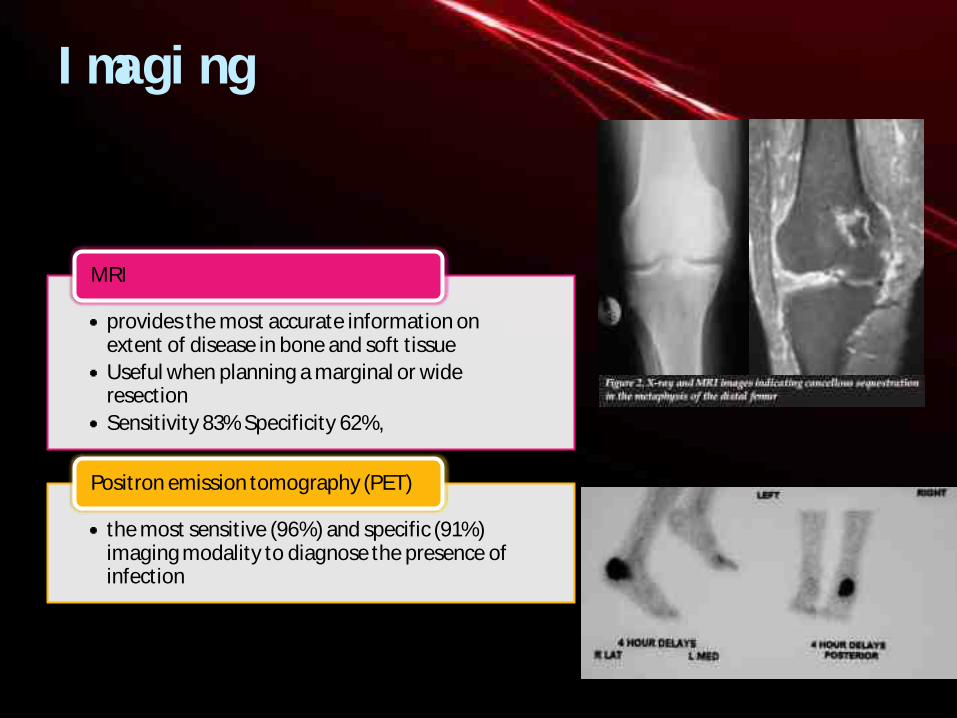
Imaging
• provides the most accurate information on extent of disease in bone and soft tissue
• Useful when planning a marginal or wide resection
• Sensitivity 83% Specificity 62%,
MRI
• the most sensitive (96%) and specific (91%) imaging modality to diagnose the presence of infection
Positron emission tomography (PET)

Laboratory investigationsFull blood count
Infection markers
• WBC, ESR, CRP, Procalcitonin
Pro-inflammatory cytokine
• IL-1, IL-6,IL-8, TNF
Renal and liver function tests
Electrolyte
Nutritional profile
• To ascertain the degree of systemic compromise
• As a diagnostic tool in the confirmation of the presence of sepsis.
Aim

Pathogen identification
In cases without significant local or systemic septic complications, pathogen detection may be delayed after the primary debridement procedure
Pre-operative (‘neo-adjuvant’) antibiotics may be mandatory, for example in patients with Significant local compromise (cellulitis in the region of the incision) or Systemic compromise (systemic sepsis or septic shock).
open biopsy or deep aspiration under ultrasound guidance, prior to definitive surgery.
• an attempts be made to identify the pathogen prior to the first surgical debridement through biopsy of deep granulation tissue
Cierny recommendation

Swab culture from a sinus may offer some diagnostic benefit.
Identification of methicillin-resistant S. aureus (MRSA) or vancomycin-resistant enterococcus necessitates the implementation of stringent infection control measures during hospitalization.
Isolation of S. aureus from a superficial culture has a high degree of correlation with deep cultures
In cases without significant local or systemic septic complications, pathogen detection may be delayed to after the primary debridement procedure
Pathogen identification

Treatment
Multidisciplinary effort is needed for successful treatment.
The team should consist of
Surgeons (orthopedic and reconstructive surgery)
Infectious disease specialist
Specialist to advise on nutrition
Psychologist if needed

Management strategies
Eradication of infection and limb reconstruction
A wide array of surgical procedures and techniques in terms of debridement
Dead space management
Soft tissue cover
Skeletal reconstruction
Healing of bone segment
Preservation of limb length and function
Aim
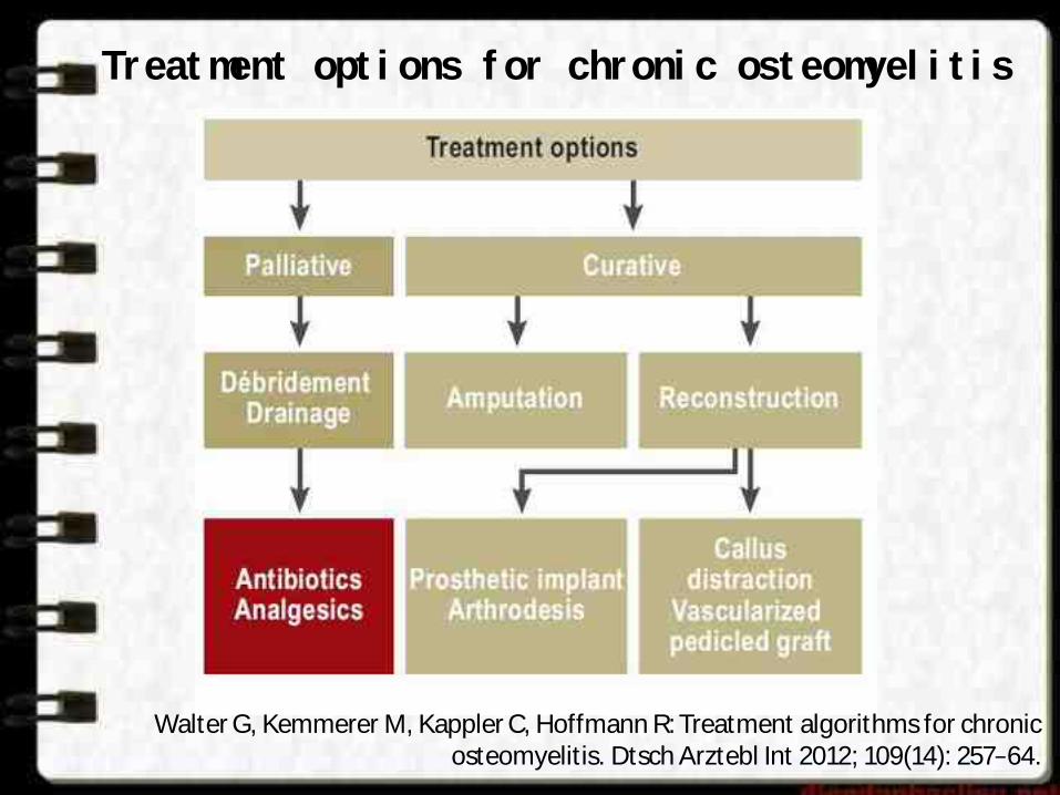
Treatment options for chronic osteomyelitis
Walter G, Kemmerer M, Kappler C, Hoffmann R: Treatment algorithms for chronic osteomyelitis. Dtsch Arztebl Int 2012; 109(14): 257‒64.

Management strategies
Approach
This decision consideration as described by Cierny C-host should be palliated A- and B-hosts may be
considered for a curative treatment protocol
• multiple surgical procedures
Curative
• Less invasive and typically involve the use of chronic suppressive antibiotic therapy
• incision and drainage, oral antibiotics, ambulatory aides, and pain medication
Palliative

TREATMENT ALGORITHM OF CIERNY-MADER STAGE-1,
OR HEMATOGENOUS, LONG-BONE
OSTEOMYELITIS

TREATMENT ALGORITHM OF CIERNY-MADER STAGE-1 LONG-
BONE OSTEOMYELITIS ASSOCIATED
WITH INFECTION AT THE SITE OF
HARDWARE
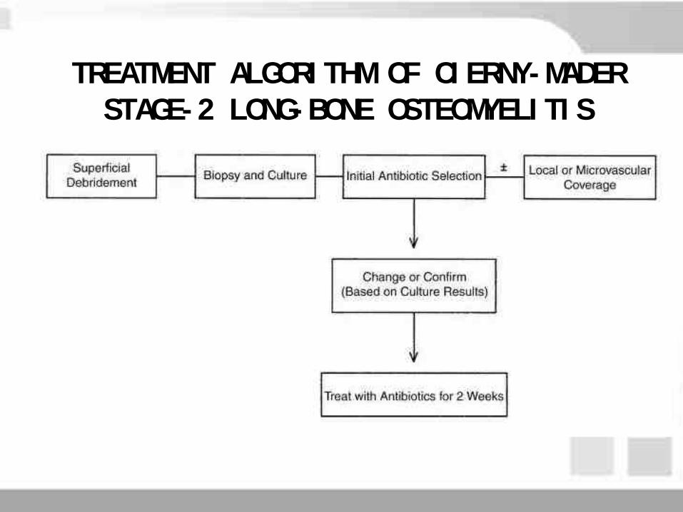
TREATMENT ALGORITHM OF CIERNY-MADER STAGE-2 LONG-BONE OSTEOMYELITIS
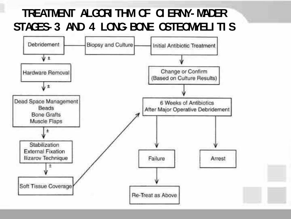
TREATMENT ALGORITHM OF CIERNY-MADER STAGES-3 AND 4 LONG-BONE OSTEOMYELITIS

ANTIBIOTIC THERAPY
Antibiotic-therapy regimen is vital This should be based on the identification of the
infective organism and its susceptibility
The challenges for successful antibiotic therapy The presence of devitalized, avascular tissue
Biofilm formation
Chemical environment at the site of infection
Effective treatment of chronic osteomyelitisrequires prolonged antimicrobial therapy

ADVANTAGES AND DISADVANTAGES OF PARENTERAL, ORAL AND LOCAL ANTIBIOTIC THERAPY
adapted from Gitelis, Brebach, 2002; Ambrose et al., 2003; Lazzarini et al., 2005

CONTROVERSIAL ISSUE A review of studies on antibiotic therapy for
osteomyelitis published between 1968 and 2000 concluded that there is inadequate evidence to recommend the best agent, route of administration, or
duration of therapy
The duration of antibiotic therapy ??? The standard recommendation of use of antibiotics
for 4–6 weeks is based on animal studies on time taken for revascularization of bone

Spellberg and Lipsky, 2012 reviewed publications from 1970 to 2011
1. Oral antibiotic therapy with agents that have high bioavailability is comparable with parenteral therapy
2. Improved cure rates with addition of rifampicin
3. The duration of antibiotic therapy should be individualized based on clinical, hematological, and radiological response, and patients should be monitored after completion of therapy
4. The cure rate of chronic osteomyelitis is increased with surgical resection of infected and devitalized tissue in conjunction with antibiotic therapy
Spellberg B, Lipsky BA. Systemic antibiotic therapy for chronic osteomyelitis in adults. Clin Infect Dis. 2012;54:393–407.

Surgical management
Debridement techniques
Pathogen detection
Dead space management
Skeletalstabilization

Debridement techniques
All necrotic or ischaemic tissues should be excised
All foreign bodies and surgical implants need to be removed
• Except of early infection following osteosynthesis where union is expected to occur
Soft tissues, and scar tissue
• should be resected to a supple, well-perfused margin
Bony debridement technique
• Simple sequestrectomy
• Intra-medullary reaming (indirect unroofing)
• Tangential excision (direct unroofing)
• Segmental resection and amputation

Problem
• Theoretically best treated with a wide resection of all infected tissues and subsequent limb reconstruction
• The reconstruction procedures requiredinvolving bone transport or extensive bone grafts
• Fraught with danger in the poor host and failure frequently results in the amputation of the limb
Compromised hosts

Pathogen detection
Routine microscopy, culture and sensitivity (MCS) of tissue, bone and
exudates taken under aseptic condition in the absence of antibiotic therapy in the preceding 10 days
Multiple samples should be acquired early in the procedure from
fluid collections, soft tissue, bone and foreign materials or sequestra
Samples should undergo aerobic and anaerobic incubation for
prolonged periods, at least 7 days, in order to increase detection of
fastidious organisms

Pathogen detection
Molecular methods have grown rapidly as the method of choice in pathogen detection
• Pyrosequencing is currently the most popular technique
• It can be performed on any specimen
• Able to reliably identify the micro-organism involved, irrespective of its phenotype (culturability), prior antibiotic therapy or metabolic state
Polymerase chain reaction (PCR)

Dead space management
Gentamycin-impregnated polymethylmethacrylate(PMMA) beads
• useful in type III lesions
Lautenbach irrigation systems
• commonly utilised in type I lesion
Antibiotic-impregnated PMMA spacers or intramedullarynails or antibiotic-loaded calcium sulphate pellets
• May be used in type I infections
• Especially in the setting of post-operative sepsis
Local or free soft tissue transfer procedures
• microvascular free-muscle transfer is considered the gold standard
• type II lesions
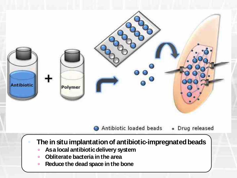
The in situ implantation of antibiotic-impregnated beads As a local antibiotic delivery system Obliterate bacteria in the area Reduce the dead space in the bone

Gentamycin-impregnated polymethylmethacrylate (PMMA) beads
Proportional weights of up to approximately 5 weight/weight % (2 g vancomycin per 40 g cement powder) have a
negligible influence on the mechanical strength of the cement
Gentamicin/vancomycin-loaded spacers
• Most effective against S.epidermidis and MRSA
Gentamicin/teicoplanin-combination spacer
• Best results against E. faecalis and S. aureus

Skeletal stabilization
Skeletal stabilization is needed for all stage 4 and some stage 3 lesions following excision of the devitalized bone
A variety of fixation options
Skeletal stability promotes
revascularizationenhanced perfusion
maximize of the host’s immune
response
• generally preferredExternal fixation
• Provide some stability
• But cannot achieve the level of stability provided by external fixation
Intramedullary PMMA nails
• Good modularity
• Minimally invasive nature
• Ability to effect bone transport and deformity correction
Circular external fixators

Ninety children (60 boys, 30 girls) were included in this study
The commonest site involved (50%) followed by tibia (45%).
A total of 112 surgical procedures were carried out in 90 patients.
Sequestrectomy (59.8%)
Ilizarov external fixator application (13.4%)
Saucerization (10.7%)
• Repeated debridements may be necessary to eradicate or control infection
• The ring fixator has been shown to be a useful tool to address pathological fracture, diaphyseal bone gaps, nonunions and stiffness and deformity
Conclusion

• To create a mechanical condition necessary for the development of distraction & compression.
• To store the new bone forming cells developed during lengthening and deposited along the line of stress and tension.
• Increase blood circulation for increased metabolic transformation of local tissue.
• Most importantly the medullary and the periosteal blood supply is not disturbed.
Ilizarov technique
Bari MM, Shahidul I, Mahfuzer R, Shetu NH, Golam M, et al. (2015) Treatment of Chronic Osteomyelitis in Children by Ilizarov Technique. MOJ Orthop Rheumatol 2(4): 00054

Surgical technique
Papineautechnique
Belfast technique
Lautenbach technique

PapineauTechnique
Stage 3 infections are best suited for this technique, as bone grafts do not provide the necessary stability.
Radical debridement
Bone grafting in stages
Delayed soft-tissue closure
Wound allowed to granulate naturally or with skin grafting

The Belfast technique Proposed by McNally et al
The reported a cure rate of 92% with this technique
Radical debridement
Early soft-tissue cover for
elimination of dead space
Delayed bone grafting
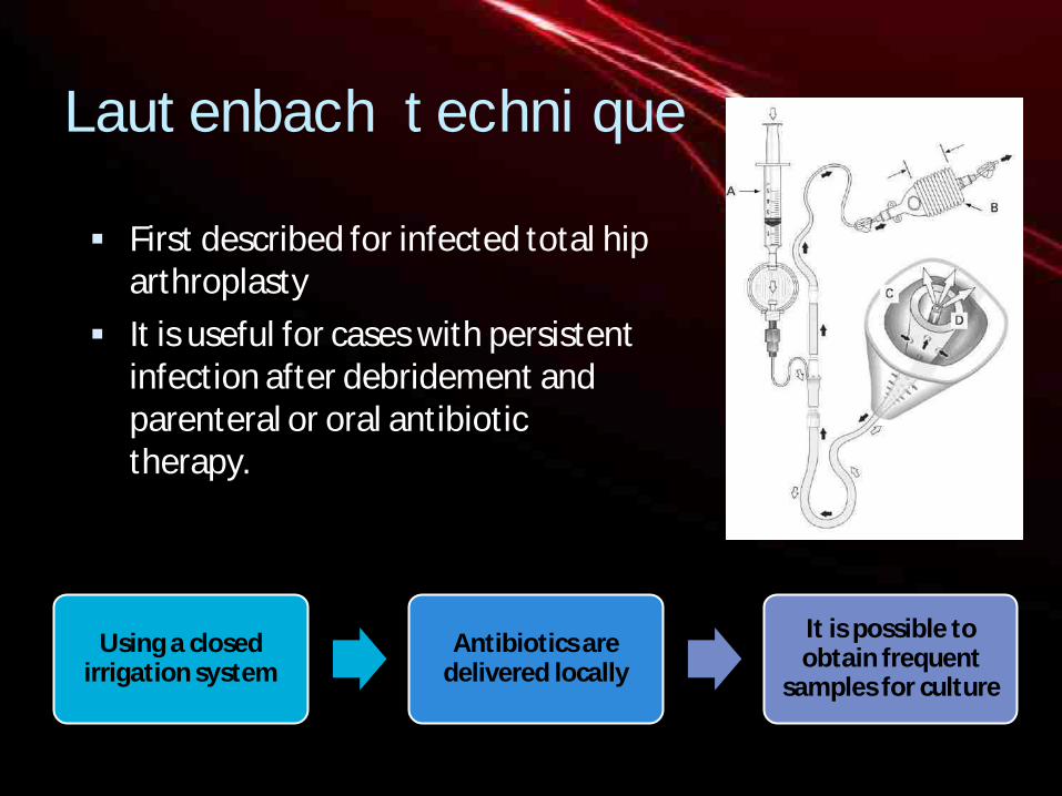
Lautenbach technique
First described for infected total hip arthroplasty
It is useful for cases with persistent infection after debridement and parenteral or oral antibiotic therapy.
Using a closed irrigation system
Antibiotics are delivered locally
It is possible to obtain frequent
samples for culture

Adjunctive therapies
Understanding of Bacterial
biofilm
Growth factors
Hyperbaric oxygen therapy

Hyperbaric oxygen therapy
There is a lack of guidelines for use as well as high-quality clinical trials for hyperbaric oxygen in chronic osteomyelitis.
The available literature however suggests great potential for this modality
Providing oxygen at high concentration and pressure in diseased tissues that are hypoxic
Improves the bactericidal ability of the neutrophils
Helps neutralize collagen synthesis and osteogenesis
Inducing angiogenesis
Suppressing anaerobic organisms
Enhancing antibiotic activity
Promoting oxygen-dependent osteoclastic resorption of necrotic bone

Growth Factors
Bone morphogenic protein
• Accelerate osteogenesis and bone healing
Platelet-rich plasma
• Promote bone and soft-tissue healing
Bacterial biofilm
The formation of biofilm is an important pathogenic factor for bacterial resistance and persistence of infection in chronic osteomyelitis
The alternative strategies resulting from understanding of the role of biofilm in chronic infection include
quorum-sensing inhibitors
bacteriophages
interspecies interaction
biofilm disruptors (sonication)
specific antibiofilm molecules

COMPLICATIONS
Pathologic fracture
Septic arthritis with joint destruction
Physeal damage
Nonunion or segmental bone loss
Leg length discrepancy (shortening or overgrowth)
Conclusion Management of chronic
osteomyelitis is challenging and prolonged
Proper staging and identification of causative organism remains vital to the success of treatment.
Newer treatment modalities are being developed to address the role of biofilm in chronic osteomyelitis

THANK YOU