Embed Size (px)
Citation preview

Chemokine responses and T cell
recruitment in central nervous system alphaherpesvirus infections
Liza Lind
Department of Rheumatology and Inflammation Research Institute of Medicine
Sahlgrenska Academy, University of Gothenburg
Gothenburg 2018

Cover illustration: Herpes virion by Servier Medical Art
Chemokine responses and T cell recruitment in central nervous system alphaherpesvirus infections © Liza Lind 2018 [email protected] ISBN 978-91-7833-081-2 (PRINT) ISBN 978-91-7833-082-9 (PDF) Printed in Gothenburg, Sweden 2018 Printed by BrandFactory

Here I stand, atoms with consciousness, matter with curiosity
A universe of atoms, an atom in the universe
Richard Feynman


Till Max och Harry


Chemokine responses and T cell recruitment in central nervous system alphaherpesvirus infections
Liza Lind
Department of Rheumatology and Inflammation Research, Institute of Medicine Sahlgrenska Academy, University of Gothenburg, Gothenburg, Sweden
Abstract
This thesis is focused on members of the alphaherpesvirus family; the two herpes simplex viruses and varicella zoster virus. Herpes simplex virus type 1 mainly causes cold sores and type 2 gives rise to genital blisters. Varicella zoster causes chickenpox in children and shingles in adults. The viruses are chronic and incurable, and very common. Both herpes simplex and varicella zoster virus are mostly harmless but can in rare cases cause inflammations of the central nervous system with various degree of severity. We have studied the immune system in neuroinflammation caused by alphaherpesviruses, with a special focus on how T cells, an important cell type in fighting off infections, reach the central nervous system. The central nervous system is immune privileged, thus immune cells entry is highly restricted and mediated by several factors. Interferons and interleukins amplify the immune response. Chemokines are attracting cells to the site of inflammation. Matrix metalloproteinases rearrange the membranes separating the central nervous system from peripheral blood.
Herpes simplex virus type 2 meningitis (HSM), an inflammation of the membranes surrounding the brain, is a more benign disease compared to herpes simplex virus type 1 encephalitis (HSE), an inflammation of the entire brain. In Paper I we found that in the cerebrospinal fluid of patients with HSE or HSM chemokines

CXCL8, CXCL9 and CXCL10 are strongly induced. In HSM two additional chemokines were induced; CCL8 and CXCL11. In Paper II we characterized T cells in the central nervous system of mice infected with herpes simplex virus type 2. We found a subset of T cells expressing both CD4 and CD8 surface markers which were highly virus-specific and activated. Returning to humans we examined levels of chemokines and matrix metalloproteinases in patients with neuroinflammations caused by varicella zoster virus (Paper III). We found that CXCL11 increased in meningitis patients, but not in patients with encephalitis or Ramsay Hunt (facial palsy). Matrix metalloproteinases 2 increased in all patient groups, whereas other matrix metalloproteinases were disease exclusive. In Paper IV we further investigated chemokine and cytokine contribution to inflammatory response. We found that CXCL11 levels correlated with expression of IL-1β, which was higher in cerebrospinal fluid of HSM patients. We also wanted to investigate the source of CXCL11 and found that neither astrocytes nor microglia produced CXCL11 upon stimulation with any herpes simplex virus, but microglia upregulated CXCL11 mRNA.
In this thesis we suggest that CXCL11 is a marker of viral meningitis, induced by interferon-γ and IL-1β. We find CD4 and CD8 double positive T cells in the CNS of mice with herpes simplex virus type 2 infection, which are degranulated, highly virus-specific and secreting cytotoxic factors.
Keywords: herpes, varicella, encephalitis, meningitis, chemokines, matrix metalloproteinases, central nervous system
ISBN 978-91-7833-081-2 (PRINT) ISBN 978-91-7833-082-9 (PDF)

Kemokinrespons och T cell-migration vid infektioner i centrala nervsystemet orsakade av alfaherpesvirus
Sammanfattning på svenska
Familjen herpesvirus består av många olika virus som infekterar såväl människor som djur. Den här avhandlingen fokuserar på två medlemmar i familjen alfaherpesvirus; herpes simplex och varicella zoster. Det finns två typer av herpes simplex virus, typ 1 och typ 2. Det är väldigt vanliga infektioner som oftast inte ger några symptom. Ibland kan dock viruset ge upphov till munsår (typ 1) eller genitala blåsor (typ 2). Varicella zoster är en infektion som kanske är mer känd som vattkoppor hos barn, eller bältros hos vuxna. Vid vuxen ålder bär de allra flesta människor på varicella zoster-viruset. Alla herpesvirus har gemensamt att när man väl smittats av dem så finns de kvar livet ut i en så kallad latent infektion. Då och då kan viruset aktiveras och orsaka symptom. I väldigt sällsynta fall kan de här tre virusen ge upphov till inflammationer i det centrala nervsystemet; i hjärna och ryggmärg. Herpes simplex virus typ 1 orsakar då vanligen encefalit, en inflammation av hela hjärnan, medan typ 2 orsakar meningit, en inflammation i membranen som omsluter hjärnan. Varicella zoster kan ge upphov till både encefalit och meningit, och också till en sjukdom som kallas Ramsay Hunt, där infektionen drabbar ansiktsnerven och leder till förlamning av delar av ansiktet.
I den här avhandlingen har jag studerat molekylära och immunologiska mekanismer i det centrala nervsystemet vid de nämnda sjukdomarna orsakade av herpes simplex och varicella zoster. Jag har fokuserat på T-celler, kemokiner och matrix metalloproteaser.

Hjärnan kallas för immunpriviligierad. Det betyder att inte vilka celler eller molekyler som helst kan ta sig till hjärnan, eftersom de kan vara skadliga för den känsliga hjärnan. Infiltrationen av celler och molekyler är kontrollerad av olika barriärer, bland annat blod-hjärnbarriären. Matrix metalloproteinaser är involverade i processer som påverkar blod-hjärnbarriärens genomsläpplighet, vilket bidrar till att celler och molekyler kan ta sig till hjärnan vid specifika situationer, som till exempel inflammation. Vi studerade matrix metalloproteinaser i ryggmärgsvätska och blod hos patienter med varicella zoster. Här kunde vi se att olika matrix metalloproteinaser var mer framträdande beroende på om patienten hade encefalit, meningit eller Ramsay Hunt, vilket kan ge oss viktig information om de olika tillstånden.
Kemokiner är molekyler som attraherar immunceller till inflammerade vävnader. De är intressanta eftersom jag velat ta reda på vilka immunceller som tar sig till det centrala nervsystemet vid inflammation orsakade av herpes simplex eller varicella zoster. Specifikt kunde vi se att ett kemokin, CXCL11, verkar finnas hos patienter med meningit orsakad av antingen herpes simplex typ 2 eller varicella zoster, men inte hos patienter med encefalit eller Ramsay Hunt. Alltså ser dessa sjukdomar lite olika ut, även om de orsakas av likartade eller till och med samma virus. Vi har också studerat hur just CXCL11 induceras i olika celltyper i hjärnan.
Den celltyp som jag studerat mest har varit T-cellen. T-celler är centrala i det adaptiva immunförsvaret; det som gör att kroppen kan känna igen patogener som den redan stött på. T-celler är mycket viktiga för att bekämpa virus. I en musmodell av herpes simplex typ 2 kunde vi se att en specifik och ovanlig T-cell var vanligt förekommande i det centrala nervsystemet hos djur med meningit.

List of papers This thesis is based on the following studies, referred to in the text by their Roman numerals.
I. CXCL11 production in cerebrospinal fluid distinguishes herpes simplex meningitis from herpes simplex encephalitis Lind L, Studahl M, Persson Berg L, Eriksson K Journal of Neuroinflammation. 2017; 14(1):134
II. Chemokine production and T cell trafficking into the central nervous system of mice with herpes simplex meningitis: accumulation of recently degranulated cytotoxic CD4 CD8 double positive T cells Lind L, Thörn K, Krzyzowska M, Svensson A, Eriksson K Manuscript
III. Chemokines and matrix metalloproteinases in cerebrospinal fluid of patients with central nervous system complications caused by Varicella zoster virus Lind L, Eriksson K, Grahn A Manuscript
IV. Role of interferon-γ and interleukin-1β in CXCL11 induction in herpes simplex neuroinflammation Lind L, Svensson A, Thörn K, Studahl M, Schlüter K, Eriksson K Manuscript


Preface
This thesis was conducted at the Department of Rheumatology and Inflammation Research, University of Gothenburg under the supervision of Professor Kristina Eriksson and co-supervisor Dr. Alexandra Svensson between April 2014 and May 2018.
The thesis consists of four papers which are presented and summarized in six chapters; introduction, objective, methods, results and discussion, conclusion and future perspectives.
I hope you will enjoy reading my thesis as much as I had fun working on it!


Content 1 INTRODUCTION ......................................................................................... 1
Alphaherpesviruses ....................................................................... 3 1.1 Epidemiology .................................................................................. 4 1.2 Infection and transmittance ......................................................... 5 1.3 Basic virology .................................................................................. 6 1.4 Central nervous system diseases ................................................ 9 1.5 The immune system in viral infection ....................................... 11 1.6 T cells ............................................................................................. 15 1.7 Chemokines .................................................................................. 18 1.8 The central nervous system ....................................................... 20 1.9
Immune cell migration into the CNS........................................ 26 1.102 OBJECTIVE ............................................................................................... 31 3 METHODS ............................................................................................... 33
Patients .......................................................................................... 33 3.1 Mouse model ................................................................................ 34 3.2 Cell analysis .................................................................................. 35 3.3 Cell culturing ................................................................................. 36 3.4 Protein analysis ............................................................................ 36 3.5 Gene expression analysis ........................................................... 37 3.6 Statistical methods ...................................................................... 38 3.7
4 RESULTS AND DISCUSSION ....................................................................... 39 Paper I ........................................................................................... 39 4.1 Paper II .......................................................................................... 42 4.2 Paper III ......................................................................................... 45 4.3 Paper IV ......................................................................................... 47 4.4
5 CONCLUSION .......................................................................................... 49 6 FUTURE PERSPECTIVES ............................................................................. 51 ACKNOWLEDGEMENTS ................................................................................ 53 REFERENCES ............................................................................................... 55


Abbreviations AIM2
APC
BBB
BCSFB
CAM
CD
cGAS
CSF
CNS
CTL
DC
HSE
HSM
HSV
IFI16
IFN
IL
IPSC
IR
IRSE
ISF
Absent in melanoma 2
Antigen-presenting cell
Blood-brain barrier
Blood-CSF barrier
Cellular adhesion molecule
Cluster of differentiation
Cyclic guanosine monophosphate adenosine monophosphate synthase
Cerebrospinal fluid
Central nervous system
Cytotoxic T cell
Dendritic cell
Herpes simplex type 1 encephalitis
Herpes simplex type 2 meningitis
Herpes simplex virus
IFN-γ-inducible protein
Interferon
Interleukin
Induced pluripotent stem cells
Internal repeat
Interferon-stimulated response element
Interstitial fluid

ISG
LAT
MHC
MIF
MMP
NK
PFU
STING
TBE
TH
TLR
TR
Treg
UL
US
Interferon-stimulated genes
Latency associated transcript
Major histocompatibility complex
Macrophage-migration inhibitory factor
Matrix metalloproteinase
Natural killer
Plaque-forming unit
Stimulator of interferon genes
Tick-borne encephalitis
T helper cell
Toll-like receptor
Terminal repeat
Regulatory T cell
Unique long region
Unique short region
vhs VZV
VZE VZM VZRH
Virion host shutoff protein
Varicella zoster virus
Varicella zoster virus encephalitis
Varicella zoster virus meningitis
Varicella zoster virus Ramsay Hunt syndrome

Chapter 1 Introduction
1
1 Introduction
Herpes viruses are a large group of viruses infecting animals as well as humans. In this thesis we have focused on the two members of the alphaherpesvirus family that infects humans; herpes simplex and varicella zoster. There are two types of herpes simplex virus; type 1 that causes cold sores and type 2 that give rise to genital blisters. Varicella zoster causes chickenpox in children and shingles in adults. The viruses are chronic and incurable yet mostly harmless. However, in rare cases these viruses can cause inflammations of the central nervous system (CNS) with various degree of severity.
In this thesis we have studied the mechanisms of the immune system in neuroinflammation caused by alphaherpesviruses. We have especially focused on how T cells, an important cell type in fighting off infections, moves from the blood and into the CNS. The CNS is sensitive to inflammation, thus immune cells must enter the CNS through a controlled process mediated by several factors. The blood-brain barrier (BBB) and the blood cerebrospinal fluid barrier (BCSFB) are the membranes separating the CNS from circulating blood. MMPs are involved in rearranging the barriers

2
Figure 1. Schematic overview of the content and focus of this thesis; chemokine responses and T cell recruitment in central nervous system alphaherpesvirus infections.
making it possible for peripheral immune cells to gain access to the CNS. Cells migrate towards the CNS by binding small molecules that form chemotactic gradients. These molecules are called chemokines. Other important molecules of the immune system are interferons (IFN) and interleukins (IL). These small proteins are involved in cell signaling and can warn their neighboring cells of an impending infection. The following chapter will describe the background to the papers presented in this thesis.
Chapter 1 Introduction
3
Alphaherpesviruses 1.1
Already in ancient Greece diseases caused by herpes viruses were described, both cold sores from HSV-1 as well as genital blisters from HSV-2 and VZV shingles. The name herpes originates from the Greek term for lesions that were spreading in a creeping or crawling fashion in the affected area [1]. Roman emperor Tiberius tried to ban kissing in an attempt to stop an outbreak of HSV cold sores. HSV is even documented in the classic books when William Shakespeare wrote in Romeo and Juliet [2]:
“O’er ladies’ lips, who straight on kisses dream, which oft the angry Mab with blister plagues, because their breaths with sweetmeats tainted are”
About 100 years later Jean Astruc, the doctor of French king Louis XIV was concerned with determining the difference between genital blisters caused by HSV-2 and syphilis, eventually leading to the documentation of HSV-2 genitalis [1]. In the 19th century herpes kept being explored; it was proven that it was transmissible and a book about genital herpes was written even before the discovery of viruses by Russian botanist Dmitri Ivanovsky in 1892 [1-3]. In 1913 Wilhelm Grüter, an ophthalmologist, was able to isolate HSV and transmit it between rabbits and in the 1930s Andrews and Carmichael could prove that cold sores was only found in persons with antibodies against HSV. It took another 20 years before VZV was isolated, and subsequently the other members of the herpesvirus family were discovered [1].
To be a member of the herpes virus family you need to fulfill some criteria. Primarily you need to be a spherical virion with a core, a capsid, a tegument and an envelope. Your genomic material is double-stranded DNA, and you mostly infect one species only [4,

4
5]. Herpesviruses are divided into three subfamilies; alpha-, beta- and gamma [4]. There are more than 130 known herpes viruses, infecting mammals, fish, birds, reptiles, amphibians and molluscs [5, 6]. Of these only eight infect humans [6]. HSV and VZV are part of the alphaherpesvirus subfamily, which originated from the same ancestor about 180-220 million years ago [1, 4, 7]. About 40 conserved genes involved in viral replication is common for the members of the herpes virus family [8]. HSV infects through mucosal surfaces, orally or genitally, while VZV is airborne or spread through fomites like clothing or bedding [9-11]. All herpesviruses have the ability to establish a lifelong latent infection in the host [1].
Epidemiology 1.2
Globally over 4 billion people are infected with any of the two herpes simplex viruses; type 1 (HSV-1) or type 2 (HSV-2), with the majority carrying HSV-1 (3.7 billion) [12, 13]. The prevalence pattern for HSV-1 and HSV-2 is the same, with more carriers in developing countries [12, 13]. HSV-1 prevalence is lowest in Americas (45%), increasing in Europe (65%) and are highest in Africa (87%). Males and females are equally affected with some regional differences, for example females are affected to a higher degree in Americas [12]. The global prevalence of HSV-2 is estimated to be 11%, with lower numbers in Europe (7%) compared to Americas (14%) and Africa (32%) [13]. In some regions in Africa HSV-2 carriers reaches 80% of the population, which is problematic since HSV-2 is a risk factor of obtaining HIV [14, 15]. HSV-2 has a heavy sex bias where females are more affected then males [13, 14]. The reasons for this have been discussed. Women have an increased biological susceptibility, where genital mucosal membranes are more directly accessible to virus. HSV-2

Chapter 1 Introduction
5
seropositivity increases with age, peaking in the 40s. Women are more frequently in sexual relationships with older partners compared to men, thus more likely to encounter a HSV-2 infected partner [14].
VZV have been easier to epidemiologically characterize compared to HSV, since the primary infection results in varicella/chickenpox and reactivation in herpes zoster/shingles, which are easily recognizable even by nonprofessionals [16]. Unlike HSV there is a VZV vaccine, altering the epidemiology drastically [16, 17]. The vaccine is part of the vaccination program in the United States since 1995, but for most European countries it is recommended to risk groups only [17]. Before vaccinations started almost every adult person was infected by VZV [16, 18]. VZV have a seasonal pattern in temperate regions, with disease incidence increasing in the winter [11, 16]. In tropical climates disease are as most active in the cold and dry months. In most regions a cyclic pattern of outbreak is observed, with a regularity of 2-5 years [16].
Infection and transmittance 1.3
HSV-1 mainly infects through epithelial cells of oropharyngeal mucosal membranes, and propagates to the CNS through the trigeminal and/or olfactory ganglia [19]. Epithelial cells in genital mucosa is the main entry route for HSV-2, spreading to the sacral ganglia in the CNS [20]. Following viral replication at the initial infection site HSV-1 and HSV-2 are transported through neurons to the respective ganglion. After a second round of viral replication latency is established in ganglion, where virus can reactivate and cause relapsing symptoms [21]. About 70-80% of HSV carriers are asymptomatic, although transmittance occurs even without visible symptoms [14, 22]. Primary infection is not necessarily associated

6
with clinical disease, with some patients having HSV antibodies before their first episode [9]. Coinfection of HSV-1 and HSV-2 occurs. In a study from the United States almost three quarters of persons with antibodies against HSV-2 did also have antibodies against HSV-1. Interestingly persons with antibodies towards HSV-2 only reported genital manifestations more frequently than persons with HSV coinfection [23].
VZV main infection site is respiratory tract mucosal epithelium or direct contact with skin lesions, where a first round of replication takes place in mononuclear cells in nearby lymph nodes [10, 24]. It is then suggested that T cells deliver virus to the skin where the classical rash is observed within 10-21 days of infection [24, 25]. VZV reach the sensory neurons by the nerve endings in the skin, or by spreading through the blood [10]. Establishment of latency has been demonstrated in the cranial nerve as well as ganglia throughout the neuraxis [26]. VZV can reactivate as herpes zoster, which is more common in elderly and immunocompromised persons. The mechanism for this is not completely elucidated, but it is believed to be because of a decline in cytotoxic T cells (CTL) that during immunocompetence hinders reactivation [27]. Varicella is more severe if contracted by adults compared to children, with 9-15 times increasing risk of hospitalization [28]. Varicella is also more transmissible than herpes zoster, with a 90% transmission rate if you live under the same roof as the affected [11, 16].
Basic virology 1.4
Herpesviruses are relatively large viruses with 125-240 kbp linear DNA contained in an icosahedral capsid composed of 162 capsomers [6]. The tegument, consisting of a dense protein network of which eleven are conserved among the

Chapter 1 Introduction
7
alphaherpesviruses, is the link between the capsid and the envelope – the viral membrane. The functions of the tegument proteins are diverse; proteins are involved in transcriptional activation upon host cell entry, can degrade host cell mRNA and proteins involved in recognition of viral DNA, deliver viral DNA into the host cell nucleus and more [8]. The envelope resembles a pincushion, full of glycoproteins forming the pin-like structure [6]. HSV enters the host cell through attachment of glycoproteins to host cell surface, where the virion undergoes endocytosis and subsequent fusion with the endosome, or by directly fusing to the plasma membrane of the host cell. The mechanism of VZV host cell entry is believed to be similar to that of HSV, although much less is known [29].
Figure 2. The HSV virion consists of double-stranded DNA protected by a nucleocapsid. Tegument proteins are the link between the capsid and the envelope, which is covered in spikey glycoproteins.
8
When the envelope has fused with the target membrane the capsid and a number of tegument proteins are released into the cell cytoplasm. For replication to occur the viral DNA needs to reach the host cell nucleus, which require an active transport facilitated by the microtubule cytoskeleton. By passive diffusion it would have taken a HSV capsid 231 years to move 10 mm in the cytoplasm [29, 30]. Upon reaching the nucleus gene expression and DNA replication can begin. The genome structures of alphaherpesviruses are similar, consisting of two unique regions; unique long (UL) and unique short (US), flanked by internal (IR) or terminal (TR) repeating regions [31]. The immediate early genes expressed are priming the host cell for further expression of viral genes. Once the host cells machinery is available for transcription of viral genes the early genes are expressed, which are needed for DNA replication. In the late phase mostly structural proteins are expressed [32]. Virus assembly and envelopment is a complex process that has been discussed [33]. Probably envelopment takes part in a two-step process where the capsid first buddies with the nuclear membrane to be released into the cytoplasm. Here the yet again free capsid assembles with tegument proteins before fusing with the plasma membrane. The complete virion is now ready for finding a new host cell [29, 33]. HSV-1 and HSV-2 are genetically alike, with about 50% sequence homology and equal G + C content [31, 34]. VZV are smaller than HSV, and G + C content is only 46% [31]. The larger HSV express around 80 genes, whereas VZV encodes about 70 [32].
An ongoing viral replication will eventually kill the host cell. Herpesviruses have the ability to go into a latent state instead of keeping up a productive infection. Little is known about latency and reactivation of alphaherpesviruses. For latency to occur the viral DNA are circularized and packed into histones, and upon

Chapter 1 Introduction
9
reactivation linearized [35]. A small number of genes are transcribed during latency, in HSV called latency associated transcripts (LAT), where at least one transcript are involved in inhibition of host cell apoptosis [6]. In VZV no LATs are transcribed, and very little is known about the mechanism of latency [27]. Reactivation occurs upon cellular stress, but the exact mechanisms are unknown [36].
Central nervous system diseases 1.5
Even though alphaherpesvirus infection is most often harmless it can, in rare cases, cause neuroinflammation. HSV-1 do most often manifest as encephalitis (HSE), an inflammation of the whole brain, while HSV-2 cause meningitis (HSM), with inflammation of the membranes surrounding the brain. VZV have the potential to cause encephalitis (VZE), meningitis (VZM) as well as Ramsay Hunt syndrome (VZRH), which is palsy of the peripheral facial nerve [11, 20, 37-39]. The incidence of HSE is 2-4/million and HSM 2-4/100 000 [14, 40, 41]. Neurological complications caused by VZV are estimated to be 1-3/10 000, and even more common in children [42-44]. The incidence on population level is not fully known, but VZE seems to be more common than VZM [45, 46]. VZRH is the second most common facial palsy with an incidence of 5/100 000 [47]. HSM is six time more likely to affect women, whereas no sex predilection is found for HSE or any of the neurological disorders caused by VZV [11, 14, 47-49].
Disorders caused by alphaherpesviruses are treated with acyclovir, a drug discovered in 1977. Acyclovir remains inactive until it is converted to its active form by viral thymidine kinase, therefor targeting virus-infected cells only. The active acyclovir is attached to the viral DNA chain, functioning as a stop signal since it lacks the

10
hydroxyl group necessary for continuing the DNA replication process [50]. The severity of encephalitis may require additional treatment like steroids and anti-inflammatory medication, but effects are not fully characterized. Steroids decrease immunological responses, but enhance viral replication, which might be harmful since initial damage is mediated by viral replication. Long-term outcome is improved upon steroid treatment [51, 52].
HSE spreads from ganglia to the contralateral temporal lobe, where the majority of HSE patients get inflammatory lesions causing symptoms like high fever, headaches, seizures, disorientation and speech deficiencies [53]. Antiviral treatment has diminished mortality immensely, from 70% to around 5-20%, but neurological complications of varying severity are present in many patients for extended periods or throughout life [37, 54]. VZV is the second most common causative agent of encephalitis, after HSV-1, with similar mortality and symptoms, except for seizures which are not as frequent in VZE as in HSE [55]. In a French study the outcome of HSE and VZE within a three-year period was equally bad, with impairment of cognitive and memory functions and behavioral changes [55, 56].
HSM is associated with fever, headaches, stiff neck and photosensitivity. Very few patients contract more severe symptoms, and HSM is usually resolved within a week [20]. The main issue for persons affected by HSM is that it is characterized by recurrent episodes of disease, which HSE and VZV neurological disorders are not [20, 38, 40]. Reports of patients with recurrent episodes for more than 20 years have been described [57]. HSE is considerably more severe compared to HSM, with exception of neonates affected by HSV-2. The infection is transferred from

Chapter 1 Introduction
11
mother to child primarily in the period around birth. About 2/3 of neonates are asymptomatic. Before acyclovir treatment was available mortality was high in neonatal HSM, and has decreased from 50-60% to about 4%. Still around 14 000 neonates are estimated to contract HSV-2 during birth, with 2/3 of cases in Africa [58-60]. VZM are similar to HSM in symptoms and severity. Adults mainly contract VZM during herpes zoster, while children can be affected both with primary or reactivated VZV [55]. For both HSV and VZV meningitis patients tend to be younger than encephalitis patients [55, 61].
Ramsay Hunt syndrome (VZRH) is a condition affecting the peripheral facial nerve (also known as the VII cranial nerve). The symptoms are facial palsy, inner ear dysfunction, pain around the ear and lesions on the ear. Mostly young people are affected [47]. The outcome of VZRH is good if diagnosed in an early stage and treatment are commenced immediately upon diagnosis, with a recovery rate of 75-90% [47, 55]. Without treatment the facial nerve is affected by VZV leading to compression, inflammation and edema of the nerve and even demyelination can occur. Lasting sequels include facial deformity, tinnitus and vertigo [47].
The immune system in viral infection 1.6
The human body is designed to detect and destroy intruding pathogens. In this chapter the immune response to viral infections will be described, with a clear focus on alphaherpesvirus infections. Herpes viruses have an ability to evade the immune system, and some of these mechanisms will also be discussed. Most studies on the immunological response in HSV infections have been carried out in mouse models [62].

12
The initial defense upon infection is provided by the innate immune system consisting of phagocytes, natural killer (NK) cells, innate lymphoid cells and the complement system. The innate immune system reacts within hours on the invading pathogen [63]. The most important cells in innate immunity of herpesvirus infections are probably NK cells and plasmacytoid dendritic cells (pDC) because of their ability to produce large quantities of interferons (IFN). The interest in IFNs began when experiments on different mouse strains revealed that BALB/c mice were more susceptible to HSV infection compared to c57bl/6. The later strain mounts a more rapid type I IFN (IFN-α/IFN-β) response compared to the former. Mice missing IFN-α/β receptor or signaling pathways cannot fight of HSV and humans with deficits in type I IFN responses have more severe HSV infections [64].
Several intracellular receptors recognize different parts of herpesvirus. Several toll-like receptors (TLR) are involved with TLR2 recognizing viral glycoproteins on the envelope, TLR3 double-stranded RNA and TLR9 CpG-rich DNA. Cytoplasmic receptors of double-stranded DNA include absent in melanoma 2 (AIM2), stimulator of interferon genes (STING), cyclic guanosine monophosphate adenosine monophosphate synthase (cGAS) and IFN-γ-inducible protein (IFI16) [64, 65]. After recognition of the virus a signal cascade is initiated resulting in expression of type I IFNs [64]. IFN-α and IFN-β functions by triggering interferon-stimulated genes (ISG) in both the producing cell and nearby cells, leading to expression of several important proteins for fighting the infection [62].
Monocytes are more abundant in the blood than pDCs and do also produce IFN-α, and together with macrophages they produce many cytokines involved in altering the immune system in

Chapter 1 Introduction
13
response to HSV, for example TNF-α and IL-12, with the latter contributing to T cell polarization [62]. HSV have ways to interfere with IFN production, mainly by viral protein virion host shutoff protein (vhs), which functions by degrading mRNA [65]. In HSV-2 vhs inhibits IFN-β with simultaneous downregulation of several TLRs and other recognition receptors, and in HSV-1 this has been shown to lead to lower maturation of dendritic cells (DC) [66, 67]. Neutrophils, another innate immune cell, are normally not the most important player in fighting viral infections. However, in HSV infections neutrophils are known to migrate to vaginal tissue in HSV-infected mice, and depletion exacerbate disease course [62, 68, 69].
NK cells function by direct killing of virus-infected cells [64, 70]. They are of fundamental importance as a second line of defense when HSV tries to avoid cytotoxic T cell responses by targeting major histocompatibility complex (MHC) class I. In HSV viral protein ICP47 blocks the protein responsible for transporting viral peptides from the cytosol to the endoplasmic reticulum where peptides are loaded onto MHC class I for subsequent antigen presentation. Since no peptide is loaded there is no antigen presentation to cytotoxic T cells, and the immune response is downregulated [62, 71]. VZV does not have an ICP47 homologue, but express immediate-early genes involved in MHC class I inhibition [72]. VZV can also retain MHC peptides in the Golgi complex inhibiting MHC assembly, probably by the action of ORF66 [73]. However NK cells have the ability to recognize and kill cells lacking MHC class I [74, 75]. Another important function of NK cells is the expression of IFN-γ, which mediates a range of immuno-modulatory functions in both the innate and adaptive immune response [64, 70]. IFN-γ has protective roles in both direct response towards herpes simplex viruses, but also in inhibiting

14
reactivation. Mice given neutralizing antibodies towards or lacking IFN-γ get a more severe infection, and have more frequent reactivation. IFN-γ often works in synergistic fashion with IFN-α/IFN-β [76-83]. For VZV studies are scarce, but it is known that IFN-γ protects neurons again VZV infection [84].
Another important cytokine in innate immunity is IL-1β, which is produced upon inflammasome activation. IL-1β activates further cytokines, like TNF and IL-6, and are responsible for the acute phase response, including fever and synthesis of essential anti-viral proteins [85]. IL-1β has shown to have a protective role in HSE, but the inflammasome is not activated upon HSV-1 infection in macrophages, suggesting that HSV-1 have a way of interfering with inflammasome activation [65, 86, 87]. HSV-1 protein ICP0 have been shown to degrade IFI16, which is a part of the inflammasome complex, but the full mechanism of inflammasome inhibition is not known [88, 89]. Inflammasome assembly by AIM2 is inhibited by HSV-2 protein VP22, indicating that there are several ways that HSV affect the inflammasome pathway [90]. VZV express ORF61, which is a homologue to ICP0, with strong sequence homology in the important RING finger domain, but less similarity elsewhere [91]. HSV-2 also express ICP0 and HSV-2 mutants lacking ICP0 are less virulent [92]. The VP22 homologue in VZV is ORF10. It is not clear if ORF10 have the same properties as ICP10 since the inflammasome complex is activated by VZV [93].
The adaptive immune system, consisting of T and B cells, reacts with delayed kinetics, after priming of the innate immune system [63]. Upon activation B cells that are specific for the invading pathogen proliferate and differentiate into antibody-producing plasma cells [63]. B cells are especially important in clearing acutely cytopathic viruses, while non-cytopathic viruses like

Chapter 1 Introduction
15
alphaherpesviruses have found mechanisms to co-exist with B cells and avoid being eliminated by them [94]. The involvement of T cells in alphaherpesvirus infections, which is of interest in this thesis, is described in the following chapter.
T cells 1.7
T cells functions by activating phagocytes, direct killing of virus-infected cells and assisting B cells. T cells need to “see” the invading pathogen, which is achieved by recognition of viral peptides presented by MHCs on antigen-presenting cells (APC); mainly DCs, macrophages and B cells, but most cells are able to express MHCs. Cytotoxic T cells (CTL) that are involved in direct killing of virus-infected cells recognize MHC class I peptides and T helper (TH) cells involved in regulating immune responses recognize MHC class II [63].
T cells are divided into several subsets. CTLs are positive for cell surface marker CD8 and are often referred to as CD8+ T cells. They kill virus-infected cells by secretion of granzymes and perforins that induce target cell apoptosis [95]. CD8+ T cells need to be activated by APCs, and when the APC itself is not directly infected, it needs to ingest viral antigens from the infected cell and present on MHC class I, a mechanism known as cross-presentation [96]. In alphaherpes infections CD8+ T cells are involved in both the acute and latent phase [62]. CD8+ T cells have been found in in VZV varicella and herpes zoster and in HSV lesions in humans as well as in the draining lymph nodes associated with HSV-1 and HSV-2 infections in mice [97-103]. Latency is also associated with CD8+ T cells infiltrations of ganglia, by suppressing reactivation by a chronic CD8+ T cell response [104-106]. Alphaherpesviruses evade CD8+ T cells immunity by several mechanism; MHC class I peptide

16
Figure 3. T cell subsets of interest; classical CD4+ and CD8+ T cells are well characterized with regards to secretion of cytokines and cytotoxic molecules. CD4+CD8+ T cell characteristics differ between reports. Tregs are CD4+ T cells with suppressive abilities.
presentation inhibition by HSV and inhibition of MHC class I assembly by VZV (both described in the previous chapter), and by inhibition of DC maturation and function leading to decreased antigen-presentation [107].
T helper (TH) cells, characterized by the expression of surface marker CD4, are as the name suggests involved in helping other cells’ anti-viral activity by the secretion of cytokines [63]. TH cells are divided into several subsets, of which TH1 cells are the most important in viral infections. They are defined by secretion of IFN-γ and TNF, and by the expression of transcription factor T-bet [108]. CD4+ T cells are, as CD8+ T cells, found in both human and murine HSV and VZV acute infections [97, 102, 109]. Both CD4+ and CD8+ T cells secrete IFN-γ in response to alphaherpesviruses [64].
Even though the CD4+ and CD8+ T cell lineages are often separated by expression of their specific surface markers there are

Chapter 1 Introduction
17
sometimes co-expression of CD4 and CD8 on the same cell. These cells are of importance in this thesis. These cells have been described in the blood of healthy persons as well as in autoimmune diseases and infections [110-117]. In human blood stimulated with cytomegalovirus (HCMV) (a betaherpesvirus) or HIV antigens CD4+ CD8+ double positive T cells were antigen-specific, highly cytokine-producing and dependent on MHC class II for cytokine production [110]. In autoimmune diseases like systemic sclerosis CD4+ CD8+ T cells had both cytolytic and helper activity and were IL-4-producing, a cytokine mainly associated with TH2 responses [112]. IL-4 production was also described in CD4+ CD8+ T cells in response to HIV, whereas low levels of IFN-γ, IL-2 and IL-10 was observed [113]. CD4+ CD8+ T cells are found in the CNS of mice infected with neurotropic HIV, and had potent anti-HIV activity [114]. CD4+ CD8+ T cells have not previously been described in alphaherpesvirus infections.
To maintain immune tolerance and downregulate immune responses a subset of T cells called regulatory T cells (Tregs) come into play. These cells are expressing transcription factor Foxp3 and cell surface marker CD25. Tregs have many functions and depending on setting they secrete different cytokines; in a suppressive anti-inflammatory state most often IL-10 [118]. Tregs may have opposing functions during infection. They can interfere with important proinflammatory cell responses leading to increased viral replication, but can also inhibit tissue damage caused by a strong prolonged immune response [119]. The two sides are demonstrated in a mouse model of HSV-1 where immune responses are enhanced upon Treg depletion in acute infection [120]. However, in HSV-1 lesions mice lacking Tregs fail to control the tissue damage leading to more severe lesions [121].
18
Chemokines 1.8
Inflammatory cells migrate to peripheral tissues in response to chemotactic cytokines termed chemokines. These will be discussed in this chapter, with a focus on chemokine expression in the CNS. Chemokines are divided into four groups, CXC, CC, CX3C and XC, depending on the presence and spacing of N-terminal cysteine residues. Signaling occurs through interaction with G protein-coupled transmembrane receptors [122]. Expression of chemokines in inflamed tissues creates a gradient to which cells with the chemokine binding partner migrate towards. The expression of chemokine receptors differs between cell types, cell type subsets and their tissue localization. It also differs between resting and activated cells [123]. About 20 known chemokine receptors bind approximately 50 chemokines, either in exclusive receptor-ligand pairs or promiscuously (Figure 4). Several chemokines can also signal through more than one receptor [124].
In HSV-1 lesions CCL3, CCL4 and CCL5 was detected shortly after appearance [125]. CXCL10, a potent pro-inflammatory chemokine binding receptor CXCR3, are critical for CD8+ T cell responses in HSV-1 [126]. Depletion of CXCL9 and CXCL10 increase susceptibility to HSV-2 infection in mice, which also is observed in mice lacking CXCR3 [127, 128]. In murine vaginal tissue CCL2 and CCL5 was observed following infection, with heightened immune responses upon CCL5 accumulation but CCL2 correlating with inflammation and tissue damage [129].
Observations on chemokine expression in the CNS both in health and disease are relatively scarce [122]. As indicated in the previous section CXCL9 and CXCL10, the binding partners of CXCR3, are the most described also in the CNS. CXCR3 is primarily expressed on T cells and NK cells [130, 131]. The less well-characterized CXCL11 is

Chapter 1 Introduction
19
Figure 4. Chemokine receptor/ligand overview. Some chemokine receptors and chemokines can interact with several binding partners, whereas some receptor/ligand interactions are exclusive. Chemokine receptors are involved in homeostatic processes (yellow), and are upregulated upon inflammation (blue).
also a CXCR3 binding partner. CXCL9, 10 and 11 is strongly induced by IFN-γ and weakly induced by TNF-α. CXL10 is known to be induced by IFN-α and IFN-β, but not as strongly as for IFN-γ. In CXCL11 there is another relationship, with both IFN-γ and IFN-β as strong inducers, followed by IFN-α. The difference in induction pattern lies within the promotor elements of the chemokines where CXCL10 and CXCL11, but not CXCL9, promotors contain
20
interferon-stimulated response elements (ISRE). CXCL11 signals through the STAT3-STAT1pathway, whereas CXCL9 and CXCL10 induce STAT1-STAT 2 signaling [131]. This is just one example of the diverse mechanisms of a few chemokines binding the same receptor. IL-1β in synergy with IFN-γ has in a few studies increased CXCL10 or CXCL11 expression [132, 133].
Most studies of chemokines in the CNS in HSV infections are conducted in mice. In this thesis CXCL11 have a prominent role. Murine studies of CXCL11 are problematic since the C57bl/6 mouse strain has lost CXCL11 expression because of a frameshift error [134]. Functional studies of CXCR3 ligands in mice are also contradicting. Mice lacking CXCL10 had less spread of HSV-1 from cornea to trigeminal ganglia [135]. Mice are also protected from fatal HSV-1 CNS disease when lacking CXCR3, and have increased viral clearance [136]. In HSV-2 trafficking of CD8+ T and NK cells were impaired in mice lacking CXCL9 and CXCL10, indicating that these chemokines are essential for recruitment of these cell types, even though there was a robust expression of CCL2, CCL3, CCL5 and CXCL1 in spinal cord [127]. In humans CCL2, CCL3, CCL6 and CXCL8 has been described in the CSF of HSE patients [137]. In this thesis we add to the knowledge by describing 5 chemokines in CSF of patients with VZE, VZM and VZRH as well as 30 chemokines in CSF from healthy individuals as well as HSE and HSM patients [61].
The central nervous system 1.9
The central nervous system (CNS) is an immensely complex entity consisting of the brain and the spinal cord. In this chapter the anatomy of the CNS is briefly described with the focus on the immune system of the CNS.

Chapter 1 Introduction
21
The CNS is a sensitive part of the body and need to be protected from external impact as well as from prolonged inflammations from the body itself. The brain is protected by the cranium and the spinal cord by vertebrae, and they are surrounded by meninges, connective membranes. The meninges consist of three layers; the dura mater closest to the bone and the pia mater that connects the meninges to the brain or spinal cord. The middle section is the thin arachnoid layer, and under it the subarachnoid space is found containing the cerebrospinal fluid (CSF) [138].
The first known description of CSF was found on a papyrus scroll dating back to 1500 BC, where 48 cases of CNS injury are described in detail. In one case report the patient was found with a wound in the scull and rupture of the meninges, where both the meninges and cerebrospinal fluid was described in detail. In ancient Greece Hippocrates (460-375 BC) described hydrocephalus; accumulation of CSF in the brain leading to increased pressure. The Greek physician Galen (129-216 AD) whose work medical science rested on for more than 1000 years, did also described CSF and that it was produced in the choroid plexus, which is true. After the medieval era, when natural sciences one again started to thrive, interest rose in understanding the human body. It was once again allowed to carry out human dissections, leading to extensive new knowledge in the field of medicine. In this era Leonardo da Vinci, Andreas Vesalius and Costanzo Varolio provided insight into the CSF riddle. Finally Domenico Cotugno (1736-1822), Emanuel Swedenborg (1688-1772) and Albrect von Haller (1708-1777) described CSF as a part of the CNS systematically, although Swedenborg referred to it as “highly gifted juice” [139-141].

22
Figure 5. Cerebrospinal fluid is produced in the choroid plexus and flows from the lateral ventricle through the brain and spinal cord.
CSF is produced in the choroid plexus and flows from the lateral ventricle through the interventricular foramina to the third ventricle, further into the cerebral aqueduct and into the fourth ventricle (Figure 5). CSF keeps circulating through the central canal of the spinal cord or into the subarachnoid space. Villi or granulations absorbs the CSF keeping the equilibrium between secretion and resorption [142].
The CNS is considered immune privileged, which means that influx of peripheral cells and molecules are tightly regulated by controlled processes [142]. CSF is clear and colorless and protein concentration is only 0.025 g/100 mL compared to 7 g/100 mL in serum. The cellular content of CSF is almost non-existent compared to serum, with no red blood cells [143]. However, we know that T cells patrol the CNS looking for intruding pathogens. T

Chapter 1 Introduction
23
Figure 6. The structure of a neuron.
cells make up 90% of cellular content in healthy CSF, whereas 5% are B cells, 5% are monocytes and <1% is DCs [142]. Less than 5000 mononuclear cells/L and 3000 polymorphonuclear cells/L are considered normal in healthy CSF of adults, whereas normal blood cell content is 4000-11000 leukocytes/microliter [144, 145]. Influx of large amounts of peripheral cells occurs upon CNS inflammations through complex mechanisms and will be described in the next chapter. Hereon we will focus on the specialized cells of the CNS.
The human brain consists of 86 billion neurons, compared to the modest number of 950 000 in the honey bee [146, 147]. Neurons (Figure 6) are cells that receive information from different sources,
24
process it and forward the information through electric or chemical signaling [138]. They are also the preferential household for alphaherpes-viruses [1]. It was before believed that neurons could not mount an anti-viral response, but studies have shown that infection with HSV-1 increase the transcription of 263 genes, of which 13 genes were involved in innate immune responses [148]. However, it is still unclear what impact this response has on herpesvirus infection.
Neurons need a support network in the CNS, which is achieved by glial cells. They are divided into several subtypes, which are summarized in Figure 7. The two cell types of special interest in this thesis are microglia and astrocytes, which are both activated upon neuroinflammation [133]. Astrocytes have functions both in homeostasis; maintaining pH, metabolism of neurotransmitters and production of cytokines and chemokines to regulate synaptic activity [149]. Astrocytes also are important for BBB maintenance. Astrocyte endfeet associate with endothelial cells of the BBB, providing mechanical and physiological support by secretion of various molecules [150, 151]. Upon neuro-inflammation astrocytes have both protective as well as damaging activity [149]. Microglia are the macrophages of the CNS, and are usually living out their existence in a resting state. When inflammation hits the CNS microglia functions similar to a classical macrophage by phagocytosis, antigen-presentation and cytotoxicity [152]. Many cell types are producing chemokines in the CNS, whereas both astrocytes and microglia are secreting CCL2, CXCL1 and CXCL8 in a resting state and CCL2, CCL3, CCL5, CXCL8, CXCL10 and CXCL16 upon activation [149, 153, 154]. Astrocytes and microglia also seems to be the main source for CXCL11 in the CNS, which peripheral T cells respond to and migrate towards [155-157]. This will be described in the next chapter.

Chapter 1 Introduction
25
Figure 7. Cell types and their function in the central nervous system. Abbreviations: CNS Central nervous system, BBB Blood-brain barrier CSF Cerebrospinal fluid, BCSFC Blood CSF barrier
26
Immune cell migration into the CNS 1.10
The CNS is separated from the blood on three levels; the blood-brain barrier (BBB) where endothelial cells separate blood from interstitial fluid (ISF) in the brain, the blood-CSF barrier (BCSFB) in the choroid plexus where epithelial cells separate blood from ISF and at the arachnoid barrier [158]. With all these barriers the CNS might seem like an impenetrable castle. However, both T cells and other peripheral immune cells need to help out when the CNS are under attack from pathogens or other harmful substances.
Peripheral cells can enter the CNS on three locations. The first pathway is from blood to CSF through the choroid plexus and seems to be the main route for T cells to enter the CNS in homeostasis. Cells pass from the blood vessel endothelium, through the choroid plexus stroma and epithelium to end up in the CSF. The second route is from the blood to the subarachnoid space, where T cells travel from the internal carotid artery into the subarachnoid space and the Virchow-Robin perivascular spaces. In this space T cells have the possibility to encounter antigen-presenting cells as well as to move on further into the CSF, which is in direct contact with the Virchow-Robin space. The third pathway is from blood to parenchymal perivascular space, where cells pass through the BBB and directly into the parenchyma. It has been suggested that this route is used for activated immune cells only [159, 160].
We have already established that chemokines are important for T cell migration from peripheral tissues and into the CNS. Especially IFN-γ can induce CXCL9, CXCL10 and CXCL11 migration strongly [131]. There are many other molecules important in the process of mediating cell migration into the CNS. First of all there are the selectins and integrins, which are involved in the T cell

Chapter 1 Introduction
27
Figure 8. The blood is separated from the CSF in the choroid plexus by connective tissue (stroma) lined by endothelium (blood) and epithelium (CSF). In the blood-brain barrier CSF is separated from blood by interstitial fluid (ISF) where astrocytes provide stability to endothelium. Ependymal cells separate CSF from ISF.
In the left figure T cell extravasation is depicted, moving towards the chemokine gradient and being squeezed through the endothelium when interacting with selectins and integrins on endothelial cells.
extravasation process. When chemokines and cytokines signals “danger – come here”, endothelial cells upregulate selectins that binds to carbohydrate ligands, like PSGL-1, on T cell surfaces, which slows down the cell [159, 161, 162]. As the cell roles in proximity to the endothelium, the chemokine environment causes a switch on the T cell integrins from a low-affinity to a high-affinity state. Integrins binds strongly to endothelial cell surface receptors, which arrests the cell. The final step is the transmigration, which is still not fully understood, but where cellular adhesion molecules (CAM) have an important role [159, 161].

28
Another group of molecules involved in peripheral cell access to the CNS are the matrix metalloproteinases (MMP). MMPs are enzymes with the ability to degrade extracellular matrix, cleavage of cell surface receptors inactivation of chemokines and cytokines and involvement in apoptosis [163, 164]. MMPs were first discovered 1962 in tadpole, when an enzyme was found with the ability to degrade fibrillary collagen. Subsequently studies were performed in humans where the existence of MMPs was confirmed [164].
Breakdown of endothelial and epithelial barriers is both a stimuli and a result of inflammation. The main reason for barrier breakdown is to allow immune cells to reach the infected tissue [163]. MMPs are a key regulator of barrier breakdown, with the ability to disrupt tight junctions and basal lamina. Tight junctions are protein complexes that holds cells together allowing them to form a barrier [165]. For example, MMP-2 and MMP-9 can cleave occludin, which is a part of the tight junction complex of the BBB [163]. To use the migration routes to the perivascular spaces requires activity of MMP-2 and MMP-9 [166, 167]. Levels of reactive oxygen species (ROS) are correlated with increased MMP activity, increasing BBB permeability. Viral infections can induce ROS, which also may lead to BBB disruption [165]. The main MMPs involved in neuroinflammation seem to be MMP-2, MMP-3 and MMP-9 [165, 168]. MMPs can also partake in restoration of epithelial barriers by promoting attachment of epithelial cells. For example MMP-7, which is involved in healing damaged airway mucosa [163]. Thus, the role of MMPs is complex.
In HSE it is known that MMP-9 increase with degradation of neurovascular matrix as a result, potentially leading to influx of cells and tissue damage [169]. This theory is supported by a study

Chapter 1 Introduction
29
where MMP-9 was knocked-down in mice, with increased survival and lower levels of pro-inflammatory cytokines [170]. In VZV patients with vasculitis (inflammation of cerebral arteries) had increased levels of MMP-1, MMP-2, MMP-3, MMP-9 and MMP-10, but very little is known about VZV neuroinflammatory conditions in general [171].


Chapter 2 Objective
31
2 Objective
The objective of this thesis was to characterize and identify parameters that control the immunological response in neuroinflammation induced by alphaherpesvirus. We were interested in how immune cells, and specifically T cells, reach the central nervous system upon infection; therefore chemokines that control cell trafficking, cytokines that amplify immune responses and matrix metalloproteinases that modify the blood-brain barrier were of particular interest to us.
The specific aims of each paper included in this thesis were to:
Paper I
Characterize the chemokine and cytokine response in the CNS of patients with herpes simplex type 1 encephalitis or herpes simplex type 2 meningitis, and determine if the two related viruses had different chemokine expression profiles.
Paper II
Determine the T cell response in the CNS of mice infected with HSV-2 and investigate factors affecting T cell trafficking.
Paper III
Characterize the expression of chemokines, cytokines and matrix metalloproteinases in the CNS of patients with encephalitis, meningitis or Ramsay Hunt syndrome caused by VZV.
Paper IV
Investigate molecular mechanisms leading to induction of CXCL11 in HSM but not HSE.


Chapter 3 Methods
33
3 Methods
In this chapter methods utilized throughout this thesis are briefly described and discussed. Both human and animal material has been used, both directly from individuals as well as cells of human origin.
Patients 3.1
Patients with encephalitis or meningitis caused by HSV were a part of Paper I and IV. Patients with encephalitis, meningitis or Ramsay Hunt syndrome caused by VZV were a part of Paper III. Patients were admitted to the Sahlgrenska University hospital upon suspicion of neurological disorder. They displayed a range of symptoms, from headaches to disorientation. A lumbar puncture was performed, where a needle is inserted in the spinal canal to collect cerebrospinal fluid for diagnostic purpose.
The presence of pathogens was determined by quantitative polymerase chain reaction (qPCR), a technique where the genetic material of the pathogen is detected. Blood samples were taken to test for tick-borne encephalitis (TBE) and Borreliosis. Both CSF and

34
blood were cultured to monitor bacterial infection. The CSF and blood was analyzed in Paper I, III and IV, if the CSF was positive for HSV or VZV. The CSF of patients negative for HSV or VZV was used as control material. Once neurological disorder caused by HSV or VZV was confirmed patients were treated, commonly with acyclovir [50].
Mouse model 3.2
Animal models are commonly used to map molecular mechanisms in different diseases. In Paper II a mouse model for herpes simplex type 2 was used. The model has been utilized for more than 50 years, making it well-known, although not perfect. Female mice are treated with medroxyprogesterone, a hormone putting mice in the diestrus phase of the estrous cycle, where they are most susceptible to infection. Six days post-treatment mice are infected with HSV-2 intravaginally under anesthesia. In these experiments we have used a high dose of HSV-2, 40 000 plaque-forming units (PFU) in total, which ensures that at least 75% of mice get neurological symptoms from the infection. Mice are scored according to the following: 0, no symptoms; 1, mild genital redness; 2, moderate genital inflammation; 3, obvious damage of the genital mucosa, impaired general condition; 4, hind leg paralysis (meningitis); 5, death. Mice were monitored daily and euthanized at latest at score 4.
In this model, mice get a severe inflammation of the CNS reminding of meningitis. However, it is much more pronounced, and unlike humans, this inflammation always leads to death. It is known that inbred mice have an undeveloped immune system resembling human newborns more than adults [172]. The mice strain used for the HSV-2 model is called C57bl/6. It is one of the

Chapter 3 Methods
35
most widely used mouse strains in science, and is suitable for investigation of viral infections because of its TH1 dominant immune response [173]. However, it is worth noting that C57bl/6 mice do not express a functional form of CXCL11 [174], which is a crucial chemokine in this thesis. CXCL11 have been shown to have a role in tolerance induction by generating a regulatory phenotype in T cells diminishing TH1 polarization [174]. This could potentially influence the severity of HSV-2 neurological disorder in mice.
Cell analysis 3.3
In Paper II cells were analyzed using flow cytometry, a technique where fluorescently conjugated markers bind proteins of interest visualizing cells positive for the specific proteins. In the animal experiments cells from spinal cord and spleen was used. The spinal cord was used since it is a part of the CNS, and spleen was used as a peripheral control organ. Cells were made into single-cell suspensions by mashing the organs and treating cells with ammonium chloride to lyse red blood cells. Cells were
Figure 9. The mouse model of HSV-2 meningitis. Mice of strain C57bl/6 are treated subcutaneously with progesterone to increase susceptibility for HSV-2, which is given intravaginally 7 days post-treatment. After additional 7-9 days mice get neurological symptoms leading to meningitis.

36
subsequently stained with fluorescently conjugated markers for different proteins. If the protein of interest was located within the cell (and not on the cell surface) cells was permeabilized and fixed to allow the marker to enter the cell. Flow cytometry was performed using BD FACSVerse (BD Bioscience).
Cell culturing 3.4
In Paper IV cell cultures were used to investigate the role of interferons, chemokines and HSV on cells from the CNS; astrocytes and microglia. Human astrocytes of two origins were used; primary astrocytes from spinal cord and astrocytes derived from induced pluripotent stem cells (IPSC). There are no well-documented astrocyte cell lines. The reason we chose to use astrocytes from two origins were that primary spinal cord astrocytes from particular donors can have specific traits that are not applicable on a population. IPSC-derived cells are still a relatively new system, and it is not fully elucidated that these cells have exactly the same traits as human mature astrocytes, even though they seem to have the same surface markers. For culturing microglia cell line HMC3 (ATCC) was used. This cell line is SV40-transformed, meaning that it can be cultured indefinitely, in contrast to primary cells. The cell line is transformed from fetal brain-derived primary microglia, having kept traits from the original host that may not always correspond to primary microglia from another host.
Protein analysis 3.5
Chemokines, cytokines and matrix metalloproteinases are all proteins that we wanted to detect and quantify in Paper I-IV. We have used two different techniques in order to do this; enzyme-

Chapter 3 Methods
37
linked immunosorbent assay (ELISA) and Luminex. Traditional ELISAs have been used for more than 50 years, and detects and quantifies one protein at a time by attaching the protein of interest to antibodies conjugated to a chromogenic reporter. It is relatively cheap and can detect reasonable low quantities of the protein. Luminex, a newer technique, can detect and quantify a variety of proteins simultaneously. As ELISA, a chromogenic detection technique is used. However, in Luminex beads with different properties are used binding specific proteins, making it possible to separate the beads and detect and quantify up to 100 proteins at the same time. Luminex can detect low quantities of proteins, but are expensive and less flexible compared to ELISA; i.e. 48 or 96 samples needs to be ran at the same time, In ELISA it is easier to run smaller batches of samples.
Gene expression analysis 3.6
In this thesis ELISA and Luminex was used to detect and quantify protein expression. However it is sometimes necessary to detect and quantify DNA, which we did in Paper IV, using quantitative PCR (qPCR). We measured relative mRNA levels of CXCL10 and CXCL11 and levels of the intracellular DNA receptors IFI16, STING and cGAS. We used the comparative ΔΔCT method of relative quantification to calculate the differences in gene expression between control and antigen stimulated cells. As an endogenous control, GAPDH was used to correct for variations in sample loading. The samples were normalized to a standard consisting of a pool of cDNA from 10 adults that were set to 1.

38
Statistical methods 3.7
All statistical analysis in this thesis was performed using GraphPad Prism (GraphPad Software). For Paper II and IV unpaired t-test was used when two groups were compared. When three groups were compared one-way ANOVA with Tukey’s post-test was used. For Paper I, III and IV, normal distribution of patient samples was tested using Shapiro-Wilk normality test. Since normal distribution could not be assumed, non-parametric tests were used; for two groups Mann-Whitney U test and for three groups Kruskal-Wallis’ non-parametric test with Dunn’s post-test. Correlation was performed using non-parametric Spearman’s correlation coefficient. In Paper I analysis of paired samples of CSF and serum was performed using Wilcoxon matched-pairs signed rank test.

Chapter 4 Results and Discussion
39
4 Results and Discussion
The main findings of Paper I-IV are presented in this chapter. Both published and unpublished data of relevance is included. In Paper I and III patient material is used; mostly blood and CSF from patients with HSV or VZV neuroinflammation. In Paper II we use an animal model for HSV-2 to get a better understanding of T cell trafficking into the CNS. In Paper IV the focus is in vitro studies of CNS cellular response to HSV.
Paper I 4.1
Very little is known about the inflammatory response in the CNS in HSV-induced neuroinflammation. We wanted to investigate mediators important in cell trafficking and inflammatory response. Therefore, we determined the presence and quantity of 30 chemokines and 10 cytokines in the CSF and serum of patients with confirmed HSE or HSM, or healthy control patients. CSF and serum was collected within 21 days of symptom onset, thus our data reflects the acute phase of HSE and HSM. For cells to traffic into the CNS concentrations of chemokines in the CSF need to exceed those in peripheral blood, creating a gradient that cells

40
move toward. Throughout this paper we present data where the CSF/serum factor >1 and there is a significant difference between the two parameters.
First we examined chemokine and cytokine expression in the CSF of healthy control patients, and found that 13 out of 30 chemokines was expressed during homeostasis, but only five had higher expression in the CSF compared to serum; CCL2, CXCL8, CXCL10, CXCL12 and CXCL16. We suggest that these five chemokines are involved in attracting cells that monitor the CNS in homeostasis. Only one cytokine, macrophage-migration inhibitory factor (MIF) was expressed during homeostasis. MIF is known to be constitutively expressed in a wide spectrum of tissues, including the CNS, by a wide range of cells, including ependymal cells making up the lining of the spinal cord [175].
In HSE and HSM patients there were three chemokines that all increased immensely in the CSF (factor for HSE/HSM in brackets); CXCL8 (3/40), CXCL9 (4/12) and CXCL10 (64/66). CXCL8 induce neutrophil migration by binding CXCR2 [176], which was curious since very few polymorphonuclear cells were found in the CSF of both HSE and HSM patients. We suggest that an additional signal is needed for neutrophil migration into the CNS upon HSV neuro-inflammation. CXCL9 and CXCL10 are well-known proinflammatory chemokines that are upregulated in a number of virally induced CNS inflammations (HSV, arboviral encephalitis, lymphocytic choriomeningitis virus, West Nile virus, measles virus, coronavirus etc.) [177-179]. CXCL10 is responsible for the migration of T cells and NK cells into the CNS.
Our main finding in this paper was that CXCL11 and CCL8 increased in HSM, but not HSE. In HSM CCL8 was expressed 23

Chapter 4 Results and Discussion
41
times higher in CSF compared to serum, and the factor for CXCL11 was 31. Among cytokines most measured cytokines increased in HSE and more pronounced in HSM, but IFN-γ increased in HSM only. We hypothesize that the high levels of CXCL11 in HSM could be responsible for attracting mononuclear cells expressing IFN-γ into the CNS. Mononuclear cell counts were higher in HSM compared to HSE.
CXCL11 binds CXCR3; the same receptor as CXCL9 and CXCL10 [130, 131]. There is a difference in induction of each chemokine depending on promotor elements and/or transcription factor binding to promotor regions [131]. Thus, CXCL9, CXCL10 and CXCL11 may display different functions despite binding the same receptor. CXCL11 have also been shown to have a role in inducing tolerance by generation of T cells with regulatory phenotype, whilst CXCL10 instead potentiated a strong proinflammatory phenotype [174].
Figure 10. Schematic summary of the results of Paper I. In homeostasis CCL2, CXCL8, CXCL10, CXCL12 and CXCL16 seems to be most important for immune cell surveillance. In HSE and HSM CXCL8, CXCL9 and CXCL10 increase in the CSF. In HSM but not HSE CCL8 and CXCL11 increase in the CSF.

42
To summarize
• Five chemokines (CCL2, CXCL8, CXCL10, CXCL12 and CXCL16) were expressed at higher levels in CSF than serum in healthy individuals
• CXCL8, CXCL9 and CXCL10 increased immensely in CSF of HSE and HSM patients
• CCL8 and CXCL11 increased in HSM but not HSE patients, distinguishing these diseases
• Overall HSM patients had a more pronounced inflammatory response in the CSF compared to HSE patients
Paper II 4.2
In this paper we wanted to focus on a well-known player in viral infections; the T cell. Although the role of T cells is well established in many infections, little is known about the migration from peripheral blood into the CNS, and T cell function in situ. It is hard to directly investigate what is going in the brain of humans with HSV-2-induced neuroinflammation. First of all it is challenging to get access to biopsies of the brain, and secondly; even if we had the possibility to take biopsies we would need to take several to be able to observe changes over time. Therefor using mice as an experimental model is a reasonable approach which we chose to utilize in this paper.
We infected mice with HSV-2 and collected spinal cord and spleen that we analyzed for T cells. We could establish that T cells infiltrated the CNS upon neuroinflammation and that numbers of both CD4+ and CD8+ T cells increased, although CD8+ T cell frequency was low. Interestingly, we also found a large number, and proportion, of T cells positive for both CD4 and CD8. These

Chapter 4 Results and Discussion
43
CD4+CD8+ T cells were located close to the spinal cord meninges. Investigating these cells further revealed that they were highly activated, recently degranulated and with high specificity to HSV-2. We suggest that CD4+CD8+ T cells functions as a replacement for CD8+ T cells that for some reason do not reach the CNS or expand to large numbers in situ.
The migration of T cells into the CNS depends on what chemokine receptors they express, and the chemokines secreted in the CNS. We found that CCL2, CCL5, CCL8, CCL12 and CXCL10 were expressed at high levels in the CNS of mice with HSV-2 meningitis. Chemokine expression levels related well to those we observed in human CSF in Paper I. The expressed chemokines also correlated well with chemokine receptors found on CD4+CD8+ T cells, i.e. CCR2 (binds CCL2, CCL8), CCR4 (CCL5), CCR5 (CCL5) and CXCR3 (CXCL10). CCR5 and CXCR3 are receptors commonly found on proinflammatory T cells; TH1 and CD8+ [180, 181]. CCR2 is mainly considered a monocyte marker, but is found in 2-15% of T cells from human peripheral blood [182]. Expression of CCR4 is more peculiar, since it is mainly considered a TH2 marker [180]. However, CCR4 have also been suggested to function as a retention mechanism keeping cells at the site of inflammation [183, 184]. The characteristics of CD4+, CD4+CD8+ and CD8+ T cells are summarized in Figure 11.
We were also interested in the role of regulatory T cells in the inflammation process because of their ability to down-regulate inflammation. We found very few regulatory T cells in the CNS of mice with HSV-2-induced meningitis. Upon injections with IL-2/IL-2 monoclonal antibodies, a process known to increase regulatory T cells, frequencies increased in spinal cord but not spleen. We

44
suggest that the lack of regulatory T cells can contribute to the lethal meningitis specific for mice. Another interesting feature is that mice lack functional CXCL11 [174], which was highly expressed in the CSF of human HSM (Paper I).
To summarize
• T cell numbers increased in mice with HSV-2-induced meningitis
• Large frequencies of CD4+ and CD4+CD8+ T cells were found, while CD8+ T cells frequencies were low
• CD4+CD8+ T cells were found to be highly active, recently degranulated and HSV-2-specific
• Expression of chemokines in the CNS correlated well with both human data and with chemokine receptors found on CD4+CD8+ T cells
• Regulatory T cell numbers and frequencies in the CNS were low, and did not increase upon stimulation
Figure 11. Schematic summary of the results of Paper II. CD4+CD8+ cells have high expression of chemokine receptors, are degranulated and contain granzyme A, but not B.

Chapter 4 Results and Discussion
45
Paper III 4.3
From Paper I we learned that there was a difference in chemokine expression, and specifically CXCL11, between HSE and HSM patients. This indicate that CXCL11 production is due to either a difference between herpes simplex type 1 and type 2 virus, or the localization of the inflammation (whole brain versus meninges). To investigate this further we analyzed CSF from patients with VZV neuroinflammation. VZV have the potential to induce either encephalitis (VZE) or meningitis (VZM), or Ramsay Hunt syndrome (VZRH). We also got the possibility to study matrix metalloproteinases (MMPs), which are involved in the rearrangement of the BBB, which is interesting because of its role in cell migration into the CNS.
Comparing the three patient groups VZRH had a tendency to lower viral loads and less mono- and polymorphonuclear cells compared to VZVE and VZM. As in HSE and HSM (Paper I) most chemokines increased in the CSF of the VZV patient groups, but only CCL19, CXCL8, CXCL9 and CXCL10 had levels in CSF exceeding those in serum. CXCL11 increased in VZM, but not VZE or VZRH, indicating that CXCL11 is a hallmark of viral meningitis.
We analyzed 9 different MMPs in CSF and serum of VZV patients. MMP-2 increased 10 000-fold in all VZV patient groups, and did also increase in serum. VZVE patients had increased expression of MMP-3, MMP-8 and MMP-12, while VZM patients had elevated levels of MMP-9. Interestingly, MMP-9 levels correlated with the number of mononuclear cells (MNCs) in CSF. MMP-2 and MMP-9 function by opening the tight junctions in the epithelium of the BBB, making it possible for cells to slip through and migrate to the CNS. T cells and monocytes secrete MMP-2 and MMP-9, thus might be self-sufficient in making their way through the BBB [185]. We
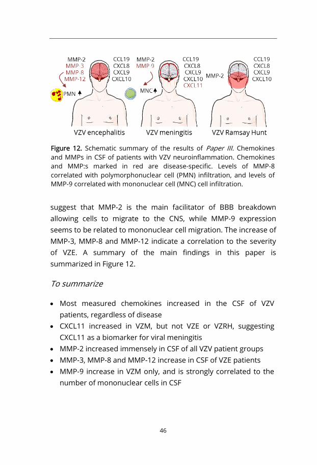
46
suggest that MMP-2 is the main facilitator of BBB breakdown allowing cells to migrate to the CNS, while MMP-9 expression seems to be related to mononuclear cell migration. The increase of MMP-3, MMP-8 and MMP-12 indicate a correlation to the severity of VZE. A summary of the main findings in this paper is summarized in Figure 12.
To summarize
• Most measured chemokines increased in the CSF of VZV patients, regardless of disease
• CXCL11 increased in VZM, but not VZE or VZRH, suggesting CXCL11 as a biomarker for viral meningitis
• MMP-2 increased immensely in CSF of all VZV patient groups • MMP-3, MMP-8 and MMP-12 increase in CSF of VZE patients • MMP-9 increase in VZM only, and is strongly correlated to the
number of mononuclear cells in CSF
Figure 12. Schematic summary of the results of Paper III. Chemokines and MMPs in CSF of patients with VZV neuroinflammation. Chemokines and MMP:s marked in red are disease-specific. Levels of MMP-8 correlated with polymorphonuclear cell (PMN) infiltration, and levels of MMP-9 correlated with mononuclear cell (MNC) cell infiltration.

Chapter 4 Results and Discussion
47
Paper IV 4.4
Summarizing Paper I-III we learned that CXCL11 is an important factor for understanding neuroinflammation induced by HSV or VZV. However, we knew very little about CXCL11 on the molecular level. What cells in the CNS produce CXCL11, and what is the induction pathway? We wanted to investigate this further, and decided to expose human astrocytes and microglia to various cytokines and HSV.
It is previously known that CXCL11 is induced by IFN-β and IFN-γ [131], thus we wanted to see if there were any differences in HSE and HSM patients. We observed IFN-α, IFN-β, IFN-γ and IFN-λ production in the CSF of HSE and HSM patients. We found that HSE patients had increased levels of IFN-α, IFN-γ and IFN-λ compared to healthy controls, while HSM had increased levels of IFN-γ only. When we studied cytokine IL-1β, which is known for its synergy with IFN to induce CXCL11 production, we did see that expression of CXCL11 correlated with IL-1β levels. Interestingly, IL-1β expression was higher in HSM compared to HSE patients, although a low absolute difference (HSE median value 3 pg/ml, HSM median value 7 pg/ml).
In the in vitro system, we could see that in astrocytes CXCL11 was mainly induced by IFN-γ, but to some extent also IFN-α and IFN-β. Adding IL-1β increased levels of CXCL11 significantly when co-stimulated with IFN-α, and a tendency when co-stimulated with IFN-β. There was no difference when co-stimulated with IFN-γ, indicating that IFN-γ itself, but not IFN-α and IFN-β, can induce a potent CXCL11 production. Neither HSV-1 nor HSV-2 induced CXLC11 expression in astrocytes. When investigating cytosolic receptors responding to viral DNA we could conclude that astrocytes do not have the ability to recognize HSV DNA.

48
Since astrocytes did not respond directly to HSV, we moved on to another cell type in the CNS; microglia. In this cell type we could see CXCL11 expression on transcriptional but not protein level. No upregulation of CXCL11 was observed when treating cells with IFN-α or IFN-β, but IFN-γ upregulated CXCL11 more than 20 times. However, no protein could be detected.
From our initial findings it seems like astrocytes may be the main source of CXCL11 in the CNS, but not upon direct stimulation by HSV. We hypothesize that despite the low levels, IL-1β in synergy with interferons has an impact on CXCL11 production in HSM. The source of interferons and IL-1β in HSV is still unclear, but main sources of IFN-γ are usually TH1 CD4+ T cells, CD8+ T cells and NK cells [186]. IL-1β is mainly produced by macrophages and monocytes, but several other cells like T cell and endo- and epithelial cells can also secrete IL-1β [187]. The full mechanisms for production of these cytokines and their potential involvement in CXCL11 expression needs to be further investigated.
To summarize
• HSE and HSM have differential expression of interferons • IL-1β levels are significantly higher in HSM compared to HSE
and correlates to expression of CXCL11 • Astrocytes produce CXCL11 upon interferon, but not HSV,
stimulation • Microglia upregulates CXCL11 upon IFN-γ and HSV stimulation,
but do not produce any protein

Chapter 5 Conclusion
49
5 Conclusion
In this thesis we map several important players in neuroinflammation induced by alphaherpesvirus.
On the immunological level we identify the CD4+CD8+ T cell as a critical component of the immune response in the CNS in a mouse model of HSV-2 meningitis. CD4+CD8+ T cells are highly specific to HSV-2 and secrete immune mediators that are involved in killing virus-specific cells. They are highly activated indicating that they have an important role in fighting off the infection. They also express several chemokine receptors interacting with ligands found in high levels in the CNS.
Patients with HSV or VZV neuroinflammation have increased levels of most chemokines in the CSF, but those that exceed levels in serum are most important for cell migration. These are CXCL8, CXCL9 and CXCL10 in encephalitis, meningitis and Ramsay Hunt, whereas CXCL11 stands out as a biomarker for meningitis.
MMPs are important for the regulation of the BBB making it possible for cells to infiltrate the immune restricted CNS. We conclude that MMP-2 increase in all neuroinflammatory disorders

50
caused by VZV. MMP-9 is associated with meningitis and correlates with levels of mononuclear cells. MMP-3, MMP-8 and MMP-12 are associated with encephalitis, and MMP-8 correlates with levels of polymorphonuclear cells.
We identify IFN-γ and IL-1β as regulators of CXCL11 expression. HSM patients did not only have exclusive CXCL11 production, but also increased levels of IL-1β. In the in vitro system CXCL11 was secreted by astrocytes upon interferon stimulation but not HSV infection. Microglia upregulated CXCL11 mRNA levels upon HSV infection, but did not produce actual protein.

Chapter 6 Future Perspectives
51
6 Future Perspectives
So much to do, so little time. There are yet a lot to discover in this research field. The main question is if the genetic differences of the herpesviruses contribute to the different neurological manifestations, or if there are other factors, like route of entry into the CNS. Thus, the cellular response of each virus (of interest) would need to be characterized on proteome level. This is an extensive approach, but would give great insight into CNS disorders caused by alphaherpesviruses. If someone who does not have a million dollar would like to keep digging more characterization of both resident cells of the CNS and peripheral cells needs to be done. It would be possible to characterize fresh peripheral cells from humans from lumbar punctures taken for diagnostic purposes. For resident CNS cells the murine model is a good alternative. The extensive characterization would also provide insight into treatment alternatives, primarily for encephalitis patients that often suffer from severe sequelae. Even though acyclovir is great for fighting herpes virus, treatments targeting the inflammation caused by alphaherpesviruses are lacking.


Acknowledgements
53
Acknowledgements Det finns ett antal fantastiska människor som har bidragit till att den här avhandlingen har blivit vad den har blivit. Tack!
Min huvudhandledare Kristina Eriksson. Tack för att du trodde att jag var rätt person för den här doktorandplatsen den där decemberdagen 2013. Du är en inspiration, både när det kommer till forskning, kvinnokamp och vin. Tack för att du låtit mig testa alla mina idéer, oavsett om de har varit rimliga eller totalt vimsiga. Och tack för att du har låtit mig ha så mycket frihet som gjort att jag kunnat hitta rätt i livet.
Alexandra Svensson, du har varit min vän lika mycket som du varit min bihandledare. Tack för att du gjort det mycket roligare att vara på jobbet, tack för alla vinkvällar och allt prat.
Min partner in crime, Karolina Thörn. Efter att ha bott på samma gata och gått i samma skola, två gånger, men med tio års mellanrum, så var det nog ödet att vi skulle föras samman i Göteborg. Och vad skulle jag ha gjort utan dig, både på jobbet och i livet? Alla borde ha en Karolina i sitt liv, och jag hoppas att jag får ha kvar min länge till, även om vi inte kommer jobba tillsammans.
Thanks to everyone in our research group, past and present; Inger, Marianela, Kerstin, Malgosha and all the students. It has been a pleasure working with you!
Tack till alla mina medförfattare för roliga samarbeten och intressanta diskussioner; Marie Studahl, Anna Grahn och Linn Persson Berg. Tack också till alla ni som på olika sätt hjälpt till; Charlotta Eriksson, Maria Johansson, Tomas Bergström, Kerstin Andersson och Christina Björklund.
Tack till alla på avdelningen för Reumatologi och Inflammationsforskning som gör det roligt att gå till jobbet!

54
Tack till den mäktigaste föreningen som universitetsvärlden någonsin har skådat; Doktorandklubben Enhörningen. Christina, Alessandro, Malene, Caroline, Jonas, Jauquline, André och alla andra som kommit och gått under åren.
Mina vänner, som tror på mig i ur och skur, vilka märkliga saker jag än tar mig för. Tack för det Teed, Mattias, Simon, Erik, Robin, Mathias.
Det har varit oerhört kul att få göra den här resan tillsammans med dig Kristina, hela vägen från Linköping via Malmö och till Göteborg. Till SF följer jag inte med, men kommer såklart och hälsar på. Den dagen det är dags för Blå Hallen hoppas jag att du smugglar in mig på festen.
Lillie, tillsammans förändrar vi världen. Tack för att du står ut med mig och har skapat världens bästa grej med mig. Tack också till alla andra i Female Legends, och speciellt Josefine, Amanda, Jennifer, Olivia, Mary, Pauline, Sanna och Ania. Även ni i Pink Orange har en speciell plats i mitt hjärta.
Tack till min familj för att ni alltid finns där; min mamma Ingela och min pappa Björn, och bonusarna Göran och Birgitta. Tack till världens bästa syster Cecilia som också har gett mig det bästa som finns i mitt liv; Max och Harry. Jag älskar er.
Max, min största inspiration. Vi utmanar varandra, vi stöder varandra, vi upptäcker tillsammans och vi gör varandra bättre. Vad vi än tar oss för i livet så har vi roligt, och tillsammans kan vi göra allt. Tack för att du finns för mig, och för att jag får vara din. Jag hoppas att jag får älska dig länge till.

References
55
References 1. Arvin, A., et al., Human herpesviruses: biology, therapy, and
immunoprophylaxis. 2007: Cambridge University Press. 2. Shors, T., Understanding viruses. 2011: Jones & Bartlett
Publishers. 3. Lustig, A. and A.J. Levine, One hundred years of virology. J Virol,
1992. 66(8): p. 4629-31. 4. Davison, A.J., Overview of classification, in Human Herpesviruses:
Biology, Therapy, and Immunoprophylaxis, A. Arvin, et al., Editors. 2007: Cambridge.
5. Brown, J.C. and W.W. Newcomb, Herpesvirus capsid assembly: insights from structural analysis. Curr Opin Virol, 2011. 1(2): p. 142-9.
6. Carter, J., V. Saunders, and V.A. Saunders, Virology: principles and applications. 2007: John Wiley & Sons.
7. McGeoch, D.J., et al., Molecular phylogeny and evolutionary timescale for the family of mammalian herpesviruses. J Mol Biol, 1995. 247(3): p. 443-58.
8. Owen, D.J., C.M. Crump, and S.C. Graham, Tegument Assembly and Secondary Envelopment of Alphaherpesviruses. Viruses, 2015. 7(9): p. 5084-114.
9. Whitley, R., D.W. Kimberlin, and C.G. Prober, Pathogenesis and disease, in Human Herpesviruses: Biology, Therapy, and Immunoprophylaxis, A. Arvin, et al., Editors. 2007: Cambridge.
10. Moffat, J., et al., VZV: pathogenesis and the disease consequences of primary infection, in Human Herpesviruses: Biology, Therapy, and Immunoprophylaxis, A. Arvin, et al., Editors. 2007: Cambridge.
11. Grahn, A., Varicella-zoster virus infections of the central nervous system. 2013.
12. Looker, K.J., et al., Global and Regional Estimates of Prevalent and Incident Herpes Simplex Virus Type 1 Infections in 2012. PLoS One, 2015. 10(10): p. e0140765.
13. Looker, K.J., et al., Global estimates of prevalent and incident herpes simplex virus type 2 infections in 2012. PLoS One, 2015. 10(1): p. e114989.
14. Wald, A. and L. Corey, Persistence in the population: epidemiology, transmission, in Human Herpesviruses: Biology,

56
Therapy, and Immunoprophylaxis, A. Arvin, et al., Editors. 2007: Cambridge.
15. Anuradha, K., et al., Herpes simplex virus 2 infection: a risk factor for HIV infection in heterosexuals. Indian J Dermatol Venereol Leprol, 2008. 74(3): p. 230-3.
16. Seward, J. and A. Jumaan, VSV: persistence in the population, in Human Herpesviruses: Biology, Therapy, and Immunoprophylaxis, A. Arvin, et al., Editors. 2007: Cambridge.
17. Carrillo-Santisteve, P. and P.L. Lopalco, Varicella vaccination: a laboured take-off. Clin Microbiol Infect, 2014. 20 Suppl 5: p. 86-91.
18. Varicella and herpes zoster vaccines: WHO position paper, June 2014. Wkly Epidemiol Rec, 2014. 89(25): p. 265-87.
19. Jennische, E., et al., The anterior commissure is a pathway for contralateral spread of herpes simplex virus type 1 after olfactory tract infection. J Neurovirol, 2015. 21(2): p. 129-47.
20. Bergstrom, T., et al., Primary and recurrent herpes simplex virus type 2-induced meningitis. J Infect Dis, 1990. 162(2): p. 322-30.
21. Kramer, M.F., et al., Latent herpes simplex virus infection of sensory neurons alters neuronal gene expression. J Virol, 2003. 77(17): p. 9533-41.
22. Wald, A., et al., Reactivation of genital herpes simplex virus type 2 infection in asymptomatic seropositive persons. N Engl J Med, 2000. 342(12): p. 844-50.
23. Xu, F., et al., Seroprevalence and coinfection with herpes simplex virus type 1 and type 2 in the United States, 1988-1994. J Infect Dis, 2002. 185(8): p. 1019-24.
24. Ku, C.C., et al., Varicella-zoster virus transfer to skin by T Cells and modulation of viral replication by epidermal cell interferon-alpha. J Exp Med, 2004. 200(7): p. 917-25.
25. Arvin, A.M., et al., Varicella-zoster virus T cell tropism and the pathogenesis of skin infection. Curr Top Microbiol Immunol, 2010. 342: p. 189-209.
26. Eshleman, E., A. Shahzad, and R.J. Cohrs, Varicella zoster virus latency. Future Virol, 2011. 6(3): p. 341-355.
27. Cohen, J.I., VZV: molecular basis of persistence (latency and reactivation), in Human Herpesviruses: Biology, Therapy, and Immunoprophylaxis, A. Arvin, et al., Editors. 2007: Cambridge.
28. Malavige, G.N., et al., Viral load, clinical disease severity and cellular immune responses in primary varicella zoster virus infection in Sri Lanka. PLoS One, 2008. 3(11): p. e3789.

References
57
29. Campadelli-Fiume, G. and L. Menotti, Entry of alphaherpesviruses into the cell, in Human Herpesviruses: Biology, Therapy, and Immunoprophylaxis, A. Arvin, et al., Editors. 2007: Cambridge.
30. Sodeik, B., Mechanisms of viral transport in the cytoplasm. Trends Microbiol, 2000. 8(10): p. 465-72.
31. Baines, J.D. and P.E. Pellett, Genetic comparison of human alphaherpesvirus genomes, in Human Herpesviruses: Biology, Therapy, and Immunoprophylaxis, A. Arvin, et al., Editors. 2007: Cambridge.
32. Sandri-Goldin, R.M., Initiation of transcription and RNA synthesis, processing and transport in HSV and VZV infected cells, in Human Herpesviruses: Biology, Therapy, and Immunoprophylaxis, A. Arvin, et al., Editors. 2007: Cambridge.
33. Mettenleiter, T.C., Herpesvirus assembly and egress. J Virol, 2002. 76(4): p. 1537-47.
34. Corey, L., et al., Differentiation of herpes simplex virus types 1 and 2 in clinical samples by a real-time taqman PCR assay. J Med Virol, 2005. 76(3): p. 350-5.
35. Grinde, B., Herpesviruses: latency and reactivation - viral strategies and host response. J Oral Microbiol, 2013. 5.
36. Preston, C.M. and S. Efstathiou, Molecular basis of HSV latency and reactivation, in Human Herpesviruses: Biology, Therapy, and Immunoprophylaxis, A. Arvin, et al., Editors. 2007: Cambridge.
37. Raschilas, F., et al., Outcome of and prognostic factors for herpes simplex encephalitis in adult patients: results of a multicenter study. Clin Infect Dis, 2002. 35(3): p. 254-60.
38. Gordon, B., et al., Long-term cognitive sequelae of acyclovir-treated herpes simplex encephalitis. Arch Neurol, 1990. 47(6): p. 646-7.
39. Sweeney, C.J. and D.H. Gilden, Ramsay Hunt syndrome. J Neurol Neurosurg Psychiatry, 2001. 71(2): p. 149-54.
40. Kallio-Laine, K., et al., Recurrent lymphocytic meningitis positive for herpes simplex virus type 2. Emerg Infect Dis, 2009. 15(7): p. 1119-22.
41. Franzen-Rohl, E., et al., High diagnostic yield by CSF-PCR for entero- and herpes simplex viruses and TBEV serology in adults with acute aseptic meningitis in Stockholm. Scand J Infect Dis, 2008. 40(11-12): p. 914-21.
42. Gnann, J.W., Jr., Varicella-zoster virus: atypical presentations and unusual complications. J Infect Dis, 2002. 186 Suppl 1: p. S91-8.

58
43. Frenos, S., et al., An increasing incidence of chickenpox central nervous system complications in children: what's happening in Tuscany? J Clin Virol, 2007. 38(4): p. 358-61.
44. Rack, A.L., et al., Neurologic varicella complications before routine immunization in Germany. Pediatr Neurol, 2010. 42(1): p. 40-8.
45. Ziebold, C., et al., Severe complications of varicella in previously healthy children in Germany: a 1-year survey. Pediatrics, 2001. 108(5): p. E79.
46. Cameron, J.C., et al., Severe complications of chickenpox in hospitalised children in the UK and Ireland. Arch Dis Child, 2007. 92(12): p. 1062-6.
47. Monsanto, R.D., et al., Treatment and Prognosis of Facial Palsy on Ramsay Hunt Syndrome: Results Based on a Review of the Literature. Int Arch Otorhinolaryngol, 2016. 20(4): p. 394-400.
48. Berger, J.R. and S. Houff, Neurological complications of herpes simplex virus type 2 infection. Arch Neurol, 2008. 65(5): p. 596-600.
49. Simko, J.P., et al., Differences in laboratory findings for cerebrospinal fluid specimens obtained from patients with meningitis or encephalitis due to herpes simplex virus (HSV) documented by detection of HSV DNA. Clin Infect Dis, 2002. 35(4): p. 414-9.
50. Hodge, R.A.V. and H.J. Field, Antiviral agents for herpes simplex virus, in Advances in pharmacology. 2013, Elsevier. p. 1-38.
51. Openshaw, H. and E.M. Cantin, Corticosteroids in herpes simplex virus encephalitis. J Neurol Neurosurg Psychiatry, 2005. 76(11): p. 1469.
52. Ramos-Estebanez, C., K.J. Lizarraga, and A. Merenda, A systematic review on the role of adjunctive corticosteroids in herpes simplex virus encephalitis: is timing critical for safety and efficacy? Antivir Ther, 2014. 19(2): p. 133-9.
53. Domingues, R.B., et al., Evaluation of the range of clinical presentations of herpes simplex encephalitis by using polymerase chain reaction assay of cerebrospinal fluid samples. Clin Infect Dis, 1997. 25(1): p. 86-91.
54. Jouan, Y., et al., Long-term outcome of severe herpes simplex encephalitis: a population-based observational study. Crit Care, 2015. 19: p. 345.

References
59
55. Grahn, A. and M. Studahl, Varicella-zoster virus infections of the central nervous system - Prognosis, diagnostics and treatment. J Infect, 2015. 71(3): p. 281-93.
56. Mailles, A., et al., Long-term outcome of patients presenting with acute infectious encephalitis of various causes in France. Clin Infect Dis, 2012. 54(10): p. 1455-64.
57. Mirakhur, B. and M. McKenna, Recurrent herpes simplex type 2 virus (Mollaret) meningitis. J Am Board Fam Pract, 2004. 17(4): p. 303-5.
58. Looker, K.J., et al., First estimates of the global and regional incidence of neonatal herpes infection. Lancet Glob Health, 2017. 5(3): p. e300-e309.
59. Kimberlin, D.W., Neonatal herpes simplex infection. Clin Microbiol Rev, 2004. 17(1): p. 1-13.
60. Purewal, R., et al., Congenital Herpes Simplex Virus in the Newborn: A Diagnostic Dilemma. J Pediatric Infect Dis Soc, 2016. 5(3): p. e21-3.
61. Lind, L., et al., CXCL11 production in cerebrospinal fluid distinguishes herpes simplex meningitis from herpes simplex encephalitis. J Neuroinflammation, 2017. 14(1): p. 134.
62. Koelle, D.M., Immunobiology and host response, in Human Herpesviruses: Biology, Therapy, and Immunoprophylaxis, A. Arvin, et al., Editors. 2007: Cambridge.
63. Abbas, A.K., A.H. Lichtman, and S. Pillai, Basic immunology: functions and disorders of the immune system. 2014: Elsevier Health Sciences.
64. Chew, T., K.E. Taylor, and K.L. Mossman, Innate and adaptive immune responses to herpes simplex virus. Viruses, 2009. 1(3): p. 979-1002.
65. Ma, Y. and B. He, Recognition of herpes simplex viruses: toll-like receptors and beyond. J Mol Biol, 2014. 426(6): p. 1133-47.
66. Cotter, C.R., et al., The virion host shut-off (vhs) protein blocks a TLR-independent pathway of herpes simplex virus type 1 recognition in human and mouse dendritic cells. PLoS One, 2010. 5(2): p. e8684.
67. Yao, X.D. and K.L. Rosenthal, Herpes simplex virus type 2 virion host shutoff protein suppresses innate dsRNA antiviral pathways in human vaginal epithelial cells. J Gen Virol, 2011. 92(Pt 9): p. 1981-93.

60
68. Milligan, G.N., Neutrophils aid in protection of the vaginal mucosae of immune mice against challenge with herpes simplex virus type 2. J Virol, 1999. 73(8): p. 6380-6.
69. Milligan, G.N., N. Bourne, and K.L. Dudley, Role of polymorphonuclear leukocytes in resolution of HSV-2 infection of the mouse vagina. J Reprod Immunol, 2001. 49(1): p. 49-65.
70. Chesler, D.A. and C.S. Reiss, The role of IFN-gamma in immune responses to viral infections of the central nervous system. Cytokine Growth Factor Rev, 2002. 13(6): p. 441-54.
71. Orr, M.T., et al., Inhibition of MHC class I is a virulence factor in herpes simplex virus infection of mice. PLoS Pathog, 2005. 1(1): p. e7.
72. Arvin, A. and A. Abendroth, VZV: immunobiology and host response, in Human Herpesviruses: Biology, Therapy, and Immunoprophylaxis, A. Arvin, et al., Editors. 2007: Cambridge.
73. Abendroth, A., et al., Varicella-zoster virus retains major histocompatibility complex class I proteins in the Golgi compartment of infected cells. J Virol, 2001. 75(10): p. 4878-88.
74. Bessoles, S., et al., Adaptations of Natural Killer Cells to Self-MHC Class I. Front Immunol, 2014. 5: p. 349.
75. Long, E.O. and S. Rajagopalan, Stress signals activate natural killer cells. J Exp Med, 2002. 196(11): p. 1399-402.
76. Sainz, B., Jr. and W.P. Halford, Alpha/Beta interferon and gamma interferon synergize to inhibit the replication of herpes simplex virus type 1. J Virol, 2002. 76(22): p. 11541-50.
77. Cantin, E., B. Tanamachi, and H. Openshaw, Role for gamma interferon in control of herpes simplex virus type 1 reactivation. J Virol, 1999. 73(4): p. 3418-23.
78. Cantin, E., et al., Gamma interferon (IFN-gamma) receptor null-mutant mice are more susceptible to herpes simplex virus type 1 infection than IFN-gamma ligand null-mutant mice. J Virol, 1999. 73(6): p. 5196-200.
79. Lekstrom-Himes, J.A., et al., Gamma interferon impedes the establishment of herpes simplex virus type 1 latent infection but has no impact on its maintenance or reactivation in mice. J Virol, 2000. 74(14): p. 6680-3.
80. Smith, P.M., et al., Control of acute cutaneous herpes simplex virus infection: T cell-mediated viral clearance is dependent upon interferon-gamma (IFN-gamma). Virology, 1994. 202(1): p. 76-88.

References
61
81. Svensson, A., et al., STAT4 regulates antiviral gamma interferon responses and recurrent disease during herpes simplex virus 2 infection. J Virol, 2012. 86(17): p. 9409-15.
82. Milligan, G.N. and D.I. Bernstein, Interferon-gamma enhances resolution of herpes simplex virus type 2 infection of the murine genital tract. Virology, 1997. 229(1): p. 259-68.
83. Petrera, E. and C.E. Coto, The synergistic effect of IFN-alpha and IFN-gamma against HSV-2 replication in Vero cells is not interfered by the plant antiviral 1-cinnamoyl-3, 11-dihydroxymeliacarpin. Virol J, 2006. 3: p. 45.
84. Baird, N.L., et al., Interferon Gamma Prolongs Survival of Varicella-Zoster Virus-Infected Human Neurons In Vitro. J Virol, 2015. 89(14): p. 7425-7.
85. Netea, M.G., et al., IL-1beta processing in host defense: beyond the inflammasomes. PLoS Pathog, 2010. 6(2): p. e1000661.
86. Sergerie, Y., S. Rivest, and G. Boivin, Tumor necrosis factor-alpha and interleukin-1 beta play a critical role in the resistance against lethal herpes simplex virus encephalitis. J Infect Dis, 2007. 196(6): p. 853-60.
87. Miettinen, J.J., S. Matikainen, and T.A. Nyman, Global secretome characterization of herpes simplex virus 1-infected human primary macrophages. J Virol, 2012. 86(23): p. 12770-8.
88. Johnson, K.E., L. Chikoti, and B. Chandran, Herpes simplex virus 1 infection induces activation and subsequent inhibition of the IFI16 and NLRP3 inflammasomes. J Virol, 2013. 87(9): p. 5005-18.
89. Orzalli, M.H., N.A. DeLuca, and D.M. Knipe, Nuclear IFI16 induction of IRF-3 signaling during herpesviral infection and degradation of IFI16 by the viral ICP0 protein. Proc Natl Acad Sci U S A, 2012. 109(44): p. E3008-17.
90. Maruzuru, Y., et al., Herpes Simplex Virus 1 VP22 Inhibits AIM2-Dependent Inflammasome Activation to Enable Efficient Viral Replication. Cell Host Microbe, 2018. 23(2): p. 254-265 e7.
91. Parkinson, J. and R.D. Everett, Alphaherpesvirus proteins related to herpes simplex virus type 1 ICP0 affect cellular structures and proteins. J Virol, 2000. 74(21): p. 10006-17.
92. Halford, W.P., R. Puschel, and B. Rakowski, Herpes simplex virus 2 ICP0 mutant viruses are avirulent and immunogenic: implications for a genital herpes vaccine. PLoS One, 2010. 5(8): p. e12251.

62
93. Nour, A.M., et al., Varicella-zoster virus infection triggers formation of an interleukin-1beta (IL-1beta)-processing inflammasome complex. J Biol Chem, 2011. 286(20): p. 17921-33.
94. Dorner, T. and A. Radbruch, Antibodies and B cell memory in viral immunity. Immunity, 2007. 27(3): p. 384-92.
95. Zhang, N. and M.J. Bevan, CD8(+) T cells: foot soldiers of the immune system. Immunity, 2011. 35(2): p. 161-8.
96. Joffre, O.P., et al., Cross-presentation by dendritic cells. Nat Rev Immunol, 2012. 12(8): p. 557-69.
97. Steain, M., et al., Analysis of T cell responses during active varicella-zoster virus reactivation in human ganglia. J Virol, 2014. 88(5): p. 2704-16.
98. Frey, C.R., et al., Identification of CD8+ T cell epitopes in the immediate early 62 protein (IE62) of varicella-zoster virus, and evaluation of frequency of CD8+ T cell response to IE62, by use of IE62 peptides after varicella vaccination. J Infect Dis, 2003. 188(1): p. 40-52.
99. Nash, A.A., R. Quartey-Papafio, and P. Wildy, Cell-mediated immunity in herpes simplex virus-infected mice: functional analysis of lymph node cells during periods of acute and latent infection, with reference to cytotoxic and memory cells. J Gen Virol, 1980. 49(2): p. 309-17.
100. Simmons, A. and D.C. Tscharke, Anti-CD8 impairs clearance of herpes simplex virus from the nervous system: implications for the fate of virally infected neurons. J Exp Med, 1992. 175(5): p. 1337-44.
101. Cunningham, A.L., et al., Evolution of recurrent herpes simplex lesions. An immunohistologic study. J Clin Invest, 1985. 75(1): p. 226-33.
102. Koelle, D.M., et al., Clearance of HSV-2 from recurrent genital lesions correlates with infiltration of HSV-specific cytotoxic T lymphocytes. J Clin Invest, 1998. 101(7): p. 1500-8.
103. Koelle, D.M., et al., Direct recovery of herpes simplex virus (HSV)-specific T lymphocyte clones from recurrent genital HSV-2 lesions. J Infect Dis, 1994. 169(5): p. 956-61.
104. Theil, D., et al., Latent herpesvirus infection in human trigeminal ganglia causes chronic immune response. Am J Pathol, 2003. 163(6): p. 2179-84.
105. Hufner, K., et al., Latency of alpha-herpes viruses is accompanied by a chronic inflammation in human trigeminal ganglia but not in
References
63
dorsal root ganglia. J Neuropathol Exp Neurol, 2006. 65(10): p. 1022-30.
106. Verjans, G.M., et al., Selective retention of herpes simplex virus-specific T cells in latently infected human trigeminal ganglia. Proc Natl Acad Sci U S A, 2007. 104(9): p. 3496-501.
107. Gewurz, B.E., J.M. Vyas, and H.L. Ploegh, Herpesvirus evasion of T-cell immunity, in Human Herpesviruses: Biology, Therapy, and Immunoprophylaxis, A. Arvin, et al., Editors. 2007: Cambridge.
108. Swain, S.L., K.K. McKinstry, and T.M. Strutt, Expanding roles for CD4(+) T cells in immunity to viruses. Nat Rev Immunol, 2012. 12(2): p. 136-48.
109. Verjans, G.M., et al., Identification and characterization of herpes simplex virus-specific CD4+ T cells in corneas of herpetic stromal keratitis patients. J Infect Dis, 1998. 177(2): p. 484-8.
110. Suni, M.A., et al., CD4(+)CD8(dim) T lymphocytes exhibit enhanced cytokine expression, proliferation and cytotoxic activity in response to HCMV and HIV-1 antigens. Eur J Immunol, 2001. 31(8): p. 2512-20.
111. Parel, Y. and C. Chizzolini, CD4+ CD8+ double positive (DP) T cells in health and disease. Autoimmun Rev, 2004. 3(3): p. 215-20.
112. Parel, Y., et al., Presence of CD4+CD8+ double-positive T cells with very high interleukin-4 production potential in lesional skin of patients with systemic sclerosis. Arthritis Rheum, 2007. 56(10): p. 3459-67.
113. Zloza, A., et al., CD8+ T cells that express CD4 on their surface (CD4dimCD8bright T cells) recognize an antigen-specific target, are detected in vivo, and can be productively infected by T-tropic HIV. Blood, 2003. 102(6): p. 2156-64.
114. Richards, M.H., et al., Migration of CD8+ T Cells into the Central Nervous System Gives Rise to Highly Potent Anti-HIV CD4dimCD8bright T Cells in a Wnt Signaling-Dependent Manner. J Immunol, 2016. 196(1): p. 317-27.
115. Munschauer, F.E., et al., Circulating CD3+ CD4+ CD8+ T lymphocytes in multiple sclerosis. J Clin Immunol, 1993. 13(2): p. 113-8.
116. Bang, K., et al., CD4+ CD8+ (thymocyte-like) T lymphocytes present in blood and skin from patients with atopic dermatitis suggest immune dysregulation. Br J Dermatol, 2001. 144(6): p. 1140-7.

64
117. Hirao, J. and K. Sugita, Circulating CD4+CD8+ T lymphocytes in patients with Kawasaki disease. Clin Exp Immunol, 1998. 111(2): p. 397-401.
118. Sakaguchi, S., et al., Regulatory T cells and immune tolerance. Cell, 2008. 133(5): p. 775-87.
119. Veiga-Parga, T., S. Sehrawat, and B.T. Rouse, Role of regulatory T cells during virus infection. Immunol Rev, 2013. 255(1): p. 182-96.
120. Suvas, S., et al., CD4+CD25+ T cells regulate virus-specific primary and memory CD8+ T cell responses. J Exp Med, 2003. 198(6): p. 889-901.
121. Sehrawat, S., et al., In vitro-generated antigen-specific CD4+ CD25+ Foxp3+ regulatory T cells control the severity of herpes simplex virus-induced ocular immunoinflammatory lesions. J Virol, 2008. 82(14): p. 6838-51.
122. Melik-Parsadaniantz, S. and W. Rostene, Chemokines and neuromodulation. J Neuroimmunol, 2008. 198(1-2): p. 62-8.
123. Griffith, J.W., C.L. Sokol, and A.D. Luster, Chemokines and chemokine receptors: positioning cells for host defense and immunity. Annu Rev Immunol, 2014. 32: p. 659-702.
124. Ransohoff, R.M., Chemokines and chemokine receptors: standing at the crossroads of immunobiology and neurobiology. Immunity, 2009. 31(5): p. 711-21.
125. Mikloska, Z., et al., In vivo production of cytokines and beta (C-C) chemokines in human recurrent herpes simplex lesions--do herpes simplex virus-infected keratinocytes contribute to their production? J Infect Dis, 1998. 177(4): p. 827-38.
126. Srivastava, R., et al., CXCL10/CXCR3-Dependent Mobilization of Herpes Simplex Virus-Specific CD8(+) TEM and CD8(+) TRM Cells within Infected Tissues Allows Efficient Protection against Recurrent Herpesvirus Infection and Disease. J Virol, 2017. 91(14).
127. Thapa, M., et al., CXCL9 and CXCL10 expression are critical for control of genital herpes simplex virus type 2 infection through mobilization of HSV-specific CTL and NK cells to the nervous system. J Immunol, 2008. 180(2): p. 1098-106.
128. Thapa, M. and D.J. Carr, CXCR3 deficiency increases susceptibility to genital herpes simplex virus type 2 infection: Uncoupling of CD8+ T-cell effector function but not migration. J Virol, 2009. 83(18): p. 9486-501.
129. Harandi, A.M., et al., Protective vaccination against genital herpes simplex virus type 2 (HSV-2) infection in mice is associated with a

References
65
rapid induction of local IFN-gamma-dependent RANTES production following a vaginal viral challenge. Am J Reprod Immunol, 2001. 46(6): p. 420-4.
130. Muller, M., et al., Review: The chemokine receptor CXCR3 and its ligands CXCL9, CXCL10 and CXCL11 in neuroimmunity--a tale of conflict and conundrum. Neuropathol Appl Neurobiol, 2010. 36(5): p. 368-87.
131. Groom, J.R. and A.D. Luster, CXCR3 ligands: redundant, collaborative and antagonistic functions. Immunol Cell Biol, 2011. 89(2): p. 207-15.
132. Lee, C.R., et al., Secretion of IL-1beta from imatinib-resistant chronic myeloid leukemia cells contributes to BCR-ABL mutation-independent imatinib resistance. FEBS Lett, 2016. 590(3): p. 358-68.
133. Michlmayr, D. and C. McKimmie, Role of CXCL10 in central nervous system inflammation. International Journal of Interferon, Cytokine and Mediator Research, 2014. 6: p. 18.
134. Zhang, B., et al., CXCR3 mediates region-specific antiviral T cell trafficking within the central nervous system during West Nile virus encephalitis. J Immunol, 2008. 180(4): p. 2641-9.
135. Carr, D.J., et al., Effect of anti-CXCL10 monoclonal antibody on herpes simplex virus type 1 keratitis and retinal infection. J Virol, 2003. 77(18): p. 10037-46.
136. Zimmermann, J., et al., Enhanced viral clearance and reduced leukocyte infiltration in experimental herpes encephalitis after intranasal infection of CXCR3-deficient mice. J Neurovirol, 2017. 23(3): p. 394-403.
137. Rosler, A., et al., Time course of chemokines in the cerebrospinal fluid and serum during herpes simplex type 1 encephalitis. J Neurol Sci, 1998. 157(1): p. 82-9.
138. Brodal, P., The central nervous system: structure and function. 2004: Oxford University Press.
139. Matsumae, M., et al., Research into the physiology of cerebrospinal fluid reaches a new horizon: intimate exchange between cerebrospinal fluid and interstitial fluid may contribute to maintenance of homeostasis in the central nervous system. Neurologia medico-chirurgica, 2016. 56(7): p. 416-441.
140. Hajdu, S.I., A note from history: discovery of the cerebrospinal fluid. Ann Clin Lab Sci, 2003. 33(3): p. 334-6.

66
141. Deisenhammer, F., et al., Cerebrospinal fluid in clinical neurology. 2015: Springer.
142. Lun, M.P., E.S. Monuki, and M.K. Lehtinen, Development and functions of the choroid plexus-cerebrospinal fluid system. Nat Rev Neurosci, 2015. 16(8): p. 445-57.
143. Spector, R., S. Robert Snodgrass, and C.E. Johanson, A balanced view of the cerebrospinal fluid composition and functions: Focus on adult humans. Exp Neurol, 2015. 273: p. 57-68.
144. Bremell, D., et al., Automated cerebrospinal fluid cell count--new reference ranges and evaluation of its clinical use in central nervous system infections. Clin Biochem, 2014. 47(1-2): p. 25-30.
145. Chernecky, C.C. and B.J. Berger, Laboratory Tests and Diagnostic Procedures-E-Book. 2012: Elsevier Health Sciences.
146. Azevedo, F.A., et al., Equal numbers of neuronal and nonneuronal cells make the human brain an isometrically scaled-up primate brain. J Comp Neurol, 2009. 513(5): p. 532-41.
147. Menzel, R. and M. Giurfa, Cognitive architecture of a mini-brain: the honeybee. Trends Cogn Sci, 2001. 5(2): p. 62-71.
148. Prehaud, C., et al., Virus infection switches TLR-3-positive human neurons to become strong producers of beta interferon. J Virol, 2005. 79(20): p. 12893-904.
149. Choi, S.S., et al., Human astrocytes: secretome profiles of cytokines and chemokines. PLoS One, 2014. 9(4): p. e92325.
150. Cabezas, R., et al., Astrocytic modulation of blood brain barrier: perspectives on Parkinson's disease. Front Cell Neurosci, 2014. 8: p. 211.
151. Alvarez, J.I., T. Katayama, and A. Prat, Glial influence on the blood brain barrier. Glia, 2013. 61(12): p. 1939-58.
152. Wolf, S.A., H.W. Boddeke, and H. Kettenmann, Microglia in Physiology and Disease. Annu Rev Physiol, 2017. 79: p. 619-643.
153. Semple, B.D., T. Kossmann, and M.C. Morganti-Kossmann, Role of chemokines in CNS health and pathology: a focus on the CCL2/CCR2 and CXCL8/CXCR2 networks. J Cereb Blood Flow Metab, 2010. 30(3): p. 459-73.
154. Rosito, M., et al., CXCL16 orchestrates adenosine A3 receptor and MCP-1/CCL2 activity to protect neurons from excitotoxic cell death in the CNS. J Neurosci, 2012. 32(9): p. 3154-63.
155. Guo, G., et al., Involvement of chemokine CXCL11 in the development of morphine tolerance in rats with cancer-induced bone pain. J Neurochem, 2017. 141(4): p. 553-564.

References
67
156. Rock, R.B., et al., Transcriptional response of human microglial cells to interferon-gamma. Genes Immun, 2005. 6(8): p. 712-9.
157. Cole, K.E., et al., Interferon-inducible T cell alpha chemoattractant (I-TAC): a novel non-ELR CXC chemokine with potent activity on activated T cells through selective high affinity binding to CXCR3. J Exp Med, 1998. 187(12): p. 2009-21.
158. Redzic, Z., Molecular biology of the blood-brain and the blood-cerebrospinal fluid barriers: similarities and differences. Fluids Barriers CNS, 2011. 8(1): p. 3.
159. Ransohoff, R.M., P. Kivisakk, and G. Kidd, Three or more routes for leukocyte migration into the central nervous system. Nat Rev Immunol, 2003. 3(7): p. 569-81.
160. Ousman, S.S. and P. Kubes, Immune surveillance in the central nervous system. Nat Neurosci, 2012. 15(8): p. 1096-101.
161. Takeshita, Y. and R.M. Ransohoff, Inflammatory cell trafficking across the blood-brain barrier: chemokine regulation and in vitro models. Immunol Rev, 2012. 248(1): p. 228-39.
162. Wilson, E.H., W. Weninger, and C.A. Hunter, Trafficking of immune cells in the central nervous system. J Clin Invest, 2010. 120(5): p. 1368-79.
163. Manicone, A.M. and J.K. McGuire, Matrix metalloproteinases as modulators of inflammation. Semin Cell Dev Biol, 2008. 19(1): p. 34-41.
164. Page-McCaw, A., A.J. Ewald, and Z. Werb, Matrix metalloproteinases and the regulation of tissue remodelling. Nat Rev Mol Cell Biol, 2007. 8(3): p. 221-33.
165. Spindler, K.R. and T.H. Hsu, Viral disruption of the blood-brain barrier. Trends Microbiol, 2012. 20(6): p. 282-90.
166. Engelhardt, B. and C. Coisne, Fluids and barriers of the CNS establish immune privilege by confining immune surveillance to a two-walled castle moat surrounding the CNS castle. Fluids Barriers CNS, 2011. 8(1): p. 4.
167. Shechter, R., A. London, and M. Schwartz, Orchestrated leukocyte recruitment to immune-privileged sites: absolute barriers versus educational gates. Nat Rev Immunol, 2013. 13(3): p. 206-18.
168. Candelario-Jalil, E., Y. Yang, and G.A. Rosenberg, Diverse roles of matrix metalloproteinases and tissue inhibitors of metalloproteinases in neuroinflammation and cerebral ischemia. Neuroscience, 2009. 158(3): p. 983-94.

68
169. Sellner, J., et al., Herpes-simplex virus encephalitis is characterized by an early MMP-9 increase and collagen type IV degradation. Brain Res, 2006. 1125(1): p. 155-62.
170. Zhou, Y., et al., Favorable effects of MMP-9 knockdown in murine herpes simplex encephalitis using small interfering RNA. Neurol Res, 2010. 32(8): p. 801-9.
171. Jones, D., et al., Proinflammatory cytokines and matrix metalloproteinases in CSF of patients with VZV vasculopathy. Neurol Neuroimmunol Neuroinflamm, 2016. 3(4): p. e246.
172. Beura, L.K., et al., Normalizing the environment recapitulates adult human immune traits in laboratory mice. Nature, 2016. 532(7600): p. 512-6.
173. Watanabe, H., et al., Innate immune response in Th1- and Th2-dominant mouse strains. Shock, 2004. 22(5): p. 460-6.
174. Zohar, Y., et al., CXCL11-dependent induction of FOXP3-negative regulatory T cells suppresses autoimmune encephalomyelitis. J Clin Invest, 2014. 124(5): p. 2009-22.
175. Calandra, T. and T. Roger, Macrophage migration inhibitory factor: a regulator of innate immunity. Nat Rev Immunol, 2003. 3(10): p. 791-800.
176. Nasser, M.W., et al., Differential activation and regulation of CXCR1 and CXCR2 by CXCL8 monomer and dimer. J Immunol, 2009. 183(5): p. 3425-32.
177. Melchjorsen, J., L.N. Sorensen, and S.R. Paludan, Expression and function of chemokines during viral infections: from molecular mechanisms to in vivo function. J Leukoc Biol, 2003. 74(3): p. 331-43.
178. Michlmayr, D. and J.K. Lim, Chemokine receptors as important regulators of pathogenesis during arboviral encephalitis. Front Cell Neurosci, 2014. 8: p. 264.
179. Hickey, M.J., L.N. Stiles, and C.S. Schaumburg, The Usual Suspects: Chemokines and Microbial Infection of the Central Nervous System, in Central Nervous System Diseases and Inflammation. 2008, Springer. p. 235-261.
180. O'Garra, A., L.M. McEvoy, and A. Zlotnik, T-cell subsets: chemokine receptors guide the way. Curr Biol, 1998. 8(18): p. R646-9.
181. van Aalderen, M.C., et al., Blood and beyond: properties of circulating and tissue-resident human virus-specific alphabeta CD8(+) T cells. Eur J Immunol, 2014. 44(4): p. 934-44.

References
69
182. Kivisakk, P., et al., Expression of CCR2, CCR5, and CXCR3 by CD4+ T cells is stable during a 2-year longitudinal study but varies widely between individuals. J Neurovirol, 2003. 9(3): p. 291-9.
183. Andrew, D.P., et al., C-C chemokine receptor 4 expression defines a major subset of circulating nonintestinal memory T cells of both Th1 and Th2 potential. J Immunol, 2001. 166(1): p. 103-11.
184. Al-Banna, N.A., et al., CCR4 and CXCR3 play different roles in the migration of T cells to inflammation in skin, arthritic joints, and lymph nodes. Eur J Immunol, 2014. 44(6): p. 1633-43.
185. Rempe, R.G., A.M. Hartz, and B. Bauer, Matrix metalloproteinases in the brain and blood-brain barrier: Versatile breakers and makers. J Cereb Blood Flow Metab, 2016. 36(9): p. 1481-507.
186. Schoenborn, J.R. and C.B. Wilson, Regulation of interferon-gamma during innate and adaptive immune responses. Adv Immunol, 2007. 96: p. 41-101.
187. Sims, J.E. and D.E. Smith, The IL-1 family: regulators of immunity. Nat Rev Immunol, 2010. 10(2): p. 89-102.


![Immunology of Herpes Simplex Virus Infection: …...[CANCER RESEARCH 36, 836-844, February 1976] Immunology of Herpes Simplex Virus Infection: Relevance to Herpes Simplex Virus Vaccines](https://img.dokumen.tips/doc/110x75/5e3c207dedbcb80872726a41/immunology-of-herpes-simplex-virus-infection-cancer-research-36-836-844.jpg)